Influence of ethanol on dentin roughness, surface free ... · Adriana de Jesus SOARES(a) Caio Cezar...
7
ORIGINAL RESEARCH Endodontic therapy Carlos Augusto de Morais Souto PANTOJA (a) Diogo Henrique da SILVA (a) Adriana de Jesus SOARES (a) Caio Cezar Randi FERRAZ (a) Brenda Paula Figueiredo de Almeida GOMES (a) Alexandre Augusto ZAIA (a) José Flávio Affonso de ALMEIDA (a) (a) Universidade Estadual de Campinas – Unicamp, Piracicaba Dental School, Department of Restorative Dentistry, Piracicaba, SP, Brazil Influence of ethanol on dentin roughness, surface free energy, and interaction between AH Plus and root dentin Abstract: This study aimed to evaluate the influence of different ethanol concentrations on dentin roughness, surface free energy, and contact angle between AH Plus and the root canal dentin. One hundred human maxillary anterior teeth were split longitudinally and 200 dentin specimens were polished to make the surface flatter and smoother. An acrylic bar was positioned between two dentin specimens and impression material was added to create a block, simulating an instrumented root canal space. Specimens were removed from the mold and cleaned in an ultrasonic bath for 10 min. Thereafter, dentin specimens were divided into four groups (n = 50) according to the drying methods used: a) wet: vacuum only, b) paper points: vacuum + absorbent paper points, c) 70% alcohol: 70% alcohol (1 min) + vacuum + absorbent paper points, and d) 100% alcohol: 100% alcohol (1 min) + vacuum + absorbent paper points. A rugosimeter and a goniometer were used to verify the roughness (Ra) and to measure the surface free energy and the contact angle between the AH Plus sealer and the root canal dentin. ANOVA and Tukey tests ( α = 0.05) were used for statistical analysis. The 70% and 100% ethanol groups showed significantly decreased roughness as well as increased surface free energy in the root canal dentin when compared to the wet and paper point groups. In addition, ethanol significantly reduced the contact angle between the AH Plus sealer and the root canal dentin. Ethanol solutions (70% and 100%) provide better wettability of AH Plus sealer on dentin surfaces. Keywords: Ethanol; Dental Pulp Cavity; Wettability. Introduction Obturation is supposed to provide a three-dimensional filling of the root canal system, 1,2 preventing bacterial infiltration from the oral cavity and from periradicular tissues. 1,3,4 Filling materials can fill root canal wall irregularities, apical ramifications, and dentinal tubules, 5,6 and entomb irritants that were not removed during chemomechanical preparation. 7,8 Thus, the physicochemical properties of the filling material and dentin wall characteristics may affect root canal sealing 9,10 and treatment outcomes. Wettability is an important thermomechanical property 11 that represents the interactions between solids and liquids. It is strongly dependent on Declaration of Interests: The authors certify that they have no commercial or associative interest that represents a conflict of interest in connection with the manuscript. Corresponding Author: José Flávio Affonso de Almeida E-mail: [email protected] https://doi.org/10.1590/1807-3107bor-2018.vol32.0033 Submitted: November 06, 2017 Accepted for publication: February 21, 2018 Last revision: March 13, 2018 1 Braz. Oral Res. 2018;32:e33
Transcript of Influence of ethanol on dentin roughness, surface free ... · Adriana de Jesus SOARES(a) Caio Cezar...

Original research
Endodontic therapy
Carlos Augusto de Morais Souto PANTOJA(a) Diogo Henrique da SILVA(a) Adriana de Jesus SOARES(a) Caio Cezar Randi FERRAZ(a) Brenda Paula Figueiredo de Almeida GOMES(a) Alexandre Augusto ZAIA(a) José Flávio Affonso de ALMEIDA(a)
(a) Universidade Estadual de Campinas – Unicamp, Piracicaba Dental School, Department of Restorative Dentistry, Piracicaba, SP, Brazil
Influence of ethanol on dentin roughness, surface free energy, and interaction between AH Plus and root dentin
Abstract: This study aimed to evaluate the influence of different ethanol concentrations on dentin roughness, surface free energy, and contact angle between AH Plus and the root canal dentin. One hundred human maxillary anterior teeth were split longitudinally and 200 dentin specimens were polished to make the surface flatter and smoother. An acrylic bar was positioned between two dentin specimens and impression material was added to create a block, simulating an instrumented root canal space. Specimens were removed from the mold and cleaned in an ultrasonic bath for 10 min. Thereafter, dentin specimens were divided into four groups (n = 50) according to the drying methods used: a) wet: vacuum only, b) paper points: vacuum + absorbent paper points, c) 70% alcohol: 70% alcohol (1 min) + vacuum + absorbent paper points, and d) 100% alcohol: 100% alcohol (1 min) + vacuum + absorbent paper points. A rugosimeter and a goniometer were used to verify the roughness (Ra) and to measure the surface free energy and the contact angle between the AH Plus sealer and the root canal dentin. ANOVA and Tukey tests (α = 0.05) were used for statistical analysis. The 70% and 100% ethanol groups showed significantly decreased roughness as well as increased surface free energy in the root canal dentin when compared to the wet and paper point groups. In addition, ethanol significantly reduced the contact angle between the AH Plus sealer and the root canal dentin. Ethanol solutions (70% and 100%) provide better wettability of AH Plus sealer on dentin surfaces.
Keywords: Ethanol; Dental Pulp Cavity; Wettability.
Introduction
Obturation is supposed to provide a three-dimensional filling of the root canal system,1,2 preventing bacterial infiltration from the oral cavity and from periradicular tissues.1,3,4 Filling materials can fill root canal wall irregularities, apical ramifications, and dentinal tubules,5,6 and entomb irritants that were not removed during chemomechanical preparation.7,8 Thus, the physicochemical properties of the filling material and dentin wall characteristics may affect root canal sealing9,10 and treatment outcomes.
Wettability is an important thermomechanical property11 that represents the interactions between solids and liquids. It is strongly dependent on
Declaration of Interests: The authors certify that they have no commercial or associative interest that represents a conflict of interest in connection with the manuscript.
Corresponding Author:José Flávio Affonso de Almeida E-mail: [email protected]
https://doi.org/10.1590/1807-3107bor-2018.vol32.0033
Submitted: November 06, 2017 Accepted for publication: February 21, 2018 Last revision: March 13, 2018
1Braz. Oral Res. 2018;32:e33

Inf luence of ethanol on dentin roughness, surface free energy, and interaction between AH Plus and root dentin
dentin roughness12 and on surface free energy.13 The surface with a lower contact angle (greater surface free energy) presents high wettability. In solids with high surface free energy, the sealer spreads and interacts better, creating a low contact angle.14,15
Dentin surface wettability can be changed by using ethanol solutions at different concentrations. Osorio et al.16 and Pei et al.17 verified a significant decrease in dentin roughness after all dentin specimens were etched with phosphoric acid and rinsed with water. Based on that, the authors suggested that ethanol might offer potential benefits to root dentin bonding with a hydrophobic adhesive.
Additional ethanol application had been previously proposed – in a technique called ethanol wet-bonding - in order to lure hydrophobic monomers into demineralized dentin collagen matrices.18,19 There is a higher compatibility between ethanol-saturated dentin and hydrophobic resin monomers, preventing collagen shrinkage and allowing for higher impregnation.20 This technique has been shown to produce adhesive interfaces with higher bond strength, reduced interface nanoleakage, and increased stability over time when compared to the “water-wet bonding technique.”21,22
In endodontics, it is suggested that a final rinse of dentin walls with different substances23 or with ethanol may alter root dentin properties, enhancing endodontic sealer penetration into dentinal tubules.24,25 The interaction between a hydrophobic sealer and a surface with higher hydrophobic characteristics would provide a low contact angle, higher sealer penetration, greater mechanical interlock into the tubules, retention, sealing ability and, consequently, in vivo antibacterial effectiveness.7,26
However, further investigation is needed to evaluate the role of physicochemical properties of dentinal surfaces in ethanol application. Therefore, the aim of this study was to evaluate the influence of different ethanol concentrations on dentin roughness, surface free energy, and contact angle between the AH Plus sealer and dentin walls. The null hypothesis tested was that no ethanol concentration would interfere with dentin surface roughness, surface free energy, and interaction between the AH Plus sealer and the root canal dentin.
Methodology
Specimen preparationAfter approval by the Research Ethics Committee of
the Piracicaba Dental School, University of Campinas – FOP-UNICAMP (process no. 029/2013), one hundred recently extracted human maxillary anterior teeth were selected. All teeth were cleaned with an ultrasonic scaler, washed with saline solution, and stored in 0.2% thymol at 4°C. The tooth crowns were removed using a 0.3-mm saw microtome (Isomet 1000, Buehler, Lake Bluff, IL, USA). Each root was sectioned longitudinally into two parallel dentin slices with a rotary diamond disk 7020 (KG Sorensen, Cotia, São Paulo, Brazil) at low speed under cool water. An acrylic bar (4 x 4 x 20 mm) was positioned between two dentin specimens and impression material was added to create a block, simulating an instrumented root canal space (Figure 1) and standardizing irrigant volumes. The specimens were removed from the mold and cleaned in an ultrasonic bath for 10 min. The samples were polished with wet 400-, 600- and 1200-grit silicon carbide (SiC) abrasive paper (Carbimet Disc Set, no. 305178180, Buehler, Coventry, UK) to make the surface flatter and smoother, according to Hu et al.11 All samples were treated with 17% ethylenediaminetetraacetic acid (EDTA) for 1 min and this procedure was repeated three times. A final rinse with 5 mL of distilled water was performed.
Two hundred dentin samples were randomly divided into four groups (n=50) according to the following drying methods: Group 1 - drying only with a capillary tip attached to a vacuum adapter (Ultradent Products Inc, South Jordan, USA) for 5 s; Group 2 - drying with a capillary tip attached to a vacuum adapter (5 s), followed by two absorbent paper points (Endo Points Industrial Amazônica, Manacapuru, Brazil); Group 3 - treatment with 70% alcohol (Prolink Indústria Química Ltda., São José do Rio Preto, Brazil) for 1 min, followed by capillary tip attachment to a vacuum adapter (5 s) and two absorbent paper points; Group 4, the same procedures used in Group 3, but the treatment was performed with 100% alcohol (Prolink Indústria Química Ltda., São José do Rio Preto, Brazil) for 1 min.
2 Braz. Oral Res. 2018;32:e33
Pantoja CAMS, Silva DH, Soares AJ, Ferraz CCR, Gomes BPFA, Zaia AA et al.
Roughness measurementsForty dentin samples (n = 10, each group) were
individually positioned in a Surfcorder SE 1700 profilometer (Kosaka Lab, Tokyo, Japan) to verify the roughness (Ra) of the root canal dentin subjected to drying methods. Three readings were performed, in the same middle area on each surface using a stylus tip (0.5 μm in diameter). The maximum track length of each reading was 1.25 mm, using a 0.25-mm cutoff filter. The mean and standard deviation of Ra were determined. The Ra parameter describes the overall roughness of a surface and can be defined as the arithmetic mean of all absolute distances of the roughness profile from the centerline within the measuring length.
Surface free energy measurements A Ramé-hart goniometer (Ramé-hart Instrument
Co., Netcong, USA) was used to measure the contact angle between dentin and the following solutions: water (polar), bromonaphthalene (nonpolar), and formamide (polar). The solutions were used to determine the dispersive/polar components and the dentin total free energy by means of the Ramé-hart DROP Image Standard software (Ramé-hart Instrument Co., Netcong, USA). A total of 120 dentin samples were used (30 samples per group and 10 samples per solution). One drop of liquid was measured in each sample. Thirty measurements were made in each drop and the arithmetic mean was then calculated. Based on the data on the three solutions above, the Ramé-hart
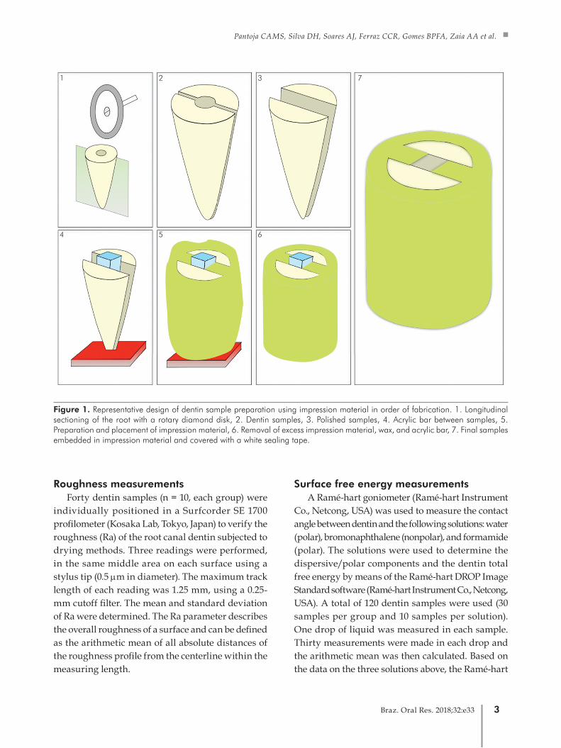
Figure 1. Representative design of dentin sample preparation using impression material in order of fabrication. 1. Longitudinal sectioning of the root with a rotary diamond disk, 2. Dentin samples, 3. Polished samples, 4. Acrylic bar between samples, 5. Preparation and placement of impression material, 6. Removal of excess impression material, wax, and acrylic bar, 7. Final samples embedded in impression material and covered with a white sealing tape.
1 2 3 7
4 5 6
3Braz. Oral Res. 2018;32:e33

Inf luence of ethanol on dentin roughness, surface free energy, and interaction between AH Plus and root dentin
software was able to measure the surface free energy of the different experimental groups.
Contact angle measurementsThe same Ramé-hart goniometer software
(Ramé-Hart Instrument Co, Netcong, USA) was used to measure the contact angle between dried dentin surfaces and a hydrophobic endodontic sealer, AH Plus (Dentsply, Petropolis, RJ, Brazil). The sealer was manipulated according to the manufacturer’s instructions. A total of 40 samples were used (10 samples per group). A drop of the sealer (0.1 mL) was deposited onto dentin surfaces with a 0.5-mL BD ultrafine syringe and a 20 x 0.55-mm needle (24G) (Becton Dickinson, Franklin Lakes, USA).23 Three drops of the sealer were evaluated for each treatment. Images of the drop were analyzed to provide the values of contact angles. Thirty measurements were made in each sample as recommended by Ramé-Hart Instrument Co. The data were computed with the Origin Pro 70 software (Origin Lab Northampton, USA).
Data analysis Since the normality assumptions of the data were
valid, the data were analyzed with one-way analysis of variance (ANOVA) and Tukey tests, with statistical significance preset at α = 0.05.
Results
Treatment with the ethanol solutions led to decreased roughness and increased surface free energy in the root canal dentin, when compared to the wet and paper point groups (Figures 2 and 3). There was no difference between different ethanol concentrations or between the wet and paper point groups. Lower contact angles between AH Plus and the dentin surface were obtained on surfaces treated with 70% and 100% ethanol (Figure 4). Again, no significant difference was found between ethanol concentrations or between the wet and paper points groups.
Discussion
Ethanol solutions showed a considerable amount of influence on the physicochemical properties of
Figure 2. Dentin surface physicochemical properties and interaction with AH Plus. Dentin surface roughness.
dent
in s
urfa
ce r
ough
ness
(µm
)
a
1.0
0.8
0.6
0.4
0.2
0.0wet paper point 70% alcohol 100% alcohol
a
b b
Figure 3. Dentin surface physicochemical properties and interaction with AH Plus. Dentin surface free energy.
dent
in s
urfa
ce fr
ee e
nerg
y (m
Jm2 ) b
50
40
30
20
10
0wet paper point 70% alcohol 100% alcohol
b
a a
Figure 4. Dentin surface physicochemical properties and interaction with AH Plus. Contact angle between AH Plus sealer and the root canal surface.
cont
act a
ngle
(deg
ree)
a
100
80
60
40
20
0wet paper point 70% alcohol 100% alcohol
a
b b
4 Braz. Oral Res. 2018;32:e33

Pantoja CAMS, Silva DH, Soares AJ, Ferraz CCR, Gomes BPFA, Zaia AA et al.
dentin, leading to the rejection of the null hypothesis. Regardless of the concentration, ethanol caused changes in dentin roughness (decreased) and in surface free energy (increased). Consequently, the contact angles between the endodontic sealer and the dentin surface also changed (decreased). Previous studies24,25 have evaluated the influence of ethanol on these physicochemical properties and found a better penetration of irrigant solution or endodontic sealer into the dentinal tubules and into small spaces of the root canal system.
Pei et al.17 found lower roughness in dentin treated with stepwise ethanol application, when compared to water-wet dentin. Our results concur with this previous study and show a decrease in roughness and an increase in surface free energy in samples treated with a final rinse of 100% and 70% ethanol. These results can be explained by the ability of ethanol solutions to promote a partial chemical dehydration of demineralized collagen fibers and contraction of dentin collagen network diameters,16 thereby increasing interfibrillar spaces27 and favoring hydrophobic material embedment.19 Pei et al.17 also pointed out that ethanol solution may reduce the intrinsic wetness of the root dentin.
The ethanol wet-bonding technique combines the reduction of polarity of the collagen network with the low polarity of highly hydrophobic resins, since hydrophobic monomers can better infiltrate into the ethanol-saturated demineralized dentin. The association between the ethanol wet-bonding technique and hydrophobic resin blends improves adhesion to the dentinal substrate, thus increasing the longevity of the bonding interface. It is already known that these hydrophobic resin blends have higher stiffness and stability than do hydrophilic ones.22,28,29,30,31 Ethanol wet-bonding can promote the infiltration of hydrophobic dimethacrylate resins into interfibrillar spaces and dentinal tubules to improve stability of resin-dentin interfaces in vitro.32 Therefore, as epoxy resin binds to collagen,33 especially in demineralized dentin, it is suggested that in the presence of alcohol instead of water, some hydrophobic materials, such as AH Plus, may have improved flow into dentinal tubules and infiltration into collagen interfibrillar spaces.
Nagas et al.9 concluded that the degree of residual moisture can affect the adhesion of root canal sealers to root dentin and suggested that slight moisture would be advantageous to the tested sealers. Taken altogether, their results seem to be completely different from ours. However, Nagas et al.9 used a 95% ethanol final rinse and stored the roots at 37°C to ensure complete dryness, leading the specimens to extreme dehydration. The moisture conditions tested by these authors are quite close to the drying protocols used by us, but in the ethanol groups, water was replaced with ethanol. Thus, the collagen network was filled with residual ethanol or at least with water/ethanol mixture, preventing its collapse17 and probably favoring hydrophobic material infiltration. It is important to emphasize that ethanol evaporates more quickly than water.34 Therefore, dentin physicochemical tests or even root canal filling should be performed right after the drying protocol to prevent ethanol evaporation and collapse of the collagen network.
The influence of alcoholic solution on periapical tissues should also be considered. A few studies have demonstrated this relationship. Oguntebi et al.35 evaluated the effect of 95% ethanol on rat teeth with periapical lesions. After daily injections of 0.1 to 0.2 mL of alcoholic solution for 6 weeks, inflammatory cells were found in the apical periodontal ligament. Yesilsoy et al.36 evaluated the cytotoxic effects of root canal irrigants and found that an 11.6% alcoholic solution in contact with subcutaneous tissue of an in vivo animal model (guinea pig) at different times (2 h, 2 days, and 2 weeks) did not cause inflammatory reactions. Further research should focus on the in vivo assessment of the effect of alcoholic solutions at different concentrations and time intervals on apical periodontal ligament structures.
Taken together, the results showed that 70% and 100% ethanol can improve wettability of AH Plus sealer on the dentin surface, reducing AH Plus contact angles and favoring the interaction between this sealer and the root canal dentin. Further studies should be performed to evaluate whether ethanol solutions could enhance endodontic sealer penetration into dentinal tubules.
5Braz. Oral Res. 2018;32:e33
Inf luence of ethanol on dentin roughness, surface free energy, and interaction between AH Plus and root dentin
Conclusion
Ethanol solutions (70% and 100%) enhance dentin characteristics (roughness and surface free energy), thereby providing better wettability of AH Plus sealer on dentin surfaces.
Acknowledgments
The authors deny any conflict of interest in this study. This study was supported by Capes.
1. Schilder H. Filling root canals in three dimensions. Dent Clin
North Am. 1967 Nov;1:723-44.
2. Hulsmann M, Peters OA, Dummer PM. Mechanical
preparation of root canals: shaping goals, techniques
and means. Endod Topics. 2005;10(1):30-76.
https://doi.org/10.1111/j.1601-1546.2005.00152.x
3. Buckley M, Spångberg LS. The prevalence and
technical quality of endodontic treatment in an
American subpopulation. Oral Surg Oral Med Oral
Pathol Oral Radiol Endod. 1995 Jan;79(1):92-100.
https://doi.org/10.1016/S1079-2104(05)80081-2
4. Saunders WP, Saunders EM. Coronal leakage as
a cause of failure in root-canal therapy: a review.
Endod Dent Traumatol. 1994 Jun;10(3):105-8.
https://doi.org/10.1111/j.1600-9657.1994.tb00533.x
5. Epley SR, Fleischman J, Hartwell G, Cicalese C. Completeness
of root canal obturations: epiphany techniques versus
gutta-percha techniques. J Endod. 2006 Jun;32(6):541-4.
https://doi.org/10.1016/j.joen.2005.10.059
6. Wu MK, Fan B, Wesselink PR. Diminished leakage along
root canals filled with gutta-percha without sealer over time:
a laboratory study. Int Endod J. 2000 Mar;33(2):121-5.
https://doi.org/10.1046/j.1365-2591.2000.00274.x
7. Heling I, Chandler NP. The antimicrobial effect within dentinal
tubules of four root canal sealers. J Endod. 1996 May;22(5):257-
9. https://doi.org/10.1016/S0099-2399(06)80144-5
8. Kokkas AB, Boutsioukis AC, Vassiliadis LP, Stavrianos
CK. The influence of the smear layer on dentinal tubule
penetration depth by three different root canal sealers:
an in vitro study. J Endod. 2004 Feb;30(2):100-2.
https://doi.org/10.1097/00004770-200402000-00009
9. Nagas E, Uyanik MO, Eymirli A, Cehreli ZC, Vallittu PK,
Lassila LV et al. Dentin moisture conditions affect the adhesion
of root canal sealers. J Endod. 2012 Feb;38(2):240-4.
https://doi.org/10.1016/j.joen.2011.09.027
10. Zmener O, Pameijer CH, Serrano SA, Vidueira M, Macchi
RL. Significance of moist root canal dentin with the use
of methacrylate-based endodontic sealers: an in vitro
coronal dye leakage study. J Endod. 2008 Jan;34(1):76-9.
https://doi.org/10.1016/j.joen.2007.10.012
11. Hu X, Ling J, Gao Y. Effects of irrigation solutions on dentin
wettability and roughness. J Endod. 2010 Jun;36(6):1064-7.
https://doi.org/10.1016/j.joen.2010.03.007
12. Rosales JI, Marshall GW, Marshall SJ, Watanabe
LG, Toledano M, Cabrerizo MA et al. Acid-etching
and hydration influence on dentin roughness and
wettability. J Dent Res. 1999 Sep;78(9):1554-9.
https://doi.org/10.1177/00220345990780091001
13. Duncan-Hewitt W. Nature of the hydrophobic effect. In:
Doule RJ, Rosenberg M, editors. Microbial cell surface
hydrophobicity. Washington, DC: ASM; 1990. p. 39-73.
14. Kontakiotis EG, Tzanetakis GN, Loizides AL. A
comparative study of contact angles of four different
root canal sealers. J Endod. 2007 Mar;33(3):299-302.
https://doi.org/10.1016/j.joen.2006.11.016
15. Milosevic A. The influence of surface finish and in-vitro
pellicle on contact-angle measurement and surface
morphology of three commercially available composite
restoratives. J Oral Rehabil. 1992 Jan;19(1):85-97.
https://doi.org/10.1111/j.1365-2842.1992.tb01593.x
16. Osorio E, Toledano M, Aguilera FS, Tay FR, Osorio
R. Ethanol wet-bonding technique sensitivity assessed
by AFM. J Dent Res. 2010 Nov;89(11):1264-9.
https://doi.org/10.1177/0022034510376403
17. Pei D, Huang X, Huang C, Wang Y, Ouyang X,
Zhang J. Ethanol-wet bonding may improve root
dentine bonding performance of hydrophobic
adhesive. J Dent. 2012 May;40(5):433-41.
https://doi.org/10.1016/j.jdent.2012.02.005
18. Pashley DH, Tay FR, Carvalho RM, Rueggeberg FA, Agee KA,
Carrilho M et al. From dry bonding to water-wet bonding to
ethanol-wet bonding. A review of the interactions between
dentin matrix and solvated resins using a macromodel of the
hybrid layer. Am J Dent. 2007 Feb;20(1):7-20.
19. Tay FR, Pashley DH, Kapur RR, Carrilho MR,
Hur YB, Garrett LV et al. Bonding BisGMA to
dentin: a proof of concept for hydrophobic dentin
bonding. J Dent Res. 2007 Nov;86(11):1034-9.
https://doi.org/10.1177/154405910708601103
References
6 Braz. Oral Res. 2018;32:e33

Pantoja CAMS, Silva DH, Soares AJ, Ferraz CCR, Gomes BPFA, Zaia AA et al.
20. Carvalho CA, Monticelli F, Cantoro A, Breschi L, Ferrari M.
Push-out bond strength of fiber posts luted with unfilled resin
cement. J Adhes Dent. 2009 Feb;11(1):65-70.
21. Carvalho CA, Breschi L, Navarro MF, Atta MT, Ferrari
M. Push-out bond strength and SEM evaluation
of a new bonding approach into the root canal.
J Appl Oral Sci. 2012 Nov-Dec;20(6):613-9.
https://doi.org/10.1590/S1678-77572012000600005
22. Hosaka K, Nishitani Y, Tagami J, Yoshiyama M, Brackett WW,
Agee KA et al. Durability of resin-dentin bonds to water- vs.
ethanol-saturated dentin. J Dent Res. 2009 Feb;88(2):146-51.
https://doi.org/10.1177/0022034508328910
23. de Assis DF, Prado M, Simão RA. Evaluation of the interaction
between endodontic sealers and dentin treated with different
irrigant solutions. J Endod. 2011 Nov;37(11):1550-2.
https://doi.org/10.1016/j.joen.2011.08.014
24. Stevens RW, Strother JM, McClanahan SB. Leakage and
sealer penetration in smear-free dentin after a final rinse
with 95% ethanol. J Endod. 2006 Aug;32(8):785-8.
https://doi.org/10.1016/j.joen.2006.02.027
25. Wilcox LR, Wiemann AH. Effect of a final
alcohol rinse on sealer coverage of obturated
root canals. J Endod. 1995 May;21(5):256-8.
https://doi.org/10.1016/S0099-2399(06)80992-1
26. Zhang H, Shen Y, Ruse ND, Haapasalo M. Antibacterial
activity of endodontic sealers by modified direct contact test
against Enterococcus faecalis. J Endod. 2009 Jul;35(7):1051-
5. https://doi.org/10.1016/j.joen.2009.04.022
27. Vafaei S, Podowski MZ. Analysis of the relationship
between liquid droplet size and contact angle. Adv
Colloid Interface Sci. 2005 May;113(2-3):133-46.
https://doi.org/10.1016/j.cis.2005.03.001
28. Breschi L, Mazzoni A, De Stefano Dorigo E, Ferrari
M. Adhesion to intraradicular dentin: a review.
J Adhes Sci Technol. 2009;23(7-8):1053-83.
https://doi.org/10.1163/156856109X440957
29. Ito S, Hashimoto M, Wadgaonkar B, Svizero N,
Carvalho RM, Yiu C et al. Effects of resin hydrophilicity
on water sorption and changes in modulus of
elasticity. Biomaterials. 2005 Nov;26(33):6449-59.
https://doi.org/10.1016/j.biomaterials.2005.04.052
30. Nishitani Y, Yoshiyama M, Donnelly AM, Agee KA, Sword
J, Tay FR et al. Effects of resin hydrophilicity on dentin
bond strength. J Dent Res. 2006 Nov;85(11):1016-21.
https://doi.org/10.1177/154405910608501108
31. Sadek FT, Pashley DH, Nishitani Y, Carrilho MR, Donnelly A,
Ferrari M et al. Application of hydrophobic resin adhesives
to acid-etched dentin with an alternative wet bonding
technique. J Biomed Mater Res A. 2008 Jan;84(1):19-29.
https://doi.org/10.1002/jbm.a.31290
32. Malacarne J, Carvalho RM, de Goes MF, Svizero N,
Pashley DH, Tay FR et al. Water sorption/solubility of dental
adhesive resins. Dent Mater. 2006 Oct;22(10):973-80.
https://doi.org/10.1016/j.dental.2005.11.020
33. Neelakantan P, Sharma S, Shemesh H, Wesselink
PR. Influence of irrigation sequence on the
adhesion of root canal sealers to dentin: a Fourier
transform infrared spectroscopy and push-out bond
strength analysis. J Endod. 2015 Jul;41(7):1108-11.
https://doi.org/10.1016/j.joen.2015.02.001
34. Pashley EL, Zhang Y, Lockwood PE, Rueggeberg FA,
Pashley DH. Effects of HEMA on water evaporation from
water-HEMA mixtures. Dent Mater. 1998 Jan;14(1):6-10.
https://doi.org/10.1016/S0109-5641(98)00003-7
35. Oguntebi BR, Barker BF, Anderson DM, Sakumura
J. The effect of indomethacin on experimental dental
periapical lesions in rats. J Endod. 1989 Mar;15(3):117-21.
https://doi.org/10.1016/S0099-2399(89)80131-1
36. Yesilsoy C, Whitaker E, Cleveland D, Phillips E, Trope M.
Antimicrobial and toxic effects of established and potential
root canal irrigants. J Endod. 1995 Oct;21(10):513-5.
https://doi.org/10.1016/S0099-2399(06)80524-8
7Braz. Oral Res. 2018;32:e33