Influence of 1 Alpha, 25-Dihydroxyvitamin D3 on T Helper...
12
Iran.J.Immunol. VOL.12 NO. 2 June 2015 82 Influence of 1 Alpha, 25-Dihydroxyvitamin D3 on T Helper 17 Cells and Related Cytokines in Systemic Lupus Erythematosus Hadi Reihani 1# , Maryam Rastin 1# , Mahmoud Mahmoudi 1 , Mohsen Ghoryani 1 , Nafiseh Abdollahi 3 , Nafiseh Sadat Tabasi 1 , Shahrzad Zamani Taghizadeh Rabe 1 , Maryam Sahebari 2* 1 Immunology Research Center, BuAli Research Institute, 2 Rheumatic Diseases Research Center, Ghaem Hospital, School of Medicine, Mashhad University of Medical Sciences, Mashhad, 3 Bone, Joint and Connective Tissue Research Center, Golestan University of Medical Sciences, Gorgan, Iran ABSTRACT Background: Systemic lupus erythematosus (SLE) is a multisystem autoimmune disease. Emerging data suggests that T helper 17 (Th17) cells play a pathogenic role in SLE and the increased number of these cells correlates with disease activity. In recent years, 1α, 25-dihydroxyvitamin D3 (1,25VitD3) has been considered as an immunomodulatory factor. Objective: To investigate the effect of 1,25VitD3 on Th17 cells and on the expression of related cytokines in SLE patients. Method: Thirty SLE patients (newly diagnosed or in remission) were sampled for 10 ml whole blood to isolate peripheral blood mononuclear cells (PBMCs) using Ficoll-Hypaque density gradient centrifugation. Isolated cells were cultured in the presence and absence of 50 nM 1,25VitD3. After incubation, cells were harvested and stimulated for 4-5 hours with phorbol myristate acetate (PMA) and ionomycin in the presence of brefeldin A. IL-17 secreting cells were analyzed by flowcytometry. RNA was extracted from cultured cells, cDNA was synthesized, and the expression levels of IL-6, IL-17, IL-23 and TGF- β genes were assessed by real-time PCR. Results: The percentage of Th17 cells (CD3 + CD8 - IL-17 + T cells) decreased significantly in 1,25VitD3-treated cells (3.67 ± 2.43%) compared to untreated cells (4.65 ± 2.75%) ( p=0.003). The expression of TGF- β up regulated (1.38-fold) and the expression of IL-6 (50%), IL-17 (27%) and IL-23 (64%) down regulated after 1,25VitD3 treatment. Conclusion: This study showed that 1,25VitD3 modulates Th17 related pathways in SLE patients and revealed the immunomodulatory effect of 1,25VitD3 on the Th17 mediated autoimmunity. Reihani H,et al. Iran J Immunol. 2015; 12(2):82-93 Keywords: IL-17, IL-23, Systemic Lupus Erythematosus, Th17, TGF-β, Vitamin D --------------------------------------------------------------------------------------------------------------------------------------------------------------- *Corresponding author: Dr. Maryam Sahebari, Rheumatic Diseases Research Center, Ghaem Hospital, School of medicine, Mashhad University of Medical Sciences, Mashhad, Iran, Tel: (+) 98 51 38012753, Fax: (+) 98 51 38410136, e-mail: [email protected] # ”These authors equally contributed to this study.
Transcript of Influence of 1 Alpha, 25-Dihydroxyvitamin D3 on T Helper...

Iran.J.Immunol. VOL.12 NO. 2 June 2015 82
Influence of 1 Alpha, 25-Dihydroxyvitamin D3 on T Helper 17 Cells and Related
Cytokines in Systemic Lupus Erythematosus
Hadi Reihani1#, Maryam Rastin1#, Mahmoud Mahmoudi1, Mohsen Ghoryani1, Nafiseh Abdollahi3, Nafiseh Sadat Tabasi1, Shahrzad Zamani Taghizadeh Rabe1, Maryam Sahebari2*
1Immunology Research Center, BuAli Research Institute, 2Rheumatic Diseases Research Center, Ghaem Hospital, School of Medicine, Mashhad University of Medical Sciences, Mashhad, 3Bone, Joint and Connective Tissue Research Center, Golestan University of Medical Sciences, Gorgan, Iran
ABSTRACT Background: Systemic lupus erythematosus (SLE) is a multisystem autoimmune disease. Emerging data suggests that T helper 17 (Th17) cells play a pathogenic role in SLE and the increased number of these cells correlates with disease activity. In recent years, 1α, 25-dihydroxyvitamin D3 (1,25VitD3) has been considered as an immunomodulatory factor. Objective: To investigate the effect of 1,25VitD3 on Th17 cells and on the expression of related cytokines in SLE patients. Method: Thirty SLE patients (newly diagnosed or in remission) were sampled for 10 ml whole blood to isolate peripheral blood mononuclear cells (PBMCs) using Ficoll-Hypaque density gradient centrifugation. Isolated cells were cultured in the presence and absence of 50 nM 1,25VitD3. After incubation, cells were harvested and stimulated for 4-5 hours with phorbol myristate acetate (PMA) and ionomycin in the presence of brefeldin A. IL-17 secreting cells were analyzed by flowcytometry. RNA was extracted from cultured cells, cDNA was synthesized, and the expression levels of IL-6, IL-17, IL-23 and TGF-β genes were assessed by real-time PCR. Results: The percentage of Th17 cells (CD3+CD8- IL-17+ T cells) decreased significantly in 1,25VitD3-treated cells (3.67 ± 2.43%) compared to untreated cells (4.65 ± 2.75%) ( p=0.003). The expression of TGF-β up regulated (1.38-fold) and the expression of IL-6 (50%), IL-17 (27%) and IL-23 (64%) down regulated after 1,25VitD3 treatment. Conclusion: This study showed that 1,25VitD3 modulates Th17 related pathways in SLE patients and revealed the immunomodulatory effect of 1,25VitD3 on the Th17 mediated autoimmunity. Reihani H,et al. Iran J Immunol. 2015; 12(2):82-93
Keywords: IL-17, IL-23, Systemic Lupus Erythematosus, Th17, TGF-β, Vitamin D ---------------------------------------------------------------------------------------------------------------------------------------------------------------*Corresponding author: Dr. Maryam Sahebari, Rheumatic Diseases Research Center, Ghaem Hospital, School of medicine, Mashhad University of Medical Sciences, Mashhad, Iran, Tel: (+) 98 51 38012753, Fax: (+) 98 51 38410136, e-mail: [email protected] # ”These authors equally contributed to this study.

Reihani H, et al.
Iran.J.Immunol. VOL.12 NO. 2 June 2015 83
INTRODUCTION Interleukin17 (IL-17) producing T cells are functionally distinct from T helper-1 (Th1) and T helper-2 (Th2) cell lineages, though they have recently been classified as a new subset of effector TCD4+ cells known as T helper17 (Th17) cells. Several cytokines are involved in the promotion and development of Th17 cells including IL-1β, IL-6, IL-21, IL-23 and TGF-β (1,2). Each of these cytokines have different roles in optimizing the differentiation and regulation of Th17 cells (3,4).Th17 cells produce IL-17A (IL-17), IL-17F, IL-21 and IL-22 cytokines (5). It is proposed that Th17 cells produce strong proinflammatory cytokines and play an important role in the development of systemic lupus erythematosus (SLE). SLE is a multifactorial autoimmune disease in which auto reactive T cells become activated against mainly nuclear antigens. Subsequently, B cells start producing autoantibodies. The major pathologic finding in SLE is immune complex deposition. SLE related clinical manifestations are different depending on the tissue involvement (6). Several independent studies have shown that IL-17 producing cells or CD4+ T cells expressing Th17 related chemokine receptors (CCR4 and CCR6) are increased in lupus. Increased frequency of these cells and higher production of IL-17 are correlated with disease activity (7-9). Other studies indicated that imbalance between T regulatory (Treg) and Th17 cell subsets is responsible for some inflammatory responses in SLE. Moreover, exacerbation of symptoms in lupus may be related to increased population of Th17 cells and IL-17 versus decreased population of regulatory T cells (10-12). The exact cause of SLE is unknown (6), but environmental factors are highly suspected of being conducive to the preparation of conditions for the development of the disease. Vitamin D (VitD3) deficiency was recently believed to be a contributing factor in SLE development (13,14).VitD3 deficiency is common in SLE, however, the relationship between low VitD3 levels and disease activity is controversial (15-16). Although it has been revealed that 1,25VitD3 inhibits conversion of CD4+ T cells to Th17 cells (17), the immunomodulatory influence of 1,25VitD3 on Th17 cells is still under investigation. Cytokine milieu could affect T helper cells conversion toward regulatory (Tregs) or Th17 cells (1,3). TGF- and IL-6 have play roles in the development of these cells; increased presence of TGF- in the absence of IL-6 promotes T helper cells differentiation toward generation of regulatory T cells, while presence of TGF- in concert with IL-6 induces the generation of Th17 cells (18-19). In the present study, by evaluating the influence of vitamin D on the expression of IL-6 and TGF- we aimed to estimate the mechanisms by which 1,25VitD3 influences production and function of Th17 cells and related cytokines in systemic lupus erythematosus patients. MATERIALS AND METHODS Study Population and Cell Preparation. In this study, 30 patients with SLE (27 women and 3 men) were enrolled based on the 1997 revised American College of Rheumatology (ACR) diagnostic criteria. Patients were newly diagnosed and had not yet received any medications (12 new cases) or were treated by a maximum dose of 5 mg per day of prednisolone and/ or a dose of 200 mg hydroxychloroquine (18 patients). After obtaining informed consent from patients, 10 ml of peripheral venous blood was sampled from each patient. Peripheral blood mononuclear cells (PBMCs) were isolated

Vitamin D3 modulates Th17 cells
Iran.J.Immunol. VOL.12 NO. 2 June 2015 84
0
2
4
6
8
10
10 nM (vitD) 50 nM (vitD) 100 nM (vitD)
Treg cells
per
cen
t Series1
using Ficoll-Hypaque (Cedarlane, Hornby, Ontario, Canada) density gradient centrifugation and after washing with PBS, cells were re-suspended in 1 ml RPMI 1640 medium (Biosera, East Sussex, UK). In newly diagnosed patient’s blood was taken before starting treatment. In patients who were in remission only the patients who took only a maximum dose of 10 mg/day of prednisolone and/or 10 mg/kg/day of hydroxychloroquine were included in the study The study was approved by the ethics committee of Mashhad University of Medical Sciences, and written informed consent was obtained from all patients before taking blood. Optimization of 1,25VitD3 Concentration for Treatment. In our lab we performed some serial studies to explore the effects of vitamin D on different features of the immune responses in systemic lupus erythematosus patients. In order to determine the optimum concentration of 1,25VitD3 (Sigma-Aldrich, St. Louis, MO, USA) in the culture medium, vitamin D concentration was first optimized. In the set up process we used different concentrations of vitamin D (10, 50 and 100 nM) according to some previous studies on this purpose (18-20) and studied the following properties:
A. Toxicity effects of different concentrations of vitamin D (by MTT method) on the PMBCs of newly diagnosed SLE patients.
B. Apoptotic effects of different concentrations of vitamin D (using Annexin V/PI by Flowcytometry) on the PMBCs of newly diagnosed SLE patients.
C. Immune modulatory effects of different concentrations of vitamin D by determining regulatory T cells using flowcytometry (Figure 1).
Figure 1. Determination of the optimum concentration of vitamin D. To determine the optimum concentration of vitamin D we cultured the PBMCs of 3 new case lupus patients with 10, 50 and 100 nM of vitamin D. The number of Regulatory T cells in each patient was evaluated in the cells treated with and without vitamin D by flow cytometer using anti-CD4, anti-CD25 and anti-Foxp3. 50 nM was chosen as the optimum concentration of vitamin D in this study.
After that 50 nM concentration was selected as the best concentration of vitamin D for our further studies.

Reihani H, et al.
Iran.J.Immunol. VOL.12 NO. 2 June 2015 85
In the set up we preferred to study the cells of newly diagnosed patients to remove the probable interference of the factors other than vitamin D. It should be mentioned that in lower doses there was not any immunomodulatory effect on selected cells. PBMCs Culture in the Presence of 1,25VitD3. PBMCs were cultured in 24 well plates under sterile conditions (2 ×10⁶ cells/well) in the presence and absence of 50 nM of 1,25VitD3 in a total volume 1 ml RPMI 1640 supplemented with 10% heat inactivated Fetal Calf Serum (FCS; Gibco Invitrogen, Paisley, U.K). Cells were incubated overnight at 37°C in a 5% CO2, humidified incubator. Cell Stimulation for Intracellular Cytokine Production. Some of the cells were harvested from the culture medium (cells with and without 1,25VitD3).The cells (1×10⁶) were cultured with 50 ng/ml PMA and 1µg/ml ionomycin (eBioscience, San Diego, CA, USA) for 4 to 5 hours in the presence of brefeldin A (eBioscience, USA) at 37°C and 5% CO2, for intracellular cytokine production. Surface Antigens and IntracellularCytokine Staining. After stimulating the cells for 4 to 5 hours, cell surface staining was performed with anti-CD3 conjugated with PE-Cy5 and anti-CD8 conjugated with FITC ( BD Biosciences, San Diego, CA, USA). The cells were fixed/permeabilized using the fixation/permeabilization and permeabilization buffers according to the manufacturer’s protocol (eBioscience, San Diego, CA, USA) and Intracellular staining was performed using anti-IL17 conjugated with PE or isotype control (BD Biosciences, San Diego, CA, USA).
Figure 2. Flowcytometric analysis of a SLE patient sample. (A) Isotype control. (B) The percentage of Th17 cells in PBMCs cultured in the absence of 1,25 VitD3. (C) The percentage of Th17 cells in PBMCs cultured in presence of 50 nM 1,25 VitD3. The percentage of IL-17 producing cells shows a decrease in the 1,25 VitD3-treated cells compared with untreated cells. Upper left quadrant shows percentage of IL-17 secreting cells. Flowcytometric Analysis. The stained cells were acquired on a BD FACSCalibur system. The collected data were analyzed using the CellQuest software (Becton Dickinson, San Jose, CA, USA). To identify Th17 cells, lymphocytes and then CD3+ cells were gated and the percentage of TCD3+ CD8- IL-17+ cells were obtained in a CD3+ Gate (Figure 2).
Vitamin D3 modulates Th17 cells
Iran.J.Immunol. VOL.12 NO. 2 June 2015 86
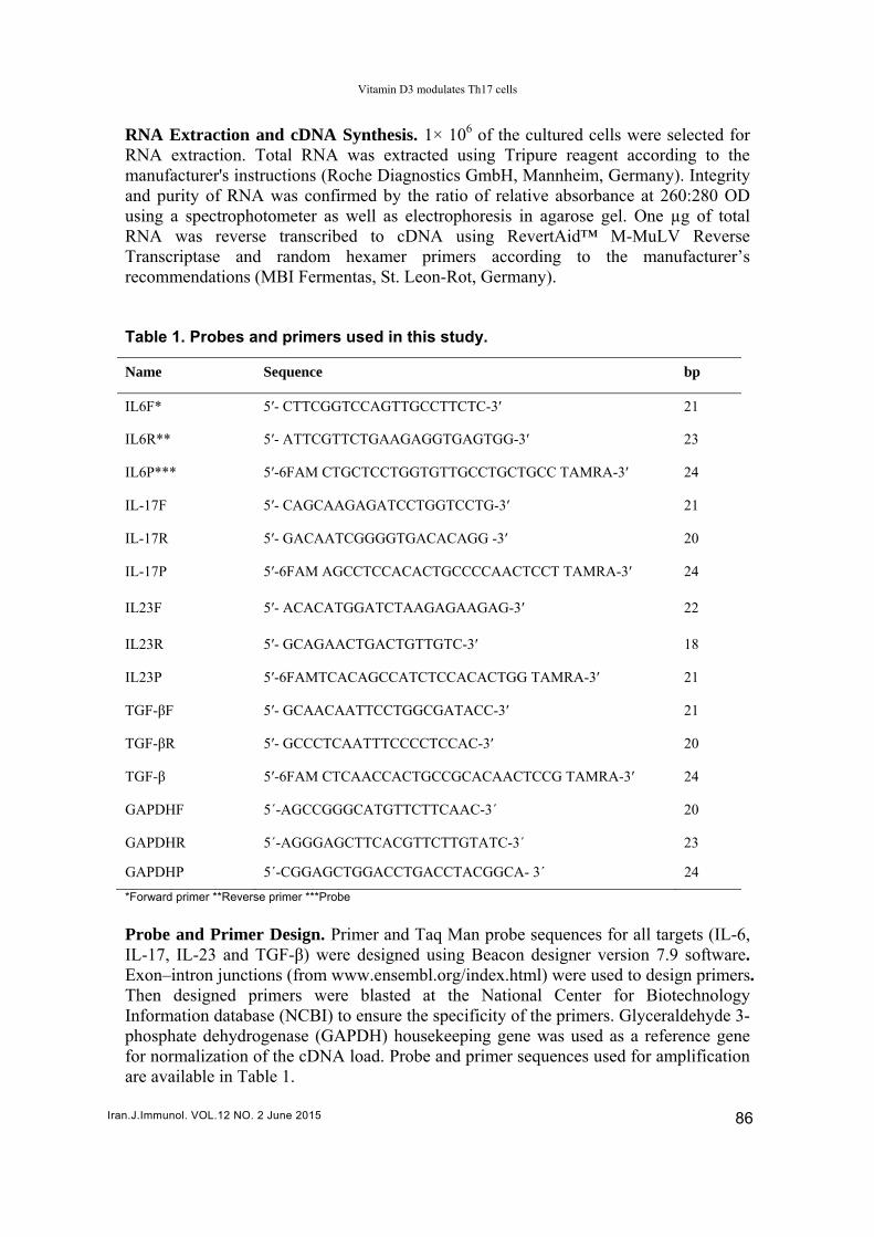
RNA Extraction and cDNA Synthesis. 1× 10⁶ of the cultured cells were selected for RNA extraction. Total RNA was extracted using Tripure reagent according to the manufacturer's instructions (Roche Diagnostics GmbH, Mannheim, Germany). Integrity and purity of RNA was confirmed by the ratio of relative absorbance at 260:280 OD using a spectrophotometer as well as electrophoresis in agarose gel. One µg of total RNA was reverse transcribed to cDNA using RevertAid™ M-MuLV Reverse Transcriptase and random hexamer primers according to the manufacturer’s recommendations (MBI Fermentas, St. Leon-Rot, Germany). Table 1. Probes and primers used in this study.
Name Sequence bp
IL6F* 5′- CTTCGGTCCAGTTGCCTTCTC-3′ 21
IL6R** 5′- ATTCGTTCTGAAGAGGTGAGTGG-3′ 23
IL6P*** 5′-6FAM CTGCTCCTGGTGTTGCCTGCTGCC TAMRA-3′ 24
IL-17F 5′- CAGCAAGAGATCCTGGTCCTG-3′ 21
IL-17R 5′- GACAATCGGGGTGACACAGG -3′ 20
IL-17P 5′-6FAM AGCCTCCACACTGCCCCAACTCCT TAMRA-3′ 24
IL23F 5′- ACACATGGATCTAAGAGAAGAG-3′ 22
IL23R 5′- GCAGAACTGACTGTTGTC-3′ 18
IL23P 5′-6FAMTCACAGCCATCTCCACACTGG TAMRA-3′ 21
TGF-βF 5′- GCAACAATTCCTGGCGATACC-3′ 21
TGF-βR 5′- GCCCTCAATTTCCCCTCCAC-3′ 20
TGF-β 5′-6FAM CTCAACCACTGCCGCACAACTCCG TAMRA-3′ 24
GAPDHF 5´-AGCCGGGCATGTTCTTCAAC-3´ 20
GAPDHR 5´-AGGGAGCTTCACGTTCTTGTATC-3´ 23
GAPDHP 5´-CGGAGCTGGACCTGACCTACGGCA- 3´ 24
*Forward primer **Reverse primer ***Probe
Probe and Primer Design. Primer and Taq Man probe sequences for all targets (IL-6, IL-17, IL-23 and TGF-β) were designed using Beacon designer version 7.9 software. Exon–intron junctions (from www.ensembl.org/index.html) were used to design primers. Then designed primers were blasted at the National Center for Biotechnology Information database (NCBI) to ensure the specificity of the primers. Glyceraldehyde 3-phosphate dehydrogenase (GAPDH) housekeeping gene was used as a reference gene for normalization of the cDNA load. Probe and primer sequences used for amplification are available in Table 1.

Reihani H, et al.
Iran.J.Immunol. VOL.12 NO. 2 June 2015 87
Real Time PCR and Analysis of Gene Expression. Quantitative Real time PCR was performed using Premix Ex Taq (Takara Bio, Otsu, Shiga, Japan) and specific primers on a Rotor-Gene® Q system (QIAGEN, Germany). After the completion of the cycles, the data were analyzed using the same system software. 2 –ΔΔCT (Livak) Method was used for the relative quantification of the target gene expression in each sample (21).The data were normalized to GAPDH as reference gene and Relative quantification of target gene in 1,25VitD3 treated sample was expressed as a fold change compared to the untreated sample. ΔCT treated=CT (target, treated)-CT (reference, treated)
CT untreated=CT (target, untreated)-CT (reference, untreated)Δ ΔΔCT=ΔCT treated-ΔCT untreated
Normalized expression ratio =2 –ΔΔCT
Statistical Analysis. IBM SPSS Statistics version 21 software was used for statistical analysis. Quantitative data were reported as Mean ± SD and the difference between the two groups was determined using the parametric paired t-test. P value <0.05 was considered statistically significant. RESULTS Demographic, Clinical and Laboratory Data. From 30 cases with SLE (27 women and 3 men), 12 patients were new cases (sampling performed before medical therapy) and 18 patients were in remission. The mean duration of disease in old cases was 5.2 ± 2.8 years. The mean age of the participants was 34.45 ± 9.33 years. Table 2. Collective demographic and serologic characteristics of the SLE patients in this study.
No of SLE patients 30
No of female patients 27
No of male patients 3
No of newly diagnosed patients 12
No of in remission patients 18
Mean age at time of study 34.45 ± 9.33 years
Positive Anti-dsDNA 88.4 %
C3 deficiency 23.0 %
C4 deficiency 26.9 %
Leucopenia 37.9 %
Lymphopenia 24.1 %
Thrombocytopenia 6.89 %
Proteinuria more than 500 mg/day 20.6 %

Vitamin D3 modulates Th17 cells
Iran.J.Immunol. VOL.12 NO. 2 June 2015 88
Table 2 shows the important demographic data of patients. Moreover, in the newly diagnosed patients the three main clinical presentations were arthritis (70.7%), nephritis (20%), and skin involvement (33.6%). 1,25VitD3 Decreases Percentage of Th17 Cells in Vitro. To assess the effect of 1,25VitD3 on the number of IL-17 producing cells, after cell culture and stimulation with PMA and ionomycin in the presence of brefeldin A, cells were analyzed using flowcytometry. Percentage of TCD3+ CD8- IL-17+cells in the PBMCs cultured in the presence of 1,25VitD3 significantly decreased (3.67 ± 2.43%) compared with PBMCs cultured in the absence of 1,25VitD3 (4.65 ± 2.75%; p=0.003)(Figure 3).
Figure 3. Percentage of Th17 cells in PBMCs of SLE patients, cultured in the presence of 50 nM 1,25VitD3 showed a statistically significant decrease compared with PBMCs cultured in the absence of 1,25VitD3.
1,25VitD3 Modulates the Expression of Th17 Related Cytokine Genes in Vitro. We studied whether the expression of Th17 related cytokine genes could be regulated by 1,25VitD3 in vitro. After real time PCR, relative expression levels (Relative Quantification) of IL-6, IL-17, IL-23 and TGF-β genes were calculated using the 2 –ΔΔCT method. With evaluation of gene expression level in PBMCs cultured in the presence and absence of 1,25VitD3, the relative expression level of TGF-β up regulated (1.38 fold) and the relative expression levels of IL-6 (50%), IL-17 (27%) and IL-23 (64%) down regulated after 1,25VitD3 treatment (Figure 4). Comparison of the Effects of 1,25VitD3 in Newly Diagnosed and in Remission SLE Patients. 1,25VitD3 treatment decreased the percentage of Th17 cells in PBMCs of both newly diagnosed and in SLE patients who were in remission, this decrease was significant in patients who were in remission (p=0.008) (Figure 5).

Iran.J.Imm
Figurrelativexpreof cytβ (1.3 Figurpatie
1,25VTGF(p=0expre(0.36Diffenot s
munol. VOL.12 N
0
1
2
3
4
5
6
7
Th
17 c
ells
(%
)
re 4. Relativve expressioession of TGtokines was 38 ± 0.24) fo
re 5. Percennts, cultured
VitD3 treatm- significa.008). In Session level6 fold) (p=0erence in thsignificant b
NO. 2 June 2015
p=0.21
New caspatients
ve expressioon of IL-6(50
GF-β (1.34-foas follows: Ild.
tage of Th17 in the prese
ment in newantly (2.11 fLE patientsl of IL-6 sig
0.023). he expressiobetween new
5
16
ses
on levels of 0%), IL-17(27old) up regulaL-6 (0.50 ± 0
7 cells in PBMence and abs
wly diagnosfold) in coms who weregnificantly
on levels ofwly diagnos
Reihani H, et a
I
genes after 7%), and IL-ated after 1,0.07), IL-17 (
MCs of newlysence of 50 n
sed SLE patmparison wie in remissi(0.85 fold)
f IL-17 andsed and in re
l.
p=0.008
n remissionpatients
treatment w-23(64%) do,25VitD3 trea(0.73 ± 0.02)
y diagnosed nM 1,25VitD3
tients increaith the in reon 1,25VitDcompared t
d IL-23 afteemission SL
n
vitami
Untrea
with 50 nM oown regulateatment. Rela), IL-23(0.36
and in remis3.
ased the expemission patD3 treatmeto newly di
er 1,25VitDLE patients
n D treate
ated
of 1,25VitD3d and the re
ative quantifi± 0.09) and
ssion SLE
pression levtients (1.04 nt decreaseagnosed pa
D3 treatmen(Figure 6).
89
3. The elative cation TGF-
vel of fold)
ed the atients
nt was

Vitamin D3 modulates Th17 cells
Iran.J.Immunol. VOL.12 NO. 2 June 2015 90
Figure 6. Relative expression levels of genes after treatment with 50 nM of 1,25VitD3 in newly diagnosed and in SLE patients who are in remission. In SLE patients who are in remission 1,25 VitD3 treatment decreased the expression level of IL-6 significantly compared to newly diagnosed patients (p=0.023). 1,25 VitD3 treatment in newly diagnosed SLE patients increased the expression level of TGF- significantly in comparison to in remission patients (p=0.008). Difference in the expression levels of IL-17 and IL-23 after 1,25VitD3 treatment was not sinificant between newly diagnosed and in remission SLE patients. DISCUSSION The main outcome of this study was that the active metabolite of vitamin D (1, 25(OH)D) strongly modulates Th17 cell-mediated immune responses. As a result, 1, 25 VitD3 can inhibit Th17 cells through different mechanisms in both gene expression and cytokine expression pathways. 1,25VitD inhibits differentiation and maturation of dendritic cells as well as B cells. 1,25VitD3 suppresses T cell proliferation and shifts Th1 cell phenotype to Th2 and also Th17 cell phenotype to Treg. To sum up, 1,25VitD3 changes inflammatory milieu to anti-inflammatory ones (22,23). However, there is still little knowledge about the molecular mechanisms underpinning the effects of 1,25VitD3 on the immune system. In this study, 1,25VitD3 significantly decreased the percentage of Th17 cells in vitro, which is consistent with previous studies. A Study demonstrated that culturing mouse cells in the presence of 1,25VitD3 reduced the number of IL-17 producing cells from 11 to 7% in Th17-inducing conditions and from 14 to 7% in Treg-inducing conditions (24). Direct regulatory effects of 1,25VitD3 on differentiation of T cells and inhibition of the generation of Th17 cells in vitro depends on vitamin D receptors (VDRs) expressed in the TCD4 cells and dose of 1,25VitD3 (25). 1,25VitD3 modulates development of Th17, Th1 and Th9 cells through inhibitory actions on these inflammatory cells and advances regulatory T cell development (26).

Reihani H, et al.
Iran.J.Immunol. VOL.12 NO. 2 June 2015 91
Some studies have shown that the influence of 1,25VitD3 on Th17 cells can be conducted by two mechanisms. The first one is direct inhibition of IL-17 production from TCD4 cells; the second one is modulating cytokine milieu of IL-1, IL-6, TGF-β1, TNF-α and IL-23 for indirect induction of Th17 cells (27). The present study showed that expression level of TGF-β is up regulated and conversely IL-6, IL-17 and IL-23 are down regulated relatively by 1,25VitD3 in vitro. These results are in agreement with previous reports about inhibitory effects of 1,25VitD3 on Th17-related cytokines. There are some other data on inhibitory effects of 1,25VitD3 on IL-17A production in rheumatoid arthritis, multiple sclerosis, and mouse model of MS (Experimental Autoimmune Encephalomyelitis) (28). 1,25VitD3 supresses IL-6 expression (29). IL-6 is a key cytokine for B cell differentiation into plasma cells. Linker-Israeli et al. in their study specified that reduced IgG production in SLE in the presence of 1,25VitD3 is mediated by reducing IL-6 (30). It has been found that imbalance between Th17 and Treg cells is a part of SLE pathogenesis. IL-6 plays a pivotal role in the interaction between Th17 and Treg and interacts with TGF-β to promote the development of Th17 cells (11). 1,25VitD3 and VDRs are a component of Th17/iTreg axis regulators. In the absence of 1,25VitD3 or its receptor, TCD4 cells produce more IL-17 (24). Mahon et al. proposed that 1,25VitD3 supplementation increases the serum levels of circulating TGF-β (31). Another study showed that 1,25VitD3 increases the expression level of TGF-β in the mouse model (32). In aggregate, it seems that 1,25VitD3 promotes Th17 cells to Treg cells by decreasing IL-6 and TGF-β. The current study also suggested that 1,25VitD3 reduces IL-6 gene expression in contrast to its effect on TGF- β gene. IL-23/IL-17 axis is also important in the pathogenesis of SLE and lupus nephritis (33,34). One study showed that T cells cultured by TGF-β1 and IL-6 are not pathogenic in the absence of IL-23. Our result was in accordance with other studies in which the inhibitory effect of 1,25VitD3 on IL-23 production was documented (35). The current study was not without limitations,however. Sample size was relatively small, but we enrolled 12 new cases who did not receive any medications. Moreover, other patients were treated with a minimum dose of immunomodulatory medications without any immunosuppressive drug. Assessment of the effects of 1,25VitD3 on Th17 cells in both gene expression and cytokine production levels using sensitive techniques can be considered a strong point in the scientific merit of this study. In conclusion, our data showed that 1,25VitD3 effectively modulates Th17 cell-mediated cytokines. Moreover, an optimal immunomodulatory dose of 1,25VitD3 without its potential side effects, especially hypercalcemia, is still a matter of debate (36). This study suggested that 50 nM of 1,25VitD3 has significant suppressive effects on Th17 cells and related cytokines. Therefore, further research on the mechanisms underlying the effects of 1,25VitD3 on cells involved in autoimmune disease along with the determination of effective doses will be warranted to designe therapeutic strategies to prevent and treat Th17 cell-mediated autoimmune diseases. ACKNOWLEDGEMENTS This article was extracted from the thesis prepared by Mr. Hadi Reihani to fulfill the requirements for earning the Immunology MS degree. The Research Council of

Vitamin D3 modulates Th17 cells
Iran.J.Immunol. VOL.12 NO. 2 June 2015 92
Mashhad University of Medical Sciences, Mashhad, Iran is appreciated for financially supporting this study, grant number [89782]. REFERENCES
1. Bettelli E, Carrier Y, Gao W, Korn T, Strom TB, Oukka M, et al. Reciprocal developmental
pathways for the generation of pathogenic effector TH17 and regulatory T cells. Nature. 2006; 441:235-8.
2. Laurence A, Tato CM, Davidson TS, Kanno Y, Chen Z, Yao Z, et al. Interleukin-2 signaling via STAT5 constrains T helper 17 cell generation. Immunity. 2007; 26:371-81.
3. Wong CK, Lit LC, Tam LS, Li EK, Wong PT, Law CW. Hyperproduction of IL-23 and IL-17 in patients with systemic lupus erythematosus: implications for Th17-mediated inflammation in auto-immunity. Clin Immunol. 2008; 127:385-93.
4. Yang L, Anderson DE, Baecher-Allan C, Hastings WD, Bettelli E, Oukka M, et al. IL-21 and TG-beta are required for differentiation of human TH17 cells. Nature. 2008; 454:350-2.
5. Shin MS, Lee N, Kang I. Effector T-cell subsets in systemic lupus erythematosus: update focusing on Th17 cells. Curr Opin Rheumatol. 2011; 23:444-8.
6. Mok CC, Lau CS. Pathogenesis of systemic lupus erythematosus. J Clin Pathol. 2003; 56:481-90. 7. Shah K, Lee WW, Lee SH, Kim SH, Kang SW, Craft J, et al. Dysregulated balance of Th17 and
Th1 cells in systemic lupus erythematosus. Arthritis Res Ther. 2010; 12: R53. 8. Crispin JC, Tsokos GC. IL-17 in systemic lupus erythematosus. J Biomed Biotechnol. 2010;
2010:943254. 9. Brkic Z, Corneth OB, van Helden-Meeuwsen CG, Dolhain RJ, Maria NI, Paulissen SM, et al. T
helper 17 cell cytokines and interferon type I: partners in crime in systemic lupus erythematosus? Arthritis Res Ther. 2014; 16:R62.
10. Yang J, Chu Y, Yang X, Gao D, Zhu L, Yang X, et al. Th17 and natural Treg cell population dynamics in systemic lupus erythematosus. Arthritis Rheum. 2009; 60:1472-83.
11. Kleczynska W, Jakiela B, Plutecka H, Milewski M, Sanak M, Musial J. Imbalance between Th17 and regulatory T-cells in systemic lupus erythematosus. Folia Histoc Cytobiol. 2011; 49:646-53.
12. Vincent FB, Northcott M, Hoi A, Mackay F, Morand EF. Clinical associations of serum interleukin-17 in systemic lupus erythematosus. Arthritis Res Ther. 2013; 15: R97.
13. Szodoray P, Nakken B, Gaal J, Jonsson R, Szegedi A, Zold E, et al. The complex role of vitamin D in autoimmune diseases. Scand J Immunol. 2008; 68:261-9.
14. Orbach H, Zandman-Goddard G, Amital H, Barak V, Szekanecz Z, Szucs G, et al. Novel biomarkers in autoimmune diseases: prolactin, ferritin, vitamin D, and TPA levels in autoimmune diseases. Ann N Y Acad Sci. 2007; 1109:385-400.
15. Attar SM, Siddiqui AM. Vitamin d deficiency in patients with systemic lupus erythematosus. Oman Med J. 2013; 28:42-7.
16. Sahebari M, Nabavi N, Salehi M. Correlation between serum 25(OH)D values and lupus disease activity: an original article and a systematic review with meta-analysis focusing on serum VitD confounders. Lupus. 2014; 23:1164-77.
17. Arnson Y, Amital H, Shoenfeld Y. Vitamin D and autoimmunity: new aetiological and therapeutic considerations. Ann Rheum Dis. 2007; 66:1137-42.
18. Wahono CS, Rusmini H, Soelistyoningsih D, Hakim R, Handono K, Endharti AT, et al. Effects of 1, 25 (OH) 2D3 in immune response regulation of systemic lupus erithematosus (SLE) patient with hypovitamin D. Int J Clin Exp Med. 2014; 7:22-31.
19. Feng X, Lv C, Wang F, Gan K, Zhang M, Tan W. Modulatory Effect of 1, 25-Dihydroxyvitamin D 3 on IL1β-Induced RANKL, OPG, TNFα, and IL-6 Expression in Human Rheumatoid Synoviocyte MH7A. Clin Dev Immunol. 2013; 2013:160123.
20. Jeffery LE, Burke F, Mura M, Zheng Y, Qureshi OS, Hewison M, et al. 1, 25-Dihydroxyvitamin D3 and IL-2 combine to inhibit T cell production of inflammatory cytokines and promote development of regulatory T cells expressing CTLA-4 and FoxP3. J Immunol. 2009; 183:5458-67.
21. Livak KJ, Schmittgen TD. Analysis of relative gene expression data using real-time quantitative PCR and the 2(-Delta Delta C(T)) Method. Methods. 2001; 25:402-8.
22. Aranow C. Vitamin D and the immune system. J Investig Med. 2011; 59:881-6.

Reihani H, et al.
Iran.J.Immunol. VOL.12 NO. ̀2 June 2015 93
23. Lang CL, Wang MH, Chiang CK, Lu KC. Vitamin D and the Immune System from the Nephrologist's Viewpoint. ISRN Endocrinol. 2014; 2014:105456.
24. Bruce D, Yu S, Ooi JH, Cantorna MT. Converging pathways lead to overproduction of IL-17 in the absence of vitamin D signaling. Int Immunol. 2011; 23:519-28.
25. Chang JH, Cha HR, Lee DS, Seo KY, Kweon MN. 1,25-Dihydroxyvitamin D3 inhibits the differentiation and migration of T(H)17 cells to protect against experimental autoimmune encephalomyelitis. PLoS One. 2010; 5:e12925.
26. Terrier B, Derian N, Schoindre Y, Chaara W, Geri G, Zahr N, et al. Restoration of regulatory and effector T cell balance and B cell homeostasis in systemic lupus erythematosus patients through vitamin D supplementation. Arthritis Res Ther. 2012; 14:R221.
27. Tian Y, Wang C, Ye Z, Xiao X, Kijlstra A, Yang P. Effect of 1,25-dihydroxyvitamin D3 on Th17 and Th1 response in patients with Behcet's disease. Invest Ophthalmol Vis Sci. 2012; 53:6434-41.
28. Joshi S, Pantalena LC, Liu XK, Gaffen SL, Liu H, Rohowsky-kochan C, et al. 1,25-dihydroxyvitamin D(3) ameliorates Th17 autoimmunity via transcriptional modulation of interleukin-17A. Mol Cell Biol. 2011; 31:3653-69.
29. Xue ML, Zhu H, Thakur A, Willcox M. 1 alpha,25-Dihydroxyvitamin D3 inhibits pro-inflammatory cytokine and chemokine expression in human corneal epithelial cells colonized with Pseudomonas aeruginosa. Immunol Cell Biol. 2002; 80:340-5.
30. Linker-Israeli M, Elstner E, Klinenberg JR, Wallace DJ, Koeffler HP. Vitamin D(3) and its synthetic analogs inhibit the spontaneous in vitro immunoglobulin production by SLE-derived PBMC. Clin Immunol. 2001; 99:82-93.
31. Mahon BD, Gordon SA, Cruz J, Cosman F, Cantorna MT. Cytokine profile in patients with multiple sclerosis following vitamin D supplementation. J Neuroimmunol. 2003; 134:128-32.
32. Cantorna MT, Woodward WD, Hayes CE, DeLuca HF. 1,25-dihydroxyvitamin D3 is a positive regulator for the two anti-encephalitogenic cytokines TGF-beta 1 and IL-4. J Immunol. 1998; 160:5314-9.
33. Zhang Z, Kyttaris VC, Tsokos GC. The role of IL-23/IL-17 axis in lupus nephritis. J Immunol. 2009; 183:3160-9.
34. Wong CK, Lit LC, Tam LS, Li EK, Wong PT, Lam CW. Hyperproduction of IL-23 and IL-17 in patients with systemic lupus erythematosus: implications for Th17-mediated inflammation in auto-immunity. Clin Immunol. 2008; 127:385-93.
35. Zhang H, Shih DQ, Zhang X. Mechanisms underlying effects of 1,25-Dihydroxyvitamin D3 on the Th17 cells. Eur J Microbiol Immunol (Bp). 2013; 3:237-40.
36. Prietl B, Treiber G, Pieber TR, Amrein K. Vitamin D and immune function. Nutrients. 2013; 5:2502-21.