Inflammation and Repair. General Vocabulary words Intracellular space Extracellular space...
84
Inflammation and Repair
-
Upload
sabina-summers -
Category
Documents
-
view
214 -
download
0
Transcript of Inflammation and Repair. General Vocabulary words Intracellular space Extracellular space...

Inflammation and Repair

General Vocabulary words
• Intracellular space• Extracellular space
– Vascular space– Interstitial space
• Read Lewis, 318 – 319– Hydrostatic Pressure– Oncotic Pressure– Fluid Shifts
• Edema


Capillary Permeability
Proteins can only leak out when there is increased capillary permeability

Lymphatics
• Lymphatic membrane increases in permeability– Allows for greater removal of interstitial fluid– Allows proteins and other substances into the
lymph drainage• Possible conduit for spreading infectious or toxic
agents

Factors Promoting Edema
• Increased Hydrostatic pressure– Hypertension– Fluid Overload (Renal, heart, or liver failure)– Increased Venous pressure (PVD, postural
blockage)• Decreased Oncotic Pressure
– Inhibited Protein production (liver disease, protein malnutrition)
– Capillary permeability (local inflammation)• Lymph obstruction

Factors Inhibiting Edema
• Hydrostatic Pressure– Compression– Drugs reducing fluid volume (diuretics)– Postural
• Oncotic Pressure– Colloids (natural or artificial albumin)– Reduce inflammation

Factors Affecting Edema

Inflammation
• Response of surrounding tissue to injury
• Allows substances in blood to enter the tissue (due to increased capillary permeability)– Antibodies, Complement, Clotting factors
• Purpose– Neutralize and eliminate offending agents– Destroy necrosed tissue– Prepare tissue for reapir

Features of Acute Inflammation
• Redness (Erythema)
• Heat
• Pain
• Swelling (Edema)
• Altered Function

Fluid Mechanism of Inflammation
• Dilation of local arterioles– Increased local blood flow and pressure
• Increase in vascular permeability– Leakage of protein
• Viscosity of local blood increases– Blood flow slows down– Allows white blood cells to enter the site of
injury

Cellular Aspects of Inflammation
• Margination and emigration (exit lane)– Allows leukocytes to exit the blood vessels
and enter the inflamed tissue– Synonyms: Extravasation, diapedesis
• Chemokines (chemoattractants)– Chemicals that attract leukocytes to the site of
inflammation– Process is called chemotaxis, gradient driven
• Cytokines– Chemicals that alter a cell’s function

Chemotaxis and Emigration

Inflammation vs Immunity
• Inflammation is nonspecific, nonadaptive• Immunity is specific (to select antigens),
adaptive• Inflammation allows immunity to happen• Immunity controls inflammation

Mediation of Inflammation
• Vasoactive amines – Histamine• Plasma enzyme products – Clotting
factors, complement, factor XII (Hageman)• Arachidonic acid metabolites –
prostaglandins, thromboxanes, leukotrienes
• Miscellaneous cell products – TNF, NO, selectins, integrins, ICAM, VCAM, interleukins

Mast Cell

Histamine Activity


Mediation Vocabulary
• Cytokine – substance that affects the way other cells function
• Zymogen – inactive storage form of an enzyme or other active substance. Examples:– Plasminogen plasmin– Fibrinogen fibrin– Pepsinogen pepsin

Leukocytes
• Common ancestor – bone marrow pluripotent hematopoeitic stem cell– Common Lymphoid Progenitor
• B cells, T cells, Natural Killer Cells
– Common Myeloid Progenitor• Erythrocytes, Macrophages, Granulocytes,
Dendritic Cells
• Progessive differentiation

Leukocytes

Monocytes-Macrophages
• Small quantities in the blood• Spend most of their life cycle in Tissues
– Tissue Macrophages may have other names• Liver – Kuppfer Cells• Nervous system – Microglial cells• Skin – Langerhans• Connective Tissue – Histiocytes
• Relatively long lived – weeks to months

Macrophage Functions
• Effector cell• Phagocytic• Antigen Presenting• Common Pathogen Feature Receptors
– Glucan, mannose, ligands, LPS
• Releases cytokines and chemokines• Granuloma – multinucleated giant cell

Antigen Processing and Presentation

Dendritic Cells
• Not to be confused with dendrites!!!• Relatively new discovery, 1973• Phagocytic and Macropinocytic
– Digest whatever is digested– Recognize digested pathogen features
including bacterial DNA, heat shock proteins, and viral RNA
• Antigen Presenting


Dendritic Cells’ Dual Role
• High levels of MHC – present antigens to T cells
• At end of life cycle or when activated, migrate to lymph nodes– Activate T cells against pathogenic antigens– Induce Tolerance to self antigens

Mast Cells
• Unknown blood precursor• Granulated cells
– Known to release at least 16 chemokines and cytokines
– Best known for Histamine
• Major function is to activate inflammation– Membrane Permeability– Leukocyte chemotaxis

Granulocytes
• Named for cytoplasmic granules– Neutrophils– Basophils– Eosinophils

Neutrophils
• Most numerous• Shortly lived – 6 hour half life in blood• Phagocytic• Primarily attack bacterial invaders• Bone marrow holds 100 times circulating
number of Neutrophils– Segmented Cells (segs) – fully mature– Banded Cells (bands) – slightly immature
• Neutropenia

Other Granulocytes
• Exocytic• Mostly distributed throughout tissues• Eosinophils
– Parasites– IgE Allergic reactions
• Basophils– Fungus

Lymphocytes
• Immune cells that control and direct inflammation
• Present in small numbers in acute exudates
• Large numbers in chronic inflammation• Destroy invaders• Prepare for tissue reparation

Lymphocytes
• B lymphocyte Plasma Cell antibodies• T lymphocytes
– CD8 cells: Cytotoxic (Killer) T Cells – kill viral infected cells
– CD4 cells: Helper T Cells (Types I and II) – direct B lymphocytes and macrophages
• (CD8 and CD4 are cell membrane proteins)

Lymphocyte Life Cycle
• Inactive (naïve) lymphocytes circulate through blood and lymph– T cells are activated by dendritic cells (and
occasionally macrophages)– B cells are activated by T cells
• Once activated, lymphocytes must – Proliferate (replicate, multiply, reproduce)– Differentiate (mature)
• Once threat is neutralized– Most undergo apoptosis– A few remain as Memory Cells

B lymphocytes
• Mature in Bone Marrow (Bone, B, B cell. Get it?)
• Naturally produce IgM antibody and display it on their cell membranes (M for Membrane, get it?)
• Proliferation and Maturation are directed by CD4 T helper cells
• Purpose of maturation is to improve the quality (affinity) of antibody produced

Antibodies
• Immunoglobulin• Variable region
– Somatic hyper-mutation
• C region– Mediates
inflammation
• Disulfide bondscan be cleaved

Immunoglobulin Polymers

Antibody Function
• Neutralization• Opsonization – “painting”• Activation of inflammation• Activation of complement• Antibody subtypes
– IgM – first produced, low affinity– IgD – no known function– IgA – crosses barriers placenta, milk, eyes– IgG – opsonin helps macrophages kill– IgE – eosinophils parasites and allergies

T Lymphocytes
• During childhood, T cells migrate to Thymus– TCR mutation and tolerance testing– Differentiation marked by CD8 and CD4 protein– CD8 binds to MHC I and marks Cytotoxic cells– CD4 binds to MHC II and marks Helper cells
• Further differentiate into Helper I and II cells

Activated T Cell Function
• Cytotoxic cells– Virally infected cells present viral antigen via
MHC I which binds to CD 8– The cytotoxic cell degranulates into the
infected cell, killing it
• Helper cells– Direct B cell maturation and Macrophages– TH1 are better at directing Macrophages– TH2 are better at directing B cells


Complement Cascade
• Consists of 9 zymogens– C1 – C9
• Three activation pathways• All end with C3 convertase• Cleaves C3 into C3a and C3b• C5 cleaves into C5a and C5b• C3b and C5b activate membrane attack
complex (MAC)• C3a and C5a act as cytokines and
chemokines

Figure 2-35


Complement activation pathways
• Classical - C1q binds– Directly to pathogen– CRP– Antibody-Antigen complex
• Mannose Binding Lectin• Alternative (spontaneous)

Complement Functions
• Kill Pathogens through MAC – (puncture them and let the guts spill out)
• Opsonize pathogens• Mediate inflammation through C3a and
C5a

Basic Immunophysiology
• Three intertwining processes– Inflammation– Adaptive response
• Cell mediated• Humoral

Non-specific response
• Pathogen recognition– Usually begins by recognizing common
pathogenic features– Initiates inflammatory response
• Brings effector cells to the site• Walls off infection• Prepares tissue for healing
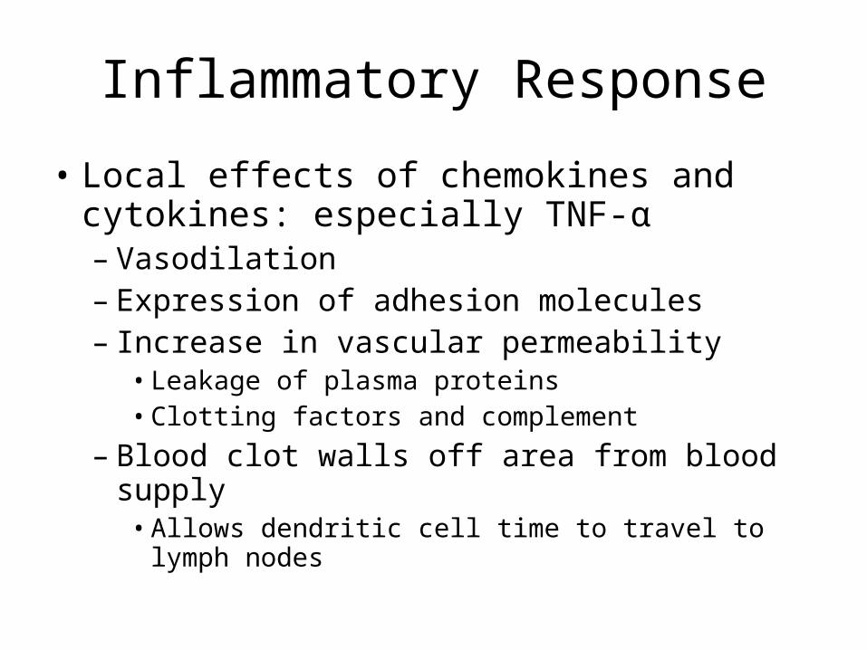
Inflammatory Response
• Local effects of chemokines and cytokines: especially TNF-α– Vasodilation– Expression of adhesion molecules– Increase in vascular permeability
• Leakage of plasma proteins• Clotting factors and complement
– Blood clot walls off area from blood supply• Allows dendritic cell time to travel to lymph nodes

Inflammatory Response
• Systemic effects – TNF-α, IL1-β, IL-6– Fever
• Inhibits pathogen growth• Enhances immune response• Protects body from TNF-α
– Acute Phase Response• Acute Phase Proteins released by liver
– CRP– MBL– Lung surfactants
– Leukocytosis– ↑ESR

Septic Shock – TNF-α run amok
• TNF-α– Vasodilation– Increases vascular permeability– Induces clotting
• TNF-α escapes into blood– Low blood pressure
• Vasodilation• Decreased plasma volume from vascular
permeability
– Disseminated intravascular coagulation (DIC)

Adaptive Immunity
• Cell Mediated – T Cells– CD8 – Always become cytotoxic T cells– CD4 – Must choose to become TH1 or TH2
• TH1 regulate macrophages– Activate macrophages– Kill infected macrophages– Regulate B cells
• TH2 regulate B cells
• Humoral Immunity – Antibodies– B cells – become Plasma cells and produce
antibodies

Memory
• Can take a month for full maturation of Plasma cells
• Memory cells are fully matured and developed effector cells– Quick response to infections– Suppress naïve immune cells– Do not require co-stimulation

Plasma Cells and Memory

Immunization
• Active – activates body’s immune system against invaders– Goal is formation of Memory cells
• Passive – injection of antibodies to offer limited support against an invader

Patterns of Inflammation
• Time factor– Acute– Chronic
• Types of Exudate– Serous (transudates)– Catarrhal (mucus)– Fibrinous (adhesions)– Purulent (furuncle, cellulitis)– Hemorrhagic (hematoma)

Inflammation vs Immunity

Fate of Inflammatory Reaction
• Resolution – Little damage• Repair – Moderate to Severe damage
– Regeneration – replacement of parenchyma– Scar formation – replacement of connective
tissue• Organization – proliferation of nearby connective
tissue into the damaged area• Granulation tissue• Collagen formation• Loss of vascularity

Inflammatory Phases

Wound Healing – Primary Intention
• Incision – Wound formation• Fibrin clot – prevents bleeding, acts as
glue to hold skin together• Inflammatory response builds
– Blood clot dissolved– Granulation tissue forms where clot was– Epithelium regenerates

Wound Healing Secondary Intention
• Skin edges cannot be held together• Similar to primary intention
– Takes longer– Involves more granulation tissue and
regeneration– May form underneath a scab– May show pinpoint bleeding


Factors affecting Inflammation
• Blood Supply– Elderly, Feet
• Bone marrow function• Protein synthesis – plasma and repair
– Liver Function– Nutrition
• Medication

Factors Affecting Wound Healing
• All from slide above• Necrotic or foreign tissue in wound• Wound infection• Excessive movement• Dehiscence – breaking open of a surgical
wound

Dehiscence

Hypersensitivity Reactions
• Damage done to the body as a result of immune reactions
• Sometimes called allergies• Four types of reactions
I. AnaphylacticII. CytotoxicIII. Immune ComplexIV. Cell-mediated

Anaphylactic
• Previously called immediate• Requires previous sensitization to antigen
– IgE is produced– IgE embeds in basophils and mast cells
• Upon subsequent exposure– Massive amounts of histamine released– Vasodilation and increased vascular perm
• Systemic– Laryngeal edema, Bronchospasm, seizures,
shock

Common Anaphylactic
• Insect stings• Penicillin• Pollen• Animal dander• Foods• Allergic rhinitis• Anigoedema and urticaria• Atopic Dermatitis• Asthma


Cytotoxic
• Antibodies bind to antigens on host cells• Host cells destroyed by
– Complement– Phagocytes (ADCC)
• Common Disorders– ABO blood rejection– Myasthenia gravis

Immune Complex
• Antibody binds with antigen – Immune complex
• Immune complex diffuses out of blood into tissue
• Complement cascade activates in the tissue causing inflammation/immune response
• Damage is collateral• Disorders: serum sickness, SLE, Stevens-
Johnson syndrome

Immune-Complex

Cell-Mediated
• TH1 cells stimulate Macrophage activity• Macrophages activity causes tissue
damage– If antigen is removed, reaction stops– If antigen persists, reaction continues and
granulomas may form• Common
– Allergic dermatitis: poison ivy, detergents, etc.– Tissue transplant rejection– Tuberculosis

Inflammation Tests
• Erythrocyte Sedimentation Rates• C-reactive protein: CRP
– hs-CRP
• Anti-nuclear antibodies (ANA)• WBC (with or without differential)• Skin tests• Ig levels

Anti-inflammatory and Anti-immune Drugs
• Anti-inflammatories– Inhibit prostaglandin – NSAIDS– Inhibit Leukotrienes – asthma drugs– Inihibt thromboxane – antiplatelet drugs– Antihistamines
• Anti-immune– Antiproliferative (Calcineurin inhibitors)– Cytotoxic
• Corticosteroids: both, depending on the dose

Calcineurin Inhibitors
• Calcineurin is needed to produce IL-2• Without IL-2, T-cells cannot proliferate, so
cannot mount an immune response• Used for transplant graft rejection• Drugs Cyclosporine: nephrotoxicity,
infection– Kidney, liver, heart transplant– Psoriasis, rheumatoid arthritis
• Tacrolimus (FK506): same

Cytotoxic Drugs
• Kill proliferating B and T cells• Are non-specific: kill all rapidly dividing
cells (red blood cells, skin, epithelial cells)• Azathioprine: Adjunct transplant• Cyclophosphamide: cancer, SLE, MS• Methotrexate: cancer, psoriasis, arthritis• Mycophenolate Mofetil: selective,
transplant

Glucocorticoids used for non-Endocrine purposes
• Pharmacologic Actions– Anti-inflammatory and Immune effects
• Inhibit prostaglandin, leukotriene, and histamine synthesis
• Suppress infiltration of phagocytes• Suppress proliferation of lymphocytes
– Effects on Metabolism and Electrolytes• Glucose levels rise• Protein synthesis suppressed• Fat deposits mobilized• Fewer electrolyte effects, but can inhibit calcium
absorption

Therapeutic Uses
• Rheumatoid Arthritis• SLE• Inflammatory Bowel Disease (IBD)• Miscellaneous Inflammatory D/Os• Allergic conditions (not acute anaphylaxis)• Asthma• Dermatologic disorders• Neoplams• Transplant rejection• Preterm infant

Immunosuppressive effect
• Cause lysis of activated B and T cells• Sequester T cells• Reduce IL-2 production• Reduce responsiveness to IL-1• Immunosuppressive doses are large, e.g.
– Methylprednisolone • Anti-immune doses: 500 – 1500mg (IV)• Anti-inflammatory doses: 5 – 60mg (IV)

Glucorticoids Adverse Effects
• Adrenal insufficiency
• Osteoporosis: long term therapy
• Infection
• Glucose intolerance
• Myopathy
• F&E disturbance
• Growth retardation
• Psychological disturbances

Glucorticoids Adverse Effects
• Cataracts and Glaucoma
• Peptic Ulcer Disease
• Iatrogenic Cushing’s Disease
• Ischemic Necrosis – especially caution with ETOH
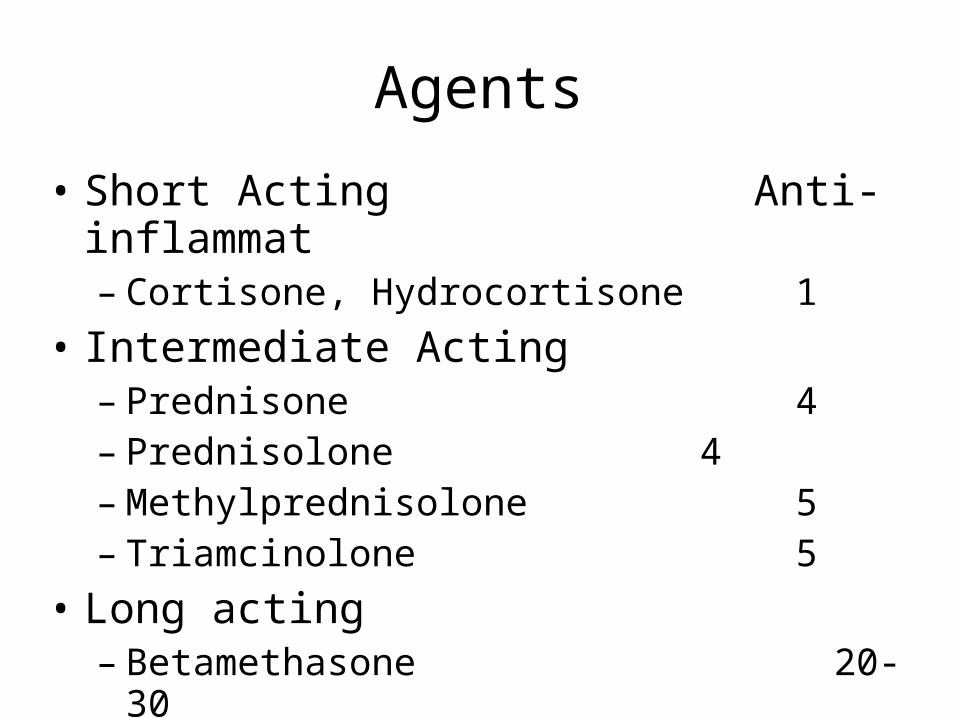
Agents
• Short Acting Anti-inflammat– Cortisone, Hydrocortisone 1
• Intermediate Acting– Prednisone 4– Prednisolone 4– Methylprednisolone 5– Triamcinolone 5
• Long acting– Betamethasone 20-30– Dexamethasone 20-30