Nuance AI-powered Intelligent Engagement Platform delivers ...

1
Proprietary & Confidential www.infervision.com
InferVision AI Research platform – InferScholar™ Empower Medical Research with Deep Learning and Radiomics
Medical Research in the Era of Big Data Recently, big data analysis has started to play a critical role in clinical research and practice. With the ever-
evolving technologies of information science, many hospitals have accomplished medical data digitalization,
which is the foundation for further development of big data research.
Big data, by definition, is not only data in large amounts, but also data possessing high dimensionality. For
example, to tell whether patients have bone fractures from 1000 cases using X-ray images, big data analysis has
to discover the relationship between the 3000 X 3000 pixels of all 1000 images and the pixel properties of
fractures. Because of high dimensionality of the data (3000 X 3000 characteristic variables, which is far beyond
the total sample amount of 1000), conventional statistical models, such as linear regression, cannot handle such
challenges. High dimensionality is usually seen in medical data and datasets such as medical imaging, electronic
medical records, and gene sequencing are all high dimensional.
Though the analysis cannot be handled by conventional statistical methods, deep learning provides advanced
solutions. Since the ImageNet competition in 2012, deep learning has become the primary method for non-
structural big data (such as image, voice and big text) analysis.
In the last 2 years, articles featuring deep learning methodology have frequently appeared in world-renowned
journals. To name a few; an article by Google using machine learning to recognize diabetes-related retinopathy
was published in JAMA in 2016; work from a Stanford team using deep learning to categorize skin cancer was
published as a cover article in Nature in 2017; and an article combining China’s doctors’ work was published as
the cover in Cell in 2018, demonstrating an AI system able to diagnose eye diseases and pneumonia based on
deep learning. According to Litjens et. al., from 2012 to 2016, over 300 articles about deep learning related to
medical imaging research have been published in journals and conferences, and the number has been increasing
exponentially (Figure 1). The methodology of deep learning has also been applied to the analysis of X-ray, CT,
MRI, pathology, electronic medical records, endoscopy and other medical big data formats; involving lesion
positions in the brain, chest, abdomen, cardiovascular system and bones.
The concept of radiomics was first proposed in 2012 by Dutch scientists. By using automated algorithms,
radiomics first obtains large amounts of feature information from the region of interests of medical images as
the research target, then extracts the critical information from the information using statistical analysis or data
mining methods (such as classical machine learning techniques including supporting vector machine, decision
tree and Bayesian algorithms), which is finally used for auxiliary diagnosis, prognosis and treatment monitoring.

2
Proprietary & Confidential www.infervision.com
The pipeline of radiomics can be summarized in the following steps: (1) imaging data collection, (2) region of
interest segmentation, (3) feature extraction, (4) classification and prediction.
Figure 1. Number of publications of medical imaging research based on deep learning.
A Brief Introduction to InferScholar™ Solid programming ability and integrative knowledge of math, statistics and computer science are usually
required for scholars to be engaged in deep learning and radiomics related research. However, accumulation of
such interdisciplinary knowledge typically requests long term and systematical academic training, which limits
the propagation and application of deep learning in different academic areas. Specifically, it limits the
application of radiomics in medical imaging related research in the era of big data.
The InferVision AI scholar platform InferScholar (Figure 2) provides a convenient visualizable big data analysis
tool for medical researchers. By integrating advanced and multiple deep learning radiomics algorithms, it
converts the modeling and programming into simplified interfaces, which enables the medical researchers who
are not familiar with computer programming the ability to launch deep learning and radiomics related research.
With the core concept of “empowering medical research with deep learning and radiomics”, InferScholar aims
to promote the methodology of deep learning and radiomics in medical imaging research.

3
Proprietary & Confidential www.infervision.com
Figure 2. InferVision AI research platform InferScholar™
InferScholar Functional Modules InferScholar integrates high performance deep learning computing servers, intelligence databases, labeling
tools, deep learning modeling systems, intelligence radiomics, deep learning training courses and customized
deep learning research services to empower hospitals to launch advanced deep learning and radiomics research;
enabling them to foster a new generation of medical researchers with interdisciplinary research abilities.

4
Proprietary & Confidential www.infervision.com
InferScholar Computation Server InferScholar’s computing server adopts the leading Pascal GPU architecture, which is equivalent to hundreds of
x86 servers and can complete tasks that would take conventional computing platforms over thousands of hours
within 1 hour.
1. The InferScholar server stack includes a deep learning framework, an NVIDIA GPU training system, a
deep learning SDK, an NVIDIA Docker, GPU drivers and CUDA, which can be used to design and evolve
deep neural networks quickly.
2. The adoption of a CUDA parallel computing platform significantly improves the efficiency of deep
learning algorithms.
3. This architecture is particularly good at handling parallel workloads, speeding up the evolving of neural
networks by 10 – 20 times and is more quiet than conventional servers.
4. With the integration of CoWoS techniques adopting HBM2 interface, RAM performance is improved by
over 300%.
5. Infervision’s unique page migration engine makes InferScholar more optimized for computation,
reducing efforts in data management.
6. InferScholar adopts the most advanced architecture from NVIDIA, which greatly improves performance
and power efficiency.
InferScholar Intelligence Database The InferScholar intelligence database (Figure 3) includes document management, DICOM information
documentation and storage, work list control, labeling information storage, information statistics, information
inquiry and monitoring, user management and log on controls. It integrates applicable programs and practical
tools for medical institutions and the core function utilizes DICOM standards. It supports CT, MR, X-ray,
mammography and other imaging modalities, with abundant and versatile functions. This product also provides
storage and inquiry services for medical imaging situations.

5
Proprietary & Confidential www.infervision.com
Figure 3. Intelligence Database.
InferScholar Intelligence Labeling Tools Supervised learning is currently the most mature deep learning technique. For supervised learning, the model
training and radiomics feature extractions are both dependent on the labeled data. This module provides
standardized, convenient and directly-visualizable labelling systems for categorization, image segmentation,
detection and radiomics analysis (Figure 4 & 5). Currently, the labeling tools can be used for:
▪ MR imaging lesion classification
▪ MR imaging lesion detection
▪ MR imaging segmentation
▪ CT imaging lesion classification
▪ CT imaging lesion detection
▪ CT imaging segmentation
▪ X-ray imaging lesion classification
▪ X-ray imaging lesion detection
▪ X-ray imaging segmentation
▪ Mammography lesion classification
▪ Mammography lesion detection
▪ Mammography segmentation
▪ Other DICOM imaging labeling tools can be customized according to needs

6
Proprietary & Confidential www.infervision.com
Figure 4. InferScholar’s CT intelligence detection labeling tool.
Figure 5. InferScholar’s CT intelligence segmentation tool
InferScholar Deep Learning Modeling System Based on the institution’s medical imaging research goal, related projects can be categorized as classification,
detection and segmentation; which are all closely related with diagnosis. Classification is usually the first
question to be answered by deep learning; that is, with the input of the whole or part of the medical images,
the computer will provide a single binary output telling whether lesions exists. Then, target or lesion detection
are the next critical steps during diagnosis, which is typically the most effort-consuming step for radiologists.
Finally, segmentation is important to precisely determine the target contour, shape and volume, which is also
key in clinical practice.

7
Proprietary & Confidential www.infervision.com
Figure 6. InferScholar’s intelligence modeling system interface.
The InferScholar modeling system integrates various classic and up-to-date deep learning algorithms, including:
▪ For classification: ResNet, Inception, DenseNet, etc.
▪ For detection: Faster-RCNN, SSD, YOLO, etc.
▪ For segmentation: Mask-RCNN, UNet, Deeplab, etc.
▪ The systems also supports customized model design, import and export.
Figure 7. Pulmonary nodule and the surrounding tissues segmented by InferScholar.
The modeling system is specifically tuned based on conventional 2D and 3D images, making it suitable for 2D
medical images such as X-ray and mammograph, and 3D modalities such as CT and MRI.

8
Proprietary & Confidential www.infervision.com
Tunable model parameters include:
▪ Training epochs
▪ Callbacks
▪ Validation interval
▪ Batch size
▪ Random seed
▪ Learning rate
▪ Optimization methods
▪ Data preprocessing
▪ Pretrained parameters
▪ Data encoding
▪ Data transformation
▪ Data augmentation
Figure 8. InferScholar’s interface for tuning deep learning model parameters.
InferScholar Intelligence Radiomics This module integrates the complete radiomics feature extraction tools, which can extract radiomics features
including, but not limited to, (1) first-order features, (2) shape features, (3) grey level co-occurrence matrix
features, (4) grey level size zone matrix features, (5) grey level run length matrix features, (6) neighboring grey

9
Proprietary & Confidential www.infervision.com
tone difference matrix features, and (7) grey level dependence matrix features. The module is suitable for both
2D and 3D images.
For the features mentioned above, except shape feature, they can be extracted from the original and processed
images. During extraction, these base features can be freely re-combined thus creating over 4000 features for
each image.
Figure 9. Pipeline of InferScholar Radiomics.
Figure 10. Pulmonary nodule malignancy prediction comparison between deep learning and radiomics using
InferScholar.
10
Proprietary & Confidential www.infervision.com
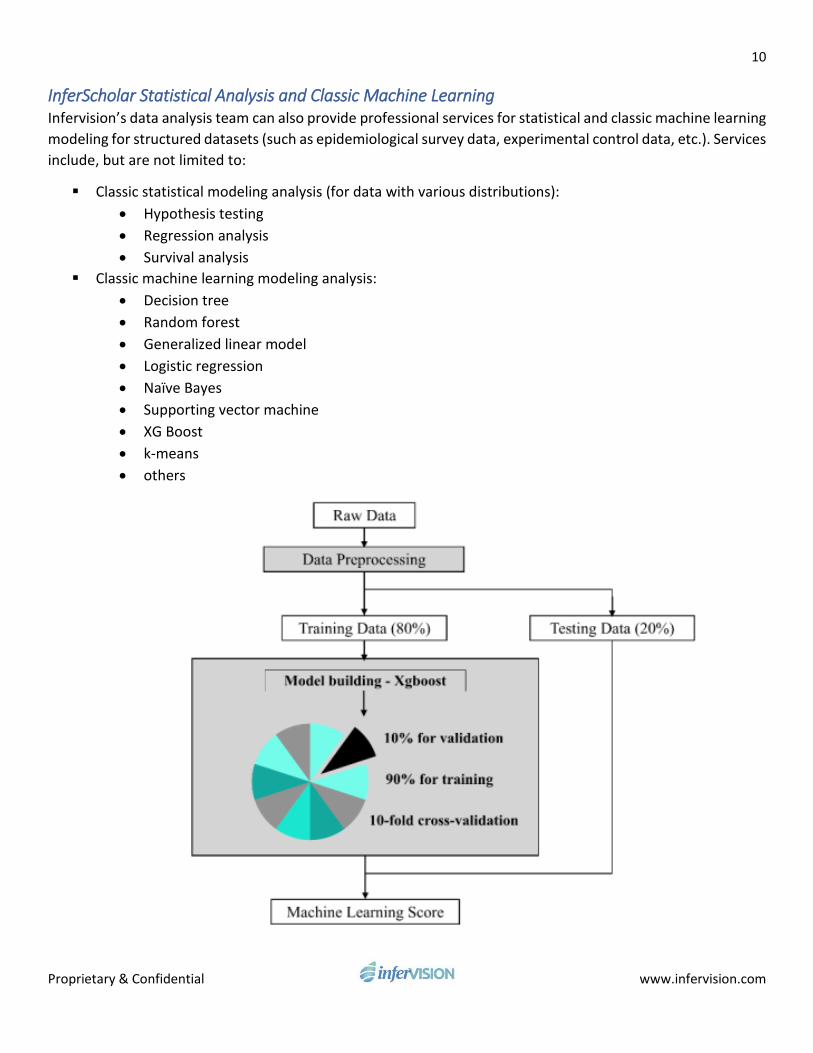
InferScholar Statistical Analysis and Classic Machine Learning Infervision’s data analysis team can also provide professional services for statistical and classic machine learning
modeling for structured datasets (such as epidemiological survey data, experimental control data, etc.). Services
include, but are not limited to:
▪ Classic statistical modeling analysis (for data with various distributions):
• Hypothesis testing
• Regression analysis
• Survival analysis
▪ Classic machine learning modeling analysis:
• Decision tree
• Random forest
• Generalized linear model
• Logistic regression
• Naïve Bayes
• Supporting vector machine
• XG Boost
• k-means
• others

11
Proprietary & Confidential www.infervision.com
Figure 11. Apply XG Boost algorithms assisting hospitals to predict coronary heart disease based on the
screening datasets.