infective Endocarditis - American Society of...
74
Infective Endocarditis Role of Echocardiography in Diagnosis and Management William K. Freeman, MD, FACC, FASE
Transcript of infective Endocarditis - American Society of...

Infective Endocarditis
Role of Echocardiography in
Diagnosis and Management
William K. Freeman, MD, FACC, FASE

DISCLOSURES
Relevant Financial Relationship(s)
None
Off Label Usage
None

Infective Endocarditis
Diagnosis
Major Duke Criteria
Positive
blood
cultures
Evidence of
endocardial
involvement
Durack DT, et al. Am J Med 1994; 96: 200
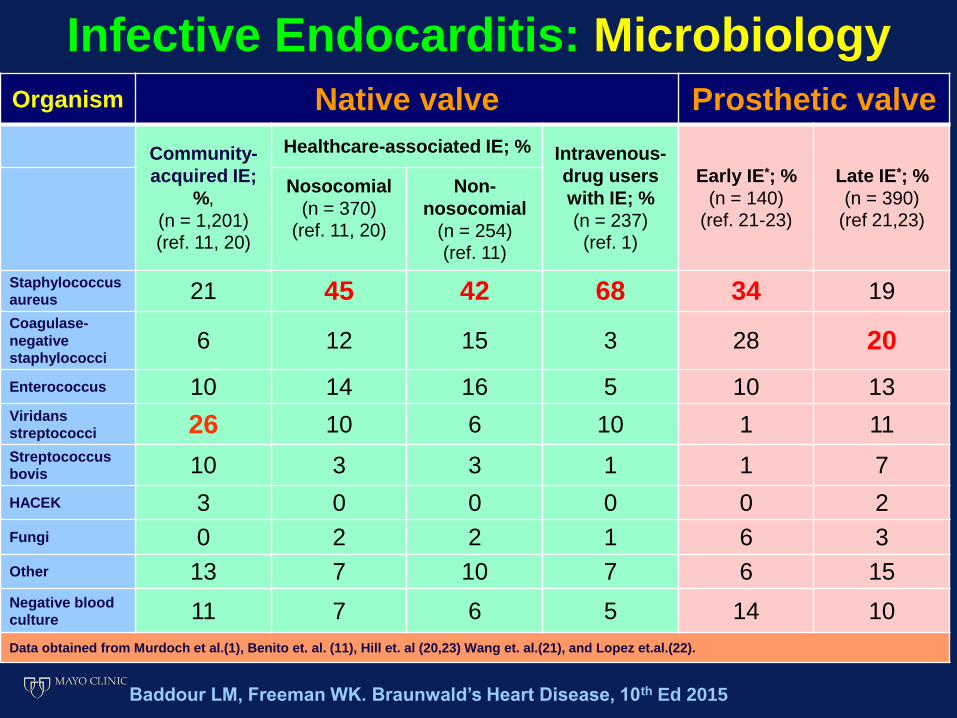
Organism Native valve Prosthetic valve
Community-
acquired IE;
%,
(n = 1,201) (ref. 11, 20)
Healthcare-associated IE; % Intravenous-
drug users
with IE; %
(n = 237) (ref. 1)
Early IE*; %
(n = 140) (ref. 21-23)
Late IE*; %
(n = 390) (ref 21,23)
Nosocomial
(n = 370) (ref. 11, 20)
Non-
nosocomial
(n = 254) (ref. 11)
Staphylococcus
aureus 21 45 42 68 34 19 Coagulase-
negative
staphylococci 6 12 15 3 28 20
Enterococcus 10 14 16 5 10 13 Viridans
streptococci 26 10 6 10 1 11 Streptococcus
bovis 10 3 3 1 1 7
HACEK 3 0 0 0 0 2
Fungi 0 2 2 1 6 3
Other 13 7 10 7 6 15 Negative blood
culture 11 7 6 5 14 10
Data obtained from Murdoch et al.(1), Benito et. al. (11), Hill et. al (20,23) Wang et. al.(21), and Lopez et.al.(22).
Baddour LM, Freeman WK. Braunwald’s Heart Disease, 10th Ed 2015
Infective Endocarditis: Microbiology

Infective Endocarditis Major Duke Criteria: Endocardial Involvement
Oscillating intracardiac mass, without
alternative anatomic explanation, involving:
• Valve
• Support structure
• Prosthetic/implanted device
• Path of regurgitant jet
Positive Echo for IE (Vegetation)
Durack DT, et al. Am J Med 1994; 96: 200

Infective Endocarditis Major Duke Criteria: Echocardiography
•Vegetation
•Peri-valvular abscess
•New partial dehiscence of prosthetic valve, or new regurgitation
Durack DT, et al. Am J Med 1994; 96: 200

Infective Endocarditis Minor Duke Criteria:
1. Predisposing cardiac conditions
2. IV drug abuse (also #1 cause for recurrent IE)
3. Fever ≥ 38.0°C; persistent, and otherwise unexplained
4. Blood culture positivity (Not meeting major criteria)
Durack DT, et al. Am J Med 1994; 96: 200
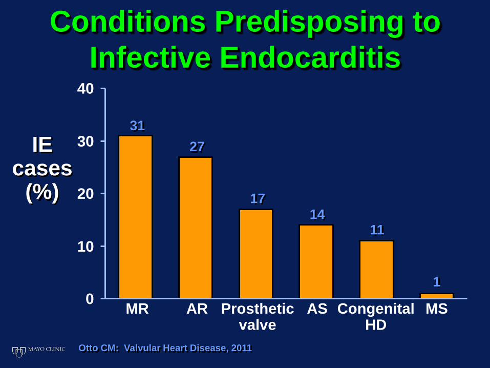
Conditions Predisposing to
Infective Endocarditis
Otto CM: Valvular Heart Disease, 2011
0
10
20
30
40
IE cases
(%)
31
MR
27
17 14
11
1
AR Prosthetic valve
AS CongenitalHD
MS

Infective Endocarditis Minor Duke Criteria:
• Conjunctival hemorrhage
• Janeway lesions
• Systemic arterial embolism
• Pulmonary embolism / infarction
• Mycotic aneurysm
• Intracranial hemorrhage
5. Vascular Phenomena
Durack DT, et al. Am J Med 1994; 96: 200

Infective Endocarditis Minor Duke Criteria:
• Diffuse glomerulonephritis
• Osler’s nodes
• Roth spots
• Rheumatoid factor +
6. Immunologic Phenomena
Durack DT, et al. Am J Med 1994; 96: 200
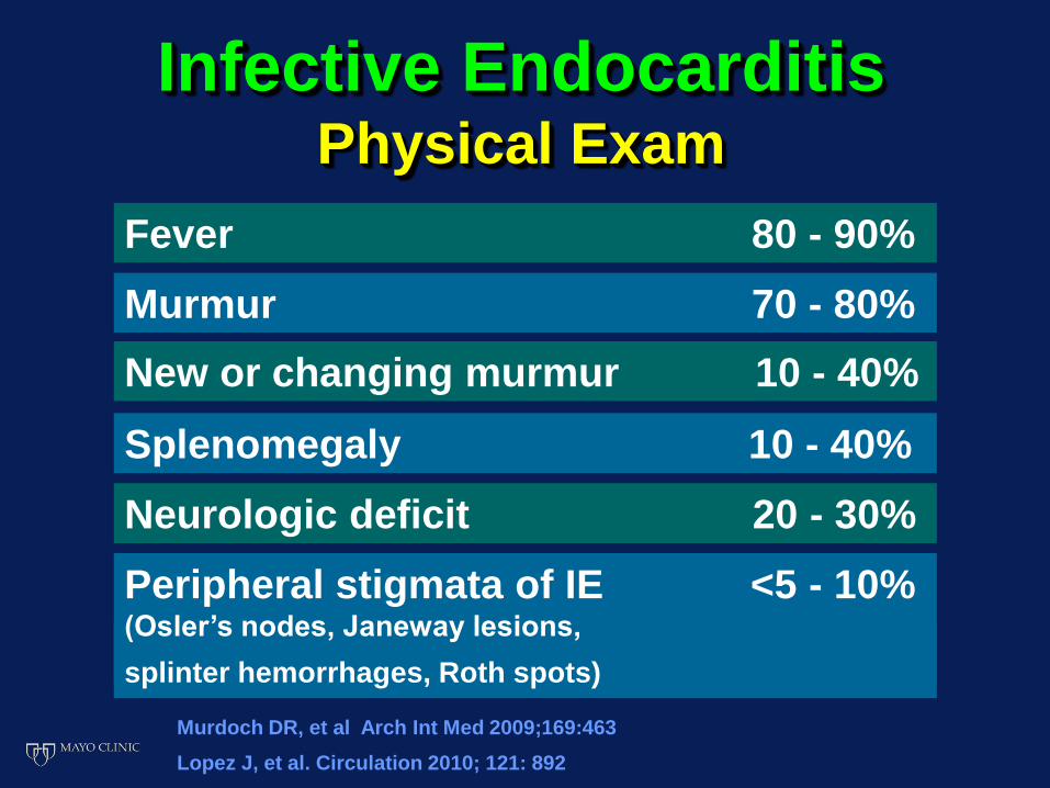
Infective Endocarditis Physical Exam
Fever 80 - 90%
Pain (chest, abdominal, back) 10-30%
Peripheral stigmata of IE <5 - 10% (Osler’s nodes, Janeway lesions,
splinter hemorrhages, Roth spots)
Murmur 70 - 80%
New or changing murmur 10 - 40%
Neurologic deficit 20 - 30%
Murdoch DR, et al Arch Int Med 2009;169:463
Splenomegaly 10 - 40%
Lopez J, et al. Circulation 2010; 121: 892
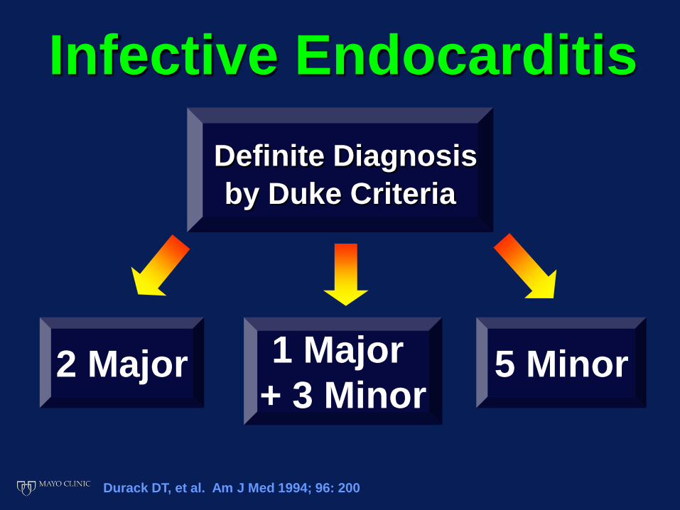
Infective Endocarditis
Definite Diagnosis
by Duke Criteria
1 Major
+ 3 Minor 2 Major 5 Minor
Durack DT, et al. Am J Med 1994; 96: 200
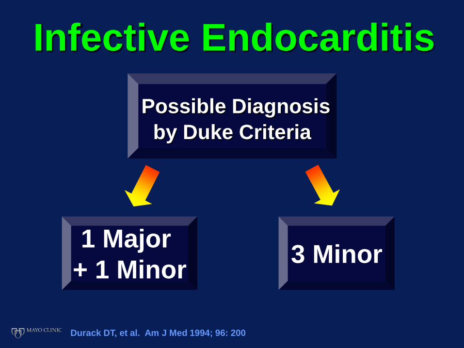
Infective Endocarditis
Possible Diagnosis
by Duke Criteria
1 Major
+ 1 Minor 3 Minor
Durack DT, et al. Am J Med 1994; 96: 200

Detection of Vegetations Transthoracic Echo (TTE)
Resolution size:
3 - 4 mm
Sensitivity: 62% - 82%*
Specificity: 91% - 100%
Pederson WR, et al. Chest 100:351, 1991
Jacob, S et al. Curr Opin Cardiol 17:478, 2002
*Casella, F et al. Echocardiography 26: 900, 2009
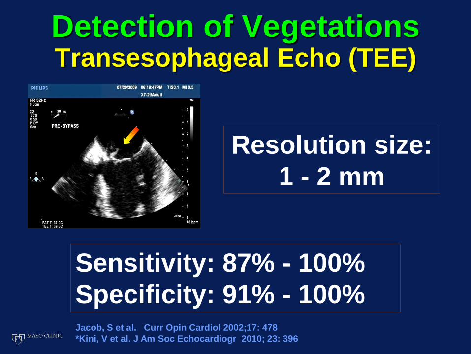
Detection of Vegetations Transesophageal Echo (TEE)
Resolution size:
1 - 2 mm
Sensitivity: 87% - 100%
Specificity: 91% - 100% Jacob, S et al. Curr Opin Cardiol 2002;17: 478
*Kini, V et al. J Am Soc Echocardiogr 2010; 23: 396
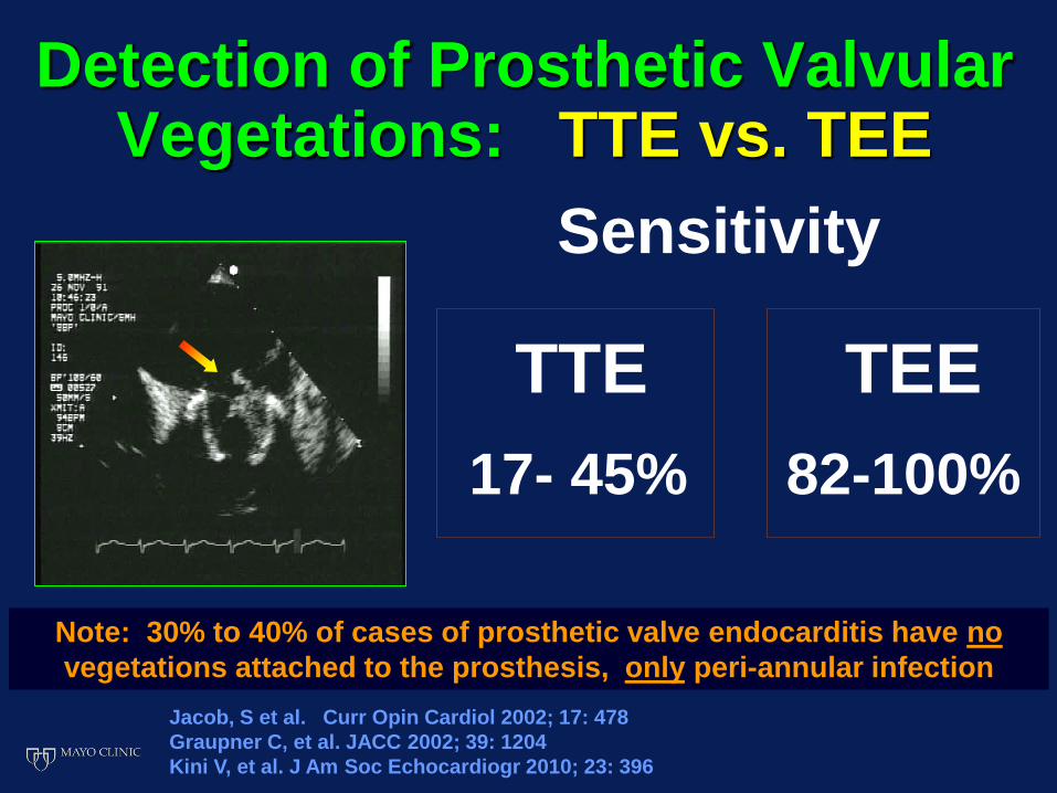
Detection of Prosthetic Valvular Vegetations: TTE vs. TEE
TTE TEE
17- 45% 82-100%
Sensitivity
Jacob, S et al. Curr Opin Cardiol 2002; 17: 478
Graupner C, et al. JACC 2002; 39: 1204
Kini V, et al. J Am Soc Echocardiogr 2010; 23: 396
Note: 30% to 40% of cases of prosthetic valve endocarditis have no
vegetations attached to the prosthesis, only peri-annular infection
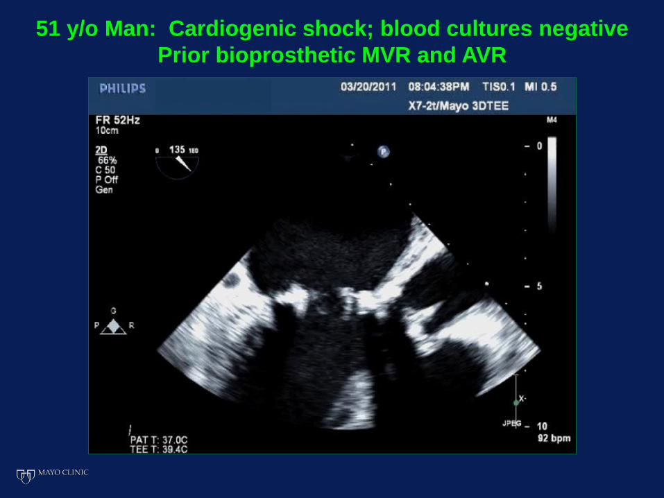
51 y/o Man: Cardiogenic shock; blood cultures negative
Prior bioprosthetic MVR and AVR

51 y/o Man: Cardiogenic shock; blood cultures negative
Prior bioprosthetic MVR and AVR

51 y/o Man: Cardiogenic shock; blood cultures negative
Prior bioprosthetic MVR and AVR

51 y/o Man: Cardiogenic shock; blood cultures negative
Prior bioprosthetic MVR and AVR

Suspected Infective Endocarditis
Low Initial
Patient Risk
High Initial
Patient Risk
Unexplained fever
Chronic murmur
No stigmata of IE
No high risk anatomy
No prosthesis or device
Significant new murmur
New heart failure
Prosthetic valve/CIED
Stigmata of IE / Prior IE
High risk anatomy
Staph aureus
Baddour LM, et al. Circulation 2005; 111:3167

Low Initial
Patient Risk High Initial
Patient Risk
Initial TTE Initial TEE
Limited Images
High Risk Findings
TEE
? LV Function
? Hemodynamics
TTE
Baddour LM, et al. Circulation 2005; 111:3167
Nishimura RA, CM Otto, et al. JACC 2014; 63: e57

High risk TTE Findings
Large / Mobile Vegetations
? Perivalvular extension of infection
Grade III-IV/IV Regurgitation
New LV dysfunction
Baddour LM, et al. Circulation 2005; 111:3167
Nishimura RA, CM Otto, et al. JACC 2014; 63: e57

1. 2.
3. 4.
Which patient most likely has infective endocarditis?

Lambl’s
excrescence,
fenestration
Mobile
Endocardial
Echodensity
Thrombus
(prosthesis)
Valvular
Strands
Vegetation:
Infective,
Noninfective
Sclerosis /
calcium
artifact
Ruptured/
retracted
chordae
Myxomatous
tissue
Papillary
fibroelastoma,
myxoma

Echocardiographic Diagnosis of Vegetation
Probable
Vegetation
Improbable
Vegetation
Texture: tissue density hyperrefractile
Location: upstream downstream
side of valve; side of valve
in jet trajectory

Echocardiographic Diagnosis of Vegetation
Probable
Vegetation
Improbable
Vegetation
Shape: lobulated filamentous,
to amorphous, discrete
multiple nodule
Assoc’d regurgitation,
findings: peri-valvular none
complications

Endothelial disruption
Valvular fibrin-platelet thrombus
Predisposing conditions
for endocarditis
Bacteremia
Infected valvular vegetation
Local tissue
destruction
Perivalvular
extension of
infection
Embolic
events
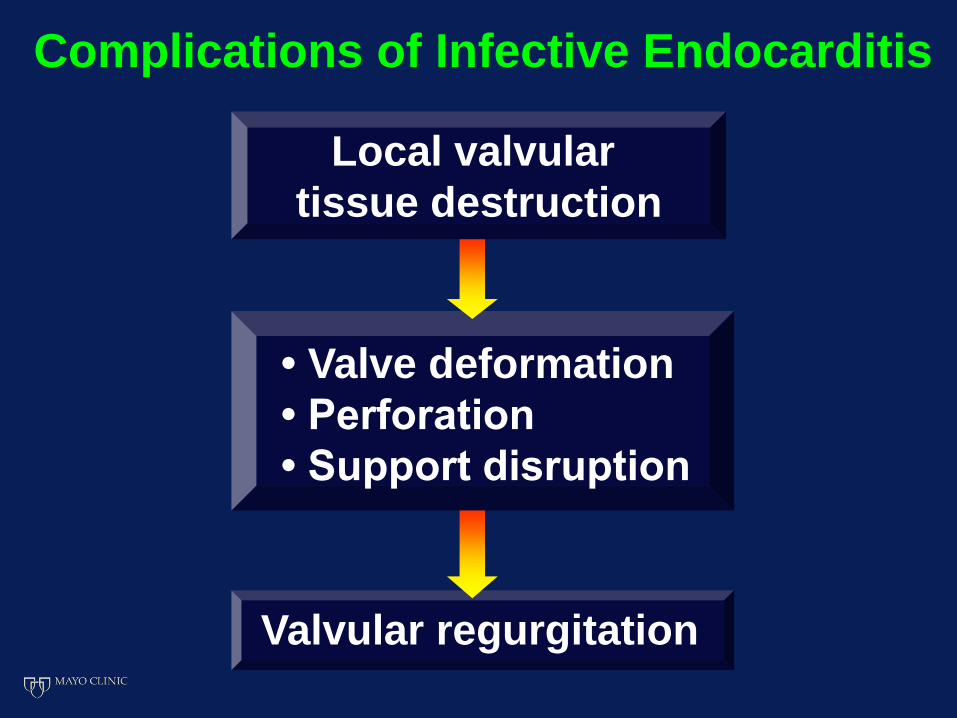
Complications of Infective Endocarditis
Local valvular
tissue destruction
• Valve deformation
• Perforation
• Support disruption
Valvular regurgitation
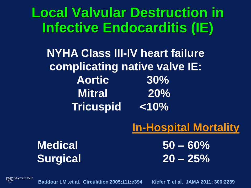
Local Valvular Destruction in Infective Endocarditis (IE)
NYHA Class III-IV heart failure
complicating native valve IE:
Aortic 30%
Mitral 20%
Tricuspid <10%
In-Hospital Mortality
Medical 50 – 60%
Surgical 20 – 25%
Baddour LM ,et al. Circulation 2005;111:e394 Kiefer T, et al. JAMA 2011; 306:2239
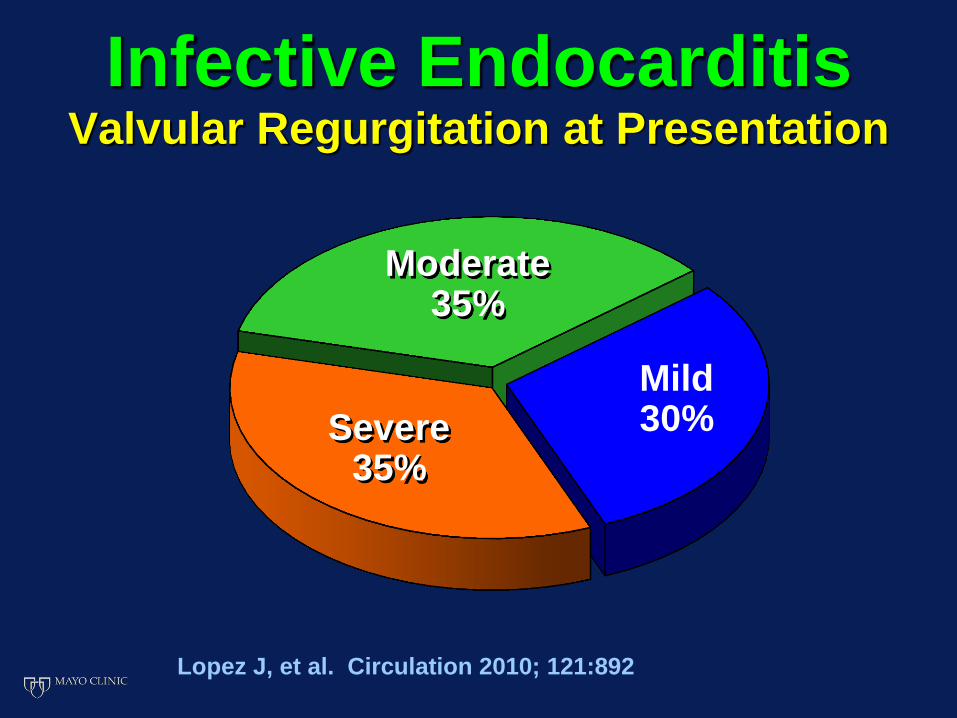
Infective Endocarditis Valvular Regurgitation at Presentation
Mild 30% Severe
35%
Moderate 35%
Lopez J, et al. Circulation 2010; 121:892

47 y/o Man: Staphylococcal bacteremia and shock

47 y/o Man: Staphylococcal bacteremia and shock

Acute Severe AR: Early mitral valve closure
MVC MVC

Valvular Perforation in Infective Endocarditis Sensitivity of TTE vs MP-TEE
Combined specificity: 98% for both TTE and MP-TEE
45
95
Combined Mitral Valve
64
100
%
100
80
60
40
20
0
Aortic Valve
27
91
TTE MP-TEE
DeCastro, S et al: Clin Infect Dis 30:825, 2000

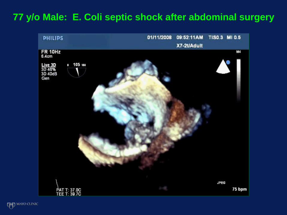
77 y/o Male: E. Coli septic shock after abdominal surgery

77 y/o Male: E. Coli septic shock after abdominal surgery
77 y/o Male: E. Coli septic shock after abdominal surgery
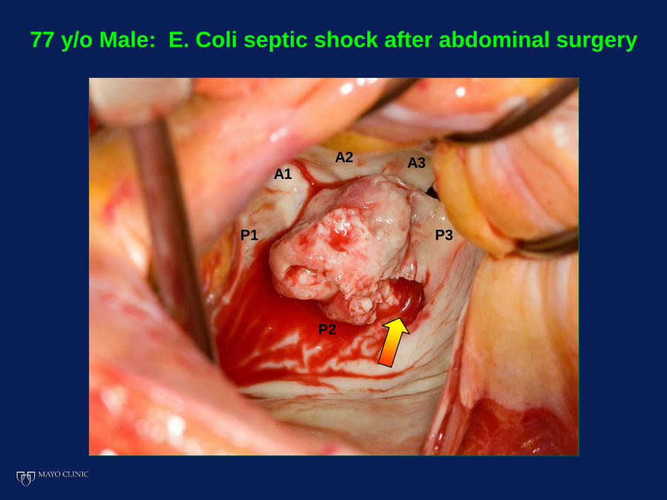
P1
P2
P3
A3 A2 A1
77 y/o Male: E. Coli septic shock after abdominal surgery

Valvular Dysfunction Complicating Infective Endocarditis
Class I Indication for Surgery
Early surgery* is indicated in patients with IE
who present with valve dysfunction (usually
severe regurgitation) resulting in symptoms
of heart failure
(*during initial hospitalization before completion
of a full therapeutic course of antibiotics)
Nishimura RA, CM Otto, et al. JACC 2014; 63: e57

Complications of Infective Endocarditis
Perivalvular Extension
of Infection (PVEI)
Phlegmon,
Abscess
Mycotic
aneurysm
Fistula,
Shunt

Perivalvular Extension
of Infection (PVEI)
Native valve IE 10 - 30%
Prosthetic valve IE 30 - 55%
Independent risk factors for PVEI:
Aortic position
Prosthetic valve
Staphylococcal infection
Graupner C, et al. JACC 2002; 39:1204
Habib G, et al. Eur Heart J 2009; 2369
Lopez J, et al. Circulation 2010; 121:892
Increased in-hospital mortality: 2-3 x
Murdoch DR, et al Arch Int Med 2009;169:463

Perivalvular Extension of Infection TTE vs TEE (118 Patients)
Daniel, WG et al: NEJM 1991; 324: 795
0
20
40
60
80
100
TTE TEE
%
28
99
87 95
Sensitivity Specificity

AV
MV
LA Ao
Peri-valvular Extension of Infection: Mitral - Aortic Intervalvular Fibrosa (MAIF)

AV
MV
LA Ao
Peri-valvular Extension of Infection: Mitral - Aortic Intervalvular Fibrosa (MAIF)

AV
MV
LA Ao
Peri-valvular Extension of Infection: Mitral - Aortic Intervalvular Fibrosa (MAIF)

Sudhakar, S et al. J Am Soc Echocardiogr 23:1009, 2010
Mitral - Aortic Intervalvular Fibrosa (MAIF)
Mycotic Pseudoaneurysm: Presentation
Persistent signs
of infection (39%)
Prosthetic valve
dysfunction (12%)
Heart
failure
(16%)
Embolism (12%)
Chest pain (10%)
Asymptomatic (9%)
Chest mass (1%)
Sudden
death (1%)

Bioprosthetic AVR: Coag Negative Staph bacteremia
MAIF phlegmon

53 y/o Male: Fever, weight loss, and dyspnea;
Aggregatibacter Aphrophilus bacteremia
10/22/13

53 y/o Male: Fever, weight loss, and dyspnea;
Aggregatibacter Aphrophilus bacteremia
Persistent fever despite antibiotic therapy
10/30/13

Infective Endocarditis: Follow-up Imaging Reevaluation with TEE/TTE
Class I Indications
• A significant change in symptoms and/or clinical findings
• Evidence of persistent infection after ≥5 days of appropriate antibiotic therapy
• Initial imaging evidence of extensive/high risk infection (i.e., large, mobile vegetations)
• Infection with aggressive/resistant organisms (staphylococcal, enterococcal, fungal)
Nishimura RA, CM Otto, et al. JACC 2014; 63: e57

48 y/o Man: Fever and syncope s/p Carbomedics
AVR one month ago; enterococcal bacteremia
MAIF mycotic aneurysm

52 y/o IV Drug Abuser: Prior CE AVR, fever/chills x 3 wks,
H. Parainfluenza bacteremia

52 y/o IV Drug Abuser: Prior CE AVR, fever/chills x 3 wks,
H. Parainfluenza bacteremia

Fever x 4 wks, SJ AVR (2005); Propionibacterium bacteremia
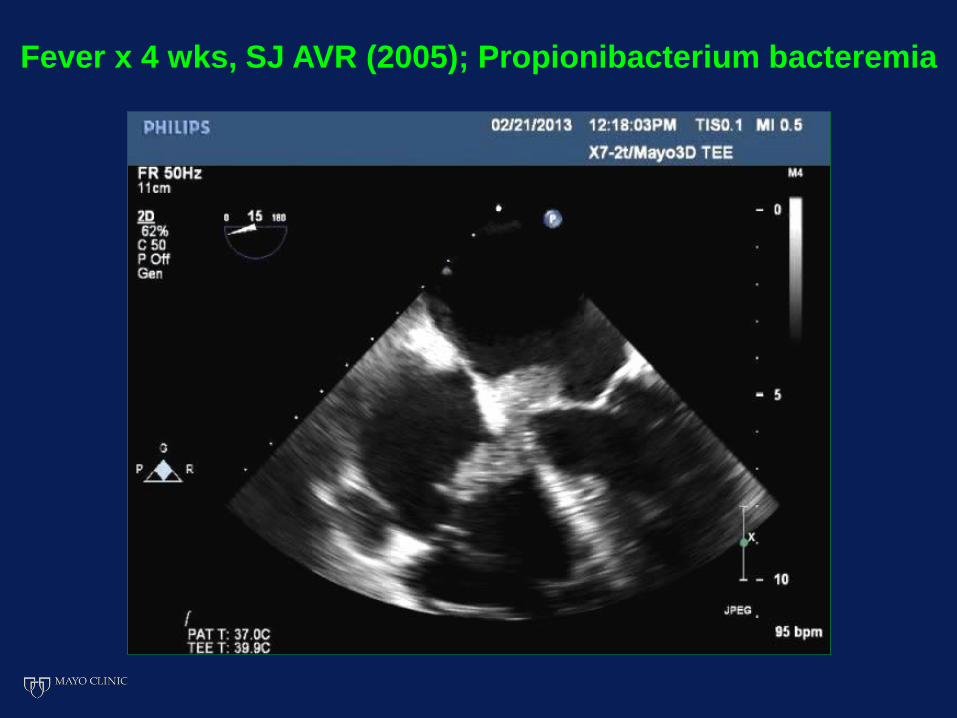
Fever x 4 wks, SJ AVR (2005); Propionibacterium bacteremia

Fever x 4 wks, SJ AVR (2005); Propionibacterium bacteremia

Perivalvular Extension of Infection Complicating Infective Endocarditis
Class I Indication for Surgery
Early surgery* is indicated in patients with IE
complicated by heart block, annular or aortic
abscess, or destructive penetrating lesions
(*during initial hospitalization before completion
of a full therapeutic course of antibiotics)
Nishimura RA, CM Otto, et al. JACC 2014; 63: e57
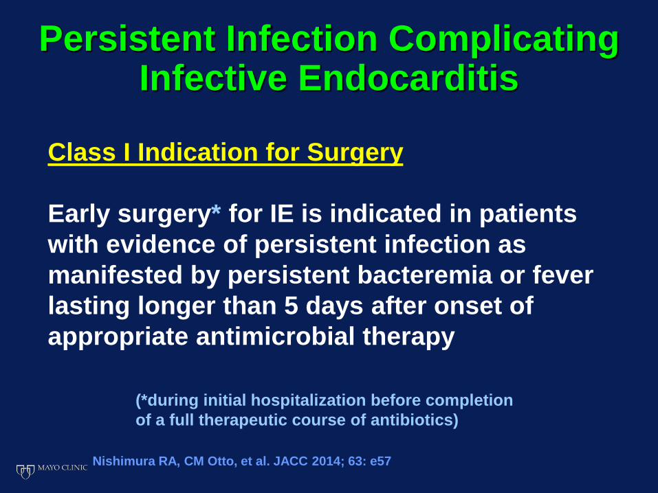
Persistent Infection Complicating Infective Endocarditis
Class I Indication for Surgery
Early surgery* for IE is indicated in patients
with evidence of persistent infection as
manifested by persistent bacteremia or fever
lasting longer than 5 days after onset of
appropriate antimicrobial therapy
(*during initial hospitalization before completion
of a full therapeutic course of antibiotics)
Nishimura RA, CM Otto, et al. JACC 2014; 63: e57

Aggressive or Resistant Organisms Complicating Infective Endocarditis
Class I Indication for Surgery
Early surgery* is indicated in patients with
left-sided IE caused by S. aureus, fungal, or
other highly resistant organisms
(e.g., Pseudomonas species, VRE, Brucella)
(*during initial hospitalization before completion
of a full therapeutic course of antibiotics)
Nishimura RA, CM Otto, et al. JACC 2014; 63: e57
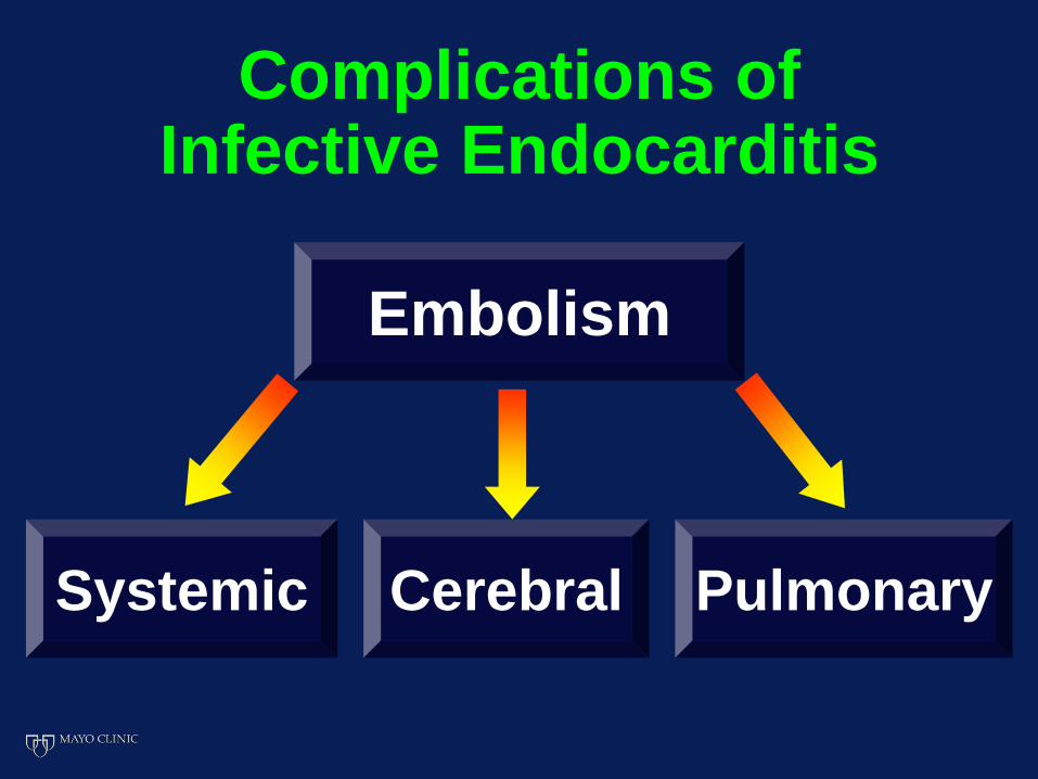
Complications of Infective Endocarditis
Systemic Cerebral Pulmonary
Embolism

Embolism in Infective Endocarditis
Clinically silent embolism: 15 – 25%
Baddour LM ,et al. Circulation 2005;111:e394
Habib G, et al. Eur Heart J 2009; 2369
Incidence of embolic events: 20 – 50%
Murdoch DR, et al Arch Int Med 2009;169:463
Thuny F, et al. Circulation 2005; 112:69
Clinically evident stroke: 10 – 20%
Screening brain MRI (12% with CNS Sxs) Acute ischemic lesions - 52%, mycotic aneurysm – 8%
Duval X, et al. Ann Intern Med 2010; 152: 497

Embolic Event Risk in Infective Endocarditis Multiplane TEE (178 Patients)
Di Salvo G et al: JACC 2001; 37: 1069
Em
bo
lic e
ve
nts
(%
)
90
80
70
60
50
40
30
20
10
0 Absent <10 10-15 >15
Vegetation size (mm)
Absent Mild Moderate Severe
Vegetation mobility

Embolic Event Risk in Infective Endocarditis
• Large vegetations (length > 10 mm)
• Highly mobile vegetation(s)
• Anterior mitral valve leaflet location
Independent structural predictors:
Independent microbiologic predictors:
• Staph aureus or Strep bovis infection
• Delay in appropriate antibiotic therapy
Habib G, et al. Eur Heart J 2009; 30:2369 Vilacosta I et al: JACC 39:1489, 2002
Thuny F et al: Circulation 112:69, 2005 Di Salvo G et al: JACC 37:1069, 2001

Embolism Complicating Infective Endocarditis Multicenter European Study (384 Patients)
34
7
27
0
10
20
30
40
%
Total Before antibiotic therapy
On antibiotic therapy
Thuny F et al: Circulation 2005; 112: 69

Systemic Embolism Complicating Infective Endocarditis
Class IIa Indication for Surgery
Early surgery* is reasonable in patients with
IE who present with recurrent emboli and
persistent vegetations despite appropriate
antibiotic therapy
(*during initial hospitalization before completion
of a full therapeutic course of antibiotics)
Nishimura RA, CM Otto, et al. JACC 2014; 63: e57
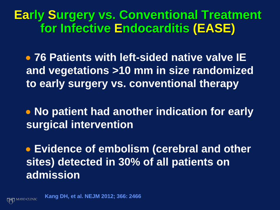
Early Surgery vs. Conventional Treatment for Infective Endocarditis (EASE)
76 Patients with left-sided native valve IE
and vegetations >10 mm in size randomized
to early surgery vs. conventional therapy
No patient had another indication for early
surgical intervention
Evidence of embolism (cerebral and other
sites) detected in 30% of all patients on
admission
Kang DH, et al. NEJM 2012; 366: 2466

Early Surgery vs. Conventional Treatment for Infective Endocarditis (EASE)
Kang DH, et al. NEJM 2012; 366: 2466
Conventional
Therapy (n=39)
Early Surgery
(n=37)
Embolic event at 6 wks 21% 0%
In-hospital death 3% 3%
Infecting organism Streptococcal species 56% 57%
Staph Aureus 13% 8%
Culture negative 18% 27%
Other 13% 8%

Systemic Embolism Complicating Infective Endocarditis
Class IIb Indication for Surgery
Early surgery* may be considered in patients
with native valve infective endocarditis who
exhibit mobile vegetations greater than
10 mm in length (with or without clinical
evidence of embolic phenomenon)
*during initial hospitalization before completion
of a full therapeutic course of antibiotics
Nishimura RA, CM Otto, et al. JACC 2014; 63: e57

74 y/o Female: Postoperative abdominal wound infection,
Corynebacterium bacteremia, no embolic events

74 y/o Female: Corynebacterium bacteremia cleared,
day 14 of antibiotic therapy; no embolic events

74 y/o Female, readmitted 7 mos later: Abdominal panniculitis
with streptococcal bacteremia; still no embolic events
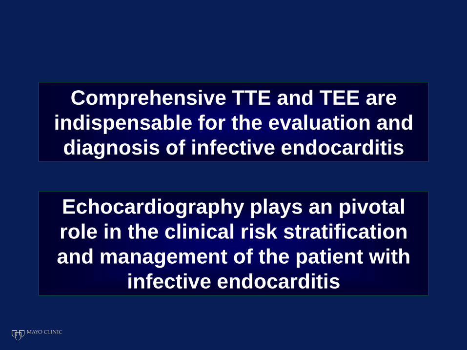
Comprehensive TTE and TEE are
indispensable for the evaluation and
diagnosis of infective endocarditis
Echocardiography plays an pivotal
role in the clinical risk stratification
and management of the patient with
infective endocarditis