Infectious myonecrosis virus has a totivirus-like, 120...
6
Infectious myonecrosis virus has a totivirus-like, 120-subunit capsid, but with fiber complexes at the fivefold axes Jinghua Tang a , Wendy F. Ochoa a,1 , Robert S. Sinkovits a , Bonnie T. Poulos b,2 , Said A. Ghabrial c , Donald V. Lightner b,3 , Timothy S. Baker a,d,3 , and Max L. Nibert e,3 a Department of Chemistry and Biochemistry and d Division of Biological Sciences, University of California at San Diego, La Jolla, CA 92093; b Aquaculture Pathology Laboratory, Department of Veterinary Science and Microbiology, University of Arizona, Tucson, AZ 85721; c Department of Plant Pathology, University of Kentucky, Lexington, KY 40546; and e Department of Microbiology and Molecular Genetics, Harvard Medical School, Boston, MA 02115 Edited by Reed B. Wickner, National Institutes of Health, Bethesda, MD, and approved September 18, 2008 (received for review July 11, 2008) Infectious myonecrosis virus (IMNV) is an emerging pathogen of penaeid shrimp in global aquaculture. Tentatively assigned to family Totiviridae, it has a nonsegmented dsRNA genome of 7,560 bp and an isometric capsid of the 901-aa major capsid protein. We used electron cryomicroscopy and 3D image reconstruction to examine the IMNV virion at 8.0-Å resolution. Results reveal a totivirus-like, 120-subunit T 1 capsid, 450 Å in diameter, but with fiber complexes protruding a further 80 Å at the fivefold axes. These protrusions likely mediate roles in the extracellular transmission and pathogenesis of IMNV, capabilities not shared by most other totiviruses. The IMNV structure is also notable in that the genome is centrally organized in five or six concentric shells. Within each of these shells, the densities alternate between a dodecahedral frame that connects the threefold axes vs. concentration around the fivefold axes, implying certain regularities in the RNA packing scheme. dsRNA virus electron cryomicroscopy nonenveloped virus penaeid shrimp Totiviridae I nfectious myonecrosis virus (IMNV) was first isolated from whiteleg shrimp, Litopenaeus vannamei, from aquaculture farms in northeast Brazil (1, 2). The associated disease was characterized by skeletal muscle necrosis, most markedly in distal abdomen and tail, with mortality over the harvest cycle nearing 70%. Purified IMNV virions reproduce this disease in pathogen-free L. vannamei (2). Diagnostics are available using reverse transcription and PCR to distinguish IMNV from other RNA viruses of shrimp (3, 4), and a survey from Pernambuco, Brazil, detected IMNV in 9 of 11 farms (5). IMNV has also been detected in L. vannamei from Indonesian farms, probably after transfer of aquaculture stocks (4). The genome sequence and many features of IMNV have been reported (2). Negatively stained virions exhibit an isometric capsid with a diameter of 400 Å. The virions contain a major capsid protein (MCP) of relative molecular weight (M r ) 106,000 and an N terminus of sequence IVSMENQSEID as shown by Edman deg- radation. The genome, a single molecule of 7,560-bp dsRNA, contains two extended ORFs in different frames of the plus strand: ORF1 in frame 1 (nucleotides 136-4953) and ORF2 in frame 3 (nucleotides 5241–7451). ORF1 encodes a 1,605-aa protein that includes the N-terminal sequence of the MCP starting at amino acid 705. The protein spanning amino acids 705-1605 have a sequence- predicted mass of 99 kDa, consistent with the M r of the MCP. The MCP thus seems to be cleaved from a larger precursor. A 60-aa region at the N terminus of ORF1 shares sequence similarities with dsRNA-binding proteins. ORF2 encodes a 736-aa protein that contains typical motifs of an RNA-dependent RNA polymerase (RdRp). Proteins representing ORF2 and the first 704 aa of ORF1 have yet to be identified, although candidate minor proteins have been seen in denaturing gels of IMNV virions. Phylogenetic anal- yses link IMNV to members of the family Totiviridae of nonseg- mented dsRNA viruses with isometric capsids, and most closely to Giardia lamblia virus (GLV) (2, 6). IMNV would be the first member of this family to infect a host other than a fungus or a protozoan (7). Further examination of the IMNV sequence has revealed other features (8). These include (i) two encoded ‘‘2A-like’’ peptide motifs (9), which specify a cotranslational autolytic or termination- reinitiation mechanism and are thus likely involved in ORF1 polyprotein ‘‘cleavage’’ into three products (the C-terminal of which includes the MCP); (ii) a 199-nt overlap between the end of ORF1 in frame 1 and the start of ORF2 in frame 3; and (iii) a ‘‘shifty heptamer’’ motif (10) and predicted RNA pseudoknot (11) in the region of ORF1-ORF2 overlap, specifying ORF2 to be translated as a fusion with ORF1 by 1 ribosomal frameshifting. Features ii and iii bring the predicted ORF2 coding strategy more in line with those of GLV (6) and several other members of family Totiviridae, including Saccharomyces cerevisiae virus L-A (ScV-L-A) (12). The 1,734-aa MCP/RdRp fusion is expected to assemble in only a few copies per IMNV virion, with the MCP region occupying a position in the capsid and the RdRp region projecting into the interior, where it can function in transcription as discussed for ScV-L-A (13, 14). Some of the N-terminal fragments of IMNV ORF1 might also be assembled into virions. Function of the IMNV-encoded 2A-like motifs at polyprotein cleavage has been demonstrated (15). Most members of family Totiviridae lack the means to be trans- mitted through extracellular media as part of their natural life cycles (7). Instead, they are passed only vertically at cell division or horizontally by hyphal anastomosis. Exceptions to date comprise only GLV and the tentative totivirus IMNV. In addition, IMNV is the only one of these viruses that is known to cause a host disease. Scrutiny of electron micrographs from Poulos et al. (2) suggested that fiber-like densities may extend from IMNV virions, a novel feature for family Totiviridae. To investigate this possibility, we undertook transmission electron cryomicroscopy (cryoTEM) and 3D image reconstruction. Results at 8.0-Å resolution reveal a totivirus-like, 120-subunit capsid, 450 Å in diameter but with fiber complexes protruding a further 80 Å at the fivefold (5f) axes. These Author contributions: J.T., W.F.O., R.S.S., B.T.P., D.V.L., T.S.B., and M.L.N. designed research; J.T., W.F.O., R.S.S., B.T.P., D.V.L., T.S.B., and M.L.N. performed research; J.T., R.S.S., B.T.P., D.V.L., and T.S.B. contributed new reagents/analytic tools; J.T., W.F.O., R.S.S., B.T.P., S.A.G., D.V.L., T.S.B., and M.L.N. analyzed data; and J.T., R.S.S., S.A.G., D.V.L., T.S.B., and M.L.N. wrote the paper. The authors declare no conflict of interest. This article is a PNAS Direct Submission. 1 Present address: Electron Microscopy Facility, Burnham Institute for Medical Research, La Jolla, CA 92037. 2 Present address: Marine Phage Laboratory, Department of Ecology and Evolutionary Biology, University of Arizona, Tucson, AZ 85721 3 To whom correspondence may be addressed. E-mail: [email protected], [email protected], or [email protected]. This article contains supporting information online at www.pnas.org/cgi/content/full/ 0806724105/DCSupplemental. © 2008 by The National Academy of Sciences of the USA 17526 –17531 PNAS November 11, 2008 vol. 105 no. 45 www.pnas.orgcgidoi10.1073pnas.0806724105
Transcript of Infectious myonecrosis virus has a totivirus-like, 120...

Infectious myonecrosis virus has a totivirus-like,120-subunit capsid, but with fiber complexesat the fivefold axesJinghua Tanga, Wendy F. Ochoaa,1, Robert S. Sinkovitsa, Bonnie T. Poulosb,2, Said A. Ghabrialc, Donald V. Lightnerb,3,Timothy S. Bakera,d,3, and Max L. Niberte,3
aDepartment of Chemistry and Biochemistry and dDivision of Biological Sciences, University of California at San Diego, La Jolla, CA 92093; bAquaculturePathology Laboratory, Department of Veterinary Science and Microbiology, University of Arizona, Tucson, AZ 85721; cDepartment of Plant Pathology,University of Kentucky, Lexington, KY 40546; and eDepartment of Microbiology and Molecular Genetics, Harvard Medical School, Boston, MA 02115
Edited by Reed B. Wickner, National Institutes of Health, Bethesda, MD, and approved September 18, 2008 (received for review July 11, 2008)
Infectious myonecrosis virus (IMNV) is an emerging pathogen ofpenaeid shrimp in global aquaculture. Tentatively assigned to familyTotiviridae, it has a nonsegmented dsRNA genome of 7,560 bp and anisometric capsid of the 901-aa major capsid protein. We used electroncryomicroscopy and 3D image reconstruction to examine the IMNVvirion at 8.0-Å resolution. Results reveal a totivirus-like, 120-subunitT � 1 capsid, 450 Å in diameter, but with fiber complexes protrudinga further 80 Å at the fivefold axes. These protrusions likely mediateroles in the extracellular transmission and pathogenesis of IMNV,capabilities not shared by most other totiviruses. The IMNV structureis also notable in that the genome is centrally organized in five or sixconcentric shells. Within each of these shells, the densities alternatebetween a dodecahedral frame that connects the threefold axes vs.concentration around the fivefold axes, implying certain regularitiesin the RNA packing scheme.
dsRNA virus � electron cryomicroscopy � nonenveloped virus �penaeid shrimp � Totiviridae
Infectious myonecrosis virus (IMNV) was first isolated fromwhiteleg shrimp, Litopenaeus vannamei, from aquaculture farms
in northeast Brazil (1, 2). The associated disease was characterizedby skeletal muscle necrosis, most markedly in distal abdomen andtail, with mortality over the harvest cycle nearing 70%. PurifiedIMNV virions reproduce this disease in pathogen-free L. vannamei(2). Diagnostics are available using reverse transcription and PCRto distinguish IMNV from other RNA viruses of shrimp (3, 4), anda survey from Pernambuco, Brazil, detected IMNV in 9 of 11 farms(5). IMNV has also been detected in L. vannamei from Indonesianfarms, probably after transfer of aquaculture stocks (4).
The genome sequence and many features of IMNV have beenreported (2). Negatively stained virions exhibit an isometric capsidwith a diameter of �400 Å. The virions contain a major capsidprotein (MCP) of relative molecular weight (Mr) 106,000 and an Nterminus of sequence IVSMENQSEID as shown by Edman deg-radation. The genome, a single molecule of 7,560-bp dsRNA,contains two extended ORFs in different frames of the plus strand:ORF1 in frame 1 (nucleotides 136-4953) and ORF2 in frame 3(nucleotides 5241–7451). ORF1 encodes a 1,605-aa protein thatincludes the N-terminal sequence of the MCP starting at amino acid705. The protein spanning amino acids 705-1605 have a sequence-predicted mass of 99 kDa, consistent with the Mr of the MCP. TheMCP thus seems to be cleaved from a larger precursor. A 60-aaregion at the N terminus of ORF1 shares sequence similarities withdsRNA-binding proteins. ORF2 encodes a 736-aa protein thatcontains typical motifs of an RNA-dependent RNA polymerase(RdRp). Proteins representing ORF2 and the first 704 aa of ORF1have yet to be identified, although candidate minor proteins havebeen seen in denaturing gels of IMNV virions. Phylogenetic anal-yses link IMNV to members of the family Totiviridae of nonseg-mented dsRNA viruses with isometric capsids, and most closely toGiardia lamblia virus (GLV) (2, 6). IMNV would be the first
member of this family to infect a host other than a fungus or aprotozoan (7).
Further examination of the IMNV sequence has revealed otherfeatures (8). These include (i) two encoded ‘‘2A-like’’ peptidemotifs (9), which specify a cotranslational autolytic or termination-reinitiation mechanism and are thus likely involved in ORF1polyprotein ‘‘cleavage’’ into three products (the C-terminal of whichincludes the MCP); (ii) a 199-nt overlap between the end of ORF1in frame 1 and the start of ORF2 in frame 3; and (iii) a ‘‘shiftyheptamer’’ motif (10) and predicted RNA pseudoknot (11) in theregion of ORF1-ORF2 overlap, specifying ORF2 to be translatedas a fusion with ORF1 by �1 ribosomal frameshifting. Features iiand iii bring the predicted ORF2 coding strategy more in line withthose of GLV (6) and several other members of family Totiviridae,including Saccharomyces cerevisiae virus L-A (ScV-L-A) (12). The1,734-aa MCP/RdRp fusion is expected to assemble in only a fewcopies per IMNV virion, with the MCP region occupying a positionin the capsid and the RdRp region projecting into the interior,where it can function in transcription as discussed for ScV-L-A (13,14). Some of the N-terminal fragments of IMNV ORF1 might alsobe assembled into virions. Function of the IMNV-encoded 2A-likemotifs at polyprotein cleavage has been demonstrated (15).
Most members of family Totiviridae lack the means to be trans-mitted through extracellular media as part of their natural life cycles(7). Instead, they are passed only vertically at cell division orhorizontally by hyphal anastomosis. Exceptions to date compriseonly GLV and the tentative totivirus IMNV. In addition, IMNV isthe only one of these viruses that is known to cause a host disease.
Scrutiny of electron micrographs from Poulos et al. (2) suggestedthat fiber-like densities may extend from IMNV virions, a novelfeature for family Totiviridae. To investigate this possibility, weundertook transmission electron cryomicroscopy (cryoTEM) and3D image reconstruction. Results at 8.0-Å resolution reveal atotivirus-like, 120-subunit capsid, 450 Å in diameter but with fibercomplexes protruding a further 80 Å at the fivefold (5f) axes. These
Author contributions: J.T., W.F.O., R.S.S., B.T.P., D.V.L., T.S.B., and M.L.N. designed research;J.T., W.F.O., R.S.S., B.T.P., D.V.L., T.S.B., and M.L.N. performed research; J.T., R.S.S., B.T.P.,D.V.L., and T.S.B. contributed new reagents/analytic tools; J.T., W.F.O., R.S.S., B.T.P., S.A.G.,D.V.L., T.S.B., and M.L.N. analyzed data; and J.T., R.S.S., S.A.G., D.V.L., T.S.B., and M.L.N.wrote the paper.
The authors declare no conflict of interest.
This article is a PNAS Direct Submission.
1Presentaddress:ElectronMicroscopyFacility,BurnhamInstituteforMedicalResearch,LaJolla,CA 92037.
2Present address: Marine Phage Laboratory, Department of Ecology and Evolutionary Biology,University of Arizona, Tucson, AZ 85721
3To whom correspondence may be addressed. E-mail: [email protected], [email protected], [email protected].
This article contains supporting information online at www.pnas.org/cgi/content/full/0806724105/DCSupplemental.
© 2008 by The National Academy of Sciences of the USA
17526–17531 � PNAS � November 11, 2008 � vol. 105 � no. 45 www.pnas.org�cgi�doi�10.1073�pnas.0806724105

protrusions likely mediate roles in the extracellular transmissionand pathogenesis of IMNV. Additionally, the virion interior con-tains five or six concentric shells of density attributed to the genome.Within each of these shells, the densities alternate between adodecahedral frame that connects the threefold (3f) axes vs.concentration around the 5f axes, implying certain regularities inthe RNA packing scheme.
ResultsPreparation of Virions. Virus particles were purified from heads ortails (see Materials and Methods) of IMNV-infected L. vannamei,using a modification of described protocols including serial cen-trifugations in sucrose and CsCl gradients (2). Estimated concen-trations of the purified particles were 2–6 mg/ml.
Denaturing polyacrylamide gels of the purified virions (Fig. 1A)confirmed the MCP as the major protein component, with Mr near100,000 (predicted mass, 99 kDa). A minor protein with Mr near200,000 likely corresponds to the MCP/RdRp fusion (predictedmass, 196 kDa). Three or four other minor proteins with Mr valuesin the 20,000–40,000 range are also visible in the gel, as has beenpreviously noted (2), and may correspond to one or more of theN-terminal fragments of ORF1.
Negative-stain electron microscopy was performed to ascertainquality of the purified preparations. As expected, virus particlesfrom shrimp heads and tails appeared very similar: well dispersedand consistently sized, with mostly unbroken capsids that excludedstain (data not shown). Particles vitrified and visualized bycryoTEM also appeared well preserved, with consistent size, shape,and density (Fig. 1B). Closer inspection suggested the presence of50- to 100-Å protrusions widely spaced on the surfaces of mostparticles (Fig. 1B, arrows).
3D Structure of the Virion. A 3D reconstruction of IMNV virionsfrom shrimp tails was computed from 5,629 particle images. Theestimated resolution was 8.0 Å [supporting information (SI) Fig.S1], based on a Fourier-shell correlation threshold of 0.5 (16). A 3Dreconstruction of IMNV virions from shrimp heads was computedto a similar resolution and yielded essentially identical conclusions(unpublished data).
Overall features of the IMNV virion were revealed in a centralsection of the 3D map (Fig. 2A). The virion contains a single-layered capsid, with a maximum thickness of �50 Å (median, �30Å) and an outer diameter of �450 Å. The capsid exhibits respec-tively coarse and smooth outer and inner surfaces. Both punctateand linear densities suggestive of secondary-structure elements are
visible within the capsid. Across much of the capsid, there appearsto be a greater concentration of density near the outer and innersurfaces, with an area of lower density between, and this appear-ance is supported by a radial density plot showing a double peak inthe capsid region (Fig. 2B).
Consistent with the protrusions seen in micrographs, additionaldensities are evident in the central section, extending �80 Å beyondthe capsid at the 5f axes (Fig. 2A). These elements have lowerdensity and resolution than the capsid, which might be explained bytheir (i) lateral flexibility, (ii) less than full occupancy of the 12 5fsites, and/or (iii) smearing due to 5f averaging of a nonpentamericstructure. A space-filling surface view shows the protrusions to becomplex fiber-like elements (Fig. 3A). The fibers comprise at leastthree morphologic domains: an outer knob; a middle stalk; and aninner, capsid-anchored foot (Fig. 3B). The total length of eachprotrusion, including the foot, is �100 Å.
Interior to the capsid, other lower-resolution densities are visibleas five or six concentric rings (Figs. 2A and 4A), typical of thepackaged genome in many dsRNA viruses. These rings are alsovisible as peaks in the radial plot (Fig. 2B). The average spacingbetween density peaks is �26 Å, similar to that in orthoreovirus(�26 Å) (17) but less than that in ScV-L-A (�30 Å) (18). The layersof RNA in IMNV virions do not appear to be perfectly spherical;instead, bulges are seen at the icosahedral twofold (2f) axes andflattenings near the 5f axes (Figs. 2A and 4A). These features ledus to examine the RNA organization in more detail. Density-coded
A B
Fig. 1. Preparation of IMNV virions. (A) Denaturing polyacrylamide gel ofpurified virions. M, molecular weight markers (values in kDa at Left); H, purifiedvirions prepared from shrimp heads; T, purified virions prepared from shrimptails. (B) Unprocessed cryomicrograph of IMNV virions purified from shrimpheads. Arrows, examples of surface protrusions; many others are evidentthroughout the micrograph.
A
B
Fig. 2. 3D reconstruction of IMNV virion at 8.0-Å resolution. (A) Central section,with density values coded in grayscale (darker, denser). Symmetry axes arelabeled in the upper right quadrant. (B) Radial density plot. Evident layers arelabeled at top.
Tang et al. PNAS � November 11, 2008 � vol. 105 � no. 45 � 17527
MIC
ROBI
OLO
GY

spherical sections at single radii identify sublayers in which thedensities span between the 3f axes, forming a dodecahedral frame(Fig. 4 B and E and data not shown), closely alternating withsublayers in which the densities concentrate around the 5f axes (Fig.4 C and F and data not shown). This interpretation is reinforced byspace-filling surface views of the two outer shells of RNA (Fig. 4 Dand G). These close alternations in density positionings betweensublayers are easy to see in a movie (Movie S1) and imply certainregularities in the RNA packing scheme.
Organization of the Capsid. When highlighted by a color-codedsurface view (Fig. 5A), the IMNV capsid is seen to be a 120-subunitT � 1, or so-called ‘‘T � 2’’ (19), icosahedron. As in all such capsids,there are 60 copies each of two chemically identical subunits,MCP-A and -B, distinguished by their nonequivalent positions (Fig.5 A and C). The A subunits form pentameric clusters around eachof the 12 5f axes, and the B subunits form trimeric clusters aroundeach of the 20 3f axes. Moreover, five B subunits lie in a staggeredarrangement around each A pentamer so that together they forma compact decamer centered at each 5f axis. The A subunits withineach decamer participate in 5f-symmetric interactions, whereas allother A:B contacts within each decamer are asymmetric. In IMNV,interactions between decamers mostly occur in two ways: (i) two Asubunits, one from each decamer in an adjacent pair, participate in2f-symmetric contacts across each icosahedral 2f axis (Fig. 5C, blueand cyan), and (ii) three B subunits, one from each of three adjacentdecamers, participate in 3f-symmetric interactions around each 3faxis (Fig. 5 A and C). These interdecamer interactions, including the
2f-symmetric A:A contacts across each 2f axis, are broadly similarto those in ScV-L-A (Fig. 5B).
The asymmetric unit of the icosahedron, 60 of which form thecapsid, includes a single MCP-A/B pair. There are quasi-2f-symmetric interactions between A and B subunits from adjacentdecamers (Fig. 5C, blue and pink, also cyan and orange), whichallow the capsid to be built from 60 such dimers. However, the A:Bcontacts within these quasisymmetric dimers are relatively limitedin terms of the buried surface areas. More extensive contacts occurbetween each asymmetric pair of A and B subunits within eachdecamer (Fig. 5C), favoring one of these asymmetric A/B dimers tobe chosen as the icosahedral asymmetric unit. There are in fact twochoices for how to pair A and B subunits in an asymmetric dimer,one in which the two subunits are less parallel to each other (Fig.5C, blue and red vs. blue and orange). In ScV-L-A (Fig. 5B), themore and less parallel A/B dimers have been variably designated asthe asymmetric unit (14, 20), and in IMNV the choice seems to besimilarly arbitrary (Fig. 5C). Which, if any, of these different typesof A/B dimer may serve as an assembly intermediate remains to bedetermined.
Although the interdecamer A:A, B:B, and A:B interactions inIMNV are broadly similar to those in ScV-L-A, there are somedifferences. In ScV-L-A the long axes of the A and B subunits pointtangentially to the 2f and 3f icosahedral axes, respectively (Fig. 5B).In IMNV, conversely, these subunit axes appear to point moredirectly toward the respective symmetry axes (Fig. 5A). Both ofthese differences may contribute to expanding the IMNV capsid,giving it a more spherical outline than ScV-L-A.
A B
Fig. 3. Fiber-like protrusions at 5f axes of IMNV virion. (A) Radially color-coded surface view of IMNV virion, in stereo, with complex protrusions evident at the 5f axes.(B) Close-up view of one protrusion and the surrounding capsid. The 3D map was calculated at 10-Å resolution for this figure, with the lowest-resolution Fouriercomponents amplified to enhance low-density features (see Materials and Methods).
A B C D
E F G
Fig. 4. RNA densities in interior of IMNV virion. (A) Central section of space-filling model, with front half of particle removed. (B, C, E, F) Spherical sections throughthetwooutermostshellsofRNA,atsingleradiias indicatedbeloweach image,withdensityvaluescodedingrayscale (darker,denser).Radiiatwhichthedensitiesdefinea dodecahedral frame connecting the 3f axes (B and E) closely alternate with other radii at which the densities concentrate around the 5f axes (C and F). (D and G)Space-filling surface views of the two outermost shells of RNA. To obtain these views, densities were zeroed above radius 164 Å (D) or 137 Å (G). The 3D map used forFig. 3 was also used for this figure.
17528 � www.pnas.org�cgi�doi�10.1073�pnas.0806724105 Tang et al.
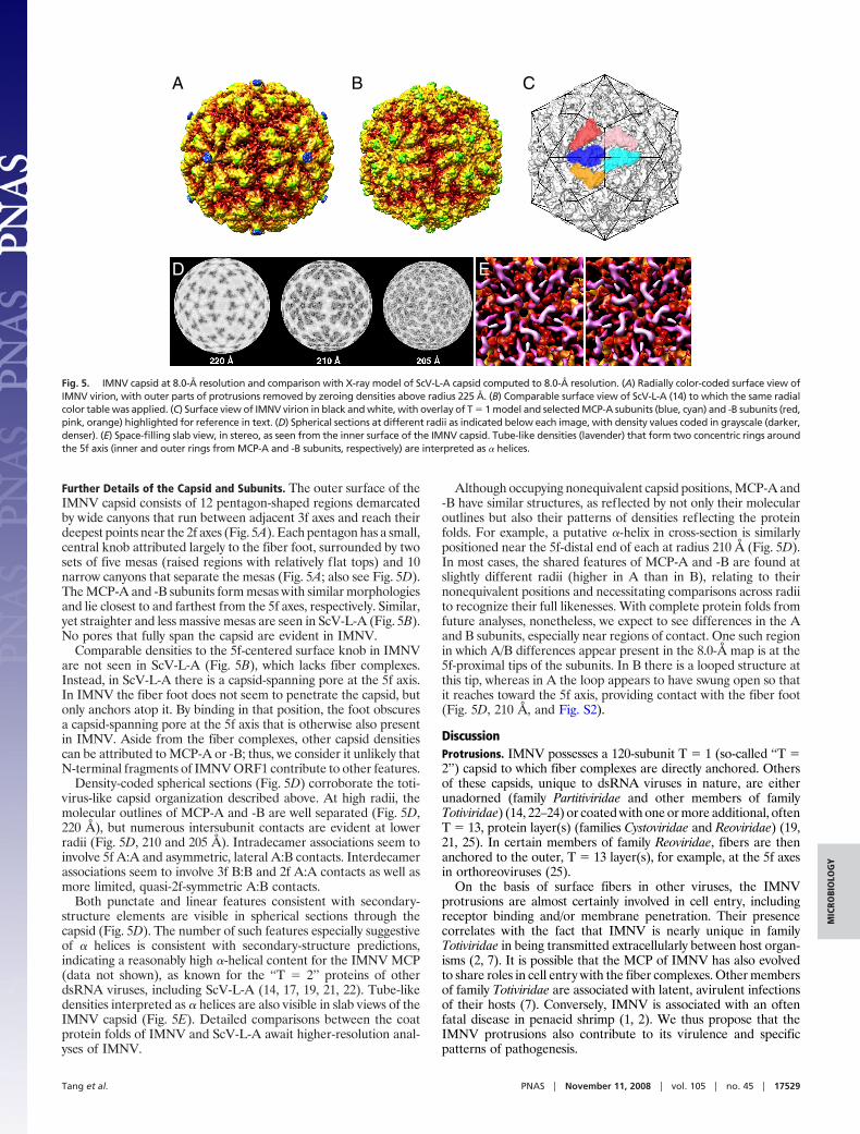
Further Details of the Capsid and Subunits. The outer surface of theIMNV capsid consists of 12 pentagon-shaped regions demarcatedby wide canyons that run between adjacent 3f axes and reach theirdeepest points near the 2f axes (Fig. 5A). Each pentagon has a small,central knob attributed largely to the fiber foot, surrounded by twosets of five mesas (raised regions with relatively flat tops) and 10narrow canyons that separate the mesas (Fig. 5A; also see Fig. 5D).The MCP-A and -B subunits form mesas with similar morphologiesand lie closest to and farthest from the 5f axes, respectively. Similar,yet straighter and less massive mesas are seen in ScV-L-A (Fig. 5B).No pores that fully span the capsid are evident in IMNV.
Comparable densities to the 5f-centered surface knob in IMNVare not seen in ScV-L-A (Fig. 5B), which lacks fiber complexes.Instead, in ScV-L-A there is a capsid-spanning pore at the 5f axis.In IMNV the fiber foot does not seem to penetrate the capsid, butonly anchors atop it. By binding in that position, the foot obscuresa capsid-spanning pore at the 5f axis that is otherwise also presentin IMNV. Aside from the fiber complexes, other capsid densitiescan be attributed to MCP-A or -B; thus, we consider it unlikely thatN-terminal fragments of IMNV ORF1 contribute to other features.
Density-coded spherical sections (Fig. 5D) corroborate the toti-virus-like capsid organization described above. At high radii, themolecular outlines of MCP-A and -B are well separated (Fig. 5D,220 Å), but numerous intersubunit contacts are evident at lowerradii (Fig. 5D, 210 and 205 Å). Intradecamer associations seem toinvolve 5f A:A and asymmetric, lateral A:B contacts. Interdecamerassociations seem to involve 3f B:B and 2f A:A contacts as well asmore limited, quasi-2f-symmetric A:B contacts.
Both punctate and linear features consistent with secondary-structure elements are visible in spherical sections through thecapsid (Fig. 5D). The number of such features especially suggestiveof � helices is consistent with secondary-structure predictions,indicating a reasonably high �-helical content for the IMNV MCP(data not shown), as known for the ‘‘T � 2’’ proteins of otherdsRNA viruses, including ScV-L-A (14, 17, 19, 21, 22). Tube-likedensities interpreted as � helices are also visible in slab views of theIMNV capsid (Fig. 5E). Detailed comparisons between the coatprotein folds of IMNV and ScV-L-A await higher-resolution anal-yses of IMNV.
Although occupying nonequivalent capsid positions, MCP-A and-B have similar structures, as reflected by not only their molecularoutlines but also their patterns of densities reflecting the proteinfolds. For example, a putative �-helix in cross-section is similarlypositioned near the 5f-distal end of each at radius 210 Å (Fig. 5D).In most cases, the shared features of MCP-A and -B are found atslightly different radii (higher in A than in B), relating to theirnonequivalent positions and necessitating comparisons across radiito recognize their full likenesses. With complete protein folds fromfuture analyses, nonetheless, we expect to see differences in the Aand B subunits, especially near regions of contact. One such regionin which A/B differences appear present in the 8.0-Å map is at the5f-proximal tips of the subunits. In B there is a looped structure atthis tip, whereas in A the loop appears to have swung open so thatit reaches toward the 5f axis, providing contact with the fiber foot(Fig. 5D, 210 Å, and Fig. S2).
DiscussionProtrusions. IMNV possesses a 120-subunit T � 1 (so-called ‘‘T �2’’) capsid to which fiber complexes are directly anchored. Othersof these capsids, unique to dsRNA viruses in nature, are eitherunadorned (family Partitiviridae and other members of familyTotiviridae) (14, 22–24) or coated with one or more additional, oftenT � 13, protein layer(s) (families Cystoviridae and Reoviridae) (19,21, 25). In certain members of family Reoviridae, fibers are thenanchored to the outer, T � 13 layer(s), for example, at the 5f axesin orthoreoviruses (25).
On the basis of surface fibers in other viruses, the IMNVprotrusions are almost certainly involved in cell entry, includingreceptor binding and/or membrane penetration. Their presencecorrelates with the fact that IMNV is nearly unique in familyTotiviridae in being transmitted extracellularly between host organ-isms (2, 7). It is possible that the MCP of IMNV has also evolvedto share roles in cell entry with the fiber complexes. Other membersof family Totiviridae are associated with latent, avirulent infectionsof their hosts (7). Conversely, IMNV is associated with an oftenfatal disease in penaeid shrimp (1, 2). We thus propose that theIMNV protrusions also contribute to its virulence and specificpatterns of pathogenesis.
A B C
D E
Fig. 5. IMNV capsid at 8.0-Å resolution and comparison with X-ray model of ScV-L-A capsid computed to 8.0-Å resolution. (A) Radially color-coded surface view ofIMNV virion, with outer parts of protrusions removed by zeroing densities above radius 225 Å. (B) Comparable surface view of ScV-L-A (14) to which the same radialcolor table was applied. (C) Surface view of IMNV virion in black and white, with overlay of T � 1 model and selected MCP-A subunits (blue, cyan) and -B subunits (red,pink, orange) highlighted for reference in text. (D) Spherical sections at different radii as indicated below each image, with density values coded in grayscale (darker,denser). (E) Space-filling slab view, in stereo, as seen from the inner surface of the IMNV capsid. Tube-like densities (lavender) that form two concentric rings aroundthe 5f axis (inner and outer rings from MCP-A and -B subunits, respectively) are interpreted as � helices.
Tang et al. PNAS � November 11, 2008 � vol. 105 � no. 45 � 17529
MIC
ROBI
OLO
GY
The IMNV structure suggests a simple evolutionary mecha-nism—adorn the capsid with fibers—for providing a new set offunctions that expands the infectivity capabilities of a virus. Evo-lution of IMNV from simpler totiviruses may have included acqui-sition of fiber-coding sequences to allow extracellular transmission.But from where were the fiber sequences acquired? Clues in answerto this question have not been apparent from database homologysearches (ref. 2 and data not shown). As for how these newsequences are positioned in the IMNV genome to allow forexpression, incorporation of ‘‘2A-like’’ sequences to provide co-translational polyprotein processing (9) has served as an elegantsolution (2, 8, 15). Alternatively, the IMNV genome organizationmay have been ancestral, with the fiber sequences lost from simplertotiviruses during adaptation to hosts in which extracellular trans-mission does not occur.
The precise morphology and structure of the IMNV protrusionsremain to be determined. In the present study 5f symmetry wasimposed on them by the reconstruction process, but they seemunlikely to be pentamers. On the basis of better-characterizedfibers, such as that of orthoreoviruses (26), the IMNV protrusionsseem more likely to be trimers. The resulting symmetry mismatch(trimer complex at 5f axis) is not uncommon in the virus world andmay promote shedding of the fibers during cell entry (see below).
Which of the three N-terminal peptides of IMNV ORF1 (2,8, 15) forms the fiber complexes? Because peptide (pep)1, the10-kDa dsRNA-binding protein homologue, probably plays arole in modulating innate immunity after expression in in-fected cells, it seems unlikely to be part of the protrusions.Pep2 (32 kDa) or pep3 (38 kDa) thus seems more likely. Bydefining the structural envelope of one fiber complex from the3D map, we estimated its molecular mass to be �90 kDa.Because the mass of a pep2 or pep3 trimer would approximate95 or 115 kDa, respectively, each of these remains a candidate.Fibrous regions of viral surface proteins can include �-helicalcoiled coils (27); however, none of the IMNV proteins havepredicted coiled-coil motifs (groups.csail.mit.edu/cb/paircoil2). On the other hand, a neighboring pair of longregions without charged residues in the N-terminal half ofpep2 (Fig. S3) may suggest a role in membrane penetration.One of these regions, amino acids 42–62, receives a significantscore for membrane-spanning potential (www.ch.embnet.org/software/TMPRED�form.html).
Capsid. Despite little or no clear sequence homology in their coatproteins, the organization of the IMNV capsid is very similar to thatof prototype totivirus ScV-L-A (14, 20, 23), including that MCP-Asubunits from adjacent A/B decamers appear to make 2f-symmetriccontacts across the icosahedral 2f axes and to ‘‘seal off’’ this axisfrom the MCP-B subunits. The latter arrangement is also like thatin members of family Reoviridae (17, 19) but appears to be unlikethat in members of families Cystoviridae and Partitiviridae, where Bsubunits from adjacent decamers appear to make 2f-symmetriccontacts across the icosahedral 2f axes (21, 22). Of note, the IMNVcapsid is less perforated than the ScV-L-A capsid (14, 18, 20, 23)(see Fig. 5 A and B), perhaps representing an adaptation to therigors of extracellular transmission. The IMNV MCP is larger thanthe ScV-L-A coat (Gag) protein (901 aa vs. 680 aa), which may helpit to form a more contiguous shell.
A fascinating feature of the ScV-L-A capsid is that it cleaves the5� caps from yeast mRNAs, facilitating translation of its ownuncapped transcripts (14, 18). Decapping is mediated by a surface-exposed ‘‘trench’’ in the ScV-L-A coat protein, located at the5f-distal end of each A and B subunit. There is no biochemicalevidence that IMNV uses decapping of shrimp mRNAs as astrategy to facilitate its own translation, and although the current3D map is insufficient to address this question definitively, we seeno clear evidence for the presence of such a trench in IMNV.
RNA. The IMNV genome (7,560 bp) (2) is �65% larger than thatof ScV-L-A (4,579 bp) (12), but the IMNV capsid diameter (450 Å)(see Fig. 2 A and B) is �3% larger than that of ScV-L-A (440 Å).In fact, the estimated volume of the IMNV interior (1.85 � 107 Å3)is �9% larger than that of ScV-L-A (1.72 � 107 Å3). Moreover, theIMNV genome is more tightly packed (spacing of density peaks,�26 Å) (see Fig. 2 A and B) than that of ScV-L-A (�30 Å) (18).Thus, ScV-L-A is less fully filled than IMNV, which is filled to asimilar extent as orthoreoviruses (17).
The pattern of densities in the IMNV interior, alternating inclosely spaced sublayers between a dodecahedral frame connectingthe 3f axes vs. concentration around the 5f axes, is intriguing. Thispattern is remarkably similar to that of the single-stranded RNAgenome of nodaviruses (28, 29), except that in IMNV the patternextends over multiple layers. In nodaviruses the pattern is proposedto reflect RNA-capsid interactions that promote both short- andlong-range base pairing within the genome, resulting in a complexRNA path dominated by formation of duplex stems that interactwith the capsid along the edges of the dodecahedron. In IMNV,however, because the genome is already dsRNA, this explanationseems unlikely. Rather, the pattern in IMNV seems more likely toreflect certain regularities in the scheme for dsRNA winding withinthe interior, in which the outer layer of dsRNA is directed byinteractions with the capsid to bind preferentially along the edgesof the dodecahedron. Repetition of this pattern in the inner layersmay then be determined by this directed pattern in the outer layer.The presence of the RdRp (as part of the MCP/RdRp fusion)projecting into the interior in IMNV may also contribute to thispattern by anchoring at least one end of the dsRNA and by directingits path, by either binding or steric exclusion, at other points in thewinding scheme.
Like those of other dsRNA viruses, the IMNV particle would beexpected to enter the cytoplasm as the outcome of cell entry, whereit would transcribe and export copies of the viral mRNA(s) fortranslation by host ribosomes (30). But where is the capsid-spanningpore through which these mRNAs are exported and/or substratenucleotides imported? The only notable pores through the IMNVcapsid are at the 5f axes, and those are covered by the protrusions.One possibility is that the 5f axis near which the MCP/RdRpsubunit is incorporated is not competent to anchor a protrusion sothat that pore is constitutively open. Another possibility is that theprotrusions are lost during or after cell entry and that the pores arethereby uncovered for transcript release and nucleotide import. Asimilar step is known for orthoreovirus (25, 31). Indeed, fiber lossmay be an essential part of the cell entry mechanism of IMNV.
Taxonomy. IMNV shares prime similarities with members of familyTotiviridae. It has a nonsegmented dsRNA genome and seems touse �1 ribosomal frameshifting to express its RdRp in fusion withthe MCP. It clusters with this family in sequence-based phylogeneticcomparisons, albeit distantly, and its closest relative is GLV (pro-totype of genus Giardiavirus). From the present results, we alsoknow that its capsid is organized much like that of ScV-L-A(prototype of genus Totivirus) as well as those of two other familymembers, Ustilago maydis virus P4 and Helminthosporium victo-riae 190S virus (23, 24), for which 3D structures are known. Itsdissimilarities are also important and include infection of a crus-tacean arthropod host; a larger genome, with coding capacitybeyond MCP and RdRp; fiber-like protrusions on the virionsurface; capability to transmit extracellularly, like GLV; and asso-ciation with host disease. On the basis of these facts, we consider itreasonable to propose a new monotypic genus to accommodateIMNV but to retain this genus in family Totiviridae. As other virusesrelated to IMNV are discovered, however, one might then argue toseparate those into a new family of phylogenetically distinct,nonsegmented dsRNA viruses that possess, in at least some cases,the machinery for extracellular transmission.
17530 � www.pnas.org�cgi�doi�10.1073�pnas.0806724105 Tang et al.

Materials and MethodsVirion Purification. Virions were purified from IMNV-infected L. vannamei by aprocedure modified from Poulos et al. (2). Gnathothoraces (‘‘heads’’) and tailmuscles (‘‘tails’’) from 30 subadult shrimp were treated separately in an effort tooptimize yields and purity from either portion. There is no evidence that virionsisolated from heads or tails are different, and preparations from each have beenused to induce IMNV infection in the lab. Tissues were homogenized in 20 mMTris/HCl (pH 7.5), 400 mM NaCl (TN buffer), and large debris were removed bycentrifugationat8,000�g.Aftercentrifugationfor3hat205,000�g, thepelletswereresuspendedinTNbuffer.LipidswereremovedwithFreon(heads)orfumedsilica(tails),andthesuspensionsweremixedwith1gcharcoalfor10min.Charcoalwas removed with Celite 535. After pelleting as above, the pellets were againresuspended in TN buffer, layered onto a 15–40% (wt/wt) sucrose gradient, andcentrifuged for 2 h at 286,000 � g. Peak fractions in the lower third of thegradient were collected and washed with TN buffer. The peak suspension fromtails was treated on a second such sucrose gradient for further purification. A20–50% (wt/wt) CsCl gradient was used for final purification. This gradient wascentrifuged for 16 h at 136,000 � g, the peak fractions were collected andwashed, and pellets were resuspended in TN buffer. The purified virions weresubjected to denaturing polyacrylamide gel electrophoresis followed by stainingwith Coomassie blue. They were also negatively stained with 2% phosphotung-stic acid and examined using a Philips CM12 microscope.
CryoTEM. Small (3-�l) aliquots of purified virions from shrimp tails (�2.5 mg/ml)were vitrified for cryoTEM via standard, rapid freeze-plunging procedures (32).Samples were applied to Quantifoil holey grids that had been plasma-cleaned for25 sec in an E.A. Fischione 1020 unit. Grids were then transferred into a precooledGatan626cryotransferholder,whichmaintainedthespecimenat liquid-nitrogentemperature in the FEI Sphera microscope. Micrographs were recorded on KodakSO-163 electron-image film at 200 keV under low-dose conditions (�15 e/Å2) andat a nominal magnification of �50,000.
3D Reconstruction. Twenty-four micrographs, exhibiting minimal specimen driftand image astigmatism and recorded at underfocus settings of between 0.7 and2.0 �m, were digitized on a Zeiss scanner at 7-�m intervals representing 1.4-Åpixels at the specimen. RobEM (cryoem.ucsd.edu/programDocs/runRobem.txt)was used to extract individual particle images, each 449 � 449 pixels in size, andto preprocess them as described (32). Processing began with a 15-Å map ofhead-derivedvirions,whichhadbeengeneratedearlier inthestudy(unpublisheddata). This map served as a starting model to initiate full orientation and origindeterminations of the full set of 5,629 particle images of tail-derived virions byusing the current version of AUTO3DEM (33). The result was a 3D reconstructionat 12-Å resolution after five iterative cycles. Several cycles of orientation andorigin refinement with the PO2R and P3DR functions of AUTO3DEM then en-abled us to improve the resolution to 8.0 Å. Effects of the microscope contrast-transfer function were compensated in part by inverting image phases in spatialfrequency bands where contrast reversals occurred (34). To enhance the lowest-resolution features of protrusions and RNA, another 3D map, at 10-Å resolution,was calculated in which spatial frequency components up to the first peak in thecontrast-transfer function for each image were amplified using �1 � 0.4 (35).Graphics were generated with RobEM and Chimera (36). Contour levels wereadjustedtooptimizerelevantfeatures foreachfigure:0.6� forFig.3;2.0� forFig.4 A, D, and G; 1.5 � for Fig. 5 A-C; and 3.0 � for Fig. 5E. Handedness was assignedto match that of ScV-L-A (14).
ACKNOWLEDGMENTS. We thank N. Olson for help with microscopy, A. Rusnakfor preliminary analyses of head-prep images, and K. Chang and S. Dunn forscanning of tail-prep images. This work was supported in part by NationalInstitutes of Health (NIH) Grants R37 GM033050 (to T.S.B.) and R01 AI46440 (toM.L.N.) plus U.S. Department of Agriculture Grants NRICRP 2001-35319-10010 (toS.A.G.) and CSREES 2004-38808-02142 (to D.V.L.). The San Diego SupercomputerCenter provided partial support for R.S.S. and for access to TeraGrid computing.Shared NIH instrumentation grant 1S10 RR020016-01 as well as support from theUniversity of California-San Diego and the Agouron Foundation (all to T.S.B.)were used to establish the cryoTEM facilities used in this study.
1. Lightner DV, et al. (2004) Infectious myonecrosis: new disease in Pacific white shrimp. GlobAquac Advocate 7:85.
2. Poulos BT, Tang KF, Pantoja CR, Bonami JR, Lightner DV (2006) Purification andcharacterization of infectious myonecrosis virus of penaeid shrimp. J Gen Virol 87:987–996.
3. Andrade TPD, Srisuvan T, Tang KFJ, Lightner DV (2007) Real-time reverse transcriptionpolymerase chain reaction assay using TaqMan probe for detection and quantification ofinfectious myonecrosis virus (IMNV) Aquaculture 264:9–15.
4. Senapin S, Phewsaiya K, Briggs M, Flegel TW (2007) Outbreaks of infectious myonecrosisvirus (IMNV) in Indonesia confirmed by genome sequencing and use of an alternativeRT-PCR detection method. Aquaculture 266:32–38.
5. Pinheiro ACAS, et al. (2007) Epidemiological status of Taura syndrome and infectiousmyonecrosis viruses in Penaeus vannamei reared in Pernambuco (Brazil). Aquaculture262:17–22.
6. Wang AL, Yang HM, Shen K, Wang CC (1993) Giardiavirus double-stranded RNA genomeencodes a capsid polypeptide and a gag-pol-like fusion protein by a translational frame-shift. Proc Natl Acad Sci USA 90:8595–8599.
7. Ghabrial SA (2008) Totiviruses. Encyclopedia of Virology, eds Mahy B, van RegenmortelMHV (Elsevier/Academic, London), 3rd Ed pp 163–174.
8. Nibert ML (2007) ‘2A-like’ and ‘shifty heptamer’ motifs in penaeid shrimp infectiousmyonecrosis virus, a monosegmented double-stranded RNA virus. J Gen Virol 88:1315–1318.
9. Donnelly ML, et al. (2001) The ‘cleavage’ activities of foot-and-mouth disease virus 2Asite-directed mutants and naturally occurring ‘2A-like’’sequences. J Gen Virol 82:1027–1041.
10. Bekaert M, et al. (2003) Towards a computational model for -1 eukaryotic frameshiftingsites. Bioinformatics 19:327–335.
11. Plant EP, et al. (2003) The 9-Å solution: How mRNA pseudoknots promote efficientprogrammed -1 ribosomal frameshifting. RNA 9:168–174.
12. Icho T, Wickner RB (1989) The double-stranded RNA genome of yeast virus L-A encodes itsown putative RNA polymerase by fusing two open reading frames. J Biol Chem 264:6716–6723.
13. Ribas JC, Wickner RB (1998) The Gag domain of the Gag-Pol fusion protein directsincorporation into the L-A double-stranded RNA viral particles in Saccharomyces cerevi-siae. J Biol Chem 273:9306–9311.
14. Naitow H, Tang J, Canady M, Wickner RB, Johnson JE (2002) L-A virus at 3.4 Å resolutionreveals particle architecture and mRNA decapping mechanism. Nat Struct Biol 9:725–728.
15. Luke GA, et al. (2008) Occurrence, function and evolutionary origins of ‘2A-like’ sequencesin virus genomes. J Gen Virol 89:1036–1042.
16. Harauz G, van Heel M (1986) Exact filters for general geometry three dimensional recon-struction. Optik 73:146–156.
17. Reinisch KM, Nibert ML, Harrison SC (2000) Structure of the reovirus core at 3.6 Åresolution. Nature 404:960–967.
18. Tang J, et al. (2005) The structural basis of recognition and removal of cellular mRNA7-methyl G ‘caps’ by a viral capsid protein: A unique viral response to host defense. J MolRecognit 18:158–168.
19. Grimes JM, et al. (1998) The atomic structure of the bluetongue virus core. Nature395:470–478.
20. Caston JR, et al. (1997) Structure of L-A virus: A specialized compartment for the transcrip-tion and replication of double-stranded RNA. J Cell Biol 138:975–985.
21. Huiskonen JT, et al. (2006) Structure of the bacteriophage �6 nucleocapsid suggests amechanism for sequential RNA packaging. Structure 14:1039–1048.
22. Ochoa WF, et al. (2008) Partitivirus structure reveals a 120-subunit, helix-rich capsid withdistinctive surface arches formed by quasisymmetric coat-protein dimers. Structure16:776–786.
23. Cheng RH, et al. (1994) Fungal virus capsids, cytoplasmic compartments for the replicationofdouble-strandedRNA, formedas icosahedral shellsofasymmetricgagdimers. JMolBiol244:255–258.
24. Caston JR, et al. (2006) Three-dimensional structure and stoichiometry of Helminthospo-rium victoriae 190S totivirus. Virology 347:323–332.
25. Dryden KA, et al. (1993) Early steps in reovirus infection are associated with dramaticchanges in supramolecular structure and protein conformation. J Cell Biol 122:1023–1041.
26. Chappell JD,ProtaAE,DermodyTS,StehleT(2002)Crystal structureofreovirusattachmentprotein �1 reveals evolutionary relationship to adenovirus fiber. EMBO J 21:1–11.
27. Bassel-Duby R, et al. (1985) Sequence of reovirus haemagglutinin predicts a coiled-coilstructure. Nature 315:421–423.
28. Johnson KN, Tang L, Johnson JE, Ball LA (2004) Heterologous RNA encapsidated inPariacoto virus-like particles forms a dodecahedral cage similar to genomic RNA in wild-type virions. J Virol 78:11371–11378.
29. Tihova M, et al. (2004) Nodavirus coat protein imposes dodecahedral RNA structureindependent of nucleotide sequence and length. J Virol 78:2897–2905.
30. Lawton JA, Estes MK, Prasad BVV (1997) Three-dimensional visualization of mRNA releasefrom actively transcribing rotavirus particles. Nat Struct Biol 4:118–121.
31. ChandranK,NibertML(2003)Animal cell invasionbya largenonenvelopedvirus:Reovirusdelivers the goods. Trends Microbiol 11:374–382.
32. Baker TS, Olson NH, Fuller SD (1999) Adding the third dimension to virus life cycles:Three-dimensional reconstruction of icosahedral viruses from cryo-electron micrographs.Microbiol Molec Biol Rev 63:862–922.
33. Yan X, Sinkovits RS, Baker TS (2007) AUTO3DEM—an automated and high throughputprogram for image reconstruction of icosahedral particles. J Struct Biol 157:73–82.
34. Frank J (1996) Three-Dimensional Electron Microscopy of Macromolecular Assemblies.(Academic, San Diego), pp 12–53.
35. Bowman VD, et al. (2002) An antibody to the putative aphid recognition site on cucumbermosaic virus recognizes pentons but not hexons. J Virol 76:12250–12258.
36. Goddard TD, Huang CC, Ferrin TE (2007) Visualizing density maps with UCSF Chimera. JStruct Biol 157:281–287.
Tang et al. PNAS � November 11, 2008 � vol. 105 � no. 45 � 17531
MIC
ROBI
OLO
GY