INFECTION ANDiai.asm.org/content/45/1/41.full.pdfINFECTION ANDIMMUNITY, JUlY 1984, p. 41-46 Vol. 45,...
6
Vol. 45, No. 1 INFECTION AND IMMUNITY, JUlY 1984, p. 41-46 0019-9567/84/070041-06$02.00/0 Copyright C) 1984, American Society for Microbiology Oligosaccharide Structures Mediating Agglutination of Sheep Erythrocytes by Staphylococcus saprophyticus ALF GUNNARSSON,1 PER-ANDERS MARDH,2 ARNE LUNDBLAD,3 AND SIGFRID SVENSSON'* Department of Carbohydrate Chemistry, Chemical Center,' and Departments of Medical Microbiology,2 and Clinical Chemistry,3 University of Lund, S-220 07 Lund, Sweden Received 3 January 1984/Accepted 3 April 1984 Agglutination of sheep erythrocytes by Staphylococcus saprophyticus was used as a model system for adherence studies. Glycolipids were isolated from sheep erythrocyte membranes, and oligosaccharides were prepared by trifluoroacetolysis. The oligosaccharides were characterized by sugar analyses, methylation analyses, gas-liquid chromatography-mass spectrometry, and nuclear magnetic resonance spectroscopy. We showed that oligosaccharides containing terminal -D-galactose-p-(1-4)-I(-D-2-acetamido-2-deoxyglucose-p-(1- were good inhibitors of the hemagglutination of sheep erythrocytes by S. saprophyticus. Staphylococcus saprophyticus is a common cause of uri- nary tract infections in young women, and is second only to Escherichia coli as its most frequent causative agent (25, 31, 39). Urinary tract infections with S. saprophyticus also occur in children (U. Jodal, personal communication) and men (10). In men, such infections are most common in older age groups, particularly in those with hyperplasia of the prostate (13). That this organism may be one causative agent of nonspecific urethritis has also been considered (14). In approximately half of all cases of urinary tract infec- tions caused by S. saprophyticus, the upper urinary tract is involved, and the concentration capacity of the kidney can be reduced (13). S. saprophyticus urinary tract infections occur in the pool of 10 to 15% of all women who get urinary tract infections. The vast majority of all cases of recurrent nonobstructive urinary tract infections are found in this pool (B. Hovelius and P.-A. M?ardh, Rev. Infect. Dis., in press). S. saprophyticus has been shown to adhere in greater numbers to urothelial and periurethral cells than to other types of cells, e.g., skin and buccal cells (4). The organism is not known as a human pathogen in organ systems other than the urogenital tract. The majority of strains of S. saprophyticus have, in contrast to Staphylococcus aureus and to other coagulase- negative staphylococcal species, the ability to agglutinate sheep erythrocytes (12). The capacity of bacteria and other microorganisms to adhere to eucaryotic cells is dependent on physicochemical factors such as surface charge and hydrophobic interactions (3) and on the presence of specific carbohydrate receptors (15, 19, 20). In the present investigation, we used the agglutination of sheep erythrocytes by S. saprophyticus as a model system to investigate adherence properties of S. saprophyticus. Oligo- saccharides were isolated by trifluoroacetolysis of glycolip- ids from sheep erythrocyte membranes and tested as inhibi- tors of the agglutination. Furthermore, a number of oligosaccharides and glycoproteins were subjected to hem- agglutination-inhibition tests to examine the specificity of the adherence. MATERIALS AND METHODS Abbreviations. Abbreviations used in this paper are: NeuNAc, N-acetylneuraminic acid; Gal, galactose; Gal- * Corresponding author. NAc, 2-acetamido-2-deoxygalactose; Glc, glucose; GIcNAc, 2-acetamido-2-deoxyglucose; Man, mannose. Bacteria. The eight strains of S. saprophyticus used had been isolated from voided urine specimens as described elsewhere (11). Species identification was made by the method of Kloos and Schleifer (17). The staphylococci were maintained on solid medium consisting of blood agar base no. 2 (Oxoid Ltd., London, United Kingdom) containing 4% defibrinated horse blood. Erythrocytes. Suspensions (1%) of fresh, washed sheep erythrocytes were prepared in physiological saline. Erythrocyte membranes. Sheep blood was obtained from a slaughterhouse, with citrate buffer as the anticoagulant. Erythrocyte membranes were prepared by the method of Dodge et al. (6). The yield of lyophilized membranes was ca. 4 g/liter of blood. Oligosaccharides. I-D-Galp-(1-3)-r-D-GlcNAcp-(1-3)-,B-D- Galp-(1-4)-D-Glc (lacto-N-tetraose) and P-D-Galp-(1-4)-P-D- GlcNAcp-(1-3)-3-D-Galp-(1-4)-D-Glc (lacto-N-neo-tetraose) were obtained from human milk as previously described (18). oa-NeuNAcp-(2-3)- - D-Galp-(1-4)- D-Glc (3 '-sialyllac- tose) and ot-NeuNAcp-(2-6)-,-D-Galp-(1-4)-D-Glc (6'-sialyl- lactose) were prepared from bovine colostrum by the proce- dure described for isolation of sialyllactoses from human milk (33). P-D-Galp-(1-4)-p-D-GlcNAcp-(1-2)-cx-D-Manp- (1-6)-3-D-Manp-(1-4)-D-GlcNAc (GM,-pentasaccharide) and ,B-D-Galp-(1-4)-,8-D-GIcNAcp-(1-2)-at-D-Manp-(1-3)-[-P-D- Galp-(1-4)-,8-D-GIcNAcp-(1-2)-ot-D-Manp-(1-6)-]-p-D-Manp- (1-4)-D-GlcNAc (GM,-octasaccharide) were obtained from the urine of a patient with GM,-gangliosidosis (23). P-D- Galp-(1-4)-p-D-GIcNAcp-(1-2)-ox-D-Manp-(1-3)-[-P-D-Galp- (1-4)-3-D-GlcNAcp-(1-2)-cx-D-Manp-(1-6)-]-D-mannitol was prepared from human asialotransferrin by the trifluoroacet- olysis procedure described for the preparation of the N- glycosidically linked carbohydrate chains in asialofetuin (27). at-D-Galp-(1-4)-p-D-Galp,-(1-4)-D-Glc, P-D-GaINAcp-(1- 4)-P-D-Galp-(1-4)-D-Glc and ,-D-GalNAcp-(1-3)-(x-D-Galp- (1-4)-f3-D-Galp-(1-4)-D-Glc were produced by specific tri- fluoroacetolytic degradation of the glycolipids globotriaosylceramide, gangliotriaosylceramide, and globo- tetraosylceramide, respectively. The glycolipids were pre- pared from pig intestinal linings (globotriaosylceramide) (38), guinea pig erythrocyte membranes (gangliotriaosylcer- amide) (32), and human erythrocyte membranes (globote- traosylceramide) (24). P3-D-Galp-(1-4)-D-Glc (lactose) was purchased from Sigma Chemical Co., St. Louis, Mo. 1-D- 41 on June 6, 2018 by guest http://iai.asm.org/ Downloaded from
Transcript of INFECTION ANDiai.asm.org/content/45/1/41.full.pdfINFECTION ANDIMMUNITY, JUlY 1984, p. 41-46 Vol. 45,...

Vol. 45, No. 1INFECTION AND IMMUNITY, JUlY 1984, p. 41-460019-9567/84/070041-06$02.00/0Copyright C) 1984, American Society for Microbiology
Oligosaccharide Structures Mediating Agglutination of SheepErythrocytes by Staphylococcus saprophyticus
ALF GUNNARSSON,1 PER-ANDERS MARDH,2 ARNE LUNDBLAD,3 AND SIGFRID SVENSSON'*Department of Carbohydrate Chemistry, Chemical Center,' and Departments of Medical Microbiology,2 and Clinical
Chemistry,3 University of Lund, S-220 07 Lund, Sweden
Received 3 January 1984/Accepted 3 April 1984
Agglutination of sheep erythrocytes by Staphylococcus saprophyticus was used as a model system foradherence studies. Glycolipids were isolated from sheep erythrocyte membranes, and oligosaccharides wereprepared by trifluoroacetolysis. The oligosaccharides were characterized by sugar analyses, methylationanalyses, gas-liquid chromatography-mass spectrometry, and nuclear magnetic resonance spectroscopy. Weshowed that oligosaccharides containing terminal -D-galactose-p-(1-4)-I(-D-2-acetamido-2-deoxyglucose-p-(1-were good inhibitors of the hemagglutination of sheep erythrocytes by S. saprophyticus.
Staphylococcus saprophyticus is a common cause of uri-nary tract infections in young women, and is second only toEscherichia coli as its most frequent causative agent (25, 31,39). Urinary tract infections with S. saprophyticus alsooccur in children (U. Jodal, personal communication) andmen (10). In men, such infections are most common in olderage groups, particularly in those with hyperplasia of theprostate (13). That this organism may be one causative agentof nonspecific urethritis has also been considered (14).
In approximately half of all cases of urinary tract infec-tions caused by S. saprophyticus, the upper urinary tract isinvolved, and the concentration capacity of the kidney canbe reduced (13). S. saprophyticus urinary tract infectionsoccur in the pool of 10 to 15% of all women who get urinarytract infections. The vast majority of all cases of recurrentnonobstructive urinary tract infections are found in thispool (B. Hovelius and P.-A. M?ardh, Rev. Infect. Dis., inpress).
S. saprophyticus has been shown to adhere in greaternumbers to urothelial and periurethral cells than to othertypes of cells, e.g., skin and buccal cells (4). The organism isnot known as a human pathogen in organ systems other thanthe urogenital tract.The majority of strains of S. saprophyticus have, in
contrast to Staphylococcus aureus and to other coagulase-negative staphylococcal species, the ability to agglutinatesheep erythrocytes (12).The capacity of bacteria and other microorganisms to
adhere to eucaryotic cells is dependent on physicochemicalfactors such as surface charge and hydrophobic interactions(3) and on the presence of specific carbohydrate receptors(15, 19, 20).
In the present investigation, we used the agglutination ofsheep erythrocytes by S. saprophyticus as a model system toinvestigate adherence properties of S. saprophyticus. Oligo-saccharides were isolated by trifluoroacetolysis of glycolip-ids from sheep erythrocyte membranes and tested as inhibi-tors of the agglutination. Furthermore, a number ofoligosaccharides and glycoproteins were subjected to hem-agglutination-inhibition tests to examine the specificity ofthe adherence.
MATERIALS AND METHODSAbbreviations. Abbreviations used in this paper are:
NeuNAc, N-acetylneuraminic acid; Gal, galactose; Gal-* Corresponding author.
NAc, 2-acetamido-2-deoxygalactose; Glc, glucose; GIcNAc,2-acetamido-2-deoxyglucose; Man, mannose.
Bacteria. The eight strains of S. saprophyticus used hadbeen isolated from voided urine specimens as describedelsewhere (11). Species identification was made by themethod of Kloos and Schleifer (17). The staphylococci weremaintained on solid medium consisting of blood agar baseno. 2 (Oxoid Ltd., London, United Kingdom) containing 4%defibrinated horse blood.
Erythrocytes. Suspensions (1%) of fresh, washed sheeperythrocytes were prepared in physiological saline.
Erythrocyte membranes. Sheep blood was obtained from aslaughterhouse, with citrate buffer as the anticoagulant.Erythrocyte membranes were prepared by the method ofDodge et al. (6). The yield of lyophilized membranes was ca.4 g/liter of blood.
Oligosaccharides. I-D-Galp-(1-3)-r-D-GlcNAcp-(1-3)-,B-D-Galp-(1-4)-D-Glc (lacto-N-tetraose) and P-D-Galp-(1-4)-P-D-GlcNAcp-(1-3)-3-D-Galp-(1-4)-D-Glc (lacto-N-neo-tetraose)were obtained from human milk as previously described(18). oa-NeuNAcp-(2-3)- - D-Galp-(1-4)- D-Glc (3 '-sialyllac-tose) and ot-NeuNAcp-(2-6)-,-D-Galp-(1-4)-D-Glc (6'-sialyl-lactose) were prepared from bovine colostrum by the proce-dure described for isolation of sialyllactoses from humanmilk (33). P-D-Galp-(1-4)-p-D-GlcNAcp-(1-2)-cx-D-Manp-(1-6)-3-D-Manp-(1-4)-D-GlcNAc (GM,-pentasaccharide) and,B-D-Galp-(1-4)-,8-D-GIcNAcp-(1-2)-at-D-Manp-(1-3)-[-P-D-Galp-(1-4)-,8-D-GIcNAcp-(1-2)-ot-D-Manp-(1-6)-]-p-D-Manp-(1-4)-D-GlcNAc (GM,-octasaccharide) were obtained fromthe urine of a patient with GM,-gangliosidosis (23). P-D-Galp-(1-4)-p-D-GIcNAcp-(1-2)-ox-D-Manp-(1-3)-[-P-D-Galp-(1-4)-3-D-GlcNAcp-(1-2)-cx-D-Manp-(1-6)-]-D-mannitol wasprepared from human asialotransferrin by the trifluoroacet-olysis procedure described for the preparation of the N-glycosidically linked carbohydrate chains in asialofetuin(27). at-D-Galp-(1-4)-p-D-Galp,-(1-4)-D-Glc, P-D-GaINAcp-(1-4)-P-D-Galp-(1-4)-D-Glc and ,-D-GalNAcp-(1-3)-(x-D-Galp-(1-4)-f3-D-Galp-(1-4)-D-Glc were produced by specific tri-fluoroacetolytic degradation of the glycolipidsglobotriaosylceramide, gangliotriaosylceramide, and globo-tetraosylceramide, respectively. The glycolipids were pre-pared from pig intestinal linings (globotriaosylceramide)(38), guinea pig erythrocyte membranes (gangliotriaosylcer-amide) (32), and human erythrocyte membranes (globote-traosylceramide) (24). P3-D-Galp-(1-4)-D-Glc (lactose) waspurchased from Sigma Chemical Co., St. Louis, Mo. 1-D-
41
on June 6, 2018 by guesthttp://iai.asm
.org/D
ownloaded from

42 GUNNARSSON ET AL.
Galp-(1-4)-D-GlcNAc (N-acetyl-lactosamine) and P-D-Galp-(1-4)-1-D-GlcNAcp-(1-O)-Me (methyl-3-N-acetyl-lactosa-minide) were gifts from Goran Magnusson, ResearchDivision, Swedish Sugar Co., Arlov, Sweden.The purity of all these oligosaccharides was determined by
either paper chromatography or gas-liquid chromatography(as permethylated alditols). Each oligosaccharide (exceptthe one commercial sample) was characterized by sugar
analysis, methylation analysis, sequential techniques, gas-
liquid chromatography-mass spectrometry, and by their nu-clear magnetic resonance spectra.
Glycoproteins. Fetuin was prepared (35) from fetal calfserum, and human transferrin was prepared (34) from humanserum. Human antithrombin was obtained from Kabi-Vi-trum, Stockholm, Sweden. The glycoproteins were desialyl-ated by mild acid hydrolysis (36), and their purity was
determined by acrylamide gel eletrophoresis. The carbohy-drate chains of the native and desialylated glycoproteinswere analyzed by sugar and methylation analysis and were inaccordance with previously published structures for thecarbohydrate chains in fetuin (25a), human transferrin (34),and human antithrombin (8).
Analytical methods. Sugar analyses were performed bygas-liquid chromatography (29) and mass spectrometry (2)after hydrolysis with 90% aqueous formic acid at 100°C for 5h and also after subsequent hydrolysis with 0.25 M aqueous
sulfuric acid at 100'C for 18 h. Methylation analyses wereperformed as previously described (1).
Isolation of glycolipid fractions from sheep erythrocytemembranes. Lyophilized sheep erythrocyte membranes (230g) were extracted successively with 95% and then 85%aqueous ethanol (13 liters of each). The combined extractswere concentrated to dryness, redissolved in 750 ml ofchloroform-methanol (2:1 [vol/vol]), and filtered. The filtratewas concentrated to dryness and dissolved in 300 ml ofchloroform-methanol (2:1 [vol/vol]) to which acetone (3
liters) was added, and the mixture was left at 4°C for 20 hbefore removal of the resultant precipitate by filtration. Thisprecipitate (32.5 g) was sonicated in 50 ml of chloroform-methanol (9:1 [vol/vol]) and fractionated on a silicic acidcolumn (10 by 80 cm, Silica Gel 60; E. Merck AG, Darm-stadt, Federal Republic of Germany) eluted successivelywith 5 liters of chloroform-methanol (9:1, 2:1, 1:1, 1:2, 1:9,and 1:20 [vol/vol]) (see Fig. 1A through F). The fractionationwas monitored by thin-layer chromatography on high-pres-sure-thin-layer chromatography plates (E. Merck AG), usingchloroform-methanol-water (65:25:4 [vol/vol/vol]). The gly-colipids were visualized on the plates by the anisaldehydereagent (16). The pooled fractions 1 through 3 (see Fig. 1)were treated with 0.1 M KOH in 10% aqueous methanol (100ml/g of glycolipid mixture). The vessel was flushed withnitrogen and magnetically stirred in the dark at 4°C for 20 h.Alkaline hydrolysis was stopped with 2 M HCl, slowly addedunder stirring, until pH 2 to 3 was reached (16). Chloroformand water were added to give a final proportion of chloro-form-methanol-water of 8:4:3 (vol/vol/vol). The phasesformed were tested by thin-layer chromatography. Theglycolipids in fraction 1 were mainly in the lower phasewhich was concentrated to dryness. Fractions 2 and 3showed distribution of glycolipids in both phases. Thematerial in the lower phase was collected and added tofraction 1. The material in the upper phase of fractions 2 and3 was collected and concentrated to dryness. Fractions 1through 3 were further purified by silicic acid chromatogra-phy and preparative thin-layer chromatography (E. MerckAG).
Nitrous acid deamination. The dried glycolipid was dis-solved in 0.5 M acetic acid (1 ml of acetic acid per mg ofglycolipid) and NaNO2 (5 mg) was added. The solution wasleft at room temperature for 3 h, and the excess HNO2 wasdestroyed with ethylamine. The reaction mixture was de-gassed with nitrogen and adjusted to pH 9 with M NaOHI(37). The oligosaccharides were reduced (NaBD4) and per-methylated (9).
Trifluoroacetolysis of glycolipid fractions. The glycolipidfraction was treated with a mixture of trifluoroacetic acidand trifluoroacetic anhydride (1:100 [vol/vol]) (50 ml/g ofglycolipid mixture) at 100°C for 48 h in a sealed tube.(Caution should be used at this point as a corrosive mixtureis under pressure.) After cooling, the reaction mixture wasconcentrated to dryness, dissolved in methanol (50 ml), andagain concentrated to dryness. The resulting product wasdissolved in glacial acetic acid (25 ml), and distilled water (25ml) was added. After 1 h at room temperature, the solutionwas evaporated to dryness, and diethyl ether (100 ml) anddistilled water (50 ml) were added. The mixture was shaken,the water phase was removed, and the diethyl ether phasewas extracted three times with 50 ml of distilled water. Thecombined yellowish water phase was washed three timeswith 25 ml of diethyl ether and evaporated to dryness, andthe residue was treated with 2 M ammonia in 50% aqueousmethanol (50 ml) at room temperature for 20 h. The solutionwas evaporated to dryness, and the residue was acetylatedwith 50 ml of acetic anhydride-pyridine (1:1 [vol/vol], 100°C,30 min). The acetylated product was de-O-acetylated with 2M ammonia in 50% aqueous methanol (50 ml) at roomtemperature (20 h). After concentration to dryness, theproduct was fractionated on Sephadex G-15 (Pharmacia AB,Uppsala, Sweden). The fractionation was monitored byassaying the fractions for hexose (30).
Hemagglutination-inhibition tests. Two to four hemaggluti-nating units (12) of bacterial suspensions in 25 ,ul of physio-logical saline were incubated at 4°C for 1 h together with 50p,l of serial twofold dilutions of the test substance in saline.To this was added 25 ,ul of a 1% suspension of sheeperythrocytes, and the mixture was incubated at 4°C for 2 h.All tests were done in disposable, plastic microtiter plates(Flow Laboratories, Inc., McLean, Va.). The minimumamount of substance giving complete inhibition of hemagglu-tination on visual inspection was determined (12).
RESULTSFractionation of the glycolipid extract gave three frac-
tions, and the major glycolipid was the Forssman antigen(Fig. 1, fraction 1 [Fr 1]). Fraction 2 consisted of glycolipidscontaining six to eight sugar residues, and fraction 3 consist-ed of glycolipids with more than eight sugar residues as
judged by thin-layer chromatography.Parts of glycolipid fractions 1 through 3 were trifluoroace-
tolysed to specifically remove the ceramide portion of the
TABLE 1. Yields and carbohydrate composition of the glycolipidfractions prepared from 230 g of sheep erythrocyte membranes
Frac- Yield Relative molar proportionsation (mg) D-Gal D-Glcb D-GlcNAc D-GalNAc
1 438 1.9 1.0 0.05 1.72 111 0.9 1.0 0.1 0.053 37 1.4 1.0 1.1 0.1a Determined by gas-liquid chromatography-mass spectrometry after acid
hydrolysis, reduction, and acetylation.b Glucose was set to 1.0.
INFECT. IMMUN.
on June 6, 2018 by guesthttp://iai.asm
.org/D
ownloaded from

OLIGOSACCHARIDES AND SHEEP ERYTHROCYTE AGGLUTINATION
A B C 0 E F Solvent system for~ i i i I' ' column elution
.... I....
....A
Gci;
4 Forssman antigen
-
pi i I 0 i60 120 1Z 240
t~~ ~ ~ ~ ~ ~ ~~~~~~~~i I
Fraction numberDnnl&1 f srfinnrFe
Fr 1 Fr 2 Fr3 ruue TaETOnaFIG. 1. Separation of glycolipids from sheep erythrocyte membranes. The plate was developed in chloroform-methanol-water (65:25:4
[vol/vol/vol]). Spots were visualized by spraying with anisaldehyde reagent. Fractions 1 through 3 were pooled as indicated in the figure.Fr 1 to 3, fractions 1 to 3.
glycolipids (28), and the liberated oligosaccharides were
purified on a Sephadex G-15 column. The oligosaccharidemixtures obtained were tested for their ability to inhibit theagglutination of sheep erythrocytes by S. saprophyticus. Theoligosaccharides from fractions 1 and 3 showed inhibitoryactivity. The monosaccharide composition of the oligosac-charide mixtures are shown in Table 1. Fraction 1 containedmainly D-Glc, D-Gal, and D-GalNAc, and fraction 3 con-
tained D-Glc, D-Gal, D-GlcNAc, and trace amounts of D-GalNAc. Methylation analysis of fraction 1 showed, interalia, nonreducing terminal D-GalNAc, 3-0-substituted D-
GalNAc, 4-0-substituted D-Gal, and 4-0-substituted D-Glc,which are consistent with the oligosaccharide portion of theForssman antigen. Fraction 3 showed the presence of nonre-ducing terminal D-Gal, 4-0-substituted D-Gal, 3-0-substitut-ed D-Gal, 4-0-substituted D-GlcNAc, and 3,6-di-0-substitut-ed D-Gal by methylation analysis. Nitrous acid deaminationof fraction 3 released D-Galp-(1-4)-2,5-anhydro-D-Man. Theformation of this disaccharide shows that fraction 3 containsglycolipids with the structural element P-D-Galp-(1-4)-P-D-GlcNAcp-(1- (37).The remaining portion of glycolipid fraction 1 was further
TABLE 2. Carbohydrate structures as inhibitors of the hemagglutination of sheep erythrocytes by S. saprophyticus as determined by thehemagglutination-inhibition test
Inhibitor MIC(GgIml)P-D-Galp-(1-4)-D-GlcNAc .............................................................................................. 300,B-D-Galp-(1-4)-,-D-GIcNAcp-(1-O)-CH3 .................................................................................. 250,-D-Galp-(1-4)-f-D-GlcNAcp-(1-3)-3-D-Galp-(1-4)-D-Glc .................................................................... 250,B-D-Galp-(1-4)-3-D-GIcNAcp-(1-2)-a-D-Manp-(1-6)-,-D-Manp-(1-4)-D-GlcNAc ................................................ 2503-D-Galp-(1-4)-1-D-GlcNAcp-(1-2)-a-D-Manp-(1-6)11-D-Galp-(1-4)-13-D-GlcNAcp-(l-2)-a-D-Manp-(1-3) jD-Manp-(l-4)-D-GCNAC.............................................................. 200M-D-Galp-(1-4)-p-D-GIcNAcp-(1-2)-o-D-Manp-(1-6)}Dmannitol.200P-D-Galp-(1-3)-3-D-GIcNAcp-(1-4)-l-D-Galp-(1-4)-D-Gc.....*.-.................-.-.-.-.-.-.-.-.>2,500P-D-Ga1NAcp-(1-3)-x-D-Galp-(1-4)-,B-D-Galp-(1-4)-D-Glc .................................................................... >2,500Aa-D-Galp-(1-4)-1-D-Galp-(1-4)-D-Glc ..................................................................................... >2,500P-D-Galp-(1-4)-D-Gal................................................................................................... >2,500P-D-Galp-(1-4)-D-G lc.................................................................................................... >2,500Antithrombin III(h ) .............................................................................................. >2,500Asialoantith rombinIII (human) ......................................................................................... 250Transferrin (human)................................................................................................... >2,500Asialotransferrin (human).............................................................................................. 250Fetuin.>2hum500..................................................................................................250Asialofetuin.......................................................................................................... 125a-D-NeuNAcp-(2-6)-,-D-Galp-(1-4)-D-Glc ................................................................................ >2,500a-D-NeuNAcp-(2-3)-p-D-Galp-(1-4)-D-Glc ................................................................................ >2,500Fraction 1:111 ......................................................................................................... 300Fraction 3: III .......................................................................................................... 150
VOL. 45, 1984 43
on June 6, 2018 by guesthttp://iai.asm
.org/D
ownloaded from

44 GUNNARSSON ET AL.
purified by silicic acid chromatography and preparative thin-layer chromatography and was divided into three pools(fraction 1:1 to III). After the release of the oligosaccharideportion by trifluoroacetolysis, fraction 1:111 showed inhibi-tory activity (Table 2). This fraction was homogeneous,judged by high-pressure-thin-layer chromatography plates,and sugar and methylation analyses indicated that it consist-ed of Forssman antigen. This was verified by direct probe massspectrometry and nuclear magnetic resonance spectroscopyafter release of the oligosaccharide by trifluoroacetolysis (A.Gunnarsson, J. Lundsten, and S. Svensson. Acta Chem.Scand. Ser. B, in press). The yield of fraction 1:III was 350mg. Fraction 3 consisted of far less material, and further
aa-NeuNAc- (2-6 )-G-Q-Galp-( 1-4 ) -B-D-GlcNAcp- (1-2 )-a-D-Ma
a-NeuNAc- ( 2-6 )-13-D-GaIlp- (1 -4 )-B-D-G1cNAcp- ( -2 )-a-D-Ma
purification was done by preparative thin-layer chromatog-raphy. The fractionation was divided into three pools (frac-tion 3:1 to III), and the oligosaccharides of fraction 3;III (12mg), consisting of the largest glycolipids, showed inhibitoryactivity (Table 2). The fraction was heterogeneous, butconsisted of a major component (90% judged by high-pressure-thin-layer chromatography) and showed the sametnonosaccharide composition and structural elements bysugar and methylation analyses as fraction 3.Of the oligosaccharides tested as inhibitors of the aggluti-
nation of sheep erythrocytes by S. saprophyticus (Table 2),those containing the structural element 1-D-Galp-(1-4)-P-D-GlcNAcp-(1-, inhibited the hemagglutination at concentra-
inp
B-D-Manp-(1-4 )-B-D-G1cNAca-(1 -4 )-D-GlcNAcg-(1-N)-Asn/D
/e
ba-NeuNAc-(2-3)-B-D-Gal-(1-3)-a-D-Ga1NAcp-(1-0)-Ser or Thr
6
a-NeuNAcC
d
a-NeuNAc-(2-3 )-1-D-Galp-(1-3 )-a-D-Ga1NAcp-(1-Q)-Ser or Thr
a-NeuNAc
2
3
1B-g-Galp
G-D-GlcNAcp
,4a-NeuNAc-(2-6 )-C-D-Gala-( 1-4 )-G-D-GIcNAc-_( 1-2 )-a-D-4anp
1
6-D-Mr7(-(41 )-B-D-GlcNAcp-( 1 4 )-B-D-GIcNAcp-( 1 -N)-Asn
a-N.uNAc-(2-3 )-G-D-Galp-( 1 4)-G-D-GlcNAcR-(1 -2 )-a-D-Manr
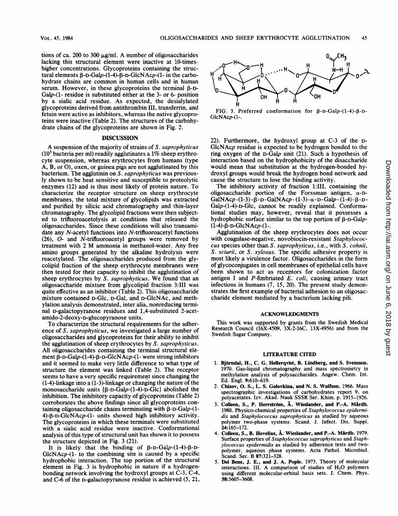
FIG. 2. Carbohydrate chains of the glycoproteins human antithrombin III and human transferrin (a) and fetuin (b, c, and d). Abbreviationsare given in the text.
INFECT. IMMUN.
on June 6, 2018 by guesthttp://iai.asm
.org/D
ownloaded from
OLIGOSACCHARIDES AND SHEEP ERYTHROCYTE AGGLUTINATION 45
tions of ca. 200 to 300 ,ug/ml. A number of oligosaccharideslacking this structural element were inactive at 10-times-higher concentrations. Glycoproteins containing the struc-tural elements P-D-Ga1p-(1-4)-P-D-GlcNAcp-(1- in the carbo-hydrate chains are common in human cells and in humanserum. However, in these glycoproteins the terminal P-D-Galp-(1- residue is substituted either at the 3- or 6- positionby a sialic acid residue. As expected, the desialylatedglycoproteins derived from antithrombin III, transferrin, andfetuin were active as inhibitors, whereas the native glycopro-teins were inactive (Table 2). The structures of the carbohy-drate chains of the glycoproteins are shown in Fig. 2.
DISCUSSIONA suspension of the majority of strains of S. saprophyticus
(105 bacteria per ml) readily agglutinates a 1% sheep erythro-cyte suspension, whereas erythrocytes from humans (typeA, B, or 0), oxen, or guinea pigs are not agglutinated by thisbacterium. The agglutinin on S. saprophyticus was previous-ly shown to be heat sensitive and susceptible to proteolyticenzymes (12) and is thus most likely of protein nature. Tocharacterize the receptor structure on sheep erythrocytemembranes, the total mixture of glycolipids was extractedand purified by silicic acid chromatography and thin-layerchromatography. The glycolipid fractions were then subject-ed to trifluoroacetolysis at conditions that released theoligosaccharides. Since these conditions will also transami-date any N-acetyl functions into N-trifluoroacetyl functions(26), 0- and N-trifluoroacetyl groups were removed bytreatment with 2 M ammonia in methanol-water. Any freeamino groups generated by the alkaline hydrolysis werereacetylated. The oligosaccharides produced from the gly-colipid fraction of the sheep erythrocyte membranes werethen tested for their capacity to inhibit the agglutination ofsheep erythrocytes by S. saprophyticus. We found that anoligosaccharide mixture from glycolipid fraction 3:11I wasquite effective as an inhibitor (Table 2). This oligosaccharidemixture contained D-Glc, D-Gal, and D-GlcNAc, and meth-ylation analysis demonstrated, inter alia, nonreducing termi-nal D-galactopyranose residues and 1,4-substituted 2-acet-amido-2-deoxy-D-glucopyranose units.To characterize the structural requirements for the adher-
ence of S. saprophyticus, we investigated a large number ofoligosaccharides and glycoproteins for their ability to inhibitthe agglutination of sheep erythrocytes by S. saprophyticus.All oligosaccharides containing the terminal structural ele-ment P-D-Galp-(1-4)-P-D-GlcNAcp-(1- were strong inhibitorsand it seemed to make very little difference to what type ofstructure the element was linked (Table 2). The receptorseems to have a very specific requirement since changing the(1-4)-linkage into a (1-3)-linkage or changing the nature of themonosaccharide units [3-D-Galp-(1-4)-D-Glc] abolished theinhibition. The inhibitory capacity of glycoproteins (Table 2)corroborates the above findings since all glycoproteins con-taining oligosaccharide chains terminating with ,-D-Galp-(1-4)-P-D-GlcNAcp-(1- units showed high inhibitory activity.The glycoproteins in which these terminals were substitutedwith a sialic acid residue were inactive. Conformationalanalysis of this type of structural unit has shown it to possessthe structure depicted in Fig. 3 (21).
It is likely that the binding of P-D-Galp-(1-4)-,B-D-GlcNAcp-(1- to the combining site is caused by a specifichydrophobic interaction. The top portion of the structuralelement in Fig. 3 is hydrophobic in nature if a hydrogen-bonding network involving the hydroxyl groups at C-3, C-4,and C-6 of the D-galactopyranose residue is achieved (5, 21,
FIG. 3. Preferred conformation for 0-D-Galp-(1-4)-P-D-GlcNAcp-(1-.
22). Furthermore, the hydroxyl group at C-3 of the D-GlcNAcp residue is expected to be hydrogen bonded to thering oxygen of the D-Galp unit (21). Such a hypothesis ofinteraction based on the hydrophobicity of the disaccharidewould mean that substitution at the hydrogen-bonded hy-droxyl groups would break the hydrogen bond network andcause the structure to lose the binding activity.The inhibitory activity of fraction 1:111, containing the
oligosaccharide portion of the Forssman antigen, a-D-GalNAcp-(1-3) - ,1-D-GalNAcp - (1-3)-a- D-Galp - (1-4)- I - D-Galp-(1-4)-D-Glc, cannot be readily explained. Conforma-tional studies may, however, reveal that it possesses ahydrophobic surface similar to the top portion of ,-D-Galp-(1-4)-P-D-GlcNAcp-(1-.
Agglutination of the sheep erythrocytes does not occurwith coagulase-negative, novobiocin-resistant Staphylococ-cus species other than S. saprophyticus, i.e., with S. cohnii,S. sciurii, or S. xylosus. The specific adhesive property ismost likely a virulence factor. Oligosaccharides in the formof glycoconjugates in cell membranes of epithelial cells havebeen shown to act as receptors for colonization factorantigen I and P-fimbriated E. coli, causing urinary tractinfections in humans (7, 15, 20). The present study demon-strates the first example of bacterial adhesion to an oligosac-charide element mediated by a bacterium lacking pili.
ACKNOWLEDGMENTSThis work was supported by grants from the Swedish Medical
Research Council (16X-4509, 3X-2-16C, 13X-4956) and from theSwedish Sugar Company.
LITERATURE CITED1. Bj6rndal, H., C. G. Hellerqvist, B. Lindberg, and S. Svensson.
1970. Gas-liquid chromatography and mass spectrometry inmethylation analysis of polysaccharides. Angew. Chem. Int.Ed. Engl. 9:610-619.
2. Chizov, 0. S., L. S. Golovkina, and N. S. Wulfson. 1966. Massspectrographic investigations of carbohydrates report 9, onpolyacetates. Izv. Akad. Nauk SSSR Ser. Khim. p. 1915-1926.
3. Colleen, S., P. Herrstrom, A. Wieslander, and P.-A. Mardh.1980. Physico-chemical properties of Staphylococcus epidermi-dis and Staphylococcus saprophyticus as studied by aqueouspolymer two-phase systems. Scand. J. Infect. Dis. Suppl.24:165-172.
4. Colleen, S., B. Hovelius, A. Wieslander, and P.-A. M&rdh. 1979.Surface properties of Staphylococcus saprophyticus and Staph-ylococcus epidermidis as studied by adherence tests and two-polymer, aqueous phase systems. Acta Pathol. Microbiol.Scand. Ser. B 87:321-328.
5. Del Bene, J. E., and J. A. Pople. 1973. Theory of molecularinteractions. III. A comparison of studies of H20 polymersusing different molecular-orbital basis sets. J. Chem. Phys.58:3605-3608.
VOL. 45, 1984
on June 6, 2018 by guesthttp://iai.asm
.org/D
ownloaded from

46 GUNNARSSON ET AL.
6. Dodge, J. T., C. Mitchell, and D. J. Hanahan. 1963. Preparationand chemical characteristics of hemoglobin: free ghosts ofhuman erythrocytes. Arch. Biochem. Biophys. 100:119-130.
7. Evans, D. G., J. D. J. Evans, Jr., and W. Tjoa. 1977. Hemagglu-tination of human group A erythrocytes by enterotoxigenicEscherichia coli isolated from adults with diarrhea: correlationwith colonization factor. Infect. Immun. 18:330-337.
8. Franzen, L. E., S. Svensson, and 0. Larm. 1980. Structuralstudies on the carbohydrate portion of human antithrombin III.J. Biol. Chem. 255:5090-5093.
9. Hakomori, S.-I. 1964. A rapid permethylation of glycolipid, andpolysaccharide catalyzed by methylsulfinyl carbanion in di-methyl sulfoxide. J. Biochem. (Tokyo) 55:205-208.
10. Hovelius, B., S. Colleen, and P.-A. Mardh. 1984. Urinary tractinfections in men caused by Staphylococcus saprophyticus.Scand. J. Infect. Dis. Suppl. 16:37-41.
11. Hovelius, B., and P.-A. Mardh. 1979. On the diagnosis ofcoagulase-negative staphylococci site with emphasis on Staphy-lococcus saprophyticus. Acta Pathol. Microbiol. Scand. Sect. B85:427-434.
12. Hovelius, B., and P.-A. Mardh. 1979. Hemagglutination byStaphylococcus saprophyticus and other staphylococcal spe-cies. Acta Pathol. Microbiol. Scand. Sect. B 87:45-50.
13. Hovelius, B., P.-A. Mardh, and P. Bygren. 1979. Urinary tractinfections caused by Staphylococcus saprophyticius-recurren-ces and complications. J. Urol. 122:645-647.
14. Hovelius, B., I. Thelin, and P.-A. Mardh. 1979. Staphylococcussaprophyticus in the aetiology of nongonococcal urethritis. Brit.J. Vener. Dis. 55:369-374.
15. Kallenius, G., R. Mollby, S. B. Svenson, J. Winberg, A. Lund-blad, S. Svensson, and B. Cedergren. 1980. The pk antigen asreceptor for the haemagglutinin of pyelonephritic Escherichiacoli. FEMS Microbiol. Lett. 7:297-302.
16. Karlsson, K. A., B. E. Samuelsson, and G. 0. Steen. 1973. Thesphingolipid composition of bovin kidney cortex, medulla andpapilla. Biochim. Biophys. Acta 316:317-335.
17. Kloos, W. E., and K. H. Schleifer. 1975. Simplified scheme forroutine identification of human Staphylococcus species. J. Clin.Microbiol. 1:82-88.
18. Kobata, A. 1972. Isolation of oligosaccharides from human milk.Methods Enzymol. 28:262-271.
19. Leffler, H., H. Lomberg, E. Gotschlich, L. Hagberg, U. Jodal, T.Korhonen, B. E. Samuelsson, G. Schoolnik, and C. Svanborg-Eden. 1982. Chemical and clinical studies on the interaction ofEscherichia coli with host glycolipid receptors in urinary tractinfection. Scand. J. Infect. Dis. Suppl. 33:46-51.
20. Leffler, H., and C. Svanborg-Eden. 1980. Chemical identifica-tion of a glycosphingolipid receptor for Escherichia coli attach-ing to human urinary tract epithelial cells and agglutinatinghuman erythrocytes. FEMS Microbiol. Lett. 8:127-134.
21. Lemieux, R. U. 1982. The binding of carbohydrate structureswith antibodies and lectins. Front. Chem. 28:3-24.
22. Lemieux, R. U., and A. A. Pavia. 1969. Substitutional andsolvation effects on conformational equilibria. Effects on theinteraction between opposing axial oxygen atoms. Can. J.Chem. 47:4441-4446.
23. Lundblad, A., S. Sjoblad, and S. Svensson. 1978. Characteriza-
tion of a penta- and an octasaccharide from urine of a patientwith juvenile GM1-gangliosidosis. Arch. Biochem. Biophys.188:130-136.
24. Marcus, D. M., and R. Janis. 1970. Localization of glycosphin-golipids in human tissues by immunofluorescense. J. Immunol.104:1530-1539.
25. Maskell, R. 1974. Importance of coagulase-negative staphylo-cocci as pathogens in the urinary tract. Lancet ii:1155-1158.
25a.Nilsson, B., N.-E. Norden, and S. Svensson. 1979. Structuralstudies on the carbohydrate portion of fetuin. J. Biol. Chem.254:4545-4553.
26. Nilsson, B., and S. Svensson. 1978. A new method for N-deacetylation of 2-acetamido-2-deoxy-sugars. Carbohydr. Res.62:377-380.
27. Nilsson, B., and S. Svensson. 1979. A new method for degrada-tion of the protein part of glycoproteins: isolation of thecarbohydrate chains of asialofetuin. Carbohydr. Res. 72:183-190.
28. Nilsson, B., and D. Zopf. 1983. Oligosaccharides released fromglycolipids by trifluoroacetolysis can be analysed by gas chro-matography-mass spectrometry. Arch. Biochem. Biophys.222:628-648.
29. Sawardeker, J. S., J. H. Sloneker, and A. R. Jeanes. 1965.Quantitative determination of monosaccharides as their alditolacetates by gas-liquid chromatography. Anal. Chem. 12:1602-1604.
30. Scott, T. A., Jr., and E. H. Melvin. 1953. Determination ofdextran with anthrone. Anal. Chem. 25:1656-1661.
31. Sellin, M., D. I. Cooke, W. A. Gillespie, D. G. H. Sylvester, andJ. D. Andersson. 1975. Micrococcal urinary tract infections inyoung women. Lancet ii:570-572.
32. Seyama, Y., and T. Yamakawa. 1974. Chemical structure ofglycolipid of guinea pig red blood cell membrane. J. Biochem.75:837-842.
33. Smith, D., D. Zopf, and V. Ginsburg. 1978. Sialyl oligosaccha-rides from milk. Methods Enzymol. 50:221-226.
34. Spik, G., R. Vandersyppe, B. Fournet, B. Bayard, P. Charet, S.Bouquelet, G. Strecker, and J. Montreuil. 1973. Structure ofglycopeptides isolated from human serotransferrin and lacto-transferrin. Methodologie Struct. Metab. Glycoconjuges221:483-499.
35. Spiro, R. G. 1960. Studies on fetuin, a glycoprotein of fetalserum. J. Biol. Chem. 235:2860-2869.
36. Spiro, R. G., and V. D. Bhoyroo. 1974. Structure of the 0-glycosidically linked carbohydrate units of fetuin. J. Biol.Chem. 249:5704-5717.
37. Strecker, G., A. Pierce-Cretel, B. Fournet, G. Spik, and J.Montreuil. 1981. Characterization by gas-liquid chromatogra-phy-mass spectrometry of oligosaccharides resulting from thehydrazinolysis-nitrous acid deamination reaction of glycopep-tides. Anal. Biochem. 111:17-26.
38. Suzuki, C., A. Makita, and Z. Yosizawa. 1968. Glycolipidsisolated from porcine intestine. Arch. Biochem. Biophys.127:140-149.
39. Wallmark, G., I. Arremark, and B. Telander. 1978. Staphylo-coccus saprophyticus: a frequent cause of acute urinary tractinfection among female outpatients. J. Infect. Dis. 138:791-797.
INFECT. IMMUN.
on June 6, 2018 by guesthttp://iai.asm
.org/D
ownloaded from