Infected non union
41
INFECTED NON-UNION DR SAGAR TOMAR llrm medical college meerut,up
-
Upload
sagar-tomar -
Category
Health & Medicine
-
view
176 -
download
10
Transcript of Infected non union

INFECTED NON-UNION
DR SAGAR TOMAR
llrm medical college meerut,up

NON UNION
Definition
A state in which healing process comes to a halt
as judged by clinical & x-ray evidence, beyond
the stipulated period of healing for a particular
bone and fracture pattern due to mechanical or
biological failure

CAUSES OF NON UNION

BASED ON THE EXTENT OF INFECTION
NON-INFECTED NON-UNION
INFECTED NON-
UNION
CLASSIFICATION OF NON UNION

CLASSIFICATION OF NON-INFECTED NON-UNION
HYPERTROHIC NON
UNION
Hypervascular
nonunions have shown
uptake of strontium-85,
which indicates a rich
blood supply in the ends
of the fragments
ATROPHIC NON
UNION
strontium-85 uptake in
these nonunions
indicate a poor blood
supply in the ends of
the fragments

HYPERTRROPHIC NON-UNION
1 “Elephant foot” nonunions
These are hypertrophic and rich in callus. They result from insecure fixation, inadequate immobilization, or premature weight bearing in a reduced fracture with viable fragments.
2 “Horse hoof” nonunions
These are mildly hypertrophic and poor in callus. They typically occur after a moderately unstable fixation with plate and screws. The ends of the fragments show some callus, insufficient for union, and possibly a little sclerosis.
3 Oligotrophic nonunions
These are not hypertrophic, but are vascular, and callus is absent. They typically occur after major displacement of a fracture, distraction of the fragments, or internal fixation without accurate apposition of the fragments.
Hypervascular nonunions.
A, “Elephant foot” nonunion.
B, “Horse hoof” nonunion.
C, Oligotrophic nonunion

AVASCULAR NON-UNION 1 Torsion wedge nonunions
These are characterized by the presence of an intermediate fragment in which the blood supply is decreased or absent. The intermediate fragment has healed to one main fragment but not to the other.
2 Comminuted nonunions
These are characterized by the presence of one or more intermediate fragments that are necrotic. The radiographs show absence of any sign of callus formation.
3 Defect nonunions
These are characterized by the loss of a fragment of the diaphysis of a bone. The ends of the fragments are viable, but union across the defect is impossible. As time passes, the ends of the fragments become atrophic.
4 Atrophic nonunions
These usually are the final result when intermediate fragments are missing and scar tissue that lacks osteogenic potential is left in their place. The ends of the fragments have become osteoporotic and atrophic.
Avascular nonunions.
A, Torsion wedge nonunion.
B, Comminuted nonunion.
C, Defect nonunion.
D, Atrophic nonunion

Paley et al.classification of non-union
Type A nonunions(<1 cm of bone loss)
A1, lax (mobile)
A2, stiff (nonmobile)
A2-1, no deformity
A2-2, fixed deformity.
Type B nonunions(>1 cm of bone loss)
B1, bony defect, no shortening
B2, shortening, no bony defect;
B3, bony defect and shortening.

WEILAND CLASSIFICATION OF INFECTED NONUNION
Based on the extend of infection
type 1
characterised by open and exposed bone without
osseous infection but with soft tissue infection
type 2
characterised by circumferential cortical and
endostesl infection with often and invlocrum
surroundind a sequestrum
type 3
characterised by cortical-endosteal infection
associated with a segmental bone defect.

CIERNY MADAR CLASSIFICATION
Cierny and Mader developed a classification system for chronic osteomyelitis, based on physiological and anatomical criteria, to determine the stage of infection.
Based on host class A- NORMAL
class B- COMPROMISED
class C-PROHIBITIVE
Based on anatomytype 1-MEDULLARY
type 2-SUPERFICIAL
type 3-LOCALISED
type 4-DIFFUSE
pairing of these forms 12 clinical stages
Clinical Stage
(Type+ Class = Clinical Stage)

UMIAROV’S CLASSIFICATION OF
INFECTED NON-UNION
based on the viability of bone ends, the presence of
limb shortening, the presence of bone, and soft
tissue defect.
- type 1 the nonunion is normotrophic without
shortening
- type 2 the nonunion is hypertrophic with shortening
- type 3 the nonunion is atrophic with shortening
- type 4 the nonunion is atrophic with bone and soft
tissue defect, in general as a result of an
open
fracture

G.S KULKARNI CLASSIFICATION OF
INFECTED NON UNION
Severity of infection
Apposition of fragments
Presence or absence of deformity.

G.S KULKARNI CLASSIFICATION OF
INFECTED NON UNION
TYPE I:
fragments in apposition with mild infection and with or with out implant
TYPE II:
Fragments in apposition with severe infection with large or small wound.
TYPE III:
Severe infection with a gap or deformity or shortening.
3A defect with loss of full circumference
3B defect in > 1/3 of cortex
3C infected nonunion with deformity.

GORDON’S CLASSIFICATION
TYPE A
Tibial defects and non unions without significant
bone
loss
TYPE B
tibial defects greater than 3 cm with an intact fibula
TYPE C
tibial defect greater than 3 cm without intact fibula

MAY’S CLASSIFICATION
-it focuses on the status of tibia after bone and soft tissue
debridement
-it helps to estimate the length of rehabilitation period before
ambulation
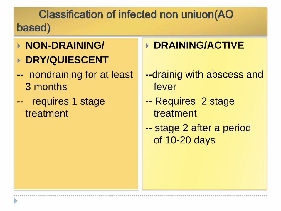
NON-DRAINING/
DRY/QUIESCENT
-- nondraining for at least
3 months
-- requires 1 stage
treatment
DRAINING/ACTIVE
--drainig with abscess and
fever
-- Requires 2 stage
treatment
-- stage 2 after a period
of 10-20 days

How infection causes non union??
1. Dissection of pus through planes and periosteum-
devascularising th ends
2. Fragmentation and dissolution of fracture
haematoma
3. Inflammatory mediators promotes fibrous tissue
formation
4. If fixation was done then implant failure occurs
destabilization the fragments
5. Increase catabolic response at # ends

PATHOGENESIS
OSTEOMYELITIS
thrombosis of blood vessel of haversian canals
bone sclerosis and dead bone.
Butterfly fragments become sequestrii, isolated & devitalized by pus & INFECTED GRANULATION TISSUE

Infection granulation tissue
OSTEOLYIS
GAP NON UNION
Osteolysis occurs around the implants loosening instability of fixation nonunion.

DIAGNOSIS OF INFECTED NON UNION
1. Pain and mobility at fracture site
2. Raised local temperature
3. Discharging sinus
4. Scar healed by secondary intension with
“puckering”
5. Irregularity of bone showing osteomyelitis

INVESTIGATIONS
Include - complete blood count
- erythrocyte sedimentation rate (ESR)
- C- reactive protein (CRP)
Plain radiogarphy
Sinography
Radionucleotide scan
MRI
CT-scan
Culture sensitivity
usg

goals
Treatment of Infected non-union…
ERADICATE INFECTION
ACHIEVE UNION
SOLVE:soft tissue problem,deformity,joint
stiffness
GOAL 1
GOAL 2
GOAL 3

GOAL 1 : ERADICATE INFECTION
INCREASE HOST RESISTANCE :
Correct host morbidity
-control blood sugar level in diabetic
-smoking cessation
-treatment of liver or renal malfunction
-optimising nutrition
-treatment of chronic disease
Antibiotic therapy according to culture sensitivity
reports.
- systemic antibiotic therapy

LOCAL CONTROL OF INFECTION
DECREASE INFECTION LOAD:
• thorough debridement of dead and necrotic tissue
• closed suction antibiotic ingress and egress irrigation systems.
• negative suction drainage system.
INCREASE LOCAL HOST RESISTANCE:
• PMMA antibiotics beads
• biodegradable antibiotic delivery system

GOAL 2 : TO ACHIEVE UNION
• ADDING BIOLOGY
– Aspirated stem cells (with or without expansion)
– Demineralized Bone Matrix
– Autogenous Cancellous Graft
– Growth Factors
• Platelet derived
• Recombinant BMPs
• Gene Therapy
EXTERNAL STIMULI
-low intensity ultrasound therapy
-electric and electromegnetic therapy

• Aspirated iliac crest stem cells has been shown to enhance the activity of osteoconductive grafts.
• There are few commercially available Recombinant BMP proved to be effective treating nonunions.

Bone grafting in infected non union
Onlay bone grafting: graft applied or laid on the surface
of a bone
Inlay bone grafting: By the inlay technique a slot or
rectangular defect is created in the
cortex of the host bone, usually A graft
the same size or slightly smaller is then
fitted
into the defect.
Single onlay dual onlay
Cancellous
Insert graft

Papinaeu method of bone grafting
Stage I: Radical debridement
Stage II: bone grafting
Stage III: skin coverage.

HARMONS’ POSTEROLATERAL GRAFT
Bone grafting on the
interosseous membrane
to obtain a long
synostosis with fibula,
spanning the tibial
defect.

Free vascularised bone transfer
Rib, fibula, iliac crest.
Isolation of a segment
of contra lateral fibula
with attached nutrient
artery and vein.
Length of graft should
be 4 cm longer than
defect to allow 2 cm
overlap at the proximal
and distal ends.

ULTRASOUND THERAPY
it cause increases in cellular activity at osteotomy
sites and increases in mineralization of the bone and
metabolic activity.
It promotes bone healing because it stimulates the
genes involved in inflammation and bone
regeneration.
It increases blood flow through dilation of capillaries
and enhancement of angiogenesis, increasing the
flow of nutrients to the fracture site.
Used : for 20 min / day

ELECTRICAL AND ELECTROMAGNETIC
STIMULATION
used for 3 or more hours per day has been
successful in healing nonunions of long and short
bones, open or closed fractures, long-standing
nonunions, infected nonunions, and those with
fracture gaps up to 1 cm.

The three methods of administering electric stimulation are shown in this diagram.
(a) Direct current (DC): A cathode is implanted at the fracture site which is attached to either a subcutaneous power source or an external power source to generate an electric field at the fracture site.
(b) Capacitive coupling(CC): Two capacitive coupled electrodes are situated on the skin on either sides of the fracture site. An external power source is then attached to the electrodes, which induces an electric field at the fracture site.
(c) Inductive coupling (IC): An electromagnetic current carrying coil is placed on the skin overlying the fracture site, which is attached to an external power source. The coil generates a magnetic field, which induces an electrical field at the fracture site.

GOAL NO.3
SOFT TISSUE PROBLEMS:
The transfer of vascularized muscle tissue improves
the local biological environment by bringing in a
blood supply that is important in the host's defense
mechanisms and for antibiotic delivery and osseous
and soft tissue healing
DEFORMITY AND SHORTENING:
- Ilizarov is the gold standard treatment to correct
deformity and shortening and eradicate infection at
the same time

TREATMENT PROTOCOL (AO BASED)
queiscent nonunion
use of short term antibiotic prophylaxis
excise dead bone and scarred ,non vascularised soft tissue
remove loose hardware
correct alignment
perform cancellous bone grafting if required
stabilise using plate or intramedullary devise or external
fixator
mobilise adjacent joints by physiotherapy
QUEISCENT NON
UNION

ACTIVE NON UNION
STAGE 1
thorough debridement
- excise and debride
- drain using closed suction irrigation
- antibiotics beads and i.v. antibiotics
- stabilize with external fixator
STAGE 2 after 10-20 days
shingling and cancellous bone auto graft.
STAGE 3
additional bone graft ,muscle or skin pedicle flap can be used

Shingling and cancellous bone
grafting
Shingling Cancellous bone graft
Shingling:a process by which ends of the fracture
Fragments are decorticated subperiosteally forming many
Small osteoperiosteal flaps.

ELIMINATION OF INFECTION IN ILIZAROV
METHOD
Resection of infected bone and subsequent
intercalary bone lengthening
gradual bone transport of one wall of the cavity
Controlled osteogenesis, filling of cavities by newly
formed tissue

A corticotomy is performed to fracture the bone into two segments, and the two bone ends of the bone are gradually moved apart during the distraction phase, allowing new bone to form in the gap. When the desired or possible length is reached, a consolidation phase follows in which the bone is allowed to keep healing.

CONCLUSION : general approach to
infection

Thank you