Induction of Rhythmic Jaw Movements by Stimulation of … Journal of Neuroscience, August 1989,...
15
The Journal of Neuroscience, August 1989, g(8): 2887-2901 Induction of Rhythmic Jaw Movements by Stimulation of the Mesencephalic Reticular Formation in the Guinea Pig Nobuyuki Hashimoto,lva Takayuki Katayama,’ Yasuo Ishiwata,2 and Yoshio Nakamural Departments of ‘Physiology and zOrthodontics II, Faculty of Dentistry, Tokyo Medical and Dental University, Tokyo 113, Japan This study was designed to investigate whether stimulation of the mesencephalic reticular formation (MRF) induces rhythmic jaw movements (RJMs) and, if it does so, to de- termine the RJM-inducing region in the MRF in ketamine- anesthetized guinea pigs. The results were as follows: (1) Repetitive electrical stimulation of the MRF at the level of the red nucleus induced rhythmic EMG bursts in the anterior digastric muscle (DIG). (2) The duration and cycle time of the rhythmic DIG EMG burst induced from the medial MRF were longer than those induced from the lateral MRF. (3) Repetitive MRF stimulation after paralysis still induced rhythmic multiple-unit activities in the anterior digastric mo- toneuron pool. (4) Neither precollicular decerebration nor cerebellectomy affected the MRF induction of RJMs. (5) Transverse hemisection at the rostra1 border of the pons abolished the RJMs induced from the contralateral, but not ipsilateral, MRF. Midline section of the midbrain abolished RJMs induced from the MRF on either side. (6) A lesion in the pontine pyramidal tract abolished the RJMs induced by stimulation of the ipsilateral cortical masticatory area (CMA), but not those induced from the contralateral MRF. (7) A uni- lateral lesion of the oral portion of the gigantocellular retic- ular nucleus, where the rhythm generator for the CMA-in- duced RJMs is located, abolished RJMs induced from not only the CMA, but also MRF on the contralateral side. (6) Microinjection of L-glutamate into the lateral, but not medial, MRF induced RJMs similar to those elicited by repetitive electrical stimulation of the same site. The results indicate that activation of MRF neurons can centrally induce RJMs via an extrapyramidal route, crossing to the contralateral side at the midbrain level and descending to the bulbar re- ticular formation. Stimulation of various subcortical structures, aswell asthe ce- rebral cortex in a number of species, induces rhythmic jaw movements (RJMs), and the cortical area that induces RJMs upon stimulation has been called the cortical masticatory area (CMA) (see Luscheiand Goldberg, 1981, for review). Lund and Dellow (197 1) reported that RJMs could be induced by repet- Received Aug. 15, 1988; revised Jan. 23, 1989; accepted Jan. 30, 1989. This study was supported by a Grant-in-Aid for Special Project Research from the Minis&v of Education. Science and Culture of Jaoan. Correspondence should be addressed to Yoshio Gaiamura, M.D., Department of Physiology, Faculty of Dentistry, Tokyo Medical and Dental University, 5-45 Yushima 1-chome, Bunkyo-ku, Tokyo 113, Japan. a Present address: 2nd Department of Prosthodontics, School of Dentistry, Na- gasaki University, Nagasaki 852, Japan. Copyright 0 1989 Society for Neuroscience 0270-6474/89/082887-15$02.00/O itive electrical stimulation of an extensive subcortical region in rabbits, including the basal ganglia, corticobulbar tract, limbic structures, thalamus, and mesencephalic reticular formation (MRF). Sumi (1971) found, however, that repetitive electrical stimulation of the MRF not only enhanced, but alsosuppressed the cortically induced RJMs in rabbits. Recently, Chandler and Tal (1987) reported that repetitive microstimulation of wide- spread areas of the rostra1 pons and midbrain produced suppres- sion of the cortically induced RJMs in guinea pigs.In addition, Tal (1987) demonstratedthat the induction of RJMs by stim- ulation of the ventral MRF was, in fact, due to current spread to the fibers in the cerebral peduncle which carried the impulses from the CMA to the masticatory rhythm generator in the bulbar reticular formation of the guinea pig (Nozaki et al., 1986a). The purposeof this study was (1) to determine whether elec- trical stimulation of the MRF can induce RJMs; (2) if it does, to delineate the exact location of the effective sites; and (3) to make clear whether activation of the MRF neuronsis involved in the RJM induction by electrical stimulation of the MRF. A part of this study was reported in abstract form (Hashimoto et al., 1987). Materials and Methods Sixty-five female albino guinea pigs were used. Thirty minutes after they were given chlorpromazine hydrochloride (2.5 mg/kg, i.m.) and atropine sulfate (0.1 mg/kg, i.p.), ketamine hydrochloride was given (100 mg/ kg, i.m.). A midline skin incision was made from the mandibular sym- physis to the top of the sternum. After the trachea was cannulated, artificial ventilation was started. The masseter (MASS) and anterior digastric muscles (DIG) were exposed bilaterally. A pair of steel needle electrodes (0.2 mm diameter, enamel-coated except for 0.5 mm at the tip) were inserted into each of these muscles for recording EMG. After lidocaine was injected to the pressure points, the head of the animal was fixed to a stereotaxic apparatus after Rossner’s method (1965; the interaural line: APO, HO). The spinal cord was transected at the C2 level. Parts of the frontal, temporal, and occipital bones were removed, and the dura was opened to expose the dorsal aspect of the cerebral cortex. The surface of the exposed cortex was covered with cotton wicks soaked with the physiological saline. An Ag-AgCl plate was placed subcutaneously in the left neck region. Anesthesia was maintained throughout the experiment by supplemental ketamine hydrochloride (10 mg/kg/hr, i.m.), except for 7 animals decerebrated at the precollic- ular level (stereotaxic coordinate A&O), with a small knife made from a razor blade and mounted on an electrode carrier through a metal tube glued to its top. The cerebellum was removed by suction in 11 animals. Midline sec- tions of the brain stem at the midbrain level (3 animals), as well as a hemisection at the rostra1 border of the pons (8 animals, including one decerebrate and one cerebellectomized animal) were performed by means of the same type of small knife as used for decerebration. The pyramidal tract was sectioned at the rostra1 pontine level (A2.0) unilaterally in 4 animals, including 1 decerebrate and 1 cerebellectomized animal, on the side contralateral to stimulation of the MRF, by passing positive
Transcript of Induction of Rhythmic Jaw Movements by Stimulation of … Journal of Neuroscience, August 1989,...
The Journal of Neuroscience, August 1989, g(8): 2887-2901
Induction of Rhythmic Jaw Movements by Stimulation of the Mesencephalic Reticular Formation in the Guinea Pig
Nobuyuki Hashimoto,lva Takayuki Katayama,’ Yasuo Ishiwata,2 and Yoshio Nakamural
Departments of ‘Physiology and zOrthodontics II, Faculty of Dentistry, Tokyo Medical and Dental University, Tokyo 113, Japan
This study was designed to investigate whether stimulation of the mesencephalic reticular formation (MRF) induces rhythmic jaw movements (RJMs) and, if it does so, to de- termine the RJM-inducing region in the MRF in ketamine- anesthetized guinea pigs. The results were as follows: (1) Repetitive electrical stimulation of the MRF at the level of the red nucleus induced rhythmic EMG bursts in the anterior digastric muscle (DIG). (2) The duration and cycle time of the rhythmic DIG EMG burst induced from the medial MRF were longer than those induced from the lateral MRF. (3) Repetitive MRF stimulation after paralysis still induced rhythmic multiple-unit activities in the anterior digastric mo- toneuron pool. (4) Neither precollicular decerebration nor cerebellectomy affected the MRF induction of RJMs. (5) Transverse hemisection at the rostra1 border of the pons abolished the RJMs induced from the contralateral, but not ipsilateral, MRF. Midline section of the midbrain abolished RJMs induced from the MRF on either side. (6) A lesion in the pontine pyramidal tract abolished the RJMs induced by stimulation of the ipsilateral cortical masticatory area (CMA), but not those induced from the contralateral MRF. (7) A uni- lateral lesion of the oral portion of the gigantocellular retic- ular nucleus, where the rhythm generator for the CMA-in- duced RJMs is located, abolished RJMs induced from not only the CMA, but also MRF on the contralateral side. (6) Microinjection of L-glutamate into the lateral, but not medial, MRF induced RJMs similar to those elicited by repetitive electrical stimulation of the same site. The results indicate that activation of MRF neurons can centrally induce RJMs via an extrapyramidal route, crossing to the contralateral side at the midbrain level and descending to the bulbar re- ticular formation.
Stimulation of various subcortical structures, as well as the ce- rebral cortex in a number of species, induces rhythmic jaw movements (RJMs), and the cortical area that induces RJMs upon stimulation has been called the cortical masticatory area (CMA) (see Luschei and Goldberg, 198 1, for review). Lund and Dellow (197 1) reported that RJMs could be induced by repet-
Received Aug. 15, 1988; revised Jan. 23, 1989; accepted Jan. 30, 1989.
This study was supported by a Grant-in-Aid for Special Project Research from the Minis&v of Education. Science and Culture of Jaoan.
Correspondence should be addressed to Yoshio Gaiamura, M.D., Department of Physiology, Faculty of Dentistry, Tokyo Medical and Dental University, 5-45 Yushima 1-chome, Bunkyo-ku, Tokyo 113, Japan.
a Present address: 2nd Department of Prosthodontics, School of Dentistry, Na- gasaki University, Nagasaki 852, Japan. Copyright 0 1989 Society for Neuroscience 0270-6474/89/082887-15$02.00/O
itive electrical stimulation of an extensive subcortical region in rabbits, including the basal ganglia, corticobulbar tract, limbic structures, thalamus, and mesencephalic reticular formation (MRF). Sumi (1971) found, however, that repetitive electrical stimulation of the MRF not only enhanced, but also suppressed the cortically induced RJMs in rabbits. Recently, Chandler and Tal (1987) reported that repetitive microstimulation of wide- spread areas of the rostra1 pons and midbrain produced suppres- sion of the cortically induced RJMs in guinea pigs. In addition, Tal (1987) demonstrated that the induction of RJMs by stim- ulation of the ventral MRF was, in fact, due to current spread to the fibers in the cerebral peduncle which carried the impulses from the CMA to the masticatory rhythm generator in the bulbar reticular formation of the guinea pig (Nozaki et al., 1986a).
The purpose of this study was (1) to determine whether elec- trical stimulation of the MRF can induce RJMs; (2) if it does, to delineate the exact location of the effective sites; and (3) to make clear whether activation of the MRF neurons is involved in the RJM induction by electrical stimulation of the MRF. A part of this study was reported in abstract form (Hashimoto et al., 1987).
Materials and Methods Sixty-five female albino guinea pigs were used. Thirty minutes after they were given chlorpromazine hydrochloride (2.5 mg/kg, i.m.) and atropine sulfate (0.1 mg/kg, i.p.), ketamine hydrochloride was given (100 mg/ kg, i.m.). A midline skin incision was made from the mandibular sym- physis to the top of the sternum. After the trachea was cannulated, artificial ventilation was started. The masseter (MASS) and anterior digastric muscles (DIG) were exposed bilaterally. A pair of steel needle electrodes (0.2 mm diameter, enamel-coated except for 0.5 mm at the tip) were inserted into each of these muscles for recording EMG. After lidocaine was injected to the pressure points, the head of the animal was fixed to a stereotaxic apparatus after Rossner’s method (1965; the interaural line: APO, HO). The spinal cord was transected at the C2 level. Parts of the frontal, temporal, and occipital bones were removed, and the dura was opened to expose the dorsal aspect of the cerebral cortex. The surface of the exposed cortex was covered with cotton wicks soaked with the physiological saline. An Ag-AgCl plate was placed subcutaneously in the left neck region. Anesthesia was maintained throughout the experiment by supplemental ketamine hydrochloride (10 mg/kg/hr, i.m.), except for 7 animals decerebrated at the precollic- ular level (stereotaxic coordinate A&O), with a small knife made from a razor blade and mounted on an electrode carrier through a metal tube glued to its top.
The cerebellum was removed by suction in 11 animals. Midline sec- tions of the brain stem at the midbrain level (3 animals), as well as a hemisection at the rostra1 border of the pons (8 animals, including one decerebrate and one cerebellectomized animal) were performed by means of the same type of small knife as used for decerebration. The pyramidal tract was sectioned at the rostra1 pontine level (A2.0) unilaterally in 4 animals, including 1 decerebrate and 1 cerebellectomized animal, on the side contralateral to stimulation of the MRF, by passing positive

2888 Hashimoto et al. - Reticular Induction of Rhythmic Jaw Movements
direct current (90 PA for 30 set) through tungsten electrodes (tip resis- tance, 0.5-l MQ). A localized electrolytic lesion was made unilaterally in the bulbar reticular formation of 4 animals by passing positive direct current (90 PA, 30 set) through a tungsten electrode (tip resistance, 3- 5 MQ) stereotaxically inserted into the oral portion of the gigantocellular reticular nucleus (P2.0-2.2, L1.5-2.0, HO), where repetitive stimulation of the contralateral cortical masticatory area (CMA) induced a rhythmic field potential in the masticatory rhythm (Nozaki et al., 1986a). These lesions of the brain stem and the pyramidal tract were made under insufflation of 0.3% halothane. Body temperature was kept at about 37°C with a heating pad under the body and an incandescent lamp from above.
An electropolished, enamel-coated tungsten microelectrode (tip re- sistance, 0.5-l .O MO) was inserted vertically into the CMA (stereotaxic coordinates A15.5-16.0, L4.0) to a depth of 2.0 mm on one side. Re- petitive electrical stimulation (40 Hz, 0.3 msec, loo-350 PA) was de- livered through this electrode, which was fixed to the point where the threshold for induction of RJMs was lowest. Another tungsten micro- electrode of the same type was inserted vertically into the brain stem for stimulation. To study the sites effective for inducing RJMs, repetitive pulses at 40 Hz were systematically applied every 500 Nrn in the ros- trocaudal and mediolateral directions and 250 p-m in the vertical di- rection. The exnlored region extended from A3.0 to A8.0. from LO to L3.5, and from-the dorsal to ventral surface of the brain stem.
After the animal was paralyzed with pancuronium bromide (1.2 mg/ kg, i.m.), the induced rhythm was monitored by the multiple unit ac- tivity (MUA) in the anterior digastric motoneuron pool on the side contralateral to the cortical and brain-stem stimulations. The electrode (tip resistance, 5-8 MR) was fixed to the point where the largest anti- dromic field potential was recorded after stimulation of the anterior digastric nerve with the electrode for recording DIG EMG.
The EMG, MUA, and field potentials were displayed on an oscillo- scope after amplification (bandwidths: 33-3000 Hz, for EMG, 150- 3000 Hz for MUA, and 0.08-3000 Hz for field potential) and recorded on an FM tape recorder (flat frequency response from DC to 2500 Hz) for later analysis. Photographic and ink-written records were made from the recorded data. RJMs were videotaped in some experiments; the distance between the upper and lower incisal edges (interincisal distance) was measured at the largest open position during the jaw movement to monitor the maximal displacement of the jaw during each jaw-opening.
Glutamate (L-glutamic acid, monosodium) dissolved in 0.9% saline (50 mM, pH 8.0) was injected into the brain stem in 7 animals with a Hamilton microsyringe mounted on an electrode carrier. The solution also contained methylene blue (0.0 1 gm/ml) to mark the injection site. The injection needle was initially lowered stereotaxically to the dor- solateral part of the MRF; it was then left in place for 10 min to allow stabilization of the tissue. Subsequently, 4.0 ~1 glutamate solution was slowly injected over a period of 3 min. The injection needle was left in place for at least 10 min after injection. As a control, the same amount of the physiological saline was injected into the same region after the same procedure.
At the termination of each experiment, an overdose of pentobarbital sodium was given intraperitoneally, and microlesions were made at the stimulated and recorded points in the brain stem by applying positive DC (70 PA, 15 set) through stimulating and recording electrodes. The brain was taken out and kept in 10% formalin solution for 1 week. Serial coronal or horizontal frozen sections, 75 km thick, were made of the brain stem, stained with cresyl violet for verification of the stimulation and recording sites.
Results RJA4s and jaw muscle EMGs induced by repetitive electrical stimulation of the mesencephalic reticular formation Tungsten stimulating electrodes were stereotaxically inserted into the MRF on one side vertically through the superior col- liculus. Repetitive electrical stimulation of the MRF induced rhythmic opening and closing movements of the jaw. The RJM was similar to that induced by repetitive electrical stimulation of the CMA in that it was accompanied by a lateral movement to the side contralateral to stimulation. The lateral displacement of the jaw was larger in the opening movement induced from the lateral MRF (L2.0-3.0) than that from the CMA, but there
was no apparent difference in the lateral displacement of the jaw between that induced from the medial MRF (LO.5-1.5) and that induced from the CMA. The vertical displacement of the jaw during the opening movement induced from the MRF, either medial or lateral (interincisal distance, 7-8 mm), far exceeded that induced from the CMA (interincisal distance, 3-4 mm). The RJM induced from the medial MRF was accompanied by a rhythmic protrusion and retraction of the tongue in coordi- nation with the RJMs, while that induced from the lateral MRF was accompanied by essentially no tongue movements.
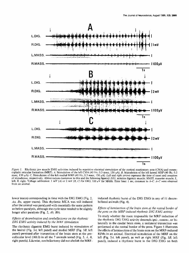
Figure 1 illustrates the rhythmic EMG burst activities of the anterior DIG and MASS muscles on both sides induced from the lateral (Fig. 1B) and medial (Fig. 1C’) MRF on the left side compared with those induced from the left CMA (Fig. 1A) in an animal.
To compare the pattern of RJMs induced from these 3 dif- ferent loci, samples of 10 consecutive DIG EMG cycles were obtained during a steady period of the induced RJMs in 5 an- imals. The cycle time and burst duration of the rhythmic DIG EMG were measured and averaged with respect to each RJM sequence. The values in Table 1 represent the mean and SD of these averaged cycle time and burst durations of each RJM sequence.
The left column of Table 1 shows the cycle time of the rhyth- mic DIG EMG burst induced by repetitive stimulation of the CMA and lateral and medial MRF. The cycle time of the RJM induced from the medial MRF was longer than that from the CMA 0, < 0.05, t test), while that induced from the lateral MRF was slightly, but significantly, shorter than that from the CMA (p < 0.05, t test).
The rhythmic burst activity appeared in the homonymous muscles on both sides, whether it was induced by stimulation of the CMA or MRF. The rhythmic EMG activity of the DIG was virtually the reciprocal of that of the MASS when induced by the CMA stimulation (Fig. 1A). When induced by stimulation of the lateral MRF (Fig. lB), however, the rhythmic burst ac- tivity of the MASS EMG occurred both in and out of phase with the DIG EMG activity. The out-of-phase burst appeared directly following the in-phase burst at the early interburst phase of the DIG EMG burst with a shorter duration and a higher amplitude than the in-phase burst. On the other hand, stimu- lation of the medial MRF induced very little MASS EMG ac- tivity (Fig. 1 c).
The right column of Table 1 shows the duration of the rhythm- ic DIG EMG burst induced by repetitive stimulation of the CMA and lateral and medial MRF. The duration of DIG EMG bursts induced from the medial MRF was longer than that in- duced from either the CMA or lateral MRF (p < 0.05, t test), while that induced from the lateral MRF was slightly shorter than that induced from the CMA (p < 0.05, t test). When the electrode track was moved mediolaterally or lateromedially, the change from one to the other of the above-mentioned 2 patterns induced from the lateral and medial MRF occurred gradually rather than abruptly: when we moved the points of insertion from the lateral to medial portion, the cycle time gradually lengthened, and this was accompanied by a gradual decrease in amplitude of the MASS EMG burst.
Effects of paralysis on the centrally induced rhythmic activity of the anterior digastric motoneurons
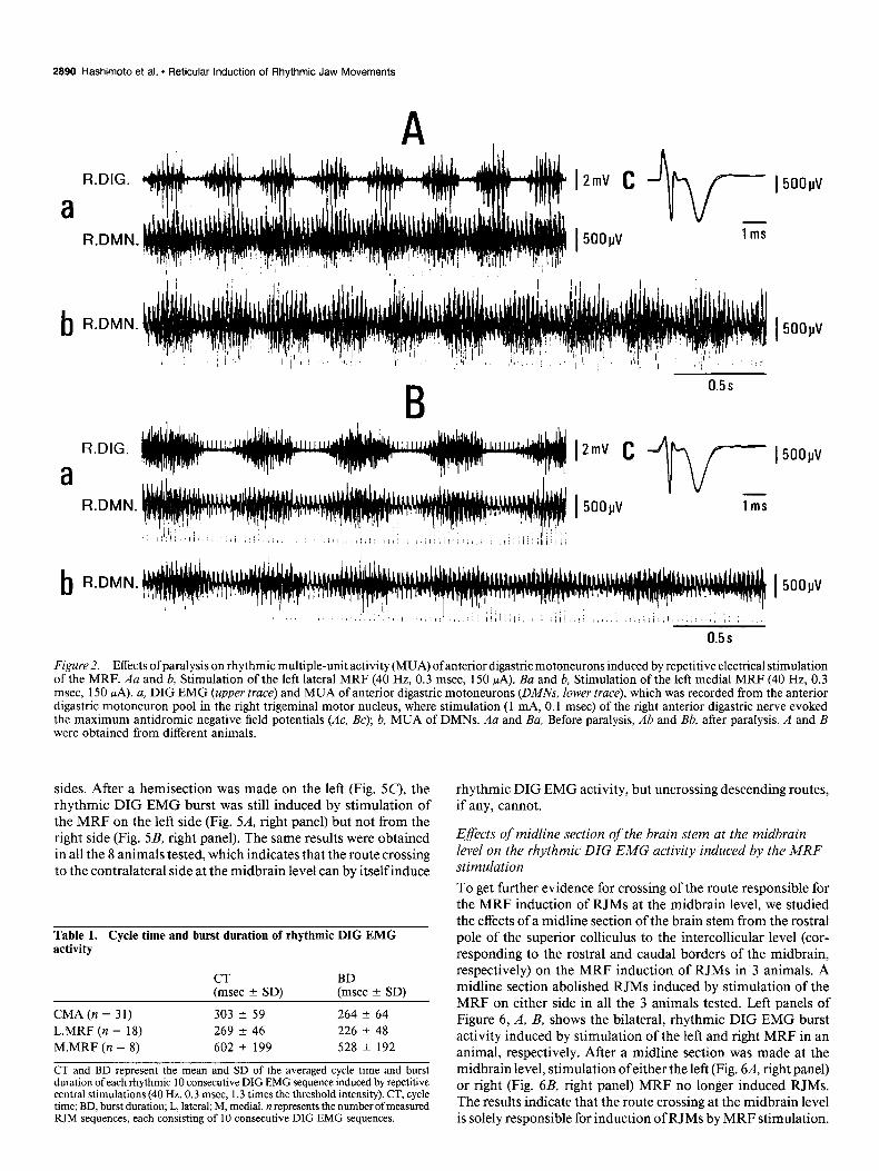
Repetitive electrical stimulation of the MRF induced a rhythmic MUA in the anterior digastric motoneuron pool (Fig. 2, Aa, Ba,
The Journal of Neuroscience, August 1989, 9(8) 2889
L.DIG. I
f?.DIG. 12mV
I ,./
IlOO~V
C 1s
1 1
L-DIG.
R.DIG.
L.MASS. I
R.MASS. “4% ,.- a IlOOyV
1s
Figure I. Rhythmic jaw muscle EMG activities induced by repetitive electrical stimulation of the cortical masticator-y area (CMA) and mesen- cephalic reticular formation (MRF). A, Stimulation of the left CMA (40 Hz, 0.3 msec, 150 PA). B, Stimulation of the left lateral MRF (40 Hz, 0.3 msec, 150 PA). C, Stimulation of the left medial MRF (40 Hz, 0.3 msec, 150 PA). Left and right arrows represent the time of onset and cessation of stimulation, respectively. Abbreviations (common to this and the following figures): DIG, anterior digastric muscle; MASS, masseter muscle; L, left; R, right. Voltage calibration: 1 mV (A) or 2 mV (B, C) for DIG, 100 NV for MASS. Time base: 1 sec. common to A-C. A-C were obtained from an animal.
lower traces) corresponding in time with the DIG EMG (Fig. 2, Au, Ba, upper traces). This rhythmic MUA was still induced after the animal was paralyzed with essentially the same pattern as before paralysis, although the cycle time tended to be slightly longer after paralysis (Fig. 2, Ab, Bb).
Efects of decerebration and cerebellectomy on the rhythmic DIG EMG activity induced by the MRF stimulation The rhythmic digastric EMG burst induced by stimulation of the lateral (Fig. 3A, left panel) and medial MRF (Fig. 3B, left panel) persisted after transection of the brain stem at the pre- collicular level (A8.0) in all the 7 animals tested (Fig. 3, A, B, right panels). Likewise, cerebellectomy did not abolish the MRF-
induced rhythmic burst of the DIG EMG in any of 11 decere- bellated animals (Fig. 4).
Eflects of hemisection of the brain stem at the rostra1 border of the pons on the MRF-induced rhythmic DIG EMG activity To study whether the route responsible for MRF induction of the rhythmic DIG EMG activity descends ipsi-, contra-, or bi- laterally in the caudal brain stem, a unilateral transection was performed at the rostra1 border of the pons. Figure 5 illustrates the effects of hemisection of the brain stem on the MRF-induced RJMs in an animal. Electrical stimulation of the MRF on the left (Fig. 5A, left panel), as well as right MRF (Fig. 5B, left panel), induced a rhythmic burst in the DIG EMG on both
2890 Hashimoto et al. * Reticular Induction of Rhythmic Jaw Movements
b R.DMN. I 5ooyv
R.DIG.
a R.DMN.
b FLDMN.
Figure 2. Effects of paralysis on rhythmic multiple-unit activity (MUA) ofanterior digastric motoneurons induced by repetitive electrical stimulation of the MRF. Au and b, Stimulation of the left lateral MRF (40 Hz, 0.3 msec, 150 WA). Ba and b, Stimulation of the left medial MRF (40 Hz, 0.3 msec, 150 PA). a, DIG EMG (upper truce) and MUA of anterior digastric motoneurons (DMA$ lower trace), which was recorded from the anterior digastric motoneuron pool in the right trigeminal motor nucleus, where stimulation (1 mA, 0.1 msec) of the right anterior digastric nerve evoked the maximum antidromic negative field potentials (AC, Bc): b. MUA of DMNs. Au and &I, Before paralysis, Ab and Bb, after paralysis. A and B were obtained from different animals.
sides. After a hemisection was made on the left (Fig. 5c), the rhythmic DIG EMG burst was still induced by stimulation of the MRF on the left side (Fig. 5.4, right panel) but not from the right side (Fig. 5B, right panel). The same results were obtained in all the 8 animals tested, which indicates that the route crossing to the contralateral side at the midbrain level can by itself induce
Table 1. Cycle time and burst duration of rhythmic DIG EMG activity
CT BD (msec + SD) (msec * SD)
CMA(n = 31) 303 * 59 264 f 64 L.MRF (n = 18) 269 k 46 226 + 48 M.MRF (n = 8) 602 + 199 528 2 192
CT and BD represent the mean and SD of the averaged cycle time and burst duration of each rhythmic 10 consecutive DIG EMG sequence induced by repetitive central stimulations (40 Hz, 0.3 msec, 1.3 times the threshold intensity). CT, cycle time; BD, burst duration; L, lateral; M, medial. n represents the number of measured RIM sequences, each consisting of 10 consecutive DIG EMG sequences.
rhythmic DIG EMG activity, but uncrossing descending routes, if any, cannot.
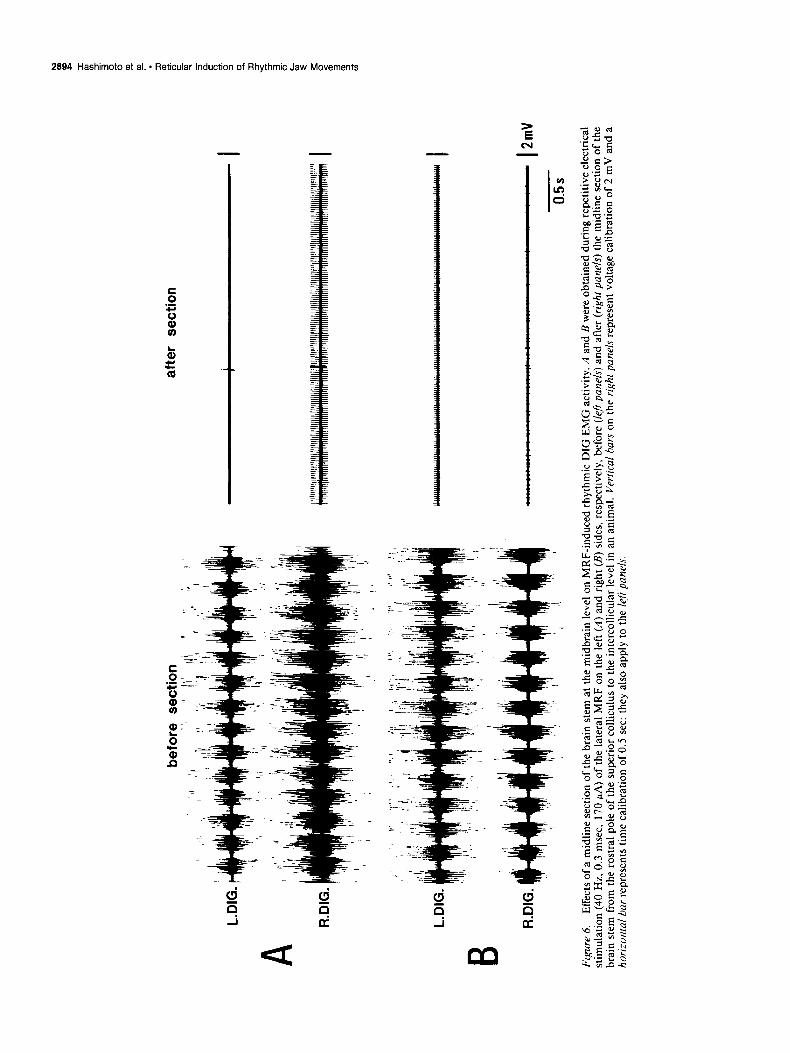
Effects of midline section of the brain stem at the midbrain level on the rhythmic DIG EMG activity induced by the MRF stimulation To get further evidence for crossing of the route responsible for the MRF induction of RJMs at the midbrain level, we studied the effects of a midline section of the brain stem from the rostra1 pole of the superior colliculus to the intercollicular level (cor- responding to the rostra1 and caudal borders of the midbrain, respectively) on the MRF induction of RJMs in 3 animals. A midline section abolished RJMs induced by stimulation of the MRF on either side in all the 3 animals tested. Left panels of Figure 6, A, B, shows the bilateral, rhythmic DIG EMG burst activity induced by stimulation of the left and right MRF in an animal, respectively. After a midline section was made at the midbrain level, stimulation of either the left (Fig. 64 right panel) or right (Fig. 6B, right panel) MRF no longer induced RJMs. The results indicate that the route crossing at the midbrain level is solely responsible for induction of RJMs by MRF stimulation.

befo
re
trans
ectio
n af
ter
trans
ectio
n
L.D
IG.
I
A R
.DIG
. I
2mV
L.D
IG.
I B
R.D
IG.
I 2m
V
0.5s
Figu
re
3.
Effe
cts
of p
reco
llicul
ar
dece
rebr
atio
n on
MR
F-in
duce
d rh
ythm
ic
DIG
EM
G
activ
ity.
A an
d B
were
obt
aine
d du
ring
repe
titiv
e st
imul
atio
n (4
0 H
z, 0
.3 m
sec,
180
PA)
of
the
late
ral
(A)
and
med
ial
(B)
MR
F on
the
left
side
befo
re (
le$
pane
& an
d af
ter
(righ
t pa
nels)
de
cere
brat
ion
in a
n an
imal
. Vo
ltage
an
d tim
e ca
libra
tions
in
the
rig
ht p
anel
al
so a
pply
to
the
lef
t.

L.D
IG.
RD
IG.
befo
re
cere
belle
ctom
y af
ter
cere
belle
ctom
y
L.M
ASS.
RM
ASS.
11
oopv
0.5s
Fi
gure
4.
E
ffect
s of
cer
ebel
lect
omy
on M
RF-
indu
ced
rhyt
hmic
jaw
m
uscl
e EM
G
activ
ity.
Rep
etiti
ve
elec
trica
l st
imul
atio
n (4
0 H
z, 0
.3 m
sec,
180
pA)
wa
s ap
plie
d to
the
rig
ht
late
ral
MR
F be
fore
(le
fr pa
nel)
and
afte
r (ri
ght
pane
l) ce
rebe
llect
omy
in a
n an
imal
. Vo
ltage
an
d tim
e ca
libra
tions
in
the
rig
ht
pane
l al
so a
pply
to
the
lef
t.

before section I
L.DIG.
A RDIG.
.DIG.
,.DIG.
The Journal of Neuroscience, August 1989, 9(E) 2892
after section
A .
mm
4.0 -
2.0 -
O-
2.0 -
4.0 -
1 P
Figure 5. Effects of a hemisection of the brain stem on MRF-induced rhythmic DIG EMG activity. A and B were obtained during repetitive electrical stimulation (40 Hz, 0.3 msec, 200 PA) of the lateral MRF on the left (A) and right (B) sides, respectively, before (left panels) and after (rzght panels) hemisection at the rostra1 border of the pons in an animal. Vertical bars on the right panels represent voltage calibration of 2 mV and a horizontal bar represents time calibration of 0.5 set; they are common to left panels. C, Photomicrograph (left) and schematic drawing (right) of horizontal histological section showing the site of hemisection on the left side. Abbreviations: BC, brachium conjunctivum; NIZZ, oculomotor nucleus; NSV, trigeminal spinal nucleus; NV, trigeminal motor nucleus; TSV, trigeminal spinal tract; V, trigeminal nerve; VZZ, facial nerve.
Effects of pontine pyramidal tract lesions on the MRF-induced rhythmic DIG EMG activity Impulses inducing RIMS from the CMA were reported to de- scend solely in the corticobulbar projection fibers (Nozaki et al., 1986b). To check whether the MRF-induced RIM was due to current spread to these corticobulbar fibers, we made a lesion in the pyramidal tract at the pontine level, i.e., caudal to the MRF stimulation site, in 4 animals. Figure 7 illustrates the results obtained from an animal in which a pyramidal tractot- omy was done on the right side. Before making this lesion, repetitive stimulation of the lateral (Fig. 7B, left panel) and medial MRF (Fig. 7C, left panel) on the left side, as well as the CMA on the right side (Fig. 7A, left panel), induced rhythmic
activity in the DIG EMG. After making a lesion in the right pyramidal tract (Fig. 7D), the rhythmic DIG EMG activity was no longer induced by stimulation of the right CMA (Fig. 7A, right panel). In contrast, stimulation of both the lateral (Fig. 7B, right panel) and medial MRF (Fig. 7C, right panel) on the left side still induced the rhythmic DIG EMG burst. The EMG pattern was essentially the same as that before making the lesion (Fig. 7, B, C, left panels).
Eflects of bulbar reticular formation lesions on A&RF-induction of RJM It was reported that the central rhythm generator responsible for induction of the rhythmic masticatory jaw movement by stimulation of the CMA of the guinea pig was located in the
afte
r se
ctio
n
I I I
A
B
befo
re
sect
ion
L.D
IG.
R.DI
G.
L.D
IG.
*I
R.DI
G.
+’
I 2m
V ,'
'I 1:
I,"
! !"
I 15
, ,
' )
i ,i
0.5s
Figu
re
6.
Effe
cts
of a
mid
line
sect
ion
of th
e br
ain
stem
at
the
mid
brai
n le
vel
on M
RF-
indu
ced
rhyt
hmic
D
IG
EMG
ac
tivity
. II
and
B we
re o
btai
ned
durin
g re
petit
ive
elec
trica
l st
imul
atio
n (4
0 H
z,
0.3
mse
c, 1
70 P
A) o
f th
e la
tera
l M
RF
on t
he l
eft
(A)
and
right
(R
) si
des,
res
pect
ivel
y,
befo
re
(/e/t
pun&
) an
d af
ter
(righ
t pa
ne/s
) th
e m
idlin
e se
ctio
n of
the
brai
n st
em f
rom
th
e ro
stra
1 po
le o
f th
e su
perio
r co
llicul
us
to t
he i
ntcr
collic
ular
le
vel
in a
n an
imal
. Ve
rtica
l ba
rs o
n th
e rig
ht
pane
ls re
pres
ent
volta
ge
calib
ratio
n of
2 m
V an
d a
horiz
onfa
l ba
r re
pres
ents
tim
e ca
libra
tion
of 0
.5 s
et:
they
also
app
ly
to t
he l
<fi p
anel
s.

The Journal of Neuroscience, August 1989,9(8) 2895
before lesion
A
L.DIG.
R.DIG.
L-DIG.
B R.DIG.
L.DIG.
C R.DIG.
after lesion
I 2mV
I 1mV
Figure 7. Effects of unilateral lesion of the pontine pyramidal tract on the rhythmic DIG EMG activity induced by stimulation of the CMA and MRF. A, CMA stimulation (40 Hz, 0.3 msec, 180 rA) on the right side. B, Stimulation (40 Hz, 0.3 msec, 200 PA) of the lateral MRF on the left side. C, Stimulation (40 Hz, 0.3 msec, 180 *A) of the medial MRF on the left side. Left and right panels of A-C were obtained during repetitive stimulation in an animal before and after lesions of the pyramidal tract, respectively. 0, Photomicrograph (left) and schematic drawing (right) of a frontal histological section showing the lesion of the right pyramidal tract. Vertical bars at right represent calibration of 1, 2, and 1 mV for A, B, and C, respectively. Time base: 0.5 set, common to A-C. Abbreviations: ZC, inferior colliculus; LC, locus coeruleus; NMV, trigeminal mesencephalic nucleus; NLL, nucleus of lateral lemniscus; NP, nuclei pontis; NPT, nucleus reticularis tegmenti pontis; PT. pyramidal tract; others as in Figure 5.
oral portion of the gigantocellular reticular nucleus (GCo) in the medulla oblongata (Nozaki et al., 1986a). The left panels of Figure 8, A, B, shows the rhythmic MUA burst in the right anterior digastric motoneuron pool (upper trace) and the rhythmic field potential in the right GCo (lower trace) induced by repetitive electrical stimulation of the left CMA and MRF, respectively, in a paralyzed animal. After a lesion was made in the right GCo region (Fig. K), stimulation of either the CMA or MRF on the left side no longer induced the rhythmic MUA in the anterior digastric motoneuron pool (Fig. 8, A, B, right
panels). The same result was obtained from all the 3 other animals tested.
Induction ofRJM by chemical stimulation of the MRF To study whether activation of the MRF neurons in the stim- ulated site is involved in induction of RIMS by electrical stim- ulation of the MRF, glutamate (4.0 ~1) was injected into the MRF of 7 animals. Injection of glutamate into the lateral MRF induced rhythmic EMG bursts similar to those induced by elec- trical stimulation of the lateral MRF in 5 out of 8 trials in 5

befo
re
lesio
n
P1.8
P2
.0
afte
r le
sion
I .,
; 1m
V Q
I1oo
jlv
0.5
s
P2.2
P2
.4
Figu
re 8
. E
ffect
s of
a le
sion
in t
he r
ostra
1 po
rtion
of
the
giga
ntoc
ellu
lar
retic
ular
nu
cleus
on
the
rhyt
hmic
M
UA
in t
he a
nter
ior
diga
stric
m
oton
euro
n po
ol
indu
ced
by r
epet
itive
el
ectri
cal
stim
ulat
ion
of th
e CM
A (4
0 H
z, 0
.3 m
sec,
270
MA)
and
MR
F (4
0 H
z, 0
.3 m
sec,
180
PA)
on
the
left
side
. A a
nd B
: Le
f? pa
nels,
Sim
ulta
neou
s re
cord
s of
the
rhyt
hmic
al
MUA
in
the
rig
ht
ante
rior
diga
stric
m
oton
euro
n po
ol
(R.D
MN
) and
th
e rh
ythm
ic
field
pot
entia
l (n
egat
ive
is d
own)
in
the
rig
ht
giga
ntoc
ellu
lar
retic
ular
nu
cleus
(o
ral
porti
on;
R.G
Cu)
induc
ed
by r
epet
itive
el
ectri
cal
stim
ulat
ion
of t
he C
MA
(A)
and
late
ral
MR
F (B
) on
the
le
ft si
de.
Rig
ht p
anel
s, Re
spon
ses
reco
rded
fro
m
the
right
an
terio
r di
gast
ric
mot
oneu
ron
pool
to
the
sam
e st
imul
atio
n of
the
left
CMA
(A)
and
late
ral
MR
F (B
) af
ter
mak
ing
a le
sion
in t
he r
ight
G
Co
by a
pplic
atio
n of
pos
itive
di
rect
cu
rrent
(9
0 PA
, 30
se
t) to
5 p
oint
s th
roug
h th
e el
ectro
de
used
for
rec
ordi
ng
of th
e fie
ld p
oten
tials
in
A a
nd B
. V
ertic
al ba
rs at
rig
ht o
f ea
ch p
anel
sh
ow
volta
ge
calib
ratio
n of
1 m
V fo
r R
.DM
N
and
100
PV f
or R
.GC
o.
Tim
e ba
se in
the
rig
ht p
anel
of B
is
com
mon
to
A a
nd B
. C
, Sc
hem
atic
dr
awin
gs
of fr
onta
l hi
stol
ogic
al
sect
ions
of
the
brai
n st
em s
howi
ng
the
loca
tion
of th
e le
sion
in t
he b
ulba
r re
ticul
ar
form
atio
n.
Alph
anum
eral
s re
pres
ent
the
ante
ropo
ster
ior
ster
eota
xic
coor
dina
tes,
Ab
brev
iatio
ns:
MLF
, m
edia
l lo
ngitu
dina
l fa
scic
ulus
; N
VII,
fa
cial
nuc
leus
; ot
hers
, sa
me
as in
Fig
ure
5.
The Journal of Neuroscience, August 1989, 9(8) 2897
R.MASS. _-1... WI
B
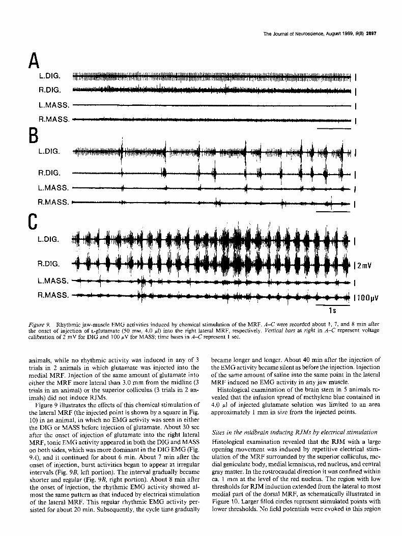
Figure 9. Rhythmic jaw-muscle EMG activities induced by chemical stimulation of the MRF. A-C were recorded about I, 7, and 8 min after the onset of injection of L-glutamate (50 mM, 4.0 ~1) into the right lateral MRF, respectively. Vertical bars at right in A-C represent voltage calibration of 2 mV for DIG and 100 WV for MASS; time bases in A-C represent 1 sec.
animals, while no rhythmic activity was induced in any of 3 trials in 2 animals in which glutamate was injected into the medial MRF. Injection of the same amount of glutamate into either the MRF more lateral than 3.0 mm from the midline (3 trials in an animal) or the superior colliculus (3 trials in 2 an- imals) did not induce RJMs.
Figure 9 illustrates the effects of this chemical stimulation of the lateral MRF (the injected point is shown by a square in Fig. 10) in an animal, in which no EMG activity was seen in either the DIG or MASS before injection of glutamate. About 30 set after the onset of injection of glutamate into the right lateral MRF, tonic EMG activity appeared in both the DIG and MASS on both sides, which was more dominant in the DIG EMG (Fig. 9A), and it continued for about 6 min. About 7 min after the onset of injection, burst activities began to appear at irregular intervals (Fig. 9B, left portion). The interval gradually became shorter and regular (Fig. 9B, right portion). About 8 min after the onset of injection, the rhythmic EMG activity showed al- most the same pattern as that induced by electrical stimulation of the lateral MRF. This regular rhythmic EMG activity per- sisted for about 20 min. Subsequently, the cycle time gradually
became longer and longer. About 40 min after the injection of the EMG activity became silent as before the injection. Injection of the same amount of saline into the same point in the lateral MRF induced no EMG activity in any jaw muscle.
Histological examination of the brain stem in 5 animals re- vealed that the infusion spread of methylene blue contained in 4.0 ~1 of injected glutamate solution was limited to an area approximately 1 mm in size from the injected points.
Sites in the midbrain inducing RJMs by electrical stimulation
Histological examination revealed that the RJM with a large opening movement was induced by repetitive electrical stim- ulation of the MRF surrounded by the superior colliculus, me- dial geniculate body, medial lemniscus, red nucleus, and central gray matter. In the rostrocaudal direction it was confined within ca. 1 mm at the level of the red nucleus. The region with low thresholds for RJM induction extended from the lateral to most medial part of the dorsal MRF, as schematically illustrated in Figure 10. Larger filled circles represent stimulated points with lower thresholds. No field potentials were evoked in this region

2898 Hashimoto et al. + Reticular Induction of Rhythmic Jaw Movements
Figure IO. Schematic representation of MRF sites inducing RIMS, projected on a frontal section of the brain stem D at A5.8. Circles show the points whose mm 1 repetitive electrical stimulation (40 Hz, 0.3 msec) induced RIMS at intensities +3.0 - not more than 180 FA, their diameters are drawn in inverse proportion to the threshold for RIM induction, as shown at bottom. Vertical and horizontal scales
+2.0 -
correspond to dorsoventral and hori- zontal axes of the stereotaxic fixation employed in the present study. Abbre-
+1.0 -
viations: CG, central gray matter; D, nucleus of Darkschewitsch; ZP, inter- peduncular nucleus; LM, medial lem- O-
niscus; MG, medial geniculate body; t NR, red nucleus; PED, cerebral pedun- cle; SC, superior colliculus; SN, sub- v
Le I I I 1 I I I -R stantia nigra. The square represents of the site of glutamate injection, the re- sults of which are shown in Figure 9.
3.0 2.0 1.0 0 mm
100<.1120<.<140< l llSO< .1180pA
by single or short-train pulses applied to either the ipsi- or contralateral CMA.
In addition to the MRF, repetitive stimulation of the sub- stantia nigra induced rhythmic DIG and MASS EMG activities at intensities comparable to those of MRF stimulation. How- ever, the pattern of induced rhythmic EMG burst was similar to that induced from the CMA (Fig. 1A) and was clearly different from that induced from the MRF (Fig. 1, B, C). Furthermore, the effective sites in the MRF were separated from those in the substantia nigra by a wide ineffective zone.
Although we also tested the effects of repetitive electrical stim- ulation of the superior colliculus in all the 65 animals used in this study, we did not succeed in inducing RJMs in any of the animals tested.
Discussion MRF region inducing RJA4s and its descending routes responsible for RJM induction The present study has demonstrated that repetitive electrical stimulation of the MRF can induce RJMs. Since the MRF- induced rhythmic burst activity of the anterior digastric mo- toneurons persists after precollicular decerebration, as well as cerebellectomy in the paralyzed preparation with the spinal cord sectioned at the C2 level, the rhythm can reasonably be assumed to be generated in the brain stem.
Chandler and Tal (1987) reported that repetitive electrical stimulation of the MRF suppressed CMA-induced RJMs in guinea pigs. On comparing the location of their MRF sites in- ducing this suppression (cf. figure 1 of their paper) with the RJM-inducing region in the MRF found in the present study, one notes that the latter is located directly rostra1 and dorsal to the former at the level of the red nucleus, and exactly corre- sponds to the area from which Chandler and Tal(1987) did not obtain suppression of the CMA-induced RJM.
Although the boundary between the superior colliculus and the MRF is not sharp (Edwards, 1980; Dean et al., 1982), the superior colliculus does not extend ventrally below the hori- zontal level of the cerebral aqueduct in rodents (Dean et al.,
1982). In the present study, no RJMs were induced by repetitive electrical stimulation of loci dorsal to the cerebral aqueduct, which supports the view that midbrain sites inducing RJMs are located in the MRF.
Similar to the MRF region inducing RJMs, a posterior mes- encephalic region is known to induce controlled locomotion on a treadmill by application of low-amplitude repetitive pulses in the precollicular-postmammillary decerebrate cat (see Garcia- Rill, 1986, for review). This physiologically defined region is called the mesencephalic locomotor region (MLR) and is mor- phologically coextensive with the pedunculopontine nucleus in the cat and rat. Thus, the MLR is located caudal to the RJM- inducing region. The threshold intensity of repetitive pulses for locomotion induction from the MLR (0.2 msec, lo-60 WA) (Garcia-Rill et al., 1985) is far lower than that for RJM induction from the MRF (0.3 msec, not less than 100 PA). This suggests that stimulation of neurons in a wider area in the MRF is re- quired to activate the central masticatory rhythm generator than to drive the central locomotor rhythm generator. Despite the difference in effective sites and threshold intensities for induc- tion of respective rhythmic movements, there is a similarity in the pattern of their emergence during stimulation. Stimulation of respective MRF regions first induces an increase in tonic muscle tone in the jaw or limb muscles. After a period of time, this tonic activity gave way to rhythmic alternation of activity between the closing and opening muscles of the jaw or between the extensors and flexors of the limb. It is suggested that ele- vation of tonic activity level of neurons composing the central rhythm generator is necessary to drive the rhythm-generating circuit.
Tal (1987) induced RJMs by repetitive electrical stimulation of the ventral MRF close to the cerebral peduncle. He ascribed this effect to current spread to the corticobulbar projection fibers from the CMA to the masticatory rhythm generator in the bulbar reticular formation (Nozaki et al., 1986b). In the present study we could also induce RJMs by repetitive stimulation of the ventral MRF and substantia nigra which were located closely dorsal to the cerebral peduncle. However, there was a distinct

The Journal of Neuroscience, August 1989, 9(8) 2999
ineffective zone between the RJM-inducing region in the dorsal MRF and the ventral MRF as well as substantia nigra. In ad- dition, the RJM-inducing effect of the dorsal MRF stimulation was not affected by the lesion of the pontine pyramidal tract that abolished the CMA-induced RJMs. Hence, the RJM in- duction by dorsal MRF stimulation cannot be ascribed to cur- rent spread to the corticobulbar fibers responsible for the CMA induction of RJMs.
Unilateral hemisection of the brain stem at the rostra1 border of the ponds abolished the RJMs induced from the MRF con- tralateral to the hemisection, but those induced from the ipsi- lateral MRF were unaffected by the hemisection. In addition, a midline section of the midbrain abolished the RJMs induced from the MRF on either side. Thus, the results indicate that a route crossing to the contralateral side at the midbrain level is responsible for the MRF induction of RJMs.
The exact route responsible for MRF induction of RJMs re- mains to be clarified, although it certainly descends outside the pyramidal tract to the bulbar reticular formation. It is possible that it descends within the RJM-suppression areas in the mes- encephalic, pontine, and bulbar reticular formation (Chandler and Tal, 1987; Chandler and Goldberg, 1988). The RJM- suppression area reported by Chandler and Tal (1987) may in- clude not only the neurons and/or fibers involved in RJM suppression, but also those involved in RJM induction; since the effects of activated RJM-suppressive component may over- whelm those of activated RJM-inducing component, suppres- sion of the CMA-induced RJMs may be produced as the overall result of simultaneous stimulation of these 2 components.
Repetitive electrical stimulation of the MRF region inducing RJMs evoked a field potential that rhythmically fluctuated in the same rhythm as the induced RJM in the medial bulbar reticular formation, including the oral portion of the GCo where repetitive electrical stimulation of the CMA also evoked a rhythmic field potential in the same rhythm as the CMA-in- duced RJM. A central rhythm generator responsible for the CMA-induced RJM was reported to be located in the GCo (Nozaki et al., 1986a). Since a lesion of this region abolished the RJM induced from the MRF as well as CMA, the central rhythm generator responsible for CMA induction of RJMs is also indispensible for the MRF induction of RJMs. It is sug- gested that the MRF and CMA may share a common rhythm generator.
Patterns of RJMs induced from medial and lateral MRF The RJM induced from the MRF was characterized by a large jaw-opening movement. This will readily be envisaged from the pattern of the MRF-induced rhythmic DIG EMG burst that is much larger in amplitude than that induced from the CMA.
A remarkable difference in the pattern between the RJM in- duced from the medial MRF and that from the lateral MRF was that the cycle time of the medial MRF-induced RJM was significantly longer than that induced from the lateral MRF. The reasons for this difference in the cycle time are, at present, unclear. However, since this difference depends on stimulated sites but not on the parameters of stimulation, there may be 2 independent input systems from the medial and lateral parts of the MRF to the central masticatory rhythm generator.
If we compare the patterns of rhythmic jaw-muscle EMG activities induced from the CMA and those from the lateral MRF, the former is characterized by a rhythmic alternation of
DIG and MASS EMG bursts, while in the latter the MASS EMG bursts occur both in and out of phase with DIG EMG bursts.
It was reported that jaw-closer EMG activities were accom- panied by jaw-opener EMG activities during jaw closing or oc- clusal phase in mastication in some species (Weijs and Dan- tuma, 1975; Gomiak and Gans, 1980; Byrd, 198 1; Oron and Crompton, 1985). On the other hand, during the MRF-induced rhythmic jaw movements the nonreciprocal burst of DIG and MASS EMG was seen during the jaw-opening phase. MASS EMG activity was also present during the burst phase of DIG EMG activity induced by the CMA stimulation in the guinea pig with local tetanus in the MASS (Enomoto et al., 1987). This nonreciprocal MASS EMG activity was demonstrated to be the result of stretch reflex of the MASS evoked by the jaw opening, due to block of all the inhibitory inputs to the masseter mo- toneurons. It could be assumed that rhythmic jaw-muscle EMG pattern induced from the lateral MRF consists of 2 components: the centrally induced reciprocal rhythmic activity in DIG and MASS EMG, and the peripherally induced MASS EMG burst in phase with the DIG EMG burst during the jaw-opening phase of an excessively large jaw displacement. Activation of gamma motoneurons of the MASS by stimulation of the MRF may also be involved in the occurrence of this stretch reflex (Shimazu et al., 1962).
Roles of mesencephalic reticular neurons in RJM induction by electrical stimulation of the MRF Glutamate induced RJMs when it was injected into the lateral portion of the MRF in 5 of 8 trials; it was ineffective when injected into the medial portion of the MRF in any of 3 trials. Because of the small number of trials of chemical stimulation in the present study, more evidence is needed to reach a definite conclusion of the effect of glutamate injection into the medial MRF. However, at least it can reasonably be assumed that ac- tivation of the neurons in the lateral MRF can induce RJMs and is involved in RJM induction by electrical stimulation of the lateral MRF.
To chemically induce RJMs from the lateral MRF, it was necessary to inject 4.0 ~1 of 50 mM glutamate (0.2 pmol). The latency of RJMs was ca. 7 min after the onset of injection over a period of 3 min. The apparently long latency of RJMs and the seemingly large volume of injection required for RJM in- duction may raise the possibility that the site of action of glu- tamate is remote from the injection site.
Sinnamon (1987) reported that injection of glutamate in a dose comparable to that used in the present study (2 M, 0.1-0.2 ~1, i.e., 0.2-0.4 pmol) into the preoptic region of the rat induced stepping after a latency of 40-200 set from the onset of injection over a period of 30 or 60 sec. This implies that locally applied glutamate requires a fairly long period to activate the central rhythm-generating circuit, although the drug rather promptly excites neurons themselves. He also reported that injection of picrotoxin into the preoptic region required even a longer la- tency of 7.8 min for induction of locomotion that continued without interruption for ca. 25 min. Likewise Garcia-Rill et al. (1985) reported that injection of picrotoxin (1.5 ~1) into the MLR of the rat elicited locomotion at a latency of 1.5-7 min after injection at rates of 0.1-1.0 pl/min, i.e., 3-22 min from the onset of injection. The duration of picrotoxin-induced loco- motion ranged from 20 to 60 min. Thus, there is a similarity in the latency and duration between the RJM induced from the lateral MRF and the locomotion induced from the MLR. By

2900 Hashimoto et al. * Reticular Induction of Rhythmic Jaw Movements
injecting the same amount of dye into the MLR, Garcia-Rill et al. (1985) estimated that the infusion spread was limited to an area approximately l-2 mm in size in the mediolateral direction. Accordingly, it could be assumed that 4.0 ~1 glutamate would spread to a region occupying 0.7-l .4 mm (i.e., 0.5-l .O mm times the cubic root of 4.011.5) from the point of injection. This es- timate of infusion spread roughly corresponds with the histo- logical data that the spread of methylene blue contained in 4.0 yl of injected glutamate solution was confined in an area ca. 1 mm from the injected point. The estimate would also be con- sistent with the results that injection ofglutamate into the medial MRF could not induce RJMs, although it was less than 2 mm apart from the lateral MRF. Thus, the long latency and duration of RJMs induced by injection of 4.0 ~1 glutamate would not necessarily indicate that the site of action is remote from the injection site.
Possible source of inputs to the A4RF involved in RJM induction
Superior colliculus. Chandler and Goldberg (1984) reported that systemic injection of apomorphine (a dopamine agonist) into guinea pigs induced RJMs and that ablation of the superior colliculus and directly underlying region blocked the ability of apomorphine to induce RJMs. The results indicate activation of the superior collicular neurons by either disinhibition or ex- citation is crucial for induction of RJMs with apomorphine and suggest that the superior colliculus may be a source of activating inputs to the RJM-inducing region in the MRF. In fact, pro- jections from the superior colliculus to wide areas in the mid- brain and lower brain stem were morphologically demonstrated in rodents (Redgrave et al., 1987).
However, neither glutamate injection into nor electrical stim- ulation of the superior colliculus in the present study could induce RJMs. The ineffectiveness of glutamate injection can be explained by assuming that the superior collicular neurons that are involved in RJM induction are not glutamate sensitive and that their activation by apomorphine is due to disinhibition, e.g., from tonic GABAergic inhibition (Redgrave et al., 1981) exerted from the pars reticulata of the substantia nigra (Rinvik et al., 1976; Graybiel, 1978; Beckstead et al., 1979).
The neurons involved in RJM induction may be dispersed in a wide region or clustered in a circumscribed region in the su- perior colliculus. If the former is the case, electrical stimulation in the superior colliculus in the present study may not have activated sufficient numbers of neurons to activate the central rhythm generator. On the other hand, if the latter is the case, we may not have hit the cluster of the neurons located in the area where we did not explore. In either case, electrical stimu- lation of the superior colliculus would be ineffective for RJM induction.
Limbic system. Kawamura and Tsukamoto (1960) induced masticatory movements by stimulation of the cerebral cortex and the amygdala in rabbits. They found that stimulation of the amygdala could induce masticatory movements after ablation of the cerebral cortex, and stimulation of the cerebral cortex could induce masticatory movements after making a lesion in the amygdala. They proposed that the routes responsible for induction of masticatory movements from the cerebral cortex and amygdala were separate from each other.
Single or short-train pulses applied to the CMA evoked no short-latency field potentials in the MRF region inducing RJMs. This suggests that there is no massive oligosynaptic projection
to this midbrain region from the CMA. On the other hand, it was reported by means of the retrograde HRP-tracing method in rats and cats that there are massive projections to the lateral portion of the MRF from wide areas in the hypothalamus, in- cluding the lateral hypothalamic area (Morrell et al., 198 1; Par- ent and Steriade, 1981), i.e., the feeding center (Anand and Brobeck, 195 1). It can be suggested that the limbic structures, including the amygdala, activate the MRF neurons to generate the masticatory movements, indirectly via the hypothalamus and/or directly via the amygdalotegmental projection (Hopkins, 1975; Hopkins and Holstege, 1978).
We conclude that the mesencephalic reticular neurons may be involved in the expression of feeding behavior as a source of impulses activating the central masticatory rhythm generator in the lower brain stem, by integrating the activating effects from the higher brain structures, including the limbic system.
References Anand, B. K., and J. R. Brobeck (195 1) Hypothalamic control of food
intake in rats and cats. Yale J. Biol. Med. 24: 123-140. Beckstead, R. M., V. B. Domesick, and W. J. H. Nauta (1979) Efferent
connections of the substantia nigra and ventral tegmental area in the rat. Brain Res. 175: 191-217.
Byrd, K. E. (198 1) Mandibular movement and muscle activity during mastication in the guinea pig (&via porcellus). J. Morphol. 170: 147- 169.
Chandler, S. H., and L. J. Goldberg (1984) Differentiation of the neural pathways mediating cortically induced and dopaminergic activation of the central pattern generator (CPG) for rhythmical jaw movements in the anesthetized guinea pig. Brain Res. 323: 297-301.
Chandler, S. H., and c. J. Goldberg (1988) Effects of pontomedullary reticular formation stimulation on the neuronal networks responsible for rhythmical jaw movements in the guinea pig. J. Neurophysiol. 59: 819-832.
Chandler, S. H., and M. Tal (1987) Brain-stem perturbations during cortically evoked rhythmical jaw movements: Effects of activation of brain-stem loci on jaw muscle cycle characteristics. J. Neurosci. 7: 463-472.
Dean, P., P. Redgrave, and L. Eastwood (1982) Suppression of apo- morphine-induced oral stereotypy in rats by microinjection of mus- cimol into midbrain. Life Sci. 30: 2 17 l-2 179.
Edwards. S. B. (1980) The deeu cell lavers of the superior colliculus: Their reticular’characteristics and structural organization. In The Re- ticular Formation Revisited, J. A. Hobson and M. A. B. Brazier, eds., pp. 193-209, Raven, New York.
Enomoto, S., N. Katakura, T. Sunada, T. Katayama, Y. Hirose, Y. Ishiwata, and Y. Nakamura (1987) Cortically induced masticatory rhythm in masseter motoneurons after blocking inhibition by strych- nine and tetanus toxin. Neurosci. Res. 4: 396-4 12.
Garcia-Rill, E. (1986) The basal ganglia and the locomotor regions. Brain Res. Rev. II: 47-63.
Garcia-Rill, E., R. D. Skinner, and J. A. Fitzgerald (1985) Chemical activation of the mesencephalic locomotor region. Brain Res. 330: 43-54.
Gomiak, G. C., and C. Gans (1980) Quantitative assay of electro- myograms during mastication in domestic cats (Felis catus). J. Mor- phol: 163: 253-281.
Gravbiel. A. M. (1978) Oraanization of the niarotectal connection: An experimental tracer studqin the cat. Brain Res. 143: 339-348.
Hashimoto, N., T. Katayama, Y. Ishiwata, and Y. Nakamura (1987) Localization of central rhythm generator and its input pathway in- volved in mesencephalically induced rhythmical masticatory move- ment in the guinea pig. J. Physiol. Sot. Jpn. 49: 406.
Hopkins, D. A. (1975) Amygdaloid projections in the rat, cat and rhesus monkey. Neurosci. Lett. 1: 263-270.
Hopkins, D. A., and G. Holstege (1978) Amygdaloid projections to the mesencephalon, pons and medulla oblongata in the cat. Exp. Brain Res. 32: 529-547.
Kawamura, Y., and S. Tsukamoto (1960) Analysis ofjaw movements from the cortical jaw motor area and amygdala. Jpn. J. Physiol. IO: 471-488.

The Journal of Neuroscience, August 1989, 9(E) 2901
Lund, J. P., and P. G. Dellow (1971) The influence of interactive stimuli on rhythmical masticatory movements in rabbits. Arch. Oral Biol. 16: 215-223.
Luschei, E. S., and L. J. Goldberg (198 1) Neural mechanisms of man- dibular control: Mastication and voluntary biting. In Handbook of Physiology, Sect. 1 The Nervous System, Vol. 2, Motor Control, J. M. Brookhart, V. B. Mountcastle, V. B. Brooks, and S. R. Geiger, eds., pp. 1237-l 274, American Physiological Society, Bethesda, MD.
Morrell, J. I., L. M. Greenberger, and D. W. Pfaff (1981) Hypotha- lamic, other diencephalic, and telencephalic neurons that project to the dorsal midbrain. J. Comp. Neurol. 201: 589-620.
Nozaki, S., A. Iriki, and Y. Nakamura (1986a) Localization of central rhythm generator involved in cortically induced rhythmical masti- catory jaw-opening movement in the guinea pig. J. Neurophysiol. 5.5: 806-825.
Nozaki, S., A. Iriki, and Y. Nakamura (1986b) Role of corticobulbar projection neurons in cortically induced rhythmical masticatory jaw- opening movement in the guinea pig. J. Neurophysiol. 55: 826-845.
Oron, U., and A. W. Crompton (1985) A cineradiographic and elec- tromyographic study of mastication in Tenrec ecaudutus. J. Morphol. 185: 155-182.
Parent, A., and M. Steriade (198 1) Afferents from the periaqueductal gray, medial hypothalamus and medial thalamus to the midbrain reticular core. Brain Res. Bull. 7: 4 1 l-4 18.
Redgrave, P., P. Dean, W. Souki, and G. Lewis (1981) Gnawing and changes in reactivity produced by microinjections of picrotoxin into the superior colliculus of rats. Psychobiology 75: 198-203.
Redgrave, P., I. J. Mitchell, and P. Dean (1987) Descending projec- tions from the superior colliculus in rat: A study using orthograde transport of wheatgerm-agglutinin conjugated horseradish peroxi- dase.-Exp. Brain Res. 68: i47-167.
Rinvik. E.. I. Grofova. and 0. P. Ottersen (1976) Demonstration of nigrotectal and nigrdreticular projections in the ‘cat by axonal trans- port of proteins. Brain Res. 112: 388-394.
Rbssner, W. (1965) Stereotaktischer Hirnatlas vom Meerschweinchen, Pallas Verlag, Lochham bei Mtinchen.
Shimazu, H., T. Hongo, and K. Kubota (1962) Two types of central influences on gamma motor system. J. Neurophysiol. 25: 309-323.
Sinnamon. H. M. (1987) Glutamate and picrotoxin iniections into the preoptic basal forebrain initiate locomotion in the anesthetized rat. Brain Res. 400: 270-277.
Sumi, T. (197 1) Modification of cortically evoked rhythmic chewing and swallowing from midbrain and pans. Jpn. J. Physiol. 21: 489- 506.
Tal, M. (1987) Neural basis for initiation of rhythmic digastric activity upon midbrain stimulation in the guinea pig. Brain Res. 411: 58-64.
Weijs, W. A., and R. Dantuma (1975) Electromyography and me- chanics of mastication in the albino rat. J. Morphol. 146: l-34.