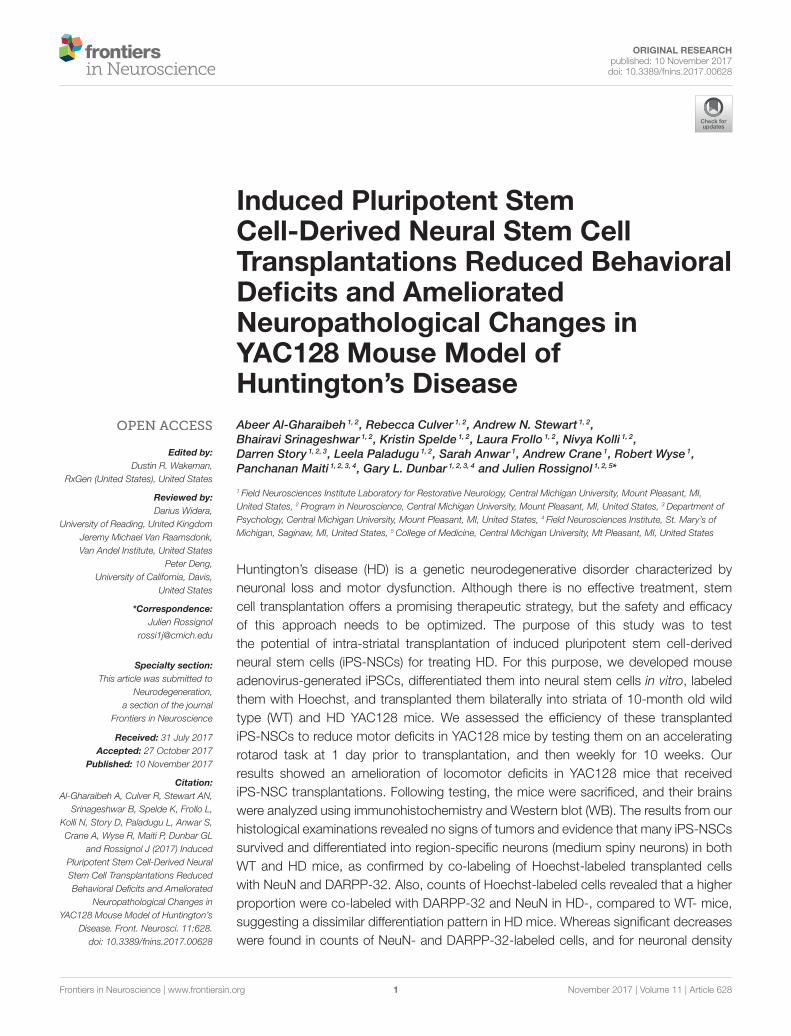
Induced Pluripotent Stem Cell-Derived Neural Stem Cell ... · Behavioral Deficits and Ameliorated...
13
ORIGINAL RESEARCH published: 10 November 2017 doi: 10.3389/fnins.2017.00628 Frontiers in Neuroscience | www.frontiersin.org 1 November 2017 | Volume 11 | Article 628 Edited by: Dustin R. Wakeman, RxGen (United States), United States Reviewed by: Darius Widera, University of Reading, United Kingdom Jeremy Michael Van Raamsdonk, Van Andel Institute, United States Peter Deng, University of California, Davis, United States *Correspondence: Julien Rossignol [email protected] Specialty section: This article was submitted to Neurodegeneration, a section of the journal Frontiers in Neuroscience Received: 31 July 2017 Accepted: 27 October 2017 Published: 10 November 2017 Citation: Al-Gharaibeh A, Culver R, Stewart AN, Srinageshwar B, Spelde K, Frollo L, Kolli N, Story D, Paladugu L, Anwar S, Crane A, Wyse R, Maiti P, Dunbar GL and Rossignol J (2017) Induced Pluripotent Stem Cell-Derived Neural Stem Cell Transplantations Reduced Behavioral Deficits and Ameliorated Neuropathological Changes in YAC128 Mouse Model of Huntington’s Disease. Front. Neurosci. 11:628. doi: 10.3389/fnins.2017.00628 Induced Pluripotent Stem Cell-Derived Neural Stem Cell Transplantations Reduced Behavioral Deficits and Ameliorated Neuropathological Changes in YAC128 Mouse Model of Huntington’s Disease Abeer Al-Gharaibeh 1, 2 , Rebecca Culver 1, 2 , Andrew N. Stewart 1, 2 , Bhairavi Srinageshwar 1, 2 , Kristin Spelde 1, 2 , Laura Frollo 1, 2 , Nivya Kolli 1, 2 , Darren Story 1, 2, 3 , Leela Paladugu 1, 2 , Sarah Anwar 1 , Andrew Crane 1 , Robert Wyse 1 , Panchanan Maiti 1, 2, 3, 4 , Gary L. Dunbar 1, 2, 3, 4 and Julien Rossignol 1, 2, 5 * 1 Field Neurosciences Institute Laboratory for Restorative Neurology, Central Michigan University, Mount Pleasant, MI, United States, 2 Program in Neuroscience, Central Michigan University, Mount Pleasant, MI, United States, 3 Department of Psychology, Central Michigan University, Mount Pleasant, MI, United States, 4 Field Neurosciences Institute, St. Mary’s of Michigan, Saginaw, MI, United States, 5 College of Medicine, Central Michigan University, Mt Pleasant, MI, United States Huntington’s disease (HD) is a genetic neurodegenerative disorder characterized by neuronal loss and motor dysfunction. Although there is no effective treatment, stem cell transplantation offers a promising therapeutic strategy, but the safety and efficacy of this approach needs to be optimized. The purpose of this study was to test the potential of intra-striatal transplantation of induced pluripotent stem cell-derived neural stem cells (iPS-NSCs) for treating HD. For this purpose, we developed mouse adenovirus-generated iPSCs, differentiated them into neural stem cells in vitro, labeled them with Hoechst, and transplanted them bilaterally into striata of 10-month old wild type (WT) and HD YAC128 mice. We assessed the efficiency of these transplanted iPS-NSCs to reduce motor deficits in YAC128 mice by testing them on an accelerating rotarod task at 1 day prior to transplantation, and then weekly for 10 weeks. Our results showed an amelioration of locomotor deficits in YAC128 mice that received iPS-NSC transplantations. Following testing, the mice were sacrificed, and their brains were analyzed using immunohistochemistry and Western blot (WB). The results from our histological examinations revealed no signs of tumors and evidence that many iPS-NSCs survived and differentiated into region-specific neurons (medium spiny neurons) in both WT and HD mice, as confirmed by co-labeling of Hoechst-labeled transplanted cells with NeuN and DARPP-32. Also, counts of Hoechst-labeled cells revealed that a higher proportion were co-labeled with DARPP-32 and NeuN in HD-, compared to WT- mice, suggesting a dissimilar differentiation pattern in HD mice. Whereas significant decreases were found in counts of NeuN- and DARPP-32-labeled cells, and for neuronal density
Transcript of Induced Pluripotent Stem Cell-Derived Neural Stem Cell ... · Behavioral Deficits and Ameliorated...
ORIGINAL RESEARCHpublished: 10 November 2017doi: 10.3389/fnins.2017.00628
Frontiers in Neuroscience | www.frontiersin.org 1 November 2017 | Volume 11 | Article 628
Edited by:
Dustin R. Wakeman,
RxGen (United States), United States
Reviewed by:
Darius Widera,
University of Reading, United Kingdom
Jeremy Michael Van Raamsdonk,
Van Andel Institute, United States
Peter Deng,
University of California, Davis,
United States
*Correspondence:
Julien Rossignol
Specialty section:
This article was submitted to
Neurodegeneration,
a section of the journal
Frontiers in Neuroscience
Received: 31 July 2017
Accepted: 27 October 2017
Published: 10 November 2017
Citation:
Al-Gharaibeh A, Culver R, Stewart AN,
Srinageshwar B, Spelde K, Frollo L,
Kolli N, Story D, Paladugu L, Anwar S,
Crane A, Wyse R, Maiti P, Dunbar GL
and Rossignol J (2017) Induced
Pluripotent Stem Cell-Derived Neural
Stem Cell Transplantations Reduced
Behavioral Deficits and Ameliorated
Neuropathological Changes in
YAC128 Mouse Model of Huntington’s
Disease. Front. Neurosci. 11:628.
doi: 10.3389/fnins.2017.00628
Induced Pluripotent StemCell-Derived Neural Stem CellTransplantations Reduced BehavioralDeficits and AmelioratedNeuropathological Changes inYAC128 Mouse Model ofHuntington’s Disease
Abeer Al-Gharaibeh 1, 2, Rebecca Culver 1, 2, Andrew N. Stewart 1, 2,
Bhairavi Srinageshwar 1, 2, Kristin Spelde 1, 2, Laura Frollo 1, 2, Nivya Kolli 1, 2,
Darren Story 1, 2, 3, Leela Paladugu 1, 2, Sarah Anwar 1, Andrew Crane 1, Robert Wyse 1,
Panchanan Maiti 1, 2, 3, 4, Gary L. Dunbar 1, 2, 3, 4 and Julien Rossignol 1, 2, 5*
1 Field Neurosciences Institute Laboratory for Restorative Neurology, Central Michigan University, Mount Pleasant, MI,
United States, 2 Program in Neuroscience, Central Michigan University, Mount Pleasant, MI, United States, 3Department of
Psychology, Central Michigan University, Mount Pleasant, MI, United States, 4 Field Neurosciences Institute, St. Mary’s of
Michigan, Saginaw, MI, United States, 5College of Medicine, Central Michigan University, Mt Pleasant, MI, United States
Huntington’s disease (HD) is a genetic neurodegenerative disorder characterized by
neuronal loss and motor dysfunction. Although there is no effective treatment, stem
cell transplantation offers a promising therapeutic strategy, but the safety and efficacy
of this approach needs to be optimized. The purpose of this study was to test
the potential of intra-striatal transplantation of induced pluripotent stem cell-derived
neural stem cells (iPS-NSCs) for treating HD. For this purpose, we developed mouse
adenovirus-generated iPSCs, differentiated them into neural stem cells in vitro, labeled
them with Hoechst, and transplanted them bilaterally into striata of 10-month old wild
type (WT) and HD YAC128 mice. We assessed the efficiency of these transplanted
iPS-NSCs to reduce motor deficits in YAC128 mice by testing them on an accelerating
rotarod task at 1 day prior to transplantation, and then weekly for 10 weeks. Our
results showed an amelioration of locomotor deficits in YAC128 mice that received
iPS-NSC transplantations. Following testing, the mice were sacrificed, and their brains
were analyzed using immunohistochemistry and Western blot (WB). The results from our
histological examinations revealed no signs of tumors and evidence that many iPS-NSCs
survived and differentiated into region-specific neurons (medium spiny neurons) in both
WT and HD mice, as confirmed by co-labeling of Hoechst-labeled transplanted cells
with NeuN and DARPP-32. Also, counts of Hoechst-labeled cells revealed that a higher
proportion were co-labeled with DARPP-32 and NeuN in HD-, compared to WT- mice,
suggesting a dissimilar differentiation pattern in HD mice. Whereas significant decreases
were found in counts of NeuN- and DARPP-32-labeled cells, and for neuronal density
Al-Gharaibeh et al. iPS-NSCs Reduced Deficits in HD-YAC128
measures in striata of HD vehicle controls, such decrements were not observed in the
iPS-NSCs-transplanted-HDmice. WB analysis showed increase of BDNF and TrkB levels
in striata of transplanted HD mice compared to HD vehicle controls. Collectively, our data
suggest that iPS-NSCs may provide an effective option for neuronal replacement therapy
in HD.
Keywords: neural stem cells, Huntington’s disease, cell transplantations, YAC128, iPSCs, iPS-NSCs
INTRODUCTION
Huntington’s disease (HD) is a progressive, neurodegenerative,genetic disorder characterized by choreic movements, behavioraland cognitive disturbances, and dementia (Craufurd et al., 2001).The disease is caused by an autosomal dominant mutation in thehuntingtin gene (HTT), and the mode of inheritance is dominantwith almost full penetration (with 40 or more CAG repeats).The genetic basis of HD was discovered in 1993 (MacDonaldet al., 1993), and it was found to be caused by an elongatedCystosine-Adenine-Guanine (CAG) repeat on the short arm ofchromosome 4p16.3 in the HTT gene. HD symptoms includepsychiatric, motor, and cognitive deficits, and are variable amongpatients during early stages of the pathology. However, asthe disease progresses, symptoms become predictable, with allpatients eventually developing same characteristic pathologies(Walker, 2007). The life expectancy for HD patients rangesbetween 15 and 20 years after the appearance of motor symptoms(Landles and Bates, 2004; Walker, 2007). The most apparent andearliest damage is seen in the neostriatum, which is composed ofthe caudate nucleus and putamen (Walker, 2007). Medium spinyneurons (MSNs) in the striatum appear to be the most vulnerableneurons to the damage in HD (Albin et al., 1990).
Different animal models, either chemically or geneticallyinduced, have been developed to study various aspects of HD.One of these models, theYAC128 HD mouse, contains the full-length human mutant HTT (mHTT) inserted into its genomewhich results in the expression of mHTT with 128 CAG repeats(Slow et al., 2003). YAC128 mice show selective, age-dependent,striatal and cortical atrophy and neurodegeneration, and developprogressive deterioration of motor and cognitive functions (VanRaamsdonk et al., 2005; Gray et al., 2008; Ehrnhoefer et al., 2009).The decline in motor abilities manifests as progressive deficits inaccelerating rotarod performance that correlates with the loss ofneurons in the striatum (Slow et al., 2003).
Abbreviations: ANOVA, analysis of variance; BCA, bicinchoninic acid assay;
BDNF, brain derived neurotrophic factor; bFGF, basic fibroblast growth factor;
DARPP32, Dopamine- and cAMP-regulated phosphoprotein of 32 kDa; DMEM,
Dulbecco’s Modified Eagles medium; EDTA, Ethylene-di-amino-tetra-acetic-
acid; EGF, epidermal growth factor; GFAP, glial fibrillary acidic protein; HBSS,
Hank’s balanced salt solution; HD, Huntington’s disease; HRP, Horseradish
peroxidase; iNSC, induced neural stem cells; iPSC, Induced pluripotent stem
cells; iPS-NSCs, induced pluripotent stem cells-derived neural stem cells; Klf4,
Kruppel-like factor 4; MSN, Medium spiny neuron; NEAA, non-essential amino
acids; NeuN, Neuronal nuclei; NSC, Neural stem cell; OCT4, Octamer-binding
transcription factor 4; OD, Optical density; PBS, Phosphate buffer saline; PFA,
Paraformaldehyde; PVDF, Polyvinylidene fluoride; QA, Quinolinic acid; RIPA,
Radio immunoprecipitation assay; SDS, Sodium dodecyl sulfate; SDS-PAGE,
sodium dodecyl sulfate polyacrylamide gel electrophoresis; SOX2, SRY (sex
Experimental approaches used to treat HD aim to decreasethe levels of the mHTT protein, improve the survivability ofneurons, as well as to replace the affected neurons. Stem celltherapy holds significant promise for treating HD, and includesthe transplantation of stem cells into the affected regions of thebrain (Cundiff and Anderson, 2011). Owing to the promisingoutcomes in animalmodels of HD, the use of stem cell transplantsin human clinical trials have been performed to test for efficacy(Clelland et al., 2008). Transplants of fetal tissue, embryonicstem cells (ESCs), mesenchymal stem cells (MSCs), and neuronalstem cells (NSCs; both adult and differentiated from ESCs) havebeen used as experimental treatments for HD, and all possessdifferent properties that may be beneficial for use as a therapy.Although some promising results have been found followingthe transplantation of any of these cell sources, the immunerejection of the graft (Bernreuther et al., 2006; Cicchetti et al.,2014) as well as some serious adverse events have been reported.Specifically, tumor formation has been found following ESCtransplants (Aubry et al., 2008), and hemorrhage with multiplesolid and cystic lesions in the brain was found following fetaltissue grafting (Keene et al., 2009). While the anti-inflammatoryproperties and ability for autologous transplantations result in asuperior survivability of MSC transplants (Rossignol et al., 2009,2011; Dey et al., 2010; Lin et al., 2011; Sadan et al., 2012; SerranoSánchez et al., 2014), these cells do not readily differentiateinto neurons (Przyborski et al., 2008), limiting their utility as asource of cellular replacement. Unlike transplants of MSCs, NSCtransplants have the potential for replacing neurons that havedegenerated within the targeted region (Vazey et al., 2006; Yangand Yu, 2009), yet the survivability of NSCs post-transplantationremains poor with signs of immune rejection (Johann et al., 2007;Rossignol et al., 2014). Similarly, the accessibility to NSCs islimited, and met with technical and ethical complications causedby the demand for using embryonic tissue for isolation.
A new source of pluripotent stem cells emerged whenTakahashi and Yamanaka (2006) generated pluripotent stemcells by reprogramming somatic cells through inserting 4 genes(OCT4, SOX2, Klf4, and c-Myc) into fibroblasts obtained fromthe skin. These cells, defined as induced pluripotent stem cells(iPSCs), were able to differentiate into any cell type in thebody (Takahashi and Yamanaka, 2006). iPSCs appear to havemuch of the same characteristics as ESCs, including morphologyand differentiation capabilities, as well as the capacity to formteratomas containing cells of all three germ lineages whentransplanted into severe combined immune deficient mice
determining region Y)-box 2; TBS, Tris buffer saline; TrkB, Tropomyosin-related
kinase B; YAC, Yeast artificial chromosome.
Frontiers in Neuroscience | www.frontiersin.org 2 November 2017 | Volume 11 | Article 628

Al-Gharaibeh et al. iPS-NSCs Reduced Deficits in HD-YAC128
(Takahashi and Yamanaka, 2006). However, there are geneticand epigenetic differences between them (Robinton and Daley,2012). The use of iPSCs offers a viable alternative to ESCsand circumvents the issues of availability and ethical concernssurrounding the use of embryos (Verma and Verma, 2011).Work in our laboratory indicated that transplanting rat-derivediPSCs into the striata of rats given 3- nitropropionic acid tomodel HD, revealed improvements in the motor function, anddifferentiation of transplanted cells into region-specific neuronsin the striatum (Fink et al., 2014a). However, the transplantationof pluripotent stem cells may pose a risk of over-proliferationif the transplants occur without prior pre-engagement down agiven germ layer, such as the neuro-ectoderm (Miura et al., 2009).Subsequently, NSCs that were derived from iPSCs obtained froma patient with juvenile onset HD, were transplanted into thestriata of YAC128 mice, and showed significant improvement inmotor functions (Jeon et al., 2014). This study demonstrated aproof-of-principle that therapeutic efficacy can still be derivedfollowing the transplantation of pre-engaged iPSCs, which mayreveal to be a safer method of therapy.
Overall, accumulating evidence suggests that stem celltransplantations hold significant promise for use as a treatmentfor HD. However, there are many technical challenges that needto be addressed before this approach can be safely translatedas a treatment in clinical cases of HD. This study aimed tofurther characterize the use of iPS-NSCs as a treatment for HD byutilizing behavioral, histological, and protein analyses as outcomemeasures for therapeutic efficacy.
We hypothesized that iPS-NSCs will provide a usefulalternative for ESCs and NSCs for HD treatment because thesecells can be generated in adequate amounts from somatic cells,and form a more personalized treatment that will promoteintegration of the transplanted cells, with less risk of immunerejection. Also, as iPS-NSCs are more restricted to the neuronallineage compared to iPSCs, we believe they will confer less of arisk for unwanted proliferation and tumor formation in vivo.
This study tested the efficacy of transplanting of iPS-NSCs asa therapy for HD after intrastriatal transplantation in YAC128HD mice by (1) assessing their effects on motor function; (2)the survivability and differentiation capabilities 10 weeks aftertransplantation; and (3) the histological and protein analyses 10weeks after transplantation.
MATERIALS AND METHODS
Cell Generation and CultureiPSC CultureiPSCs used in this study, were generated and characterized asdescribed in a previously published protocol in our laboratory(Fink et al., 2014b). In brief, iPSCs were generated fromfibroblasts that were isolated from tails of adult wild typemice. These fibroblasts were reprogrammed into iPSCs byusing two adenoviruses (ADs): one contains Oct4, Sox2, andKlf4 and another contains c-Myc which are all consideredpluripotent factors. The recombinant ADs were developed inour laboratory and described previously in a published protocol(Fink et al., 2014b). The generated iPSCs were confirmed to
express pluripotent markers using immunocytochemistry (ICC)and flow cytometry. The cells were then cryopreserved infreezing media containing 10% dimethyl sulfoxide (DMSO)[Medium is composed of 45% knock out serum and 45% ofDulbecco’s Modified Eagles Media (DMEM), and 10% DMSO].The generated iPSCs were thawed and plated on 0.1% gelatincoat, and cultured in iPSC media [DMEM (Life Technologies,Carlsbad, CA) supplemented with 10% knock-out serum, β-mercaptoethanol (Life Technologies, Carlsbad, CA), 1% 1Xnon-essential amino acids (NEAA; Life Technologies, Carlsbad,CA), 20 ng/mL basic fibroblast growth factor (bFGF; LifeTechnologies, Carlsbad, CA), 2µM L-glutamine (Sigma, St.Louis, MO), 5 mg/mL streptomycin and 5 UI/mL penicillin,and 10 ng/mL leukemia inhibitory factor (LIF; Life Technologies,Carlsbad, CA)]. Cells were passaged by dissociating them inAccutase (Sigma, St. Louis, MO), centrifuging at 250 g for 5minat 4◦C, and plating them on 0.1% gelatin coat.
iPS-NSCs GenerationThe iPS-NSCs were generated by differentiation of iPSCsfollowing the first stage in a published protocol with somemodifications (Niclis et al., 2013). Briefly, iPSCs were expandedto 80% confluency, and then, the iPSC media were replacedby neuronal induction media [Neurobasal-A (Life Technologies,Carlsbad, CA) supplemented with 1X B27-A (Life Technologies,Carlsbad, CA), 1X N2 (Life Technologies, Carlsbad, CA), 1XNEAA (Life Technologies, Carlsbad, CA), 1X Glutamax (LifeTechnologies, Carlsbad, CA), and 5 mg/mL streptomycin and5 UI/mL penicillin]. Half of the media was changed every 3days, and the cells were kept in culture until they detachedand formed neurospheres. The media containing detached cellswere centrifuged at 100 g for 5min at 4◦C, and the pellet wasdissociated in 1mL Accutase (Sigma, St.louis, Mo) for 5minat 37◦C, then suspended in 5mL phosphate buffered saline(PBS) and centrifuged another time. Cells were then re-platedin neural stem cell media [Neurobasal-A supplemented with1X B27-A, 1X N2 (Life technologies, Carlsbad, CA), 1X NEAA,1X Glutamax (Life Technologies, Carlsbad, CA), 20 ng/mLepidermal growth factor (EGF; Life Technologies, Carlsbad, CA)and 10 ng/mL bFGF (Life Technologies, Carlsbad, CA), and5 mg/mL streptomycin and 5 UI/mL penicillin] (Figure 1).
Characterization of iPS-NSCsThe iPSC-derived neurospheres were passaged every week for3 weeks and then characterized through ICC for neural lineagespecific protein expression (Nestin, Sox2, β-tubulin-III andNeuN). The cells were grown on poly-L-lysine-coated, 25mmglass coverslips for 2 days, after which cells were washedwith PBS (0.01M at pH 7.4) three times and fixed using 4%paraformaldehyde for 10min at 4◦C. Then a blocking solution(10% normal goat serum in PBS) was added to the coverslipsand incubated for 1 h at room temperature. After that, theprimary antibodies [Nestin (mouse monoclonal), Sox2 (rabbitpolyclonal), and NeuN (rabbit monoclonal); 1:500; Abcam,Cambridge, U.K, β-Tubulin III (chicken polyclonal antibody);1:300; Aves Labs Inc., Tigard, OR] diluted in PBS containing 0.1%Triton X-100 were added to the assigned wells and incubated at
Frontiers in Neuroscience | www.frontiersin.org 3 November 2017 | Volume 11 | Article 628

Al-Gharaibeh et al. iPS-NSCs Reduced Deficits in HD-YAC128
FIGURE 1 | Differentiation of iPSCs into iPS-NSCs. iPSCs were expanded to 80% confluency, and then, the iPSC media were replaced by neuronal induction media
(Neurobasal-A, 1X B27-A, 1X N2, 1X NEAA, 1X Glutamax, and 5 mg/mL streptomycin and 5 UI/mL penicillin). The cells were kept in culture until they detached and
formed neurospheres. The media containing detached cells were centrifuged, and the pellet was dissociated in Accutase. Cells were then re-plated in neural stem cell
media (Neurobasal-A, 1X B27-A, 1X N2, 1X NEAA, 1X Glutamax, 20 ng/mL EGF, 10 ng/mL bFGF, and 5 mg/mL streptomycin and 5 UI/mL penicillin). Cells were
passaged three times and then characterized using ICC.
4◦C overnight. The primary antibodies were then aspirated andthe coverslips were rinsed 3 times in PBS. Secondary antibodieswith either AlexaFluor488 (1:500; goat anti-chicken IgG, or goatanti-rabbit IgG; Invitrogen, Carlsbad, CA), or AlexaFluor594(1:500; goat anti-mouse IgG; Invitrogen, Carlsbad, CA) were thenadded and incubated at room temperature for 1 h. After that, thecoverslips were rinsed 3 times in PBS. Hoechst-33342 (1:1,000;Sigma, St. Louis, MO) was added to each coverslip for 5minat room temperature, and then, the coverslips were rinsed 3times and mounted onto glass slides using Fluoromount reagent(Sigma, St louis, MO). Slides were visualized under fluorescentmicroscopy (Leica, Germany).
AnimalsAll procedures involving animals that were used in this studyare approved by the Central Michigan University InstitutionalAnimal Care and Use Committee. Twenty eight, 10-month-oldmale and female wild type and YAC 128 mice were randomlyassigned to groups and housed in cages on a continuous 12-hday/night cycle (from 11:00 to 23:00 h). Mice had access to waterand food, ad libitum, and they were kept at the same conditionsof temperature and humidity.
TransplantationPreparation of iPS-NSCs for TransplantationOn the day of surgery, iPS-NSCs were pre-labeled with 5µg/mLof Hoechst 33342 (Sigma, St Louis, MO), and re-suspended ata density of 200,000 cells/µL in Hanks’ Balanced Salt Solution(HBSS).
SurgeriesSurgeries were performed on all mice in the study at 10months of age. Mice were randomly assigned into one ofthe following groups (n = 7): HD+HBSS, HD+iPS-NSCs,WT+HBSS, and WT+iPS-NSCs. The surgery was conductedunder aseptic conditions. Mice were anesthetized using 2.0%isoflurane with 0.8 L/min oxygen maintenance throughout theprocedure. The mice were continuously monitored throughoutsurgery, and adjustments of isoflurane and oxygen supply weremade as needed. The back of the head of each mouse was shavedfrom the line between ears to the frontal part. After that, each
anesthetized mouse was placed into the stereotaxic device (KopfInstruments, Tujunga, CA), and the surgical site of the headwas cleaned with chlorhexidine (Molnycke Healthcare, Norcross,GA). Then, a midline incision was made on the scalp, and skinwas retracted. Two burr holes were made over the neostriatum(coordinates relative to bregma: anterior +0.5mm; lateral ±1.75mm; with the tooth bar set at −3.3mm). The iPS-NSCs orHBSS were loaded into a 10 µL Hamilton micro-syringes andevery mouse received bilateral injections of cells and/or vehicleat a constant rate of 0.33 µL/min. Each hemisphere was injectedwith 200,000 cells at 2.5mm ventral to the dura. After a 3-min rest period, the micro-syringe was moved 0.1mm dorsallyand another 200,000 cells were injected, followed by another3-min rest period. The syringe was withdrawn slowly and re-positioned over the contralateral hemisphere and the procedurewas repeated. Each hemisphere received total of 400,000 cells,while the vehicle control group received 2 µL HBSS. Incisionswere closed by using 7-mm sterile wound clips, and analgesicointment was applied to the incision site. Following surgeries,mice were monitored in recovery cages and transferred to theirhome-cages when they were fully recovered.
Postoperative care over a 5-day period includedmonitoring ofvital signs, weight, movement, amount of food, water ingested aswell as the status of the tissue at the incision site. Intra-peritonealinjections of physiological saline were given for mice showingsigns of dehydration during the second post-surgical day. Clipswere removed 10 days following the surgery.
Accelerating Rotarod TestingThemotor activity of the mice was assessed using the acceleratingrotarod (San Diego Instruments; San Diego, CA). Mice were firsttrained at increasing speed starting from 5 rpm/s and acceleratedat 0.5 rpm/s up to 40 rpm on five consecutive trials for 5 daysbefore receiving the treatment. For testing, the mice were placedon the rod, which started rotating at 5 rpm/s and acceleratedat 0.5 rpm/s until they fell. Mice were given 5 trials with a 45-sinter-trial interval. Baseline measurements were performed 1 daybefore the transplantation, and then, testing was done once eachweek, for 10 weeks after surgery. Motor function was measuredby latency to fall (sec) from the accelerating rotarod.
Frontiers in Neuroscience | www.frontiersin.org 4 November 2017 | Volume 11 | Article 628

Al-Gharaibeh et al. iPS-NSCs Reduced Deficits in HD-YAC128
HistologyImmunohistochemistryFour mice from each group were anesthetized with sodiumpentobarbital by intraperitoneal injection, and then, weretranscardially perfused first with 0.01M cold PBS (pH 7.4),followed by 4% paraformaldehyde for fixation of the brains andtheir brains were extracted and kept in 4% paraformaldehydefor 24 h at 4◦C. The brains were then transferred to 30%sucrose in PBS for 48 h at 4◦C and then flash-frozen in 2-methylbutane (Sigma, St. Louis, MO) on dry ice for 3min andstored at −80◦C until processing. The brains were sectionedcoronally on a cryostat at 40µm thickness and placed in 6 serialwells.
For immuno-histochemical (IHC) analysis, primaryantibodies were used for double labeling of (1) mature neurons(mouse anti-NeuN clone A60, 1:500; Millipore, Billerica,MA) and (2) medium spiny neurons (rabbit monoclonal[EP720Y] to DARPP32, 1:500; Abcam, Cambridge, U.K).Equally spaced sections from each brain was used for labeling ofastrocyte reactivity (Rabbit polyclonal to GFAP, 1:500; Abcam,Cambridge, U.K). Tissue sections were blocked using 10%normal goat serum in PBS for 1 h at room temperature, andthen transferred to wells containing the primary antibodies inPBS with 0.1% Triton X-100, and incubated at 4◦C overnightwith continuous agitation. On the following day, the brainsections were rinsed three times in Tris-buffer saline with0.1% Tween-20 (TBST) and transferred to wells containing theappropriately conjugated secondary antibodies [AlexaFluor488(goat anti-mouse IgG), or AlexaFluor594 (goat anti-rabbit IgG);1:1,000); Invitrogen] for 1 h at room temperature. Finally, thesections were rinsed in TBST and mounted onto positivelycharged glass slides, using Fluoromount media (Sigma, St.Louis, MO).
Imaging and AnalysisFor NeuN and DARPP-32 slides from each mouse, both striatumfrom 4 randomly selected tissue sections were imaged underfluorescent microscopy, while maintaining a consistent exposuretime and fluorescent intensity for each slide. Exposure settingswere maintained at 360-, 460-, and 380-ms, respectively, forimages obtained from Hoechst, Alexafluor-488, and Alexafluor-594 labeled sections. A 20-µm Z-stack of images was collectedfrom seven individual depths, spaced 3µm apart. Each striatumwas imaged in its entirety by tiling each individual region undera 20-x objective. Following image acquisition, the completeZ-stack from each image was processed using ZEN 2.3 (CarlZeiss AG; Oberkochen, Germany) through the extended depthof focus to flatten the acquired images, and the tiled imagewas stitched. Each color channel was exported as individualTIFFfiles and subsequently analyzed, and estimation of totalneuronal profiles and area were calculated using MBF StereoInvestigator software (MBF Bioscience; Williston, VT). Countsof DARPP-32 and NeuN labeled cells were done within 200× 200 counting frames spaced evenly throughout the striatum(grid size was 1,000× 1,000mm). Random confocal images werecaptured using confocal laser microscope (Zeiss; Thornwood,NY) to look at the co-localization of Hoechst labeled cells
with DARPP-32 and NeuN. For GFAP, transplants site fromeach mouse was imaged using a fluorescence microscope (Leica,Germany).
Western BlotThree brains from each group were used for Western blot (WB)analysis at 54-weeks of age. Mice were sacrificed by cervicaldislocation and their brains were extracted and dissected. Forevery brain, striata were isolated, and lysed in cold radio-immuno-precipitation assay (RIPA) buffer [10mM Tris-Cl (pH8.0), 1mM EDTA, 0.5mM EGTA, 0.1% SDS, 140mMNaCl, 0.1%sodium deoxycholate, 1% Triton X-100, with protease inhibitors(Sigma, St. Louis, MO)]. The homogenate was centrifuged at 20g at 4◦C for 30min. The supernatant was taken and aliquoted inPCR tubes and stored at−80◦C until use. Protein concentrationsfor each sample were determined using the Pierce BCA proteinassay (Thermo Scientific, Rockford, IL). Samples were mixedwith equal amount of 2X SDS-sample buffer (125mM Tris-HCl, pH 6.8, 4% sodium dodecyl sulfate, 20% glycerol, 10%2-mercaptoethanol and 0.2% bromophenol blue) and boiledfor 2min. For assessment, equal amount of protein of eachsample was loaded and separated on gradient gel (4–20%SDS-PAGE). The SDS-PAGE was run at 100V with runningbuffer (25mM Tris-Base, 192mM glycine, 0.1% SDS, and 1mMEDTA). The proteins from gel were transferred overnight tothe PVDF membrane (Millipore, Billerica, MA) in an ice coldbuffer containing 25mM Tris–Base, 192mM glycine and 10%methanol. Following transfer, the blots were rinsed three timesin TBST, and the membranes were blocked with 5% fat-free milkin TBST for 1 h. Then, the blots were incubated with primaryantibody rabbit anti-BDNF (1:1,000; Sigma, St. Louis, MO) orrabbit anti-TrkB (1:1,000; Cell Signaling Technology, Danvers,MA), and rabbit anti- β-tubulin, (1:1,000; Abcam) in 5% fat-freemilk powder dissolved in TBST for overnight at 4◦C. Membraneswere then rinsed three times with TBST, and incubated with therespective horse radish peroxidase (HRP) conjugated secondaryantibodies (goat anti-rabbit IgG; diluted 1:10,000) in 1.5% fat-free milk powder in TBST for 1 h. The membranes were thenwashed three times with TBST. The blots were then developedwith ImmobilonTM Western Chemiluminescent HRP-substrate(Millipore, Billerica, MA), and scanned. The optical density ofeach lane of the blot was measured using ImageJ software (NIH,Bethesda, MD).
StatisticsAll statistical analyses were performed using SPSS v24.Accelerating rotarod data was analyzed using repeated measuresanalysis of variance (ANOVA). Fall latency, including thebaseline and 10 weeks following transplantation, was used forstatistical comparisons between wild type and YAC128 micetreated with iPS-NSCs, or received HBSS. One way ANOVAwas performed to analyze differences of weekly fall latencyamongst all groups. Histological and WB data were analyzedusing one way ANOVA. Tukey’s Honest Significant Difference(HSD) post-hoc test, was performed when the omnibus F-valueswere significant. The alpha level is set at p ≤ 0.05 for all analyses.
Frontiers in Neuroscience | www.frontiersin.org 5 November 2017 | Volume 11 | Article 628

Al-Gharaibeh et al. iPS-NSCs Reduced Deficits in HD-YAC128
RESULTS
iPS-NSC CharacterizationTo test the efficacy of iPS-NSC transplantations, we generatediPSCs from WT mice and differentiated them to NSCs. Afterdifferentiation of iPSCs to NSCs, ICC was performed tocharacterize the cells to confirm the expression of neural stemmarkers. Results of characterization confirmed that neurospheresshowed positive expression of SOX2 and Nestin (M =
46.1%, SD = 18.7; M = 61.9%, SD = 14.3, respectively;Figures 2A,C). Similarly, cells from the neurospheres showedpositive expression of the immature neuronal marker β-Tubulin-III (M = 11.8%, SD = 2.9; Figures 2B,C), confirmingthe immature status of neuronal cells derived from theiPS-NSCs.
Accelerating RotarodTo determine if the transplantation of iPS-NSCs could improvemotor function in YAC128 mice, we tested motor coordinationusing the accelerating rotarod. Repeated-measures ANOVA ofaccelerating rotarod (accelerod) data demonstrated a significantbetween-group effect in the performance of the mice over thecourse of the study [F(3,24) =8.461, p = 0.001]. Tukey post-hocrevealed a significant between-group difference in performanceon the accelerod, withWT groups demonstrating longer latencies
to fall thanmice in theHD vehicle control group (p= 0.001), thusconfirming themotor deficits of this animalmodel. No significantdifferences were found betweenWT group and iPS-NSCs-treatedHD mice (p = 0.077), nor between HD vehicle-control and theiPS-NSCs-treated HD mice group (p = 0.336) (Figure 3 andTables S1, S2).
One-way ANOVA of weekly accelerod data showed asignificant difference at baseline between WT and iPS-NSCs-treated HDmice (p= 0.017), but by week 4 after transplantation,the performance of iPS-NSCs-treated HD mice was notsignificantly different from that of the WT groups (p = 0.158).At 10 weeks following transplantation, the HD iPS-NSCs treatedHD group was still performing similarly toWTmice (p= 0.605),however there was not a significant difference between theirperformance and that of the HD control animals (p = 0.393)suggesting an intermediate treatment effect at this time point.(Table S2).
Histological ResultsSurvivability and Differentiation of Transplanted
iPS-NSCsHoechst-labeled cells were found in both WT and HD brains10 weeks post-transplantation. However, fewer transplantedcells (Hoechst-labeled cells) in iPS-NSCs-treated WT mice were
FIGURE 2 | Characterization of iPS-NSCs through ICC. (A) Hoechst-labeled iPS-NSCs (blue) showed positive expression of neural stem cells markers; SOX2 (green)
and Nestin (red). Upper row shows a neurosphere, and lower row shows individual cells. (B) Few cells in the neurosphere showed positive expression of immature
neuronal marker, β-tubulin-III (green). (C) Percentage of cells expressing Nestin (M = 61.9%, SD = 14.3), SOX2 (M = 46.1%, SD = 18.7), and β-tubulin-III
(M = 11.8%, SD = 2.9).
Frontiers in Neuroscience | www.frontiersin.org 6 November 2017 | Volume 11 | Article 628

Al-Gharaibeh et al. iPS-NSCs Reduced Deficits in HD-YAC128
FIGURE 3 | Accelerating rotarod testing. Accelerod testing showed a significant decrease of the fall latency in HD vehicle control compared to WT mice (p = 0.001)
but no significant decrease in the iPS-NSCs treated HD mice were found compared to WT mice (p = 0.077). At the same time, there was no overall significant
difference between HD vehicle controls and iPS-NSCs treated HD mice (p = 0.336). Weekly testing on the accelerod revealed a significant difference between
iPS-NSCs treated HD mice and WT mice at baseline (p = 0.017). However, iPS-NSCs treated HD mice showed no significant difference from WT mice starting from
Week 4 after transplantation (p = 0.159) and up to 10 weeks after transplantation (p = 0.605). Also, there was no significant difference between iPS-NSCs treated HD
mice and HD vehicle controls (p = 0.393) suggesting an intermediate treatment effect at this time point. *significant different from WT+HBSS, p < 0.05.
observed in the striatum compared to iPS-NSCs-treatedHDmice(Figure 4A).
We observed that Hoechst-labeled cells co-localized withNeuN and DARPP-32 in both WT and HD mice. However,a higher proportion of Hoechst-labeled cells were co-labeledwith DARPP-32 and NeuN in HD- (M = 18.8%, SD = 1.52;M = 46.3%, SD = 3.56, respectively) compared to WT-mice(M = 9.00%, SD = 6.63; M = 27.88%, SD = 13.39, respectively;Figures 5A–C).
Analysis of the stereologically acquired cell-count datarevealed significant between-group differences in DARPP-32and NeuN labeled cells in the striatum [F(3, 12) = 9.512,p= 0.002, F(3, 12) = 8.573, p = 0.003, respectively]. Specifically,Tukey post-hoc analysis showed that there was a significantdifference between WT and HD vehicle control groups inDARPP-32 and NeuN stained cells (p = 0.002). However,there was no significant difference between WT mice andthe iPS-NSCs-treated HD group (p = 0.573 for DARPP-32,p = 0.793 for NeuN). Furthermore, there was a significantdifference between HD vehicle control and iPS-NSCs-treatedHD mice (p = 0.014 for DARPP-32, p = 0.011 for NeuN;Figures 4A,B,D).
In addition, results of stereologically acquired densitymeasures revealed significant between-group differences inthe density of NeuN and DARPP-32 in striata [F(3, 12) =
5.031, p= 0.017, F(3, 12) = 7.392, p = 0.001]. Tukey post-hoc analysis revealed a significant reduction in NeuN- andDARPP-32 labeled cell densities in HD vehicle-control micein comparison to the WT mice (p = 0.012, p = 0.002,respectively), but these differences were not seen between
iPS-NSCs-treated HD and WT control groups (p = 0.35, p= 0.29, respectively), Moreover, the density of the DARPP-32 labeled cells was significantly elevated in iPS-NSCs-HDtreated mice compared to HD vehicle controls (p = 0.047;Figures 4A,C,E).
Astrocyte Response at the Transplant SiteReactive astrocytes were observed at the transplantation site.Higher astrocytic (GFAP; red) responses were observed iniPS-NSCs (Hoechst labeled cells; blue) transplanted brainsin comparison to vehicle controls. Also, higher astrocytesresponse was observed in iPS-NSCs-transplanted WTmice in comparison to iPS-NSCs-transplanted HD mice(Figure 6).
Western Blotting of Striatal BDNF and TrkBAnalysis of WB data showed that there were significant between-group differences in BDNF and TrkB levels in striata [F(3, 8) =4.250, p= 0.045 & F(3, 8) = 4.739, p= 0.035, respectively]. Tukeypost-hoc analysis showed that there was a significant reductionin BDNF and TrkB in HD vehicle control mice comparedto WT mice (p = 0.041, p = 0.02, respectively). However,there was no significant difference between iPS-NSCs-treatedHD mice and WT group (p = 0.59, p = 0.31, respectively;Figure 7). Although there was no significant difference betweeniPS-NSCs treated HD mice and HD vehicle controls in levelsof BDNF (p = 0.24), and TrkB (p = 0.31), levels were alsonot significantly different in iPS-NSCs-treated HD mice whencompared to WT mice, suggesting an intermediate treatmenteffect.
Frontiers in Neuroscience | www.frontiersin.org 7 November 2017 | Volume 11 | Article 628

Al-Gharaibeh et al. iPS-NSCs Reduced Deficits in HD-YAC128
FIGURE 4 | Striatal neuronal and medium spiny neurons counts and densities. (A) Stitched images for striatum in each group showing markers of iPS-NSCs (Hoechst
labeled cells; blue), mature neurons (NeuN; green) and medium spiny neurons (DARPP-32, red). (B,D) Analysis of NeuN and DARPP-32 labeled cells (mature neurons
and medium spiny neurons, respectively) in striata showed that HD+HBSS mice are significantly different from WT+HBSS mice (p = 0.002), but HD+iPS-NSCs are
not significantly different from WT+HBSS mice (NeuN, p = 0.79; DARPP-32, p = 0.57). Moreover, HD+iPS-NSCs mice are significantly different from HD+HBSS
(p = 0.01; p = 0.01). (C,E) Analysis of NeuN and DARPP-32 labeled cells densities (mature neurons and medium spiny neurons, respectively) in striata showed that
HD+HBSS mice are significantly different from WT+HBSS mice (p = 0.01, p = 0.003, respectively), but HD+iPS-NSCs are not significantly different from WT mice
(p > 0.05). Also, DARPP-32 labeled cells densities were significantly increased in iPS-NSCs-HD treated mice compared to HD vehicle controls (p = 0.047).
*Significant different from WT+HBSS,†significant different from HD+iPS-NSCs, p < 0.05.
DISCUSSION
The histological and motor deficits that are seen in the YAC128mouse model recapitulates the HD changes in human HDpatients, which makes this model ideal for assessing therapeuticinterventions (Slow et al., 2003). The onset of motor symptomsin HD patients correlates with the striatal neuronal degeneration,and typically appear before the onset of neurodegeneration(Vonsattel et al., 1985). Similarly, in the YAC128 model, motordeficits on the accelerod start at 6 months of age before theonset of neuronal loss, and progress to hypokinesis by 12months of age. Neuronal degeneration is seen in the striataas early as 9 months of age, with significant neuronal lossdeveloping by 12 months of age (Van Raamsdonk et al., 2005).In this study, we transplanted iPS-NSCs into 10 months oldmice and followed them for 10 weeks after transplantation.This age of YAC128 mice is equivalent to the middle age inhuman (Flurkey et al., 2007), which corresponds with the timeof the evident motor deficits in HD patients, and pathologicalchanges in HD brains (Walker, 2007). The defined progressiveneuronal degeneration that is accompanied with a deteriorationin accelerating rotarod performance in YAC128 mice is crucialin predicting the phenotype which helps in assessing therapeuticinterventions.
The primary findings of the present study were that: (1) iPS-NSC transplants improved motor abilities in HD-treated mice;
(2) iPS-NSCs survived for at least 10 weeks after transplantationin both WT and HD mice brains; (3) iPS-NSCs differentiatedinto mature neurons and region-specific neurons (medium spinyneurons); (4) increases in protein levels of BDNF and TrkBwere found in iPS-NSCs-treated HDmice; and (5) differentiationpatterns of the transplanted iPS-NSCs were dissimilar betweenHD and WT mice.
Consistent with previous studies in YAC128 mice, this studyfound significant motor deficits in the performance of HDmice on the accelerating rotarod, as evidenced by a decreasedlatency to fall relative to WT mice. Decreases in motor abilities,such as those necessary for performance on the accelerod,are related to striatal dysfunction that is usually precededby neuronal loss (Van Raamsdonk et al., 2005). This studyutilized iPS-NSCs as a treatment to ameliorate these motordeficits, and indicated that HD mice that received iPS-NSCtransplantation showed behavioral sparing ofmotor performanceon the accelerod. Additionally, significantly more neurons werefound within the striatum of treated YAC128 mice comparedto HD vehicle controls. Prior to our study, Jeon et al. (2014)transplanted iPSC-derived neuronal precursors generated froman HD patient into YAC128 mice, and found a significantincrease in the latency to fall during the accelerating rotarodcompared to HD vehicle controls. However, iPSCs generatedfrom HD patients were found to show the HD phenotypesboth in vitro (Hd iPSC Consortium, 2012) and in vivo (Jeon
Frontiers in Neuroscience | www.frontiersin.org 8 November 2017 | Volume 11 | Article 628
Al-Gharaibeh et al. iPS-NSCs Reduced Deficits in HD-YAC128
FIGURE 5 | (A) Transplanted iPS-NSCs survived and differentiated into mature neurons and medium spiny neurons in HD mice. Confocal images were captured from
iPS-NSCs treated HD mouse. Hoechst labeled iPS-NSCs (blue) were found in striata 10 weeks post transplantation. Also, Hoechst labeled iPS-NSCs (blue) show
co-expression of mature neurons marker (NeuN; green) and region specific neurons (DARPP-32; red). (B) Proportion of Hoechst labeled iPS-NSCs that are co-labeled
with DARPP-32 is significantly higher in HD+iPS-NSCs mice compared to WT+iPS-NSCs (p = 0.029). (C) Proportion of Hoechst labeled iPS-NSCs that are
co-labeled with NeuN is significantly higher in HD+iPS-NSCs mice compared to WT+iPS-NSCs (p = 0.038). *Significant different, p < 0.05.
et al., 2012). In this study, we transplanted NSCs derivedfrom iPSCs that were generated from WT mice. Althoughthe transplantations in our study were allogenic, the use ofimmunosuppressants could help integrate the transplanted cellsin host tissue more efficiently, and lead to even better outcomesin motor performance.
In iPS-NSCs-treated HD mice, Hoechst-labeled cells werefound co-localized with DARPP-32 and NeuN within thestriatum, which suggests a regional specificity to the patternsof differentiation in vivo. The results from this study founda significant reduction of both total neurons, as well asmedium-spiny neurons, in HD vehicle controls mice at 54weeks of age. This is consistent with previous findings andconfirms the progressive neuronal degeneration attributed toHD (Slow et al., 2003). However, in HD mice treated with iPS-NSCs, more neurons, including more medium-spiny neuronswere found within the striatum, with a significant portionof these co-labeling with Hoechst. Although both the higherneuronal count, and the appropriate regional-specific pattern ofdifferentiation could be detected following the transplantationof iPS-NSCs into HD mice, it is difficult to determine whetherthe transplanted cells were able to functionally integrate intothe neuronal architecture without further investigations usingmethods in electrophysiology. Therapeutic benefits from celltransplantations can be derived from both cellular replacement,such as neuronal differentiation and integration, or by therelease of trophic factors such as BDNF that can supportthe survival of neurons in compromised neurodegenerativeconditions such as HD (Dey et al., 2010; Serrano Sánchez et al.,2014; Pollock et al., 2016). In this study, it may also be possible
that the transplantation of these iPS-NSCs functioned to protectendogenous neurons from further degeneration by releasingneurotropic factors such as BDNF.
The role of BDNF in promoting neuronal survival andfunction has been demonstrated across several neuropathologicalconditions including HD (Strand et al., 2007; Zuccato andCattaneo, 2007). In HD, BDNF and the BDNF receptors (TrkB)are substantially reduced as a consequence of epigenetic andtranscriptional regulation (Zuccato et al., 2008), and BDNF-TrkB signaling is thought to be a major contributor to theprogressive neuronal degeneration (Ginés et al., 2006). Medium-spiny neurons are a particularly sensitive population of neuronsto BDNF dysfunction due to the necessity of maintaining itselaborate architecture (Zuccato and Cattaneo, 2007). This studyalso found a deficit in BDNF levels in HD mice, but followingthe transplantation of iPS-NSCs, a trend toward increased BDNFwas found in striata of HD mice. It is possible that this increasein BDNF could be derived either from the transplanted cells,from preservation of endogenous neurons, or most likely acombination of transplant-derived BDNF that helped preserveendogenous neurons, leading to additional endogenous-derivedBDNF. In either event, the increase of BDNF within the striatumof iPS-NSCs treated HD mice supports the utility of thistreatment to improve the microenvironment of the HD brain.We also found a trend toward increased total TrkB levels instriata of iPS-NSCs transplanted HD mice compared to HDvehicle controls. There are two major TrkB isoforms; the full-length and the truncated isoforms (Fryer et al., 1996). The fulllength isoform contains an intracellular tyrosine kinase domainand it is activated by BDNF, while the truncated form lacks the
Frontiers in Neuroscience | www.frontiersin.org 9 November 2017 | Volume 11 | Article 628

Al-Gharaibeh et al. iPS-NSCs Reduced Deficits in HD-YAC128
FIGURE 6 | Astrocyte response at the injection site following transplantation. Greater activation of astrocytes (GFAP; red) was observed in iPS-NSCs (Hoechst labeled
cells; blue) transplanted brains in comparison to vehicle controls and in iPS-NSCs transplanted WT mice in comparison to iPS-NSCs transplanted HD mice.
intracellular kinase activity and contains a unique C-terminal(Eide et al., 1996). The function of truncated TrkB receptorsis still not well described, but it was reported that they actas modulators of BDNF responsiveness; they sequester andtranslocate BDNF, and induce cascades of intracellular signaling.In addition, they play a role in enhancing neurite growthand modifying cytoskeletal structures (Carim-Todd et al., 2009;Fenner, 2012). The ultimate effectiveness of BDNF depends onthe distribution and expression of TrkB receptors including thefull-length and truncated isoforms (Fenner, 2012). The increasein total TrkB receptors in iPS-NSCs treated HD mice comparedto HD vehicle controls in this study could be as a result from theincreases in neurons, as well as BDNF levels that are found instraita of transplanted HDmice. This increase in total TrkB levelsmay have played a role in modulating BDNF-TrkB signaling andled to preservation of endogenous neurons.
With regards to the migration of iPS-NSCs post-transplantation, a dissimilar pattern of migration was observedbetweenWT and HDmice. In HD-treated mice, Hoechst-labeledcells were found predominately within the striatum. However inWT-iPS-NSCs-treated mice, Hoechst-labeled cells were foundpredominately surrounding the needle track, with some labeledcells identified within the corpus callosum and cortex. This
represents an interesting finding that suggests a differentialpattern of migration of these cells post-transplantation in theHD condition. NSCs such as iPS-NSCs are known to expresschemokine receptors such as CXCR4 (Stewart et al., 2017).The progressive neurodegeneration found in HD results ina substantially increased inflammatory response, which isaccompanied by reactive microglia as well as increased levelsof cytokines and chemokines (Träger et al., 2015). An increasein chemokines within the sriata of HD mice may functionto facilitate the distribution of the transplanted iPS-NSCswithin the striatum. WT mice on the other hand, lack thisinflammatory response and could reasonably promote a lessspecific distribution of these cells throughout the brain, withfewer total cells enticed to leave the initial site. Similar topromoting migration of transplanted iPS-NSCs, an increasein chemokines and cytokines in HD mice may also encouragealtered patterns of differentiation and survival, which couldexplain why a greater number of transplanted iPS-NSCs could befound in HD mice compared to WT mice in this study.
Although not explicitly quantified in this study, an increasein reactive astrogliosis was observed surrounding the needletrack in iPS-NSCs treated WT compared to iPS-NSCs HDtreated mice. The reason for this discrepancy is unknown,
Frontiers in Neuroscience | www.frontiersin.org 10 November 2017 | Volume 11 | Article 628
Al-Gharaibeh et al. iPS-NSCs Reduced Deficits in HD-YAC128
FIGURE 7 | Western blot analysis of BDNF and TrkB. (A,B) Western blot analysis showed a significant decrease of BDNF in striata of HD+HBSS compared to
WT+HBSS mice (p = 0.04), but HD+iPS-NSCs were not significantly different from WT mice (p = 0.59), and HD+HBSS mice (p = 0.25) suggesting intermediate
treatment effect. (A,C) Western blot analysis showed a significant decrease of total TrkB in striata of HD+HBSS compared to WT+HBSS mice (p = 0.02), but
HD+iPS-NSCs were not significantly different from WT+HBSS mice (p = 0.31), and HD+HBSS (p = 0.31) suggesting intermediate treatment effect. *Significant
different from WT+HBSS, p < 0.05.
however, several factors might account for this. First, it may bea function of an altered and dysfunctional capacity for glial scarformation in HD compared to WT mice. This could be dueto co-morbid pathologies existing in HD, such as a weakenedblood-brain barrier (Drouin-Ouellet et al., 2015). The increasein reactive gliosis may also be a function of transplanted iPS-NSCs differentiating more into astrocytes in WT mice comparedto HD mice, with more integration surrounding the immediatetransplant site. If HD mice possess a decreased ability forastrocyte function, then this may have important implications forthe role of astrocyte dysfunction in supporting and protectingneurons in the HD condition (Maragakis and Rothstein, 2006).In either regard, further characterization of both the roleof inflammation in transplant survival, differentiation, andmigration, as well as the disturbance of astrocyte function in HDneeds to be further studied.
Of important note, as iPSCs are generated through viralincorporation of oncogenes, there are concerns of insertionand subsequent mutagenesis, or of a permanent expressionof the oncogenes, that could transform the transplanted cells
into tumors (Rossignol et al., 2014). No tumors were foundin this study, which is likely a result of pre-engaging the cellsto a committed neuro-ectodermal lineage. The lack of tumorsfound in this study supports the approach of pre-differentiatingof pluripotent cell lines prior to transplantation, however,the risk of having heterogeneous population containing somepluripotent stem cells within the transplanted cells is still possible(Miura et al., 2009). The need for optimizing the differentiationprotocols, and sorting cells before transplantations to avoidundifferentiated cells is critical for the safety when translatingiPS-NSCs for clinical applications. Also, many studies haveshown that induced neural stem cells (iNSCs) can be generateddirectly by reprogramming somatic cells and bypassing thepluripotent stem cell stage (Lujan et al., 2012; Yu et al.,2015). This could also offer an alternative means of obtainingNSCs with less risk of tumor formation (Hemmer et al.,2014). However, since the protocols for direct differentiationof somatic human cells into NSCs use exogenous genes, tumorformation is still a concern when using induced cells fortransplantations (Choi et al., 2017). Whether adopting the
Frontiers in Neuroscience | www.frontiersin.org 11 November 2017 | Volume 11 | Article 628

Al-Gharaibeh et al. iPS-NSCs Reduced Deficits in HD-YAC128
iPS-NSCs or iNSCs approach, optimizing of protocols forreprogramming cells is needed to reduce the risk of tumorformation, and to provide a safe and effective treatment withclinical utility.
The current study provides an additional step in a longline of research supporting the clinical relevance of iPS-NSCtransplantation in HD. We observed survival of transplantediPS-NSCs up to 10-weeks following transplantation, withdifferentiation of transplanted cells into region specific neurons,and a behavioral sparing of motor performance. Collectively,these findings support the hypothesis that iPS-NSCs may proveto be a viable cell-replacement strategy with a high potentialfor therapeutic benefit. In order to fully assess the utility oftransplantation of iPS-NSCs for cell replacement therapies inHD, future work will focus on long-term transplantation whileanalyzing behavioral function in HD mice.
AUTHOR CONTRIBUTIONS
AA-G: conception and design, collection and/or assembly ofdata, data analysis and interpretation, manuscript writing. RC:
participated in the design of the study, collection and/or
assembly of data. AS: collection and/or assembly of data,manuscript writing. BS, KS, LF, NK, DS, LP, and SA: collectionand/ or assembly of data. AC and RW: generation ofiPSCs. PM: design, administrative support, data analysis andinterpretation, manuscript writing. GD and JR: conception anddesign, administrative support, data analysis and interpretation,manuscript writing, final approval of manuscript.
FUNDING
Support for this study was provided by the Field NeurosciencesInstitute, and the College of Humanities and Social & BehavioralSciences, College of Medicine, and the John G. KulhaviProfessorship in Neuroscience at CMU.
SUPPLEMENTARY MATERIAL
The Supplementary Material for this article can be foundonline at: https://www.frontiersin.org/articles/10.3389/fnins.2017.00628/full#supplementary-material
REFERENCES
Albin, R. L., Young, A. B., Penney, J. B., Handelin, B., Balfour, R., Anderson, K.
D., et al. (1990). Abnormalities of striatal projection neurons and N-methyl-D-
aspartate receptors in presymptomatic Huntington’s disease. New Engl. J. Med.
322, 1293–1298. doi: 10.1056/NEJM199005033221807
Aubry, L., Bugi, A., Lefort, N., Rousseau, F., Peschanski, M., and Perrier, A. L.
(2008). Striatal progenitors derived from human ES cells mature into DARPP32
neurons in vitro and in quinolinic acid-lesioned rats. Proc. Natl. Acad. Sci.
U.S.A. 105, 16707–16712. doi: 10.1073/pnas.0808488105
Bernreuther, C., Dihné,M., Johann, V., Schiefer, J., Cui, Y., Hargus, G., et al. (2006).
Neural cell adhesion molecule L1-transfected embryonic stem cells promote
functional recovery after excitotoxic lesion of the mouse striatum. J. Neurosci.
26, 11532–11539. doi: 10.1523/JNEUROSCI.2688-06.2006
Carim-Todd, L., Bath, K. G., Fulgenzi, G., Yanpallewar, S., Jing, D., Barrick, C.
A., et al. (2009). Endogenous truncated TrkB. T1 receptor regulates neuronal
complexity and TrkB kinase receptor function in vivo. J. Neurosci. 29, 678–685.
doi: 10.1523/JNEUROSCI.5060-08.2009
Choi, K. A., Choi, Y., and Hong, S. (2017). Stem cell transplantation for
Huntington’s diseases.Methods. doi: 10.1016/j.ymeth.2017.08.017. [Epub ahead
of print].
Cicchetti, F., Lacroix, S., Cisbani, G., Vallières, N., Saint-Pierre, M., St-Amour, I.,
et al. (2014). Mutant huntingtin is present in neuronal grafts in huntington
disease patients. Ann. Neurol. 76, 31–42. doi: 10.1002/ana.24174
Clelland, C. D., Barker, R. A., and Watts, C. (2008). Cell therapy in Huntington
disease. Neurosurg. Focus 24, E9. doi: 10.3171/FOC/2008/24/3-4/E8
Craufurd, D., Thompson, J. C., and Snowden, J. S. (2001). Behavioral changes in
Huntington disease. Cogn. Behav. Neurol. Behav. Neurol. 14, 219–226.
Cundiff, P. E., and Anderson, S. A. (2011). Impact of induced pluripotent stem
cells on the study of central nervous system disease. Curr. Opin. Genet. Dev. 21,
354–361. doi: 10.1016/j.gde.2011.01.008
Dey, N. D., Bombard, M. C., Roland, B. P., Davidson, S., Lu, M., Rossignol, J.,
et al. (2010). Genetically engineered mesenchymal stem cells reduce behavioral
deficits in the YAC 128 mouse model of Huntington’s disease. Behav. Brain Res.
214, 193–200. doi: 10.1016/j.bbr.2010.05.023
Drouin-Ouellet, J., Sawiak, S. J., Cisbani, G., Lagacé, M., Kuan, W. L., Saint-
Pierre, M., et al. (2015). Cerebrovascular and blood–brain barrier impairments
in Huntington’s disease: potential implications for its pathophysiology. Ann.
Neurol. 78, 160–177. doi: 10.1002/ana.24406
Ehrnhoefer, D. E., Butland, S. L., Pouladi, M. A., and Hayden, M. R. (2009). Mouse
models of Huntington disease: variations on a theme. Dis. Models Mech. 2,
123–129. doi: 10.1242/dmm.002451
Eide, F. F., Vining, E. R., Eide, B. L., Zang, K., Wang, X. Y., and Reichardt,
L. F. (1996). Naturally occurring truncated trkB receptors have dominant
inhibitory effects on brain-derived neurotrophic factor signaling. J. Neurosci.
16, 3123–3129.
Fenner, B. M. (2012). Truncated TrkB: beyond a dominant negative receptor.
Cytokine Growth Factor Rev. 23, 15–24. doi: 10.1016/j.cytogfr.2012.01.002
Fink, K. D., Crane, A. T., Lévêque, X., Dues, D. J., Huffman, L. D., Moore, A.
C., et al. (2014a). Intrastriatal transplantation of adenovirus-generated induced
pluripotent stem cells for treating neuropathological and functional deficits in
a rodent model of Huntington’s disease. Stem Cells Transl. Med. 3, 620–631.
doi: 10.5966/sctm.2013-0151
Fink, K. D., Rossignol, J., Lu, M., Lévêque, X., Hulse, T. D., Crane, A. T.,
et al. (2014b). Survival and differentiation of adenovirus-generated induced
pluripotent stem cells transplanted into the rat striatum. Cell Transplant 23,
1407–1423. doi: 10.3727/096368913X670958
Flurkey, K., Currer, J. M., and Harrison, D. E. (2007). “The mouse in aging
research,” in The Mouse in Biomedical Research, 2nd Edn., eds J. G. Fox, M. T.
Davisson, F. W. Quimby, S. W. Barthold, C. E. Newcomer, and A. L. Smith
(Burlington, MA: American College Laboratory Animal Medicine; Elsevier),
637–672.
Fryer, R. H., Kaplan, D. R., Feinstein, S. C., Radeke, M. J., Grayson, D. R., and
Kromer, L. F. (1996). Developmental and mature expression of full-length and
truncated TrkB, receptors in the rat forebrain. J. Comp. Neurol. 374, 21–40.
doi: 10.1002/(SICI)1096-9861(19961007)374:1<21::AID-CNE2>3.0.CO;2-P
Ginés, S., Bosch, M., Marco, S., Gavalda, N., Díaz-Hernández, M., Lucas, J.
J., et al. (2006). Reduced expression of the TrkB receptor in Huntington’s
disease mouse models and in human brain. Eur. J. Neurosci. 23, 649–658.
doi: 10.1111/j.1460-9568.2006.04590.x
Gray, M., Shirasaki, D. I., Cepeda, C., André, V. M., Wilburn, B., Lu, X. H.,
et al. (2008). Full-length human mutant huntingtin with a stable polyglutamine
repeat can elicit progressive and selective neuropathogenesis in BACHD mice.
J. Neurosci. 28, 6182–6195. doi: 10.1523/JNEUROSCI.0857-08.2008
Hd iPSC Consortium (2012). Induced pluripotent stem cells from patients
with Huntington’s disease show CAG-repeat-expansion-associated
phenotypes. Cell Stem Cell 11, 264–278. doi: 10.1016/j.stem.2012.
04.027
Frontiers in Neuroscience | www.frontiersin.org 12 November 2017 | Volume 11 | Article 628

Al-Gharaibeh et al. iPS-NSCs Reduced Deficits in HD-YAC128
Hemmer, K., Zhang, M., van Wüllen, T., Sakalem, M., Tapia, N., Baumuratov,
A., et al. (2014). Induced neural stem cells achieve long-term survival and
functional integration in the adult mouse brain. Stem Cell Rep. 3, 423–431.
doi: 10.1016/j.stemcr.2014.06.017
Jeon, I., Choi, C., Lee, N., Im, W., Kim, M., Oh, S. H., et al. (2014). In
vivo roles of a patient-derived induced pluripotent stem cell line (HD72-
iPSC) in the YAC128 model of Huntington’s disease. Int. J. Stem Cells 7, 43.
doi: 10.15283/ijsc.2014.7.1.43
Jeon, I., Lee, N., Li, J. Y., Park, I. H., Park, K. S., Moon, J., et al. (2012).
Neuronal Properties, in vivo effects, and pathology of a Huntington’s disease
patient-derived induced pluripotent stem cells. Stem Cells 30, 2054–2062.
doi: 10.1002/stem.1135
Johann, V., Schiefer, J., Sass, C., Mey, J., Brook, G., Krüttgen, A., et al. (2007). Time
of transplantation and cell preparation determine neural stem cell survival
in a mouse model of Huntington’s disease. Exp. Brain Res. 177, 458–470.
doi: 10.1007/s00221-006-0689-y
Keene, C. D., Chang, R. C., Leverenz, J. B., Kopyov, O., Perlman, S., Hevner,
R. F., et al. (2009). A patient with Huntington’s disease and long-surviving
fetal neural transplants that developed mass lesions. Acta Neuropathol. 117,
329–338. doi: 10.1007/s00401-008-0465-0
Landles, C., and Bates, G. P. (2004). Huntingtin and the molecular pathogenesis of
Huntington’s disease. EMBO Rep. 5, 958–963. doi: 10.1038/sj.embor.7400250
Lin, Y. T., Chern, Y., Shen, C. K., Wen, H. L., Chang, Y. C., Li, H., et al. (2011).
Human mesenchymal stem cells prolong survival and ameliorate motor deficit
through trophic support in Huntington’s disease mouse models. PLoS ONE
6:e22924. doi: 10.1371/journal.pone.0022924
Lujan, E., Chanda, S., Ahlenius, H., Südhof, T. C., and Wernig, M.
(2012). Direct conversion of mouse fibroblasts to self-renewing, tripotent
neural precursor cells. Proc. Natl. Acad. Sci. U.S.A. 109, 2527–2532.
doi: 10.1073/pnas.1121003109
MacDonald, M. E., Ambrose, C. M., Duyao, M. P., Myers, R. H., Lin, C.,
Srinidhi, L., et al. (1993). A novel gene containing a trinucleotide repeat that
is expanded and unstable on Huntington’s disease chromosomes. Cell 72,
971–983. doi: 10.1016/0092-8674(93)90585-E
Maragakis, N. J., and Rothstein, J. D. (2006). Mechanisms of disease: astrocytes in
neurodegenerative disease.Nat. Rev. Neurol. 2, 679. doi: 10.1038/ncpneuro0355
Miura, K., Okada, Y., Aoi, T., Okada, A., Takahashi, K., Okita, K., et al. (2009).
Variation in the safety of induced pluripotent stem cell lines. Nat. Biotechnol.
27, 743–745. doi: 10.1038/nbt.1554
Niclis, J. C., Pinar, A., Haynes, J. M., Alsanie, W., Jenny, R., Dottori, M.,
et al. (2013). Characterization of forebrain neurons derived from late-onset
Huntington’s disease human embryonic stem cell lines. Front. Cell. Neurosci.
7:37. doi: 10.3389/fncel.2013.00037
Pollock, K., Dahlenburg, H., Nelson, H., Fink, K. D., Cary, W., Hendrix, K., et al.
(2016). Human mesenchymal stem cells genetically engineered to overexpress
brain-derived neurotrophic factor improve outcomes in Huntington’s disease
mouse models.Mol. Ther. 24, 965–977. doi: 10.1038/mt.2016.12
Przyborski, S. A., Maltman, D. J., Hardy, S. A. (2008). Mesenchymal stem cells
as mediators of neural differentiation. Curr. Stem Cell Res. Ther. 3, 43–52.
doi: 10.2174/157488808783489471
Robinton, D. A., and Daley, G. Q. (2012). The promise of induced pluripotent stem
cells in research and therapy. Nature 481, 295–305. doi: 10.1038/nature10761
Rossignol, J., Boyer, C., Lévèque, X., Fink, K. D., Thinard, R., Blanchard, F., et al.
(2011). Mesenchymal stem cell transplantation and DMEM administration
in a 3NP rat model of Huntington’s disease: morphological and behavioral
outcomes. Behav. Brain Res. 217, 369–378. doi: 10.1016/j.bbr.2010.11.006
Rossignol, J., Boyer, C., Thinard, R., Remy, S., Dugast, A. S., Dubayle, D., et al.
(2009). Mesenchymal stem cells induce a weak immune response in the rat
striatum after allo or xenotransplantation. J. Cell. Mol. Med. 13, 2547–2558.
doi: 10.1111/j.1582-4934.2008.00657.x
Rossignol, J., Fink, K., Davis, K., Clerc, S., Crane, A., Matchynski, J., et al.
(2014). Transplants of adult mesenchymal and neural stem cells provide
neuroprotection and behavioral sparing in a transgenic rat model of
Huntington’s disease. Stem Cells 32, 500–509. doi: 10.1002/stem.1508
Sadan, O., Shemesh, N., Barzilay, R., Dadon-Nahum, M., Blumenfeld-Katzir, T.,
Assaf, Y., et al. (2012). Mesenchymal stem cells induced to secrete neurotrophic
factors attenuate quinolinic acid toxicity: a potential therapy for Huntington’s
disease. Exp. Neurol. 234, 417–427. doi: 10.1016/j.expneurol.2011.12.045
Serrano Sánchez, T., Alberti Amador, E., Lorigados Pedre, L., Blanco Lezcano,
L., Diaz Armesto, I., Bergado, J. A. (2014). BDNF in quinolinic acid
lesioned rats after bone marrow cells transplant. Neurosci. Lett. 559, 147–151.
doi: 10.1016/j.neulet.2013.11.060
Slow, E. J., Van Raamsdonk, J., Rogers, D., Coleman, S. H., Graham, R.
K., Deng, Y., et al. (2003). Selective striatal neuronal loss in a YAC128
mouse model of Huntington disease. Hum. Mol. Genet. 12, 1555–1567.
doi: 10.1093/hmg/ddg169
Stewart, A. N., Kendziorski, G., Deak, Z. M., Brown, D. J., Fini, M. N., Copely, K.
L., et al. (2017). Co-transplantation of mesenchymal and neural stem cells and
overexpressing stromal-derived factor-1 for treating spinal cord injury. Brain
Res. 1672, 91–105. doi: 10.1016/j.brainres.2017.07.005
Strand, A. D., Baquet, Z. C., Aragaki, A. K., Holmans, P., Yang, L.,
Cleren, C., et al. (2007). Expression profiling of Huntington’s disease
models suggests that brain-derived neurotrophic factor depletion plays
a major role in striatal degeneration. J. Neurosci. 27, 11758–11768.
doi: 10.1523/JNEUROSCI.2461-07.2007
Takahashi, K., and Yamanaka, S. (2006). Induction of pluripotent stem cells from
mouse embryonic and adult fibroblast cultures by defined factors. Cell 126,
663–676. doi: 10.1016/j.cell.2006.07.024
Träger, U., Andre, R., Magnusson-Lind, A., Miller, J. R., Connolly, C., Weiss,
A., et al. (2015). Characterisation of immune cell function in fragment and
full-length Huntington’s disease mouse models. Neurobiol. Dis. 73, 388–398.
doi: 10.1016/j.nbd.2014.10.012
Van Raamsdonk, J. M., Murphy, Z., Slow, E. J., Leavitt, B. R., and Hayden, M. R.
(2005). Selective degeneration and nuclear localization of mutant huntingtin
in the YAC128 mouse model of Huntington disease. Hum. Mol. Genet. 14,
3823–3835. doi: 10.1093/hmg/ddi407
Vazey, E. M., Chen, K., Hughes, S. M., and Connor, B. (2006). Transplanted
adult neural progenitor cells survive, differentiate and reduce motor function
impairment in a rodent model of Huntington’s disease. Exp. Neurol. 199,
384–396. doi: 10.1016/j.expneurol.2006.01.034
Verma, A., and Verma, N. (2011). Induced pluripotent stem cells and
promises of neuroregenerative medicine. Neurol. India 59, 555.
doi: 10.4103/0028-3886.84337
Vonsattel, J. P., Myers, R. H., Stevens, T. J., Ferrante, R. J., Bird, E.
D., and Richardson, E. P. (1985). Neuropathological classification
of Huntington’s disease. J. Neuropathol. Exp. Neurol. 44, 559–577.
doi: 10.1097/00005072-198511000-00003
Walker, F. O. (2007). Huntington’s disease. Lancet 369, 218–228.
doi: 10.1016/S0140-6736(07)60111-1
Yang, C. R., and Yu, R. K. (2009). Intracerebral transplantation of neural stem cells
combined with trehalose ingestion alleviates pathology in a mouse model of
Huntington’s disease. J. Neurosci. Res. 87, 26–33. doi: 10.1002/jnr.21817
Yu, K. R., Shin, J. H., Kim, J. J., Koog, M. G., Lee, J. Y., Choi, S. W.,
et al. (2015). Rapid and efficient direct conversion of human adult somatic
cells into neural stem cells by HMGA2/let-7b. Cell Rep. 10, 441–452.
doi: 10.1016/j.celrep.2014.12.038
Zuccato, C., and Cattaneo, E. (2007). Role of brain-derived neurotrophic
factor in Huntington’s disease. Progr. Neurobiol. 81, 294–330.
doi: 10.1016/j.pneurobio.2007.01.003
Zuccato, C., Marullo, M., Conforti, P., MacDonald, M. E., Tartari, M., and
Cattaneo, E. (2008). Systematic assessment of BDNF and its receptor levels
in human cortices affected by Huntington’s disease. Brain Pathol. 18, 225–238.
doi: 10.1111/j.1750-3639.2007.00111.x
Conflict of Interest Statement: The authors declare that the research was
conducted in the absence of any commercial or financial relationships that could
be construed as a potential conflict of interest.
Copyright © 2017 Al-Gharaibeh, Culver, Stewart, Srinageshwar, Spelde, Frollo,
Kolli, Story, Paladugu, Anwar, Crane, Wyse, Maiti, Dunbar and Rossignol. This
is an open-access article distributed under the terms of the Creative Commons
Attribution License (CC BY). The use, distribution or reproduction in other forums
is permitted, provided the original author(s) or licensor are credited and that the
original publication in this journal is cited, in accordance with accepted academic
practice. No use, distribution or reproduction is permitted which does not comply
with these terms.
Frontiers in Neuroscience | www.frontiersin.org 13 November 2017 | Volume 11 | Article 628