Coronary versus carotid blood flow and coronary perfusion pressure

http://dx.doi.org/10.4068/cmj.2013.49.2.69Ⓒ Chonnam Medical Journal, 2013 Chonnam Med J 2013;49:69-7469
Original Article-Criculation and hemodynamics
www.cmj.ac.kr
Indirect Radionuclide Coronary Angiography to Evaluate Gradients of Myocardial Blood Flow and Flow Reserve Through Coronary Stenosis Using N-13 Ammonia PET/CTHyun-Sik Kim, Sang-Geon Cho, Ju Han Kim1 and Hee-Seung Bom*
Departments of Nuclear Medicine and 1Cardiology, Chonnam National University Medical School, Gwangju, Korea
Although quantitative evaluation of myocardial blood flow (MBF) and myocardial flow reserve (MFR) has been perceived as an attractive advantage of positron emission to-mography (PET) over other cardiac imaging technologies, application of the in-formation to specific coronary lesions is a difficult task for nuclear cardiologists. We hypothesized that changes in MBF and MFR over a coronary lesion could be identified by use of a hybrid technology of CT coronary angiography (CTCA) and N-13 ammonia PET. To evaluate this hypothesis, we measured the gradient of MBF and MFR through coronary stenosis in seven patients (M:F=3:4, median age 56 years) with coronary ar-tery disease who underwent N-13 ammonia PET, CTCA, and interventional coronary angiography. Two patients had proximal left anterior descending (LAD) coronary ar-tery disease and five patients had mid to distal LAD disease. Mean global stress and rest MBF were 2.62±0.58 and 1.03±0.19 ml/min/g, respectively. Mean global MFR was 2.6±0.73. Regional stress and rest MBF in the LAD territory were 2.36±0.75 and 0.96±0.21 ml/min/g, respectively. Regional MFR in the LAD territory was 2.55±0.83 ml/ min/g. Stress MBF changed dramatically according to the location of coronary stenosis. It dropped acutely in proximal lesions, whereas it diminished gradually in mid to distal lesions. In conclusion, by use of a hybrid technology of CTCA and PET, it was feasible to make a direct correlation of coronary lesions with the gradient of MFR and CFR through coronary stenosis, which indicated the severity of the coronary lesion. We named this technique indirect radionuclide coronary angiography.
Key Words: Radionuclide imaging; Coronary angiography; Mycocardium
This is an Open Access article distributed under the terms of the Creative Commons Attribution Non-Commercial License (http://creativecommons.org/licenses/by-nc/3.0) which permits unrestricted non-commercial use, distribution, and reproduction in any medium, provided the original work is properly cited.
Article History:received 13 March, 2013revised 15 April, 2013accepted 16 April, 2013
Corresponding Author:Hee-Seung Bom Department of Nuclear Medicine, Chonnam National University Hwasun Hospital, 322 Seoyang-ro, Hwasun- eup, Hwasun 519-763, Korea TEL: +82-61-379-7270FAX: +82-61-379-7281E-mail: [email protected]
INTRODUCTION
Noninvasive evaluation of coronary stenosis severity re-mains a challenge despite vast achievements in developing new technologies of positron emission tomography with computed tomography (PET/CT), CT coronary angiog-raphy (CTCA), magnetic resonance imaging (MRI), and echocardiography. Quantitation of myocardial blood flow (MBF) and flow reserve (MFR) by use of PET confers advan-tages over other modalities related to the functional evalu-ation of the severity of coronary stenosis1; however, the ex-act localization of the culprit lesion is difficult because of the difference in imaging formats. The evaluation of blood flow from the epicardial coronary artery down to the micro-
circulation is possible only by using MFR, which therefore provides functional information on the whole myocardium including the endocardium.2 Single photon emission com-puted tomography provides relative MBF or flow hetero-geneity induced by hyperemic stress, which sometimes un-derestimates the extent of coronary artery disease (CAD) because it depends on the most severely underperfused territory.3 Absolute measurement of MBF and MFR can in-dicate the true extent of CAD, which helps in the early diag-nosis of coronary atherosclerosis. Recent publications have shown the added value of MFR using either N-13 ammo-nia4,5 or Rb-82.6,7 Although many studies have revealed the value of MBF and MFR of the global left ventricle (LV), val-ues of diseased artery territories have not been assessed

70
Indirect Radionuclide Coronary Angiography by N-13 Ammonia PET/CT
TABLE 1. Patient characteristics
Patient no. Age Sex DM HTN Smoking DyslipidemiaTypical angina
Stenotic site of LAD
Obstruction severity
TIMI flow grade
1234567
51605356725951
FFFMMMF
-+--+--
+-+--+-
----+-+
---++++
---++-+
MidDiffuseDistalProximalMidProximalMid
55%55%60%90%70%85%85%
2322333
DM: diabetes mellitus, HTN: hypertension, LAD: left anterior descending artery, TIMI: thrombolysis in myocardial infarction.
as compared with the whole LV values. PET/CT provides information on both coronary anatomy as well as MBF and MFR. Therefore, evaluation of changes in MBF and MFR are possible through coronary arteries and each specific lesion. We hypothesized that change in MBF and MFR over a coronary lesion could be identified by use of the hybrid technology of CTCA and N-13 ammonia PET. To evaluate this hypothesis, we measured the gradient of MBF and MFR through coronary stenosis in patients with CAD who underwent N-13 ammonia PET, CTCA, and interventional coronary angiography (CAG).
MATERIALS AND METHODS
1. Study population The subjects of this study were seven consecutive pa-tients (Table 1) who underwent N-13 ammonia PET, CTCA, and CAG. The study was approved by the institu-tional review board and written informed consent was re-ceived from all subjects.
2. N-13 ammonia PET/CT The image acquisition protocol for N-13 ammonia PET/CT was described previously.8,9 Briefly, the patients were instructed to fast for at least 4 hours and to stop meth-ylxanthine derivatives at least 24 hours before image acquisition. Vasodilator medications such as nitrates, beta blockers, or calcium channel blockers were also stopped for 24 hours before image acquisition. Appropriate medical history was obtained for all subjects. After a low-dose CT scan (120 kVp, 30 mA) for attenu-ation correction, 11 MBq/kg (0.3 mCi/kg) of N-13 ammonia was injected as a bolus within 15 s. A dynamic scan for 6 minutes and electrocardiogram (ECG)-gated scan for 13 minutes followed for rest imaging data. After 1 hour, 0.14 mg/kg/min of adenosine was intravenously infused for 6 minutes into the opposite arm vein for vasodilator stress. N-13 ammonia was injected at peak stress (3 minutes after the infusion had started). The image acquisition was per-formed by a dedicated PET scanner with a BGO crystal and 8-slice CT scanner (Discovery ST, GE Healthcare, USA), and image reconstruction was performed by using ordered subset expectation maximization iterative reconstruction.
Quantitative MBF was assessed by using PMOD soft-ware (version 3.2; PMOD Technologies Ltd., Zurich, Swi-tzerland).4 Briefly, a spheric region of interest was set into the blood pool of the LV. Blood pool and myocardial time-ac-tivity curves were determined from dynamic frames and corrected for radioisotope decay. MBF was assessed by model fitting of the blood pool and myocardial time-activity curves,10 with correction for partial volume and spillover as previously reported.11 MFR was determined as the ratio of hyperemic MBF to resting MBF, and a value of less than 2.0 was considered abnormal as previously suggested2 and supported by prognostic data5.
3. CT coronary angiography The CT acquisitions were performed with a two-phase, contrast-enhanced, ECG-gated, multi-detector CT scan-ner (Sensation Cardiac 64, Siemens, Forchheim, Germany). Section thickness was 0.75 mm, gantry rotation time was 330 ms, and the tube current was 800 mAs at 120 kVp. The pitch was determined as 0.2. Serial CT scanning in the axial plane was performed from the level of the left ventricular apex after a bolus injection of 60 ml of nonionic contrast me-dia (Ultravist 370Ⓡ; Bayer Schering Pharma, Berlin, Germany) followed by a 60-ml saline bolus injection, both of which were injected at 4 ml/s. The axial images were re-constructed at multiple phases that covered the cardiac cy-cle in increments of 10% of the RR interval between 5% and 95%. Multiphase reconstruction was performed by using short axis slices from the base to the apex of the heart with the use of the commercially available software ArgusⓇ (Siemens). The ECG-gated CT images were post-processed with the GE Advantage Volume Share Workstation and interpreted by expert observers blinded to all clinical data. The border-line of proximal and mid LAD was the origin of the first di-agonal branch, and obstructive CAD was defined as a diam-eter stenosis >50%.12-14
4. Fusion of CTCA and PET/CT Integration of PET perfusion findings with CTA images was performed on a designated workstation (Advantage Workstation 4.3, GE Healthcare) by using the CardIQ Fusion software package. This software allows the overlay
71
Hyun-Sik Kim, et al
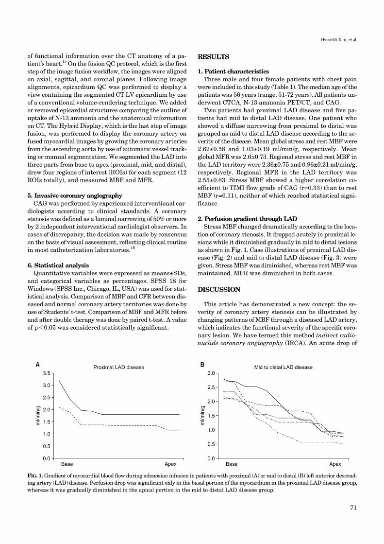
FIG. 1. Gradient of myocardial blood flow during adenosine infusion in patients with proximal (A) or mid to distal (B) left anterior descend-ing artery (LAD) disease. Perfusion drop was significant only in the basal portion of the myocardium in the proximal LAD disease group,whereas it was gradually diminished in the apical portion in the mid to distal LAD disease group.
of functional information over the CT anatomy of a pa-tient’s heart.15 On the fusion QC protocol, which is the first step of the image fusion workflow, the images were aligned on axial, sagittal, and coronal planes. Following image alignments, epicardium QC was performed to display a view containing the segmented CT LV epicardium by use of a conventional volume-rendering technique. We added or removed epicardial structures comparing the outline of uptake of N-13 ammonia and the anatomical information on CT. The Hybrid Display, which is the last step of image fusion, was performed to display the coronary artery on fused myocardial images by growing the coronary arteries from the ascending aorta by use of automatic vessel track-ing or manual segmentation. We segmented the LAD into three parts from base to apex (proximal, mid, and distal), drew four regions of interest (ROIs) for each segment (12 ROIs totally), and measured MBF and MFR.
5. Invasive coronary angiography CAG was performed by experienced interventional car-diologists according to clinical standards. A coronary stenosis was defined as a luminal narrowing of 50% or more by 2 independent interventional cardiologist observers. In cases of discrepancy, the decision was made by consensus on the basis of visual assessment, reflecting clinical routine in most catheterization laboratories.16
6. Statistical analysis Quantitative variables were expressed as means±SDs, and categorical variables as percentages. SPSS 18 for Windows (SPSS Inc., Chicago, IL, USA) was used for stat-istical analysis. Comparison of MBF and CFR between dis-eased and normal coronary artery territories was done by use of Students' t-test. Comparison of MBF and MFR before and after double therapy was done by paired t-test. A value of p<0.05 was considered statistically significant.
RESULTS
1. Patient characteristics Three male and four female patients with chest pain were included in this study (Table 1). The median age of the patients was 56 years (range, 51-72 years). All patients un-derwent CTCA, N-13 ammonia PET/CT, and CAG. Two patients had proximal LAD disease and five pa-tients had mid to distal LAD disease. One patient who showed a diffuse narrowing from proximal to distal was grouped as mid to distal LAD disease according to the se-verity of the disease. Mean global stress and rest MBF were 2.62±0.58 and 1.03±0.19 ml/min/g, respectively. Mean global MFR was 2.6±0.73. Regional stress and rest MBF in the LAD territory were 2.36±0.75 and 0.96±0.21 ml/min/g, respectively. Regional MFR in the LAD territory was 2.55±0.83. Stress MBF showed a higher correlation co-efficient to TIMI flow grade of CAG (r=0.33) than to rest MBF (r=0.11), neither of which reached statistical signi-ficance.
2. Perfusion gradient through LAD Stress MBF changed dramatically according to the loca-tion of coronary stenosis. It dropped acutely in proximal le-sions while it diminished gradually in mid to distal lesions as shown in Fig. 1. Case illustrations of proximal LAD dis-ease (Fig. 2) and mid to distal LAD disease (Fig. 3) were given. Stress MBF was diminished, whereas rest MBF was maintained. MFR was diminished in both cases.
DISCUSSION
This article has demonstrated a new concept: the se-verity of coronary artery stenosis can be illustrated by changing patterns of MBF through a diseased LAD artery, which indicates the functional severity of the specific coro-nary lesion. We have termed this method indirect radio-nuclide coronary angiography (IRCA). An acute drop of

72
Indirect Radionuclide Coronary Angiography by N-13 Ammonia PET/CT
FIG. 2. (A) Stress perfusion graph of a 59-year-old male patient with unstable angina showing an acute perfusion drop in the base ofthe left ventricle, the proximal LAD territory, whereas the rest perfusion graph shows a steady state of myocardial blood flow. (B) Myocardial flow reserve (MFR) diminished from the left ventricular base to the apex. The reference cutoff value of MFR is 2.0 at the authors’ institute. (C) Interventional coronary angiography showing a discrete stenotic lesion (arrow) in the proximal LAD. (D) Hybridimage of CT angiography and stress perfusion image of N-13 ammonia PET showing cutting of proximal left anterior descending artery(LAD), which correlated with the large perfusion defect in the LAD territory.
stress MBF was a typical finding of proximal LAD disease, whereas a gradual decrease of stress MBF was the finding in mid to distal LAD disease. The extent and degree of the perfusion defect measured by myocardial perfusion imaging (MPI) were regarded as indicators of the coronary lesion. The localization of coro-nary lesions is presumed from the location of the perfusion defect, which is frequently not matched to real coronary le-sions because of variations in coronary anatomy. Although the hybrid technology of CTCA and PET opened a possi-bility of pinpointing culprit coronary lesions, there are sev-eral barriers to overcome. First, coronary lesions with in-termediate severity detected by CTCA are false-positive in almost half of cases. Second, defining the perfusion defect from the 3D image of MPI is frequently confusing. Third, correlation of anatomical and functional lesions is arbitrary. Recently, fractional flow reserve (FFR) with the use of hyperemic coronary pressure measurement during CAG has been introduced as an accurate methodology for dis-criminating ischemic from nonischemic conditions. FFR has become an important index for decision making for re-vascularization of coronary artery lesions.17 An advantage
of FFR measurement is that coronary intervention can be done within the same session. A disadvantage of FFR is its invasiveness. We hypothesized that tracing blood flow changes through the coronary artery by using a hybrid technique of CTCA and PET would provide information on perfusion change in the same way as FFR, which traces pressure changes through the coronary artery lesion. We named this method indirect coronary angiography because it shows the coronary blood flow in an indirect way, and we added the term radionuclide because we used it. Thus, the name of the method became IRCA. IRCA has several advantages for clinical use. Most of all, it is noninvasive. It shows the severity of the coronary ar-tery lesion. It can pinpoint the location of the culprit lesion. The clinical usefulness of IRCA should be verified by a gold standard. Therefore, a direct comparison of FFR with IRCA may be a future and promising research theme. The ability of cardiac PET to measure MBF and MFR has been documented by numerous reports that showed an in-verse relationship between the severity of coronary lumi-nal narrowing and MFR. A clinical diagnostic added value of MFR over MPI was also reported.4 Adding functional da-
73
Hyun-Sik Kim, et al
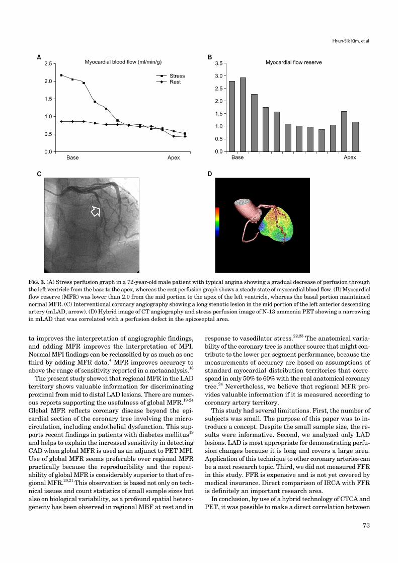
FIG. 3. (A) Stress perfusion graph in a 72-year-old male patient with typical angina showing a gradual decrease of perfusion throughthe left ventricle from the base to the apex, whereas the rest perfusion graph shows a steady state of myocardial blood flow. (B) Myocardialflow reserve (MFR) was lower than 2.0 from the mid portion to the apex of the left ventricle, whereas the basal portion maintainednormal MFR. (C) Interventional coronary angiography showing a long stenotic lesion in the mid portion of the left anterior descendingartery (mLAD, arrow). (D) Hybrid image of CT angiography and stress perfusion image of N-13 ammonia PET showing a narrowingin mLAD that was correlated with a perfusion defect in the apicoseptal area.
ta improves the interpretation of angiographic findings, and adding MFR improves the interpretation of MPI. Normal MPI findings can be reclassified by as much as one third by adding MFR data.4 MFR improves accuracy to above the range of sensitivity reported in a metaanalysis.18
The present study showed that regional MFR in the LAD territory shows valuable information for discriminating proximal from mid to distal LAD lesions. There are numer-ous reports supporting the usefulness of global MFR.19-24 Global MFR reflects coronary disease beyond the epi-cardial section of the coronary tree involving the micro-circulation, including endothelial dysfunction. This sup-ports recent findings in patients with diabetes mellitus19 and helps to explain the increased sensitivity in detecting CAD when global MFR is used as an adjunct to PET MPI. Use of global MFR seems preferable over regional MFR practically because the reproducibility and the repeat-ability of global MFR is considerably superior to that of re-gional MFR.20,21 This observation is based not only on tech-nical issues and count statistics of small sample sizes but also on biological variability, as a profound spatial hetero-geneity has been observed in regional MBF at rest and in
response to vasodilator stress.22,23 The anatomical varia-bility of the coronary tree is another source that might con-tribute to the lower per-segment performance, because the measurements of accuracy are based on assumptions of standard myocardial distribution territories that corre-spond in only 50% to 60% with the real anatomical coronary tree.24 Nevertheless, we believe that regional MFR pro-vides valuable information if it is measured according to coronary artery territory. This study had several limitations. First, the number of subjects was small. The purpose of this paper was to in-troduce a concept. Despite the small sample size, the re-sults were informative. Second, we analyzed only LAD lesions. LAD is most appropriate for demonstrating perfu-sion changes because it is long and covers a large area. Application of this technique to other coronary arteries can be a next research topic. Third, we did not measured FFR in this study. FFR is expensive and is not yet covered by medical insurance. Direct comparison of IRCA with FFR is definitely an important research area. In conclusion, by use of a hybrid technology of CTCA and PET, it was possible to make a direct correlation between

74
Indirect Radionuclide Coronary Angiography by N-13 Ammonia PET/CT
the coronary lesion and the gradient of MFR and CFR through coronary stenosis, and this indicated the severity of the coronary lesion. We named this technique indirect radionuclide coronary angiography.
ACKNOWLEDGEMENTS
This study was supported by a grant (A070001) from the Korea National Enterprise for Clinical Trials.
REFERENCES
1. Ghosh N, Rimoldi OE, Beanlands RS, Camici PG. Assessment of myocardial ischaemia and viability: role of positron emission tomography. Eur Heart J 2010;31:2984-95.
2. Kaufmann PA, Camici PG. Myocardial blood flow measurement by PET: technical aspects and clinical applications. J Nucl Med 2005;46:75-88.
3. Lima RS, Watson DD, Goode AR, Siadaty MS, Ragosta M, Beller GA, et al. Incremental value of combined perfusion and function over perfusion alone by gated SPECT myocardial perfusion imaging for detection of severe three-vessel coronary artery disease. J Am Coll Cardiol 2003;42:64-70.
4. Fiechter M, Ghadri JR, Gebhard C, Fuchs TA, Pazhenkottil AP, Nkoulou RN, et al. Diagnostic value of 13N-ammonia myocardial perfusion PET: added value of myocardial flow reserve. J Nucl Med 2012;53:1230-4.
5. Herzog BA, Husmann L, Valenta I, Gaemperli O, Siegrist PT, Tay FM, et al. Long-term prognostic value of 13N-ammonia myocar-dial perfusion positron emission tomography added value of coronary flow reserve. J Am Coll Cardiol 2009;54:150-6.
6. Fukushima K, Javadi MS, Higuchi T, Lautamäki R, Merrill J, Nekolla SG, et al. Prediction of short-term cardiovascular events using quantification of global myocardial flow reserve in patients referred for clinical 82Rb PET perfusion imaging. J Nucl Med 2011;52:726-32.
7. Ziadi MC, Dekemp RA, Williams KA, Guo A, Chow BJ, Renaud JM, et al. Impaired myocardial flow reserve on rubidium-82 positron emission tomography imaging predicts adverse outcomes in patients assessed for myocardial ischemia. J Am Coll Cardiol 2011;58:740-8.
8. Henzlova MJ, Cerqueira MD, Mahmarian JJ, Yao SS; Quality Assurance Committee of the American Society of Nuclear Cardiology. Stress protocols and tracers. J Nucl Cardiol 2006; 13:e80-90.
9. Lee BI, Kim KH, Kim JY, Kim SJ, Lee JS, Min JJ, et al. Correlation between semiquantitative myocardial perfusion score and absolute myocardial blood flow in 13N- ammonia PET. Nucl Med Mol Imaging 2007;41:194-200.
10. Muzik O, Beanlands RS, Hutchins GD, Mangner TJ, Nguyen N, Schwaiger M. Validation of nitrogen-13-ammonia tracer kinetic model for quantification of myocardial blood flow using PET. J Nucl Med 1993;34:83-91.
11. Hutchins GD, Schwaiger M, Rosenspire KC, Krivokapich J, Schelbert H, Kuhl DE. Noninvasive quantification of regional blood flow in the human heart using N-13 ammonia and dynamic
positron emission tomographic imaging. J Am Coll Cardiol 1990; 15:1032-42.
12. Chow BJ, Abraham A, Wells GA, Chen L, Ruddy TD, Yam Y, et al. Diagnostic accuracy and impact of computed tomographic coronary angiography on utilization of invasive coronary angio-graphy. Circ Cardiovasc Imaging 2009;2:16-23.
13. Chow BJ, Wells GA, Chen L, Yam Y, Galiwango P, Abraham A, et al. Prognostic value of 64-slice cardiac computed tomography severity of coronary artery disease, coronary atherosclerosis, and left ventricular ejection fraction. J Am Coll Cardiol 2010;55: 1017-28.
14. Hoffmann U, Moselewski F, Cury RC, Ferencik M, Jang IK, Diaz LJ, et al. Predictive value of 16-slice multidetector spiral computed tomography to detect significant obstructive coronary artery disease in patients at high risk for coronary artery disease: patient-versus segment-based analysis. Circulation 2004;110: 2638-43.
15. Gaemperli O, Schepis T, Kalff V, Namdar M, Valenta I, Stefani L, et al. Validation of a new cardiac image fusion software for three-dimensional integration of myocardial perfusion SPECT and stand-alone 64-slice CT angiography. Eur J Nucl Med Mol Imaging 2007;34:1097-106.
16. Tonino PA, De Bruyne B, Pijls NH, Siebert U, Ikeno F, van' t Veer M, et al; FAME Study Investigators. Fractional flow reserve versus angiography for guiding percutaneous coronary interven-tion. N Engl J Med 2009;360:213-24.
17. Pijls NH. Fractional flow reserve to guide coronary revascula-rization. Circ J 2013;77:561-9.
18. Nandalur KR, Dwamena BA, Choudhri AF, Nandalur SR, Reddy P, Carlos RC. Diagnostic performance of positron emission tomography in the detection of coronary artery disease: a meta-analysis. Acad Radiol 2008;15:444-51.
19. Schindler TH, Facta AD, Prior JO, Cadenas J, Zhang XL, Li Y, et al. Structural alterations of the coronary arterial wall are associated with myocardial flow heterogeneity in type 2 diabetes mellitus. Eur J Nucl Med Mol Imaging 2009;36:219-29.
20. Kaufmann PA, Gnecchi-Ruscone T, Yap JT, Rimoldi O, Camici PG. Assessment of the reproducibility of baseline and hyperemic myocardial blood flow measurements with 15O-labeled water and PET. J Nucl Med 1999;40:1848-56.
21. Wyss CA, Koepfli P, Mikolajczyk K, Burger C, von Schulthess GK, Kaufmann PA. Bicycle exercise stress in PET for assessment of coronary flow reserve: repeatability and comparison with adenosine stress. J Nucl Med 2003;44:146-54.
22. Austin RE Jr, Aldea GS, Coggins DL, Flynn AE, Hoffman JI. Profound spatial heterogeneity of coronary reserve. Discordance between patterns of resting and maximal myocardial blood flow. Circ Res 1990;67:319-31.
23. Chareonthaitawee P, Kaufmann PA, Rimoldi O, Camici PG. Heterogeneity of resting and hyperemic myocardial blood flow in healthy humans. Cardiovasc Res 2001;50:151-61.
24. Javadi MS, Lautamäki R, Merrill J, Voicu C, Epley W, McBride G, et al. Definition of vascular territories on myocardial perfusion images by integration with true coronary anatomy: a hybrid PET/CT analysis. J Nucl Med 2010;51:198-203.