Indian Journal of - Indian Society of Electrocardiology · Consultant Cardiologist, ... New Delhi...
48
Indian Journal of ELECTROCARDIOLOGY 2007 : No. 1 EDITORS Dr. Yash Lokhandwala Dr. Amit Vora
Transcript of Indian Journal of - Indian Society of Electrocardiology · Consultant Cardiologist, ... New Delhi...
Indian Journal ofELECTROCARDIOLOGY
2007 : No. 1
Secretariat
S. B. GUPTAHON. GENERAL SECRETARY
Indian Society of ElectrocardiologyHead, Department of Medicine and Cardiology, C. Rly, Head Quarters Hospital, Byculla, Mumbai - 400 027.
Phone : 2371 7246 (Ext. 425), 2372 4032 (ICU) • Resi: 2262 4556 • Fax : 2265 1044Mobile : 98213 64565 / 98216 38617 • e-mail : [email protected] • Website : www.iseindia.org
E D I T O R SDr. Yash LokhandwalaDr. Amit Vora
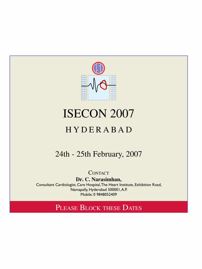
isecon 2007 H y d e r a b a d
24th - 25th February, 2007
ContaCt
Dr. C. Narasimhan, Consultant Cardiologist, Care Hospital, The Heart Institute, Exhibition Road,
Namapally, Hyderabad 500001, A.P. Mobile: 0 9848052409
Please BloCk these Dates
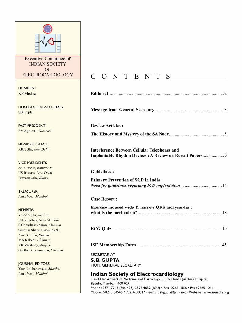
C o n t e n t s
Editorial ...................................................................................................... 2
Message from General Secretary ............................................................. 3
Review Articles :
The History and Mystery of the SA Node ................................................. 5
Interference Between Cellular Telephones and Implantable Rhythm Devices : A Review on Recent Papers ................... 9
Guidelines :
Primary Prevention of SCD in India : Need for guidelines regarding ICD implantation ..................................... 14
Case Report :
Exercise induced wide & narrow QRS tachycardia : what is the mechanism? .......................................................................... 18
ECG Quiz .................................................................................................. 19
ISE Membership Form ........................................................................... 45
executive Committee ofIndIan soCIety
ofeleCtroCardIology
PRESIDENTKP Mishra
HON. GENERAL-SECRETARYsB gupta
PAST PRESIDENTBV agrawal, Varanasi
PRESIDENT ELECT KK sethi, New Delhi
VICE PRESIDENTSss ramesh, BangaloreHs rissam, New DelhiPraveen Jain, Jhansi
TREASURERamit Vora, Mumbai
MEMBERSVinod Vijan, NashikUday Jadhav, Navi Mumbais Chandrasekharan, Chennaisushum sharma, New Delhianil sharma, KarnalMa Kabeer, ChennaiKK Varshney, Aligarhgeetha subramanian, Chennai
JOURNAL EDITORSyash lokhandwala, Mumbaiamit Vora, Mumbai
SECRETARIATS. B. GUPTAHON. GENERAL SECRETARY
Indian Society of ElectrocardiologyHead, Department of Medicine and Cardiology, C. Rly, Head Quarters Hospital, Byculla, Mumbai - 400 027.Phone : 2371 7246 (Ext. 425), 2372 4032 (ICU) • Resi: 2262 4556 • Fax : 2265 1044 Mobile : 98213 64565 / 98216 38617 • e-mail : [email protected] • Website : www.iseindia.org

Dear Colleagues,
the annual IseCon is around the corner. dr. narasimhan and his colleagues have spared no effort to provide a comprehensive eCg update in Hyderabad. the topics and the faculty promise an excellent 2 days for all who attend.
this issue of the IJe starts with an article by one of the doyens of eCg, our own dr. KP Misra and his colleagues. We are sure you will enjoy this comprehensive review, enriched with vast personal experience.
francis Johnson from Calicut has been doing an exemplary task in bringing out the web-based Indian Pacing and electrophysiology Journal (www.ipej.org). the last issue of the IJe had carried 2 articles from the IPeJ with his permission. this issue also has an article by Johnson and niehaus on cellular phones and implanted devices. this is a situation we are increasingly facing, with several queries from patients and their relatives. Hence this article will be of much practical utility.
an interesting case report has been provided by dr. I. sathyamurthy from Chennai. this report provides an excellent understanding in the mechanism of wide & narrow Qrs tachycardia.
the eCg Quiz is the core of our IJe. this time around we have covered some new ground and we hope you enjoy the exercise.
sudden death is overwhelmingly cardiac, usually arrhythmic. the ICd has emerged as a very useful device to prevent sCd in those at high risk. However, it is prohibitively expensive for the majority of Indians. a working group of the Ise has thus put together a document studying the cost-benefit of the ICD in different patient subgroups.
We once again request you for articles and eCg vignettes.
Yash Lokhandwala Amit VoraEditor Editor
Editorial

From Hon. Secretary’s Desk
dear Members,Indian society of electrocardiology is on fast track. We have tried to make the programs of Ise, a symbol of academic feasts only.dr K P Misra, dr Ulhas Pandurangi and their team organized IseCon-2006 at Chennai on 18th and 19th february 2006. there was a Pre-Conference Workshop for paramedics on 17th february 2006; both the events were astounding success. the organizers deserve heartiest congratulations. I feel Pre-Conference Workshops for paramedics shall become a regular feature.Indian society of electrocardiology organized the “2nd eCg learning Course” for postgraduate students on 22nd and 23rd July 2006 at Mumbai, which was attended by approximately 100 students. Successful candidates were awarded the Certificate of Competence for eCg reading. We would like to make it as a regular feature and would like to rotate to other cities too.the Mid-term CMe Course (aaC 2006) was organized at aligarh on 2nd and 3rd september 2006 by dr K K Varshney and his team and cudos to them for bringing another good scientific program.Ise organized an af symposium, highly successful meeting on 29th october 2006 under the leadership of dr amit Vora.and a unique program on “global approaches to Cardiac safety 2006” was organized under the banner of Ise in collaboration with Quintiles at Mumbai on 25th and 26th november 2006, coordinated by dr yash lokhandwala and dr snehal Kothari.ISE is organizing a Satellite Symposium on “Management of Atrial fibrillation” at Hotel goa Marriot, Panaji, goa on 16th february 2007 in association with euro Medical exchange foundation (eMeX). We are here to witness IseCon 2007 at Hyderabad by dr C narasimhan, a Master teacher and the organizer par excellence and his team on 24th and 25th february 2007. another grand event waits for you!My sincere thanks to dr yash lokhandwala, dr amit Vora and the editorial team for their effort to bring out the Indian Journal of electrocardiology.
long live Indian society of electrocardiology
Dr S B Gupta Hon. Secretary Indian Society of Electrocardiology


5
Review Article
The History and Mystery of the SA Node Presidential Oration – AAC 2006 in Aligarh Misra K.P.*, Gopichandran V.**, Senthamarai S.***
*senior Consultant Cardiologist **resident, Cardiology ***registrar, Cardiology
ABSTRACT
the sinoatrial node (sa node), discovered in 1907 by Keith and flack, is also referred to as the sinus node or the Keith and flack node. It is the primary pacemaker of the heart. next year being the centenary year of the discovery of the sa node it is appropriate to take a peek into the history of its discovery and the unique properties and characteristics of this very special node. located in the right atrium just beneath the pericardium in the junction between the superior vena cava and the right atrium, this collection of specialized cells has a very peculiar cellular arrangement. the action potential that is generated in the sa node is unique in that it is a self-depolarizing potential (automatic) and has a perfect rhythmicity. the role of calcium and potassium currents are dominant as compared to the dominant sodium current in the action potential of the other cells in the conduction system. there are several complicated mathematical models to explain the generation of action potential in the cells of the sa Node. The SA Node has a few diseases unique to itself. Electrophysiology has advanced much in the field of identification and treatment of sa node diseases.
Introduction:
the sinoatrial node (sa node) was discovered in 1907 and thus the next year will be the centenary of its discovery. the heart performing a very vital function namely circulation, has its own power generator, its pacemaker, the primary pacemaker being the sa node. due to its special properties it has a self-sustaining current, which drives the heart to contract and expel blood to all parts of the body in a coordinated and synchronized fashion. as one hundred years have passed since the knowledge of its existence it is fascinating to take a look at the history and mystery of this master pacemaker tissue. this article aims to give a brief insight into the structure of the sa node, its unique properties, the mechanism of action potential generation in the node and its common diseases.
History of the Discovery of the SA Node:
Keith and flack discovered the sa node in 19071. sir arthur Keith, a scottish anatomist, was the student of Charles darwin. He worked in close association with James McKenzie, a famous cardiologist and dissected the hearts of patients of dr. McKenzie, who had died of heart diseases. Martin William flack was a British physiologist. He was the student of sir arthur Keith and they discovered the node together. In their landmark paper published in the lancet, they wrote, “… an impulse arising in the sinus musculature near the termination of the superior vena cava may spread directly to the right auricle and the aV bundle…”1 It is curiously intereting that the discovery of the different tissues of the cardiac conduction tissues was in the
reverse order. the sa node, which is the master of the whole conduction system, was paradoxically the last to be discovered. The Purkinje fibres were discovered in 1845, the His bundle in 1893, AV node by Tawara in 1906 and finally the SA Node in 1907. at the time of its discovery it was called a unique collection of cells and gradually its properties were discovered which still remain mysterious.
Anatomy of the SA Node:
the sinoatrial node (sa node) consists of a cluster of specialized cells that have pacemaker activity (automaticity). these cells are responsible for initiating the electrical impulse that stimulates the heart muscles to contract sequentially and rhythmically. the sa node is located high on the surface of the right atrium close to where the superior vena cava enters the right atrium. It is located in the terminal groove between the right atrium and the superior vena cava. It is located directly under the pericardium and can be easily visualized when the pericardium is reflected. The SA nodal artery supplies the SA Node. It is the first and the largest branch of the right coronary artery arising from the first couple of centimeters of the artery. In some people the sa nodal artery arises directly from the anterior aortic sinus. In roughly about 40% of people the sa node artery arises from the left coronary artery passes behind the ascending aorta and pulmonary veins and reaches the right atrium.
Cells of the SA Node and their arrangement:
Histologically, the centre of the sa node is characterized

6 Indian Journal of Electrocardiology
by stellate or P-cells. these make multiple contacts with surrounding cells and are characterized by minimal organelles and contractile machinery within their cytoplasm. the surrounding cells show features akin to both stellate cells and contractile myocytes - these are transitional cells. the artery of the sinoatrial node traverses the node; it is purported that it may serve a physiological function by conveying aortic pressure to nodal tissue. apart from this there is a unique property of the sinus node in its cellular arrangement. This is the fibroblast network. Cardiomyocytes form a conducting network that is assumed to be electrically isolated from nonmyocytes in vivo in the SA Node. The Cardiac fibroblasts, which are interspersed in the node, can contribute to the spread of excitation via functional gap junctions with cardiomyocytes. these gap junctions help to morphologically distribute the cells in the node while making them a functional syncytium2. studies have shown that sections through the sinoatrial node double labeled with different connexins demonstrate regions, which are sharply demarcated from one another in the regions of the crista terminalis. a zone of connective tissue contributes to separation between the zones, although elsewhere the zones seem to be more closely approximated3. It is believed that this kind of arrangement might have significance in the electrophysiological properties of the node.
Innervation of the SA Node:
the sinus node is densely innervated with postganglionic adrenergic and cholinergic nerve terminals. discrete vagal efferent pathways innervate both the sinus and atrioventricular (aV) regions of the dog and nonhuman primate. the concentration of norepinephrine is two to four times higher in atrial than in ventricular tissue in canine and guinea pig hearts. although the sinus nodal region contains amounts of norepinephrine equivalent to those in other parts of the right atrium, acetylcholine, acetylcholinesterase, and choline acetyltransferase have all been found in greatest concentration in the sinus node, with the next highest concentration in the right and then the left atrium. the concentration of acetylcholine in the ventricles is only 20 to 50 percent of that in the atria4.
neurotransmitters modulate the sinus node discharge rate by stimulation of beta-adrenergic and muscarinic receptors. Both beta1 and beta2 adrenoceptor subtypes are present in the sa node. Human sa node contains a more than threefold greater density of beta-adrenergic and muscarinic cholinergic receptors than adjacent atrial tissue. The functional significance of beta adrenoceptor subtype diversity in the sinus node is unclear. Binding of receptor agonists released from sympathetic nerve terminals causes a positive chronotropic response. the negative chronotropic response of vagal stimulation is mediated by acetylcholine binding to and ensuing activation of M2 muscarinic receptors. Besides its negative chronotropic effect, acetylcholine also prolongs intranodal conduction time, at times to the point of sinus nodal exit block. acetylcholine increases
whereas norepinephrine decreases refractoriness in the center of the sinus node. the phase (timing) in the cardiac cycle at which vagal discharge occurs and the background sympathetic tone importantly influence vagal effects on the sinus rate and conduction5. after cessation of vagal stimulation, sinus nodal automatically may accelerate transiently (postvagal tachycardia). the neurotransmitters neuropeptide y (nPy) and vasoactive intestinal peptide (VIP) are localized in the sympathetic and parasympathetic nerve terminals, respectively. their exact roles are not clear.
The Unique pacemaker function of the SA Node:
Cells within the sa node are the primary pacemaker site within the heart. these cells are characterized as having no true resting potential, but instead generate regular, spontaneous action potentials. Unlike most other cells that elicit action potentials (e.g., nerve cells, muscle cells), the depolarizing current is carried primarily by relatively slow, inward Ca++ currents instead of by fast na+ currents. there are, in fact, no fast na+ currents operating in sa nodal cells. the components of sa node action potential are as shown in figure 1:
Figure 1: the sa node action Potential
The figure shows the action potential of the SA Node. Phase 0 is the phase of depolarization. Phase 3 is the phase of repolarisation and Phase 4 is the phase of slow depolarization, which is characteristic of the sa node.
• Phase 0 depolarization is primarily due to increased Ca++ conductance. Because the movement of Ca++ through their channels is not rapid, the rate of depolarization is much slower than found in other cardiac cells (e.g., Purkinje cells).
• Repolarization occurs (Phase 3) as K+ conductance increases and Ca++ conductance decreases.
• Spontaneous depolarization (Phase 4) is due to a fall in K+ conductance as potassium channels close and to a small, slow increase in Ca++ conductance. a slow inward na+ current may also contributes to Phase 4, and is thought to be responsible for what is termed as the pacemaker or “funny” current. once this spontaneous depolarization reaches threshold (about –40 to -60 mV), a new action potential is triggered4.
this action potential is unique to the sa node and drives the

7
heart contraction around 70-90 times each minute.
The mathematical models of SA Node pacemaker activity:
there are several mathematical models for the pacemaker activity of the sa node. these models are based on experimental data from voltage clamp experiments on single cells. three mathematical models of rabbit sa node pacemaker activity are the Bristow-Clark model (Bristow and Clark, 1982)6, the Irisawa-noma model (Irisawa and noma, 1982)7 and the noble-noble model (noble and noble, 1984)8 have been described. noble et al. later (1989) published a modification of the Noble-Noble model, referred to as the noble-difrancesco-denyer model9. In the Bristow – Clark model there are two main currents: Voltage dependent-time independent background Potassium current (iK1) and the pacemaker current (iK2). It was relatively simple but had its own drawbacks. the Irisawa noma model was based on the same five basic currents of the Bristow and Clark model. It simulated the spontaneous current generation more closely than the previous model. Its major disadvantage was the large artificial background current. A more comprehensive and detailed model was the noble-noble model, which was further modified by the Noble-DiFrancesco-Denyer model. The understanding of these detailed models is beyond the scope of this paper.
Diseases of the SA Node:
sinus node dysfunction (snd) is a disturbance in the normal sa node function. It may manifest as abnormal sa node impulse formation and/or propagation, which leads to rhythms that are slow (ie, bradyarrhythmias) or fast (ie, tachyarrhythmias) for the person’s age10. snd is referred to as sick sinus syndrome when accompanied by brady-tachy arrhythmias. the diseases of the sa node are the following:
• Sinus Tachycardia• Sinus Bradycardia• Sinus Arrhythmia (Figure 1)• SA Block• SA Arrest• SA Wenckebach• Sick Sinus Syndrome
the eCg criteria for the diagnosis of sa node dysfunction are11:
• Sinus bradycardia below the heart rate expected for age (figure 2)
• Sinus pause or absence of an expected P wave for more than 3 seconds, which may be due to sinus arrest or due to sinoatrial exit block (figure 3)
• Slow escape rhythms that originate within the atria, AV junction, or ventricles
• Marked sinus arrhythmia with constant variation in the P-P interval, which is likely to be accompanied by sinus bradycardia
• Presence of both bradyarrhythmias and tachyarrhythmias (ie, sn reentry tachycardia, atrial tachycardias from an ectopic focus, atrial flutter, atrial fibrillation)
Figure 3 sinus arrest
on electrophysiological studies the criteria for sa node dysfunction are11:
• Corrected SN recovery time (CSNRT) greater than 275 msec
• Sinoatrial conduction time greater than 200 msec Figure 1 sinus arrhythmia
Figure 2 sinus Bradycardia

8 Indian Journal of Electrocardiology
• Sinoatrial node arrest
• Sinoatrial exit block
• SN reentry tachycardia
Sinus Node Recovery Time (SNRT):
Measurement of snrt is achieved by pacing the right atrium. Pacing should be performed for 4 -6 trials of 30 seconds each. snrt is the time interval between the last paced atrial beat to the first spontaneous sinus beat. If the longest interval for the recovery interval or secondary pause exceeds 1500 milliseconds, the snrt is prolonged12.
Sino Atrial Conduction Time (SACT):
It is the time interval (in ms) for an impulse originating in the sn to conduct through the perinodal tissue to the adjacent ra tissue. Eight PACs are fired in the High Right Atrium (HRA) at 5-10 bpm faster than the sa node rate before they are stopped abruptly. saCt represents the time (in ms) it takes for the PaC fired in the HRA to enter and reset the SN. It also represents the time for the new spontaneous sa node impulse to reach the Hra12.
Treatment Modalities:
no treatment is required for asymptomatic patients, even if they have abnormal snrts or saCts. However, implantation of a permanent pacemaker is required if bradycardia is severe for the patient’s age or if snd is accompanied by dizziness, syncope, fatigue, CHf, chest pain and palpitations (in patients with bradyarrhythmias or tachyarrhythmias), and especially syncope. If the patient is receiving medications that can provoke sinus bradyarrhythmias (eg, beta-blockers, non-dihydropyridine calcium channel blockers and angiotensin-converting enzyme [aCe] inhibitors), the medications should be stopped if possible.
the sa node has been known for a century now and much has been understood about it and its function. But much more remains a mystery and will continue to be so. this article is a tribute to Keith and flack for their diligent work and to all the innumerable scientific minds that have contributed to the understanding of the structure and function of this enigmatic node.
References:
1. a. Keith, M. W. flack. the auriculo-Ventricular Bundle of the Human Heart. lancet. 1906, 2: 359.
2. Patrizia Camelliti, Colin r. green, Ian legrice, Peter Kohl. fibroblast network in rabbit sinoatrial node - structural and functional Identification of Homogeneous and Heterogeneous Cell Coupling. Circ res. 2004;94:828-835.
3. Coppen sr, Kodama I, Boyett Mr, et al: Connexin45, a major connexin of the rabbit sino-atrial node, is co-expressed with connexin43 in a restricted zone at the nodal-crista terminalis border. J Histochem Cytochem 47:907, 1999.
4. Schuessler RB, Boineau JP, Saffitz JE, et al: Cellular mechanisms of sinoatrial activity. In Zipes dP, Jalife J (eds): Cardiac electrophysiology: from Cell to Bedside. 3rd ed. Philadelphia, WB saunders, 1999, pp 187–195.
5. Steinberg SF: The molecular basis for distinct β-adrenergic receptor subtype actions in cardiomyocytes. Circ res 85:1101, 1999.
6. Bristow, d. g., and J. W. Clark. 1982. a mathematical model of primary pacemaking cell in sa node of the heart. am. J. Physiol. 243:H207-H218.
7. Irisawa, H., and a. noma. 1982. Pacemaker mechanisms of rabbit sinoatrial node cells. In Cardiac rate and rhythm. l. n. Bouman, and H. J. Jongsma, editors. Martinus nijhoff, london. 35-51.
8. noble d., and s. J. noble. 1984. a model of sino-atrial node electrical activity based on a modification of the DiFrancesco-Noble (1984) equations. Proc. r. soc. lond. B Biol. sci. B222:295-304.
9. noble, d., d. difrancesco, and J. C. denyer. 1989. Ionic mechanisms in normal and abnormal cardiac pacemaker activity. In neuronal and Cellular oscillators. J. W. Jacklet, editor. Marcel dekker, Inc., new york. 59-85.
10. Victor adan, loren a Crown. diagnosis and treatment of sick sinus syndrome. am fam Physician 2003;67:1725-32,1738.
11. fish fa, Benson W: disorders of Cardiac rhythm and Conduction. In: Moss and adam’s Heart disease in Infants, Children and adolescents. Vol 1. Philadelphia, Pa: lippincott Williams and Wilkins; 2000: 495-496.
12. g Breithardt, l seipel and f loogen. sinus node recovery time and calculated sinoatrial conduction time in normal subjects and patients with sinus node dysfunction. Circulation. 1977; Vol 56: 43-50.
9
Review Article
Introduction
Implantable rhythm device (Ird) is the generic name for the group of implantable devices used for treatment of cardiac arrhythmias like cardiac pacemakers and implantable cardioverter defibrillators. Since these devices have complex microelectronic circuitry and use electromagnetic waves for communication with programmers, they are susceptible to interference from most sources of electromagnetic radiation and magnetic energy1. Cellular telephones use radio frequency waves for communication and are likely to interfere with the function of implantable rhythm devices.
Cellular telephones produce both static and dynamic electromagnetic fields. The magnet in the earpiece of the phone produces a low energy static magnetic field. This static magnetic field can activate the internal reed switch causing temporary suspension of sensing function when placed in close proximity to the implanted device2. Dynamic fields with much higher intensity are produced by the radio frequency energy used for communication. today we have two basically different
Interference Between Cellular Telephones and Implantable Rhythm Devices: A Review on Recent Papers Johnson Francis, MBBs, Md, dM, fCsI1 and Michael Niehaus, Md2 1department of Cardiology, Calicut Medical College, Calicut, Kerala, India 2department of Cardiology and angiology, Medical school Hannover, germany
ABSTRACT
Background: Cardiac pacemakers and implantable defibrillators are potentially susceptible to electromagnetic interferences as they have complex circuitry for sensing and communication purposes. Cellular telephones being an important source of electromagnetic waves are likely to cause interference in the function of these devices.
Methods : a systematic analysis of studies on interaction between cellular telephones and implantable devices was done using professional databases for literature. related articles and references of relevant articles were also searched for suitable studies.
Results: Fourteen studies on pacemakers and eight studies on implantable defibrillators were identified. No dangerous malfunction was found in any of the analyzed studies, but most of the studies noted interference with device function when the phone was operated very close to the device. Interference was minimally in those devices with built in feed-through filters for eliminating electromagnetic interference. device programming and interrogation were the most susceptible phases of operation.
Summary: Cellular phones are likely to interfere with implantable rhythm devices if operated in close proximity or during programming of the device. Patients with implanted devices can safely use cellular phones if they are not carried close to the implanted devices or operated near them. Carrying the cellular phones in the belt position, receiving calls in the ear opposite to the side of the implanted device and keeping the phone as far away as possible while dialing can be considered a safe practice. Interrogation of the devices should take place exclusively in areas where utilization of cellular phones is strictly prohibited. studies on pacemakers published in the current decade have shown much lesser rates of interference, possibly due to improvement in device technology.
Keywords : pacemaker; defibrillator, implantable; electromagnetic interference; cellular phone; European; American
communication systems, analogue and digital systems that vary in their ability to produce interference with Irds.
Methods
We carried out a systematic analysis of available data on the interference of implantable rhythm devices by cellular telephones. database searches were conducted using the search words “cell phone, mobile phone, cellular telephone” in combination with “pacemaker, implantable cardioverter defibrillator, ICD” and independently. the retrieved results were checked to identify relevant studies. further studies were sought by searching for the related articles and the references of the retrieved articles. only clinical studies were included.
Studies Identified
The studies identified are listed in Table 13-24. the earliest published study dates back to 1995 and the latest was published in 2004. the largest study till date was done by Hayes et al, (1997) with 980 patients with implanted pacemakers11. the

10 Indian Journal of Electrocardiology
largest study on patients with implantable defibrillators (ICD) was published in 2002 with 97 patients19. a total of 14 studies on patients with implanted pacemakers and 8 studies on patients with ICD were identified3-24. Interestingly, majority of the studies were from europe, with only two from north america and one each from asia and australia. there were a total of 3054 patients in all studies taken together.
Cellular Phones and Networks
Various types of analogue and digital cellular phones are in use across the globe. analogue telephones transmit by modulation of the amplitude or frequency of electromagnetic waves which are transmitted continuously. on the other hand, the digital telephones transmit data in series of pulses or fast bursts. the advantage of the digital systems is that they allow simultaneous transmission of messages of different users on the same frequency which increases the capacity of the transmission channels. digital phones are more likely to interfere with Irds than analogue phones. this is because the pulse repetition rate of the devices falls within the frequency range of physiological signals.
different frequencies and technologies are in use in different parts of the globe. european system is gsM (global system for Mobile Communications) in three different frequency ranges. the digital d-net works on a carrier frequency of 900 MHz and the digital e-net works on a carrier frequency of 1800 MHz. the C-net working analogue on 450 MHz was being used in european countries earlier25. gsM networks are in use in asia and australia1,7. the nadC-phones (north american digital Cellular) work on a carrier frequency of 835 Mhz2.
Feed Through Filters
Feed through filters are broadband filters using ceramic capacitors which reduce the influence arising from radio frequency sources on pacemakers and ICDs significantly. All IRDs have a titanium can which acts as an electromagnetic shield and a hermetic barrier to protect the internal components from body fluids. the lead wires which carry the pacing pulses and sense cardiac activity may also act as an antenna that conducts undesirable radio frequency signals from cellular phones to sensitive internal electronic circuits. The EMI filter decouples and shields such signals and prevents them from interfering with pacemaker or ICd functions.
Interference With Pacemakers
Until now, pacemakers constitute the large majority of Irds and hence most of the studies have been on these devices. of the 2726 patients included in the various studies, 393 (14.4%) had some form of electromagnetic interference, when the cellular phones were operated in close proximity of the device. But there was considerable heterogeneity between the studies, with the percentage varying from 0 to 43. Inhibition of ventricular output, tracking of the interference sensed in the atrial channel and asynchronous pacing were the common problems noted. Interference could be reduced by programming to lower sensitivity levels5,15. Increasing the transmitting power of the cellular phone also increases the probability for interference5. In the practical scenario, this occurs in rural areas where access points for the gsM phones are farther apart and the cellular phone automatically steps up the output. the studies uniformly reported no interference when the phone was held in the phoning
Table 1: Clinical studies on Implantable rhythm devices and Cellular Phones
Year No. of Patients
EMI Year No. of Patients
EMI
Pacemakers ICDBarbaro et al 1995 101 26 sanmartin et al 1997 30 0Wilke et al 1996 50 2 Jimenez et al 1998 72 14naegeli et al 1996 39 7 fetter et al 1998 41 0nowak et al 1996 31 0 occhetta et al 1999 30 30sparks et al 1996 16 1-7 Barbaro et al 1999 13 3Hofgartner et al 1996 104 43 Chiladakis et al 2001 36 7Chen et al 1996 29 8 niehaus et al 2002 97 38Hayes et al 1997 980 20.00% tandogan et al 2002 9 0altamura et al 1997 200 43raden et al 1999 144 9-17elshershari et al 2002 95 1Hekmat et al 2004 100 2tandogan et al 2005 679 37trigano et al 2005 158 4

11
position over the ear. almost all the interferences occurred with the phone held directly over the device.
Pacemaker interference by cellular phones has been classified into three groups according to the clinical significance (Hayes et al11):
Class I -Clinical responses that are definitely significant. e.g. Interference associated with syncope or pre-syncope
Class II -Clinical responses that are probably significant. e.g. transient ventricular inhibition less than 3 seconds
Class III - Clinical responses that are probably not significant
In this study, 20% of the total 5533 tests carried out showed interference of some form. of these 1.7% were Class I, 4.9% Class II and 13.4% Class III interference.
the earliest series in this review was by Barbaro et al3. this study involved 101 patients with 43 pacemaker models from 11 manufacturers. 26 patients showed interference at minimum sensing thresholds, with the phone in direct contact with the patient’s chest. Pulse inhibition (9.9%), ventricular triggering (19.5%) and asynchronous pacing (7.7%) were the common interferences noted. Maximum distance at which interference occurred was 10 cm with the pacemaker programmed at its minimum sensing threshold.
the study by Hayes et al11 involving 980 patients with implanted pacemakers was the largest of the lot. It is a well designed study with five types of cellular phones (one analogue and four digital). the telephones were programmed to transmit full power, to mimic the worst case situation. of a total of 5533 tests conducted, interference was noted in 20%, of which 7.2% were symptomatic. Clinically significant interference was seen in 6.6%. No significant interference was noted when the telephone was placed in the standard phone position over the ear. as expected, interference was much higher when the phone was placed near the pacemaker. dual-chamber pacemakers were more susceptible (25.3%) than single-chamber pacemakers (6.8%; P<0.001). Pacemakers with feed-through filters were less susceptible to eMI (0.4 to 0.8%) than those without such filters (28.9 to 55.8 %, P=0.01). Marked difference was noted in the incidence of eMI between analogue and digital phones (2.5% vs 23.7%, P=0.01). Interference was higher among pacemaker dependent patients (20.9%) than those who were not (15.2%). the most common types of interferences were tracking interference (14.2%), noise reversion or asynchronous pacing (7.3%) and ventricular inhibition (6.3%). less common problems noted were atrial inhibition (2.3%), ventricular safety pacing (1.8%), undersensing (0.9%) and rate-adaptive sensor-driven pacing (0.3%). Palpitations was the most common symptom (4.5%). light headedness occurred in 1.2 % and pre-syncope in 0.2%. Pre-syncope occurred only in those patients who were pacemaker dependent.
study by altamura et al12 included 200 patients. Interference was noted in 21.5 % with gsM phones and 17.5% with total access of Communication system (taCs) telephones. Interference was much more common during ringing than on/off phase (131 vs 26 episodes, P<0.0001). Incidence of interference increased with increasing sensitivity (106 at maximum sensitivity vs 51 at basal values; P<0.0001). the authors concluded that if phones were not carried close to the pacemaker, safety was not compromised.
raden et al15 reported a study on 144 patients with implanted pacemakers (134 with single chamber and 10 with dual chamber). While 9 patients (6.25%) had intermittent pacemaker inhibition at basal settings, 17 patients (11.8%) showed inhibition on reprogramming to maximum sensitivity. the tests were conducted with the phone directly over the pacemaker site.
Hofgartner et al8 reported on 104 patients with 58 different models of pacemakers. Interference was noted in 28 different pacemaker types (48.3%) spread over 43 patients (41.3%). Pacemaker inhibition, noise reversion and triggering of pacemaker mediated tachycardia were noted.
all the above series with 100 or more patients which reported rather high incidence of interference were from the last decade (figure 1). the four studies published in the current decade report a very low incidence of interference21-24. smaller studies in the last decade have also reported low incidence4,6. In 2002, elshershari et al21 reported on 95 patients with pacemakers from 6 different manufacturers. testing was done with two models of gsM d-net phones. only one instance of brief oversensing was noted.
Hekmat et al, (2004)22 observed pacing inhibition in only 2 patients out of 100, with the phones placed directly above the pacemaker pocket. these inhibitions occurred at programmed sensitivity values of less than 0.5 mV and could be eliminated by reprogramming to 1.0 mV. Hence they recommended
Figure 1: Percentage of eMI in Major studies on Pacemakers

12 Indian Journal of Electrocardiology
programming ventricular sensitivity to 2.0 mV or higher. a change of lead configuration from unipolar to bipolar did not eliminate the interference. all pacemakers in this study were equipped with feed-through filters. All evaluated models showed significant telemetric noise when the phone was placed near the programming head, sometimes even causing loss of telemetric data.
trigano et al, (2005)24 noted interference in 1.5% of 330 tests performed in 158 patients. Interference was noted only during 5 tests in 4 unprotected pacemaker models due to interaction with gsM mobile phones. no interference was noted in 12 other tests of identical pulse generator models. the gsM phones had a maximal power output of 2 W and were operating on a 900 MHz carrier frequency.
the largest report of the current decade from tandogan et al23
included 679 patients. Interference was noted in 37 patients (5.5%). thirty-three VVI-r pacemakers were converted to asynchronous mode and 3 were inhibited. one ddd-r pacemaker developed ventricular triggering. Interference was more common when the lead polarity was unipolar (4.12% vs 1.40%, p<0.01). these interferences did not cause any symptoms and the pacemaker function returned to normal when the cell phone was removed away from the patient.
It is likely that better pacemaker technology, especially the use of feed-through filters could have contributed to the lower incidence of interference in the recent studies.
Interference With Implantable Cardioverter Defibrillators
In the last two decades, ICds are fast becoming universal arrhythmia management devices for prevention of sCd, especially after the publication of MadIt I and II results. We could identify 8 studies on the influence of cellular phones on ICds, with a total of 328 patients. 92 patients (28.09%) showed some type of interference when the activated phone was placed over the ICd. Pseudo-oversensing18, ventricular triggering17, telemetry noise16 and partial loss of telemetry13 were the types of interference noted. no inadequate shock therapy was observed. as in the case of pacemakers, interference occurred mostly when the phone was held close to the implanted device. devices were most vulnerable for interference during the time of interrogation and programming.
the largest available study was on 97 devices reported by niehaus et al in 200219. d-net (900 MHz) and e-net (1800 MHz) phones were used for testing. Interferences (loss of communication or temporary inactivation of the device during interrogation) were noticed in 38 patients. Most of these (93%) occurred while testing close to the device. Jimenez et al13
published in 1998 their study on 72 patients of which 14 showed interference. Partial loss of telemetry was found in 8 patients with analogue phones and 6 patients with digital phones. But none of these were clinically significant.
occhetta et al (1999)16 reported on thirty patients with ICds from five different manufacturers. Both TACS and GSM phones were used for testing. this study was unique in that it reported interference with all the evaluated models. the interference consisted of noise in telemetric transmission when the phone was located near the ICd and the programmer´s head. the noise was most significant during call and reception, leading to loss of telemetry in most cases.
It is important to note that there was no false arrhythmia detections during the tests, neither a delay in recognition of induced ventricular fibrillation. Hence they suggested that patients with implanted ICds may use cellular phones, but not during ICd programming and interrogation. In contrast to the above report, fetter et al14 who studied the effect of north american digital Communications (nadC)/time division Multiple access-50-Hz (tdMa-50) digital phones on ICds, reported no interference due to oversensing of the dynamic electromagnetic field in their 41 patients. However, they found that the static magnetic field of the phone’s earpiece placed over the ICd will activate the internal reed switch causing temporary suspension of ventricular tachycardia and fibrillation detection.
Chiladakis et al18 reported on 36 patients with ICds from two manufacturers. In seven devices from one manufacturer, they noted transient eMI causing 19 erroneous sensing events (pseudo-oversensing) when the phone was operated close to the programmer head. But these events were not logged as arrhythmia episodes by the counter in the device. therefore this observation has to be interpreted as adverse interaction between the phone and the telemetry function of the ICd. no interference in the function of the ICd was documented regardless of the distance, power or mode of operation of the cellular phone.
Conclusion
In summary, cellular phones are likely to interfere with implantable rhythm devices if operated in close proximity or during programming of the device. Patients with implanted devices can safely use cellular phones if they are not carried close to the devices or operated near them. Carrying the cellular phones in the belt position, receiving calls in the ear opposite to the side of the implanted device and keeping the phone as far away as possible while dialing can be considered a safe practice. Interrogation of the devices should take place exclusively in areas where utilization of mobile phones is strictly prohibited as this is the period in which maximum interference is likely. due to the heterogenic reactions of the implanted devices on cellular phones, eMI by cellular phones should be tested carefully in every new developed implantable rhythm device.
References
1 Johnson francis. electromagnetic Interference in Implantable rhythm devices -the Indian scenario. Indian Pacing electrophysiol. J. 2002;2:64-65.

13
2 Marcos de sousa, gunnar Klein, thomas Korte, Michael niehaus. electromagnetic Interference in Patients with Implanted Cardioverter-Defibrillators and Implantable Loop Recorders. Indian Pacing electrophysiol. J. 2002;2:79-84.
3 Barbaro V, Bartolini P, donato a, Militello C, altamura g, ammirati f, santini M. do european gsM mobile cellular phones pose a potential risk to pacemaker patients? Pacing Clin electrophysiol. 1995;18:1218-24.
4 Wilke A, Grimm W, Funck R, Maisch B. Influence of D-net (European gsM-standard) cellular phones on pacemaker function in 50 patients with permanent pacemakers. Pacing Clin electrophysiol. 1996 ;19:1456-8.
5 naegeli B, osswald s, deola M, Burkart f. Intermittent pacemaker dysfunction caused by digital mobile telephones. J am Coll Cardiol. 1996 ;27:1471-7.
6 nowak B, rosocha s, Zellerhoff C, liebrich a, Himmrich e, Voigtlander t, Meyer J. Is there a risk for interaction between mobile phones and single lead Vdd pacemakers? Pacing Clin electrophysiol. 1996;19:1447-50.
7 sparks PB, Mond Hg, Joyner KH, Wood MP. the safety of digital mobile cellular telephones with minute ventilation rate adaptive pacemakers. Pacing Clin electrophysiol. 1996;19:1451-5.
8 Hofgartner f, Muller t, sigel H. Could C-and d-network mobile phones endanger patients with pacemakers? dtsch Med Wochenschr. 1996;121: 646-52.
9 Chen WH, lau CP, leung sK, Ho ds, lee Is. Interference of cellular phones with implanted permanent pacemakers. Clin Cardiol. 1996;19:881-6.
10 sanmartin M, fernandez lozano I, Marquez J, antorrena I, Bautista a, silva l, ortigosa J, de artaza M. the absence of interference between GSM mobile telephones and implantable defibrillators: an in-vivo study. groupe systemes Mobiles. rev esp Cardiol. 1997 ;50:715-9.
11 Hayes DL, Wang PJ, Reynolds DW, Estes M 3rd, Griffith JL, Steffens ra, Carlo gl, findlay gK, Johnson CM. Interference with cardiac pacemakers by cellular telephones. n engl J Med. 1997 ;336:1473-9.
12 altamura g, toscano s, gentilucci g, ammirati f, Castro a, Pandozi C, Santini M. Influence of digital and analogue cellular telephones on implanted pacemakers. eur Heart J. 1997 ;18:1632-41.
13 Jimenez a, Hernandez Madrid a, Pascual J, gonzalez rebollo JM, fernandez e, sanchez a, ortega J, lozano f, Munoz r, Moro C. Electromagnetic interference between automatic defibrillators and digital and analog cellular telephones. rev esp Cardiol. 1998 ;51:375-82.
14 fetter Jg, Ivans V, Benditt dg, Collins J. digital cellular telephone interaction with implantable cardioverter-defibrillators. J Am Coll Cardiol. 1998 ;31:623-8.
15 raden g, Pavlovic P, Vucinic Z, tavciovski d, Matunovic r, djuran P, Prcovic M. the effect of cell phones on pacemaker function. Vojnosanit Pregl. 1999;56:491-7.
16 occhetta e, Plebani l, Bortnik M, sacchetti g, trevi g. Implantable cardioverter defibrillators and cellular telephones: is there any interference? Pacing Clin electrophysiol. 1999 ;22:983-9.
17 Barbaro V, Bartolini P, Bellocci f, Caruso f, donato a, gabrielli d, Militello C, Montenero as, Zecchi P. electromagnetic interference of digital and analog cellular telephones with implantable cardioverter defibrillators: in vitro and in vivo studies. Pacing Clin Electrophysiol. 1999 ;22:626-34.
18 Chiladakis Ja, davlouros P, agelopoulos g, Manolis as. In-vivo testing of digital cellular telephones in patients with implantable cardioverter-defibrillators. Eur Heart J. 2001 ;22:1337 42.
19 niehaus M, gille K, Cierpka r, Korte t, tebbenjohanns J: Interference of two common european digital cellular phones with implantable cardioverter-defibrillators. Eur Heart J. 2002;23:586-588.
20 tandogan I, Ileri M, yetkin e, temizhan a, aras d, sezgin at, Biyikoglu f, sasmaz a. do mobile telephones have adverse effects on the functions of implantable cardioverter defibrillators?. Anadolu Kardiyol Derg. 2002;2:45-8.
21 Elshershari H, Celiker A, Ozer S, Ozme S. Influence of D-net (EUROPEAN gsM-standard) cellular telephones on implanted pacemakers in children. Pacing Clin electrophysiol. 2002 ;25:1328-30.
22 Hekmat K, salemink B, lauterbach g, schwinger rH, sudkamp M, Weber HJ, Mehlhorn U. Interference by cellular phones with permanent implanted pacemakers: an update. europace. 2004 ;6:363-9.
23 tandogan I, temizhan a, yetkin e, guray y, Ileri M, duru e, sasmaz a. the effects of mobile phones on pacemaker function. Int J Cardiol. 2005; 103:51-8.
24 trigano a, Blandeau o, dale C, Wong Mf, Wiart J. reliability of electromagnetic filters of cardiac pacemakers tested by cellular telephone ringing. Heart rhythm. 2005;2:837-41.
25 Barbaro V, Bartolini P, donato a, Militello C. electromagnetic interference of analog cellular telephones with pacemakers. Pacing Clin electrophysiol. 1996;19:1410-8.
14 Indian Journal of Electrocardiology
Primary Prevention of SCD in India : Need for guidelines regarding ICD implantationNitish Naik*, Rajnish Juneja*, C Narasimhan**, Yash Lokhandwala*** - ISE Working Group for Primary Prevention Sudden Cardiac Death* department of Cardiology, Cn Center, all India Institute of Medical sciences, new delhi ** department of Cardiology, Care Hospital, Hyderabad *** arrhythmia associates, Mumbai
Guidelines
sudden cardiac death occurs in 0.1% of the population based on data collected from western literature. sudden death accounts for nearly 450,000 lives annually in the United states1. Considering an Indian population of more than 1 billion, unadjusted figures would predict about 1 million events every year in India! as survival rates from a sudden death event in the community are extremely low (less than 5% even in the developed world), intensive efforts are being made to identify and protect such sCd prone individuals from early fatal cardiac events.
a wide range of drugs including amiodarone, beta-blockers, angiotensin converting enzyme inhibitors, spironolactone and statins have been shown to either decrease recurrences of serious ventricular tachyarrhythmias or reduce arrhythmic death. However, in randomized trials comparing anti-arrhythmic drugs with the implantable cardioverter defibrillator (ICD), ICD”s have emerged superior to drugs in most patient populations, for secondary as well as primary prevention of sudden death. antiarrhythmics have not been an effective means of preventing recurrences of ventricular tachyarrhythmias and apart from amiodarone and beta-blockers most of them actually increase mortality. Consequently, the indications of the ICd have been expanded to include patients at future risk of sudden death based on certain risk markers. this “risk-marker” has been left ventricular function as assessed by left ventricular ejection fraction (lVef). earlier primary prevention studies included other risk markers in addition to lVef to better identify the patient at risk for arrhythmic death. these included nonsustained ventricular tachycardia on Holter, signal averaged eCg and electrophysiologic studies. The MADIT II trial was the first trial that investigated the role of LVEF alone as a risk stratifier for sudden death in patients with coronary artery disease. another trial that included patients with non-ischemic cardiomyopathy and heart failure with LVEF ≤ 0.35 has also shown a survival benefit with an ICD compared to amiodarone (SCDHeFT)3. Cost-effective analysis performed in Us demonstrated a cost-effectiveness of $ 40,0004. the american College of Cardiology/ american Heart association and Center for Medicare and Medicaid accepted this body of scientific data and agreed to reimburse americans for implants adhering to these indications. even though this policy has been approved numerous editorials
within and outside Us have expounded the need to better risk stratify these patients to improve cost-effectiveness. Because of the large number of patients to whom this therapy can be offered, and the enormous costs involved (a single chamber ICd costing approximately rs. 4 lakh in India), pertinent questions were raised even in developed countries as to how the society would fund these implants. sub-analysis of data from these trials have, however, also identified patient sub-groups who have only a marginal improvement in survival in both secondary and primary prevention studies. the dilemma these data have posed to physicians and cardiologists in developing countries has been even more intense – what advice do they offer to their patients?
this article is focused on primary prophylaxis of sCd in patients of coronary artery disease. It is a step towards awakening our policy decision makers to urgently formulate Indian guidelines for ICd implantation, especially in the context of reimbursement from public funds.
What should be the indications for implanting ICD”s in India?
due to the high unit cost, ICd implantation can become a major drain on health resources of developing nations. even in the developed world, the cost of implanting ICd”s as per MadIt II and sCdHeft indications would be several billion dollars annually5. for developing countries like India, the huge cost would preclude its reimbursement to majority of Indians. the current health budget of India is approximately 5% of its gdP (which is approximately $800 billion) with a per capita public expenditure on health being a paltry $ 22. on the contrary the Us economy is worth trillions of dollars ($12,000 billion) and spends approximately 7% of its wealth on funding public health care. thus, the per capita expenditure on health in India is a miniscule 2-3% of the amount spent on health by the developed nations. as tax funded health expenditure accounts for more than 96% of public expenditure on health, hard but informed decisions need to be taken when reimbursing ICd costs for primary prevention from public accounts. the WHo Commission of Macroeconomics and Health considers

15
interventions to be cost-effective if they have incremental cost-effectiveness of less than three times the gross national income (gnI) per head6. With a per capita gnI of $ 503, a cost-effective intervention in India will be roughly $ 1500. With the country unlikely to increase its expenditure on health by not more than 1-2% even in the next five-year period, public expenditure on health is going to be hugely different in these two nations. these economic disparities require appropriate changes in public health spending in developing countries to prevent wasted opportunities for disease prevention due to indiscriminate health expenditure. Cardiological societies in these nations would need to address these issues with health economists, the political class, the bureaucracy and citizen representatives to effectively address this conundrum.
What is the source of health funding in the country? What are the reimbursement options for government employees?
It is sobering to know that of the 5% of the gdP that India spends on its health, 82% is classifiable as private expenditure. Out-of-pocket expenditure accounts for more than 97% of the private expenditure on health. some form of health insurance covers only approximately 85 million Indians. the largest sector is the employees state Insurance scheme that covers approximately 25-26 million beneficiaries. The Central Government Health Scheme covers about 4-5 million beneficiaries. Other health schemes include railway employees, defense personnel, ex-servicemen, etc. thus, the vast majority of Indians depend upon self-payments to finance health care.
Why not MADIT II guidelines if Out-Of-Pocket (OOP) payments are the only means for funding?
although the MadIt II and sCdHeft trial demonstrated a survival benefit with ICD, they did also demonstrate, in sub-studies, that there are sub-groups that do not derive as much benefit. This is more obvious if one looks at the number needed to treat (nnt) to prevent one death in the MadIt and MadIt II trials. In the MADIT trial, patients with CAD and LVEF ≤ 0.30, non-sustained Vt and inducible non-suppressible arrhythmias were randomized to ICd or amiodarone. the nnt in this trial was 4.4. In the MADIT II trial, patients with CAD and LVEF ≤ 0.30 were included without the need for an electrophysiologic study or ambient ventricular ectopy. the inclusion criteria therefore represented a lower risk cohort than the earlier MadIt study. the nnt in this trial was 18 (equivalent cost of rs. 72 lakhs, even considering only a single chamber ICd!). This underscores the need for better risk stratification amongst this cohort. this has demonstrated by Buxton et al who made a predictive model for arrhythmic death based on a number of clinical and electrocardiographic variables such as advanced age (> 65 years), presence of lBBB or intraventricular conduction delay, EF ≤ 0.30, need for digitalis therapy and NSVT. Using this model, the two-year total mortality in patients whose only risk factor was LVEF ≤ 0.30 was only 6.2% and two-year risk of arrhythmic death was only 3.5%7. this data is extremely
relevant for Indian patients who have to spend hard-earned money on expensive and recurring medical technology.
Are there other sub-groups with MADIT II like criteria who have lower arrhythmic risk? Can they be further risk stratified?
data from MadIt II and other studies have also suggested that there are patients with lower arrhythmic risk in this cohort of patients with CAD and LVEF ≤ 0.30. Even in MADIT II mortality and defibrillator benefit varied depending upon QRS width. In a sub-analysis MADIT II there was no significant benefit with the ICD in patients with a QRS width of less than 120 msec. Also, the ICD did not confer any survival benefit in patients who were enrolled within 18 months from their myocardial infarction8.
these widely dispartate estimates of arrhythmic risk within the same cohort of patients with LVEF ≤ 0.30 arise due to the limitations of lVef as a risk marker of arrhythmic death. lVef ≤ 0.30 is a marker of both total mortality and sudden death and cannot accurately stratify into patients at low risk for sudden death and those at high risk for sudden death. a subgroup analysis from the MUstt investigators showed that proportion of arrhythmic death was not higher in patients with LVEF ≤ 0.30 as compared to those with lVef between 0.30 to 0.409.
Clinical factors such as nsVt, inducible ventricular arrhythmias on electrophysiologic study, nyHa class, etc also effect risk of arrhythmic death. the relevance of clinical parameters in defining risk of arrhythmic death is best understood by examining the mortality rate in the control arm of the various ICd trials. It would be presumed that patients enrolled in secondary prevention trials would have higher mortality rates than those enrolled in primary prevention trials as these patients have had a clinical event like Vt/Vf or syncope. on the contrary, it is surprising to note that the mortality rates in the control arm of all these ICd trials reveals just the converse: mortality rates in the control arm are actually higher in some of the primary prevention than in the secondary prevention trials. this is because far sicker patients were enrolled in primary prevention trials than suggested by the inclusion criteria “LVEF ≤ 0.30”. Though heart failure was not an inclusion criterion for MadIt II, it is interesting to note that the mortality of the control arm of MadIt II exceeded the mortality of the control arm of sCdHeft trial, a heart failure trial that required patients to have symptomatic heart failure for recruitment10. this demonstrates that a broad generalization of MadIt II criteria would necessarily include a low risk population that would not benefit from the ICD. In this context, it should also be noted that the ICD did not fire in nearly two-thirds of the MadIt II recipients.
these limitations of lVef in predicting sudden death have rekindled interest in additional markers for risk stratification. the role of ePs is relevant as it does have some utility in risk stratification in ischemic cardiomyopathy. Non–invasive

16 Indian Journal of Electrocardiology
markers such as baroreflex sensitivity, heart rate turbulence and microvolt t-wave alternans are being investigated for their role in further risk stratification. The ATRAMI investigators reported that abnormal baroreflex sensitivity (BRS) and heart rate variability (HrV) along with nsVt increased mortality more than 22-fold amongst patients with lV dysfunction11. they also reported that among patients with lVef < 35%, absence of non-sustained Vt and a normal Brs was associated with a very low mortality rate. as this group comprised more than half of their trial population, it is possible to identify patients at low risk for sudden death amongst patients with lVef<35%. similarly Barthel et al also demonstrated the usefulness of heart rate turbulence (Hrt) in stratifying risk of arrhythmic death amongst post MI patients12. Hrt category 2 was the strongest predictor of death followed by LVEF ≤ 0.30. In patients in HRT category 2 and LVEF ≥ 0.30, the 2-year mortality was almost 35% in patients with diabetes mellitus and age ≥ 65 years. Thus HRT was a strong risk stratifier even in patients with an LVEF ≥0.30, showing that this empiric cut-off of LVEF ≤ 0.30 is flawed in its ability to demarcate between high and low-risk patients. Other studies have further refined the risk of SCD in MADIT II eligible patients using microvolt t wave alternans (MtWa). In one such study of 122 MadIt II eligible patients, the 2-year rate of arrhythmic events in MtWa negative patients was 0% while event rate was 15.6% in MtWa non-negative patients13. In another study the 2-year mortality in MtWa negative, MadIt II eligible patients was 3.8% while it was 17.8% in MtWa non-negative patients14. However, the last word has not been spoken vis a vis these tools for risk stratification – results of these known tools need to be reproduced for a much larger population, viability of these tools when applied as public health measures has to be shown, satisfactory reproducible results of such sophisticated tools applied across the globe needs to be shown and lastly the search for better tools has to continue. thus, a more comprehensive battery of tests along with clinical assessment and lVef maybe required to correctly identify the high-risk patient.
What then, should be the Indian Perspective?
While lives of Indian patients are not any less important than those in the developed world, financial constraints would limit majority of patients from affording an ICd for primary prevention from out-of-pocket expenses. for those seeking reimbursement from government sources, a hard look needs to be taken to assess cost-effectiveness in our own country. Physicians and cardiologists need to accurately risk-stratify patients to improve cost-effectiveness. for example, patients with advanced nyHa class IV heart failure would surely not have much survival benefit with the ICD due a strong competing risk of heart failure death.
It is interesting to note that most of the health insurance schemes extended by the central government can be grouped as “Desired Benefit” schemes in which the ‘end benefit’ (eg. a
revascularization procedure or a surgical procedure) is defined and reimbursed regardless of the expenditure. the individual is not required to contribute any more than the minimum prescribed amount towards his health expenses, regardless of the actual expenses incurred. this is against the “desired Contribution” scheme (followed by private insurance organizations) where premiums vary according to the ceiling of benefit being reimbursed. a shift in stance wherein the government varies premiums according to the employees health needs would help to partly mop up the additional expenditure. this way a person invests in his/ her health. However, this would require a major change in policy that would need political support.
Conclusions
It is always difficult to take decisions when physicians try to play a dual role of a doctor and an economist. does one do justice to the individual or justice to the society? this is an extremely relevant area of health care in developing countries where a mismatch of available resources competes with increasing needs, demands and expectations. However despite these needs and expectations the state has the responsibility to deliver adequate healthcare that may not necessarily be maximal health care. In this context it is wise to visit some of the principles of the nICe guidelines where for both legal and bioethical reasons national guidelines incorporate economic considerations such as cost-utility and cost-effectiveness. this is relevant, as the decision for medical rationing should not be delegated downwards to individual physicians as this would lead to inconsistent approaches with unfair practices creeping into the health system. a fair health care model that recognizes fiscal constraints and legitimizes rationing decisions assures a transparent disbursal system. More important than the actual decision that one may arrive at when denying health opportunities to individuals, it is important that the principles on which this decision is based should be sound and justifiable.
Commentary
the MadIt II trial has put cardiologists worldwide in a quandary – what do we tell a patient with Cad and lVef < 30%? as per the study data and the CMs, patients in developed countries will be reimbursed for an ICd implanted for this indication. If we follow the same practice there is going to be an infinite number of people needing an ICd. Can our economy manage this – out of pocket payments and government reimbursed? all of us while treating a middle class patient needing such expensive devices feel relieved if the patient mentions that it will be reimbursed by the government or esI or CgHs etc. the relief is from a sense that the patient will not have to bear such an exorbitant cost, little realizing that ultimately it is our money when we are talking of state funding. It is imperative for us to feel and be a part of the government – thinking that government money is somebody else’s money is like living in a fool’s paradise. It may not hit us that hard but nevertheless the implications of this will have to be borne by someone- be it us or a patient of

17
tuberculosis who will not get his rifampicin or an rHd patient who will not be able to afford a mitral valve replacement the purpose of this critique is not to deny anybody what s/he needs, but to share our resources (the government resources) in the best possible way for all for creating proper guidelines. It will not be an understatement to say that these devices alone could ruin our entire health economy.
Acknowledgement
We are grateful to dr nitish naik, associate Professor, Cardiology, aIIMs for his contribution towards preparing this document.
References
1. Myerburg rM, Kessler KM, Castellanos a. sudden cardiac death: epidemiology, transient risk and intervention assessment. ann Intern Med 1993; 119: 1187-97.
2. Moss aJ, Zareba W, Hall WJ, et al for the Multicenter automatic Defibrillator Implantation Trial II Investigators. Prophylactic implantation of a defibrillator in patients with myocardial infarction and reduced ejection fraction. n engl J Med 2002; 346: 877-83.
3. Bardy gH, lee Kl, Mark dB, et al for the sudden Cardiac death in Heart failure (sCdHeft) Investigators. amiodarone or an implantable cardioverter-defibrillator for congestive heart failure. N Engl J Med 2005; 352: 225-37.
4. Hlatky Ma, sanders gd, owens dK. Cost-effectiveness of the Implantable Cardioverter Defibrillator. Card Electrophysiol Rev 2003; 7: 479-82.
5. McClellan MB, tunis sr. Medicare coverage of ICds. n engl J Med 2005; 352: 222-4.
6. WHo. Macroeconomics and health: investing in health for economic development – report of the commission on macroeconomics and health.
Table 1 : Major randomized Primary Prevention ICd trials
Trial Inclusion criteria Mean LVEF Symptomatic CHF (NYHA Class ≥ II)
Absolute RR Relative RR Increase in Life Years due to ICD
Numbers Needed to Treat
MadIt Cad, Prior MI, lVef ≤0.35, NSVT, non-suppressible Vt on ePs
0.26±0.07 63% 22.8% reduction(ICd 15.8%, Conventional arm 38.6%)
56% 3.64yrs 4.4
MUstt CAD, LVEF ≤ 0.40, nsVt, Inducible Vt
0.28±0.08 63% 25% ePs guided therapy, 32% no anti-arrhythmic therapy
76% 4.14yrs 16.7
CaBg Patch
CAD, LVEF ≤0.35, Undergoing CaBg, abnormal saeCg
0.27±0.06 71% ICd 22.6%, Conventional arm 20.9%
- - -
MadIt II Cad, Prior MI, lVef≤ 0.30
0.23±0.05 65% 5.6%(ICd 14.2%,Conventional arm 19.8%)
31% 2.03yrs 17.9
sCdHeft CAD, DCM, LVEF≤ 0.35 0.25 100% 7.2%(ICd 28.9% vs. Conventional arm 36.1%)
23% 1.4yrs
defInIte DCM, LVEF≤0.35, nsVt/ PVC
0.21 88.4% 6.2%(ICd 7.9% vs. Conventional arm 14.1%)
Hazard ratio 0.66 (p=NS)
2.73yrs -
dInaMIt Recent MI, LVEF ≤0.35, depressed heart rate variability
0.28±0.05 86.5% ICd 7.5% vs. Conventional arm 6.9%
Hazard ratio 1.08 (p=NS)
- -
geneva, switzerland: World Health organization; 2001.
7. Buxton AE, Lee KL, Hafley GE, et al for the MUSTT Investigators. A simple model using the MUstt database can stratify total mortality and sudden death risk of coronary artery disease patients. J am Coll Cardiol 2004; 43: 425a.
8. Wilber dJ, Zareba W, Hall J, et al. time dependence of mortality risk and defibrillator benefit after myocardial infarction. Circulation 2004; 109: 1082-4.
9. Buxton ae, Powell J, lee K, et al for the MUstt Investigators. relation of ejection fraction and inducible ventricular tachycardia to the mode of death in patients with coronary artery disease. Circulation 2002; 106: 2466-72.
10. Buxton ae. should everyone with an ejection fraction less than or equal to 30% receive an implantable cardioverter defibrillator? Not everyone with an ejection fraction less than or equal to 30% should receive an implantable cardioverter defibrillator. Circulation 2005; 111: 2537-49.
11. la rovere Mt, Pinna gd, Hohnloser sH, et al on behalf of the autonomic Tone and Reflexes After Myocardial Infarction (ATRAMI) Investigators. Baroreflex sensitivity and heart rate variability in the identification of patients at risk for life-threatening arrhythmias: implications for clinical trials. Circulation 2001; 103: 2072-7.
12. Barthel P, schneider r, Bauer a, Ulm K, schmitt C, schomig a, schmidt G. Risk stratification after acute myocardial infarction by heart rate turbulence. Circulation 2003; 108: 1221-6.
13. Hohnloser SH, Ikeda T, Bloomfield DM, Dabbous OH, Cohen RJ. T-wave alternans negative coronary patients with low ejection and benefit from defibrillator implantation. Lancet 2003; 362: 125–6.
14. Bloomfield DM, Steinman RC, Namerow PB, et al. Microvolt T-wave alternans distinguishes between patients likely and patients not likely to benefit from implanted cardiac defibrillator therapy: a solution to the Multicenter Automatic Defibrillator Implantation Trial (MADIT) II conundrum. Circulation 2004; 110: 1885–9.

18 Indian Journal of Electrocardiology
Case Report
A 30 yr-old-male, on treadmill test develops tachycardia (figure 1). at baseline the eCg was normal with no preexcitation and the patient is not known to have any cardiac disease. What is the likely mechanism of the tachycardia?
Exercise induced wide & narrow QRS tachycardia: what is the mechanism?Sathyamurthy I.Consultant Cardiologist, apollo Hospital, Chennai
therefore slower tachycardia during BBB as opposed to narrow Qrs tachycardia.
this phenomenon is most often seen in the electrophysiology laboratory and would be very uncommon as a clinical episode. In this gentleman during the exercise test the sVt was precipitated and the sudden change in rate found the lBB refractory and hence the wide Qrs at the onset. on continuation of the tachycardia there was adaptation of the left bundle branch resolving the block and thus culminating in narrow Qrs tachycardia which is faster because of the shorter tachycardia circuit. the tachycardia was hemodynamically stable; it terminated abruptly and spontaneously to sinus rhythm. Patient is advised electrophysiology study which is awaited. st segment depression during the narrow Qrs tachycardia is often mistaken as a sign of ischemia or rate related change, however the true reason is the presence of retrograde p’ in the st segment dragging the st segment downward especially in the lateral leads in a patient with aVrt using the left lateral accessory pathway in the retrograde direction which is well highlighted in this example.
Figure 1: eCg rhythm strips during exercise treadmill test.
Figure 2 : diagramatic explanation of slower tachycardia with lBBB of an orthodromic aVrt using ipsilateral accessory pathway conduction in the retrograde direction.
the simultaneous eCg rhythm strips during exercise test shows a regular, wide Qrs tachycardia with left bundle branch (lBBB) like morphology. In the latter half of the strip there is narrow Qrs, regular tachycardia. the likely differential diagnosis is a ventricular tachycardia (Vt) converting in to sVt or sVt with rate related lBBB. although vast majority of wide Qrs tachycardia are Vt, in presence of narrow & wide Qrs tachycardia in a young patient with structurally normal heart presence of both mechanism of tachycardia i.e. sVt & Vt would be very unlikely. Importantly the intrinsicoid deflection during the wide Qrs tachycardia is very brisk, especially in lead V1 of < 60 ms and therefore likely to be sVt with lBBB. However the tachycardia cycle length during lBBB is 300 ms and during the narrow Qrs tachycardia is 260 ms. this is contrary to rate related bundle branch block as the latter occurs when there is faster tachycardia. the most likely explanation therefore is that the tachycardia is an orthodromic atrio-ventricular reentrant tachycardia (aVrt) using the left sided accessory pathway in the retrograde direction i.e. ipsilateral to the BBB. This is explained in figure 2, wherein the LBBB results in a longer aVrt circuit for a left sided pathway and

eCg Quiz
Compiled by Gopi Krishna Panicker#, GR Kane#, Shantanu Deshpande#, Amit Vora*, Yash Lokhandwala*
# Quintiles eCg services* arrhythmia associates

21
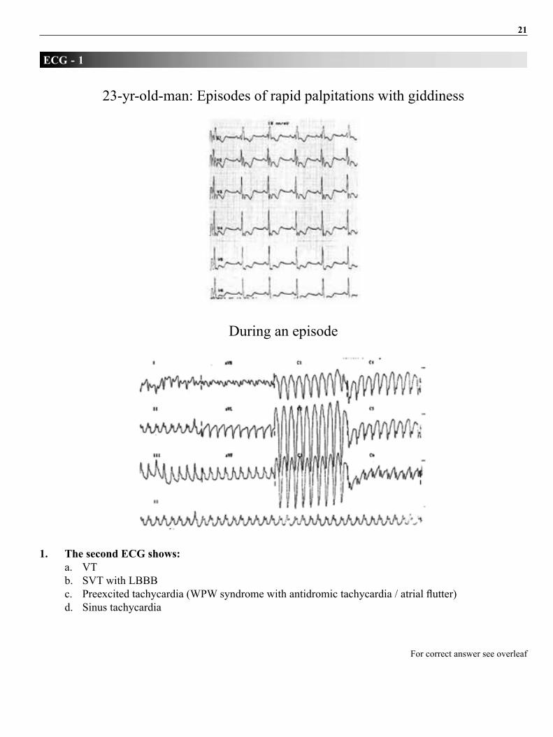
1. The second ECG shows: a. Vt b. sVt with lBBB c. Preexcited tachycardia (WPW syndrome with antidromic tachycardia / atrial flutter) d. sinus tachycardia
ECG - 1
for correct answer see overleaf
23-yr-old-man: episodes of rapid palpitations with giddiness
during an episode

22 Indian Journal of Electrocardiology
The correct answer is ‘a’ – Vt
the second eCg shows a regular wide Qrs tachycardia. the negative Qrs complexes in
leads V5 and V6 are diagnostic of Vt. the lBBB-like pattern in lead V1 suggests that the
Vt originates from the right ventricle (rV).
In the sinus rhythm eCg, the t waves are inverted from V1 to V6 with right bundle branch
block pattern. this suggests a structurally abnormal heart. the heart disease which is known to
affect the right ventricle and produce Vt is arrhythmogenic rV Cardiomyopathy (arVC).
this patient in fact was proved to have arVC by imaging techniques- echocardiography
and cardiac MrI. arVC is known to also affect the lV to some extent. Multiple foci of
monomorphic Vt may be seen at different times. the eCg below shows another Vt episode
with a different morphology, in the same patient, despite being on amiodarone and atenolol.
during another episode
ECG - 1
Hence he finally received an ICD (implantable cardioverter-defibrillator)

23
2. This ECG does not show: a. Atrial fibrillation b. artefact c. sVt d. Vt
ECG - 2
for correct answer see overleaf

24 Indian Journal of Electrocardiology
The correct answer is ‘d’ – Vt
The first 4 complexes show sinus rhythm with T inversions in the inferolateral leads. The 5th
Qrs complex is premature, preceded by an artifact. after this the ventricular rate is fast and
irregular. The baseline shows fibrillatory complexes, best seen in leads II and V2. There is a
rBBB pattern, due to a sudden increase in ventricular rate- this is rate-related phase 3 bundle
branch block. the rBBB disappears for 1 complex, due to a longer rr interval. Immediately
after this is a regular tachycardia with rBBB. this is a sVt.
Below is the intracardiac eCg during this transition, recorded at a fast speed of 100mm/sec.
The fibrillatory complexes (*) change to regular atrial activation (#) which are simultaneous
with the Qrs complexes. Hence this sVt is due to aV nodal reentry (aVnrt).
ECG - 2
SVT and atrial fibrillation (A Fib) may coexist. In such cases, cure of the SVT by radiofrequency
ablation often eliminates the “secondary” A fib also. This was precisely what happened in this
patient.

25
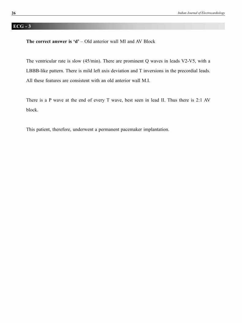
ECG - 3
for correct answer see overleaf
65 year old man. History of ‘heart attack’ and PTCA. Recent giddiness
3. This ECG shows: a. old anterior MI b. aV block c. sinus bradycardia d. a and b e. c and b
26 Indian Journal of Electrocardiology
The correct answer is ‘d’ – old anterior wall MI and aV Block
the ventricular rate is slow (45/min). there are prominent Q waves in leads V2-V5, with a
lBBB-like pattern. there is mild left axis deviation and t inversions in the precordial leads.
all these features are consistent with an old anterior wall M.I.
there is a P wave at the end of every t wave, best seen in lead II. thus there is 2:1 aV
block.
this patient, therefore, underwent a permanent pacemaker implantation.
ECG - 3
27
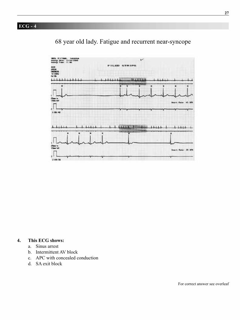
4. This ECG shows: a. sinus arrest b. Intermittent aV block c. aPC with concealed conduction d. sa exit block
ECG - 4
for correct answer see overleaf
68 year old lady. fatigue and recurrent near-syncope

28 Indian Journal of Electrocardiology
The correct answer is ‘a’ – sinus arrest
there are several long pauses, of variable duration. the longest one is 4.2 sec. during
these pauses, no P waves are seen. the pauses are not exact multiples of the P-P interval.
Hence these are not sa blocks. the pauses are terminated by escape junctional or low atrial
depolarisations. a sinus arrest is more than 3 sec long- hence this is the correct diagnosis.
these strips are during night time (see carefully the time mentioned), when the patient was
asleep. Hence in asymptomatic individuals with a high vagal tone, this could even be a normal
variation. In this patient, however, the associated symptoms of bradycardia made a pacemaker
implantation necessary. since the aV nodal conduction was normal, an aaIr pacemaker was
implanted. the paced eCg is displayed below.
ECG - 4
a small pacing artifact precedes each P wave. there is also a solitary PVC. the patient was
asymptomatic after this.
29
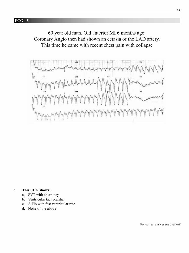
5. This ECG shows: a. sVt with aberrancy b. Ventricular tachycardia c. a fib with fast ventricular rate d. none of the above
ECG - 5
for correct answer see overleaf
60 year old man. old anterior MI 6 months ago. Coronary angio then had shown an ectasia of the lad artery.
this time he came with recent chest pain with collapse

30 Indian Journal of Electrocardiology
The correct answer is ‘b’ – Ventricular tachycardia
the eCg shows a regular wide Qrs tachycardia. the Qrs complexes are monomorphic.
Unless proved otherwise, a wide Qrs tachycardia in subjects with a history of MI must be
considered a ventricular tachycardia. Moreover, one can even discern aV dissociation in
lead V1.
Monomorphic Vt in coronary artery disease is due to an underlying scar of an old MI.
ECG - 5

31
5a. Does this patient have acute ischemia? a. yes b. no c. Can’t say d. don’t know
ECG - 5a
for correct answer see overleaf
the same patient was given a dC shock, but went on to develop a Vt storm. shown below are some of the monitor strips during these episodes,
which required repeated dC shocks.

32 Indian Journal of Electrocardiology
The correct answer is ‘a’ – yes!
the eCg strips show a wide Qrs tachycardia. the r-r intervals are irregular (measure carefully) and the Qrs pattern is changing, often from complex to complex. Hence, we are seeing polymorphic Vt.
In coronary artery disease patients, polymorphic VT is a defining arrhythmia of acute ischemia.
the Vt storm was refractory to anti-arrhythmics, electrolyte optimization, sedation and nitrates. Hence he was taken up for emergent angiography. the angiogram showed a thrombus in the ectatic segment of the lad artery. angioplasty with a covered stent immediately restored sinus rhythm, as seen below.
ECG - 5a
the eCg shows Qs complexes in leads V1-V3, consistent with an old anteroseptal MI. here is gross st elevation in leads V4-V6, due to the superimposed recent MI.
the patient subsequently stabilized and was due for discharge a week later. He was on amiodarone, atenolol, antiplatelets and ramipril.
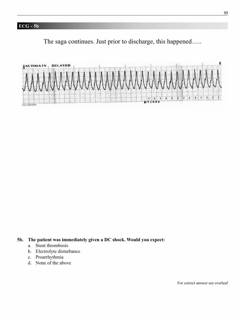
33
5b. The patient was immediately given a DC shock. Would you expect: a. stent thrombosis b. electrolyte disturbance c. Proarrhythmia d. none of the above
ECG - 5b
for correct answer see overleaf
the saga continues. Just prior to discharge, this happened…..
34 Indian Journal of Electrocardiology
The correct answer is ‘d’ – none of the above
this is a monomorphic Vt. as stated earlier, this would be due to a reentrant circuit in
and around an old scar. Ischemia as well as metabolic arrhythmias would be polymorphic.
Proarrhythmias are rare with amiodarone, especially with such short-term use.
yet this patient underwent repeat angiography- the stent was widely patent and there was no
fresh thrombus anywhere.
Thus, in coronary artery disease, monomorphic VT is rarely eliminated by
revascularization!!!
ECG - 5b

35
6. This ECG shows: a. Atrial flutter b. Ventricular tachycardia c. Junctional tachycardia d. atrial tachycardia
ECG - 6
for correct answer see overleaf
22 year old man. frequent rapid palpitations since 1 month; echo showed moderate biventricular dysfunction

36 Indian Journal of Electrocardiology
The correct answer is ‘c’ – Junctional tachycardia
there is a regular narrow Qrs tachycardia. only in lead V2 there is mild prolongation of the Qrs complex. Hence a myocardial Vt is ruled out. lead II (especially the long lead) shows tall, “nippled” t waves intermittently- these are due to superimposed P waves. Hence there is clearly aV dissociation.
the only “supraventricular” tachycardia that can show aV dissociation is junctional ectopic tachycardia. this arrhythmia is more common in children who have recently undergone cardiac surgery. However, it can also be seen in myocarditis, which was the diagnosis in this patient.
after 1 week of amiodarone loading, the eCg showed…
ECG - 6
There is now a stable junctional rhythm with retrograde P waves, better seen in the inferior leads. the deep s wave in lead V3 is due to lV enlargement.
After 10 days of amiodarone….

37
7. The likely diagnosis: a. acute coronary syndrome b. Intracerebral bleed c. Hypokalemia d. none of the above
ECG - 7
for correct answer see overleaf
53 year old man. On Indapamide for hypertension. Sudden collapse in office. resuscitated in nearby emergency room (Vf noted- shocked).
Prompt and complete recovery. eCg after 4 hrs.

38 Indian Journal of Electrocardiology
The correct answer is ‘c’ – Hypokalemia
The ECG shows an obvious QT prolongation (QT/QTc 560 ms), with bifid T waves in leads
V3-V6.
acute ischemia can produce Qt prolongation. However, absence of both st depressions/
elevations and angina make this diagnosis untenable.
raised intracranial tension can produce marked Qt prolongation. However, this is associated
with deep, symmetrical t inversion. also, such patients remain in altered sensorium.
Many diuretics can produce hypokalemia. though indapamide is considered safer in this
regard, in this patient it proved to be the culprit. Is serum potassium was only 1.8 meq/l.
after withdrawing the drug and replacing potassium, his eCg normalized.
ECG - 7

39
8. This ECG suggests: a. lad occlusion b. distal occlusion of rCa. c. Proximal occlusion of Circumflex d. Can’t say
ECG - 8
for correct answer see overleaf
52 yr old man. Chest discomfort 2 hours ago.
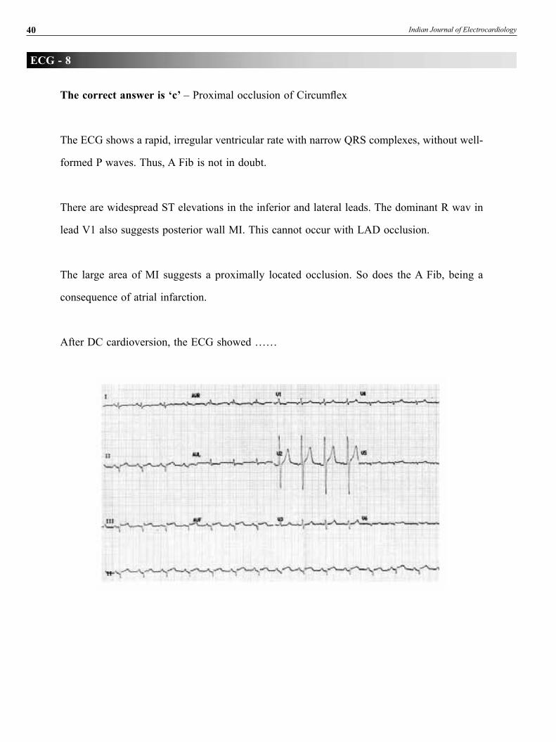
40 Indian Journal of Electrocardiology
The correct answer is ‘c’ – Proximal occlusion of Circumflex
the eCg shows a rapid, irregular ventricular rate with narrow Qrs complexes, without well-
formed P waves. thus, a fib is not in doubt.
there are widespread st elevations in the inferior and lateral leads. the dominant r wav in
lead V1 also suggests posterior wall MI. this cannot occur with lad occlusion.
the large area of MI suggests a proximally located occlusion. so does the a fib, being a
consequence of atrial infarction.
after dC cardioversion, the eCg showed ……
ECG - 8

41
9. The diagnosis: a. Brugada syndrome b. Benign early repolarisation change c. long Qt syndrome d. arVC
ECG - 9
for correct answer see overleaf
41 year old man. asymptomatic. 3 sudden deaths in family members at 35-45 years of age.

42 Indian Journal of Electrocardiology
The correct answer is ‘a’ – Brugada syndrome
the pattern seen on the eCg is st elevations in the electrocardiographic leads V1-V3.
especially lead V1 shows a coving st, steadily descending from its origin. there is also
an incomplete rBBB-like pattern. this conglomeration is the classical “Brugada pattern”.
such a pattern, when combined with familial sudden death, is nearly diagnostic of Brugada
syndrome.
Further confirmation can be obtained by provocative testing with I.V. flecainide, which
enhances the st elevation and can provoke polymorphic Vt. Conversely, the st elevation
resolves with isoprenaline; this was demonstrated in this patient.
He therefore received a prophylactic ICd.
A similar ECG pattern may be seen even in as much as 2% of normal individuals. They
should not be labeled as Brugada syndrome.
ECG - 9
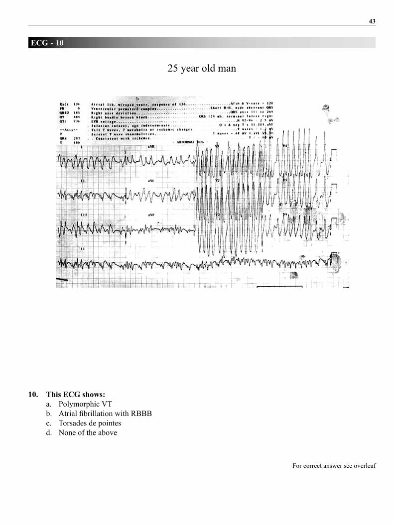
43
10. This ECG shows: a. Polymorphic Vt b. Atrial fibrillation with RBBB c. torsades de pointes d. none of the above
ECG - 10
for correct answer see overleaf
25 year old man
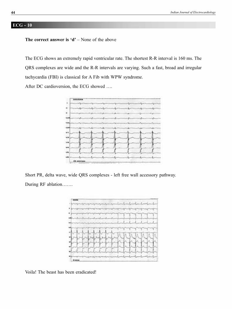
44 Indian Journal of Electrocardiology
The correct answer is ‘d’ – none of the above
the eCg shows an extremely rapid ventricular rate. the shortest r-r interval is 160 ms. the
Qrs complexes are wide and the r-r intervals are varying. such a fast, broad and irregular
tachycardia (fBI) is classical for a fib with WPW syndrome.
after dC cardioversion, the eCg showed ….
ECG - 10
short Pr, delta wave, wide Qrs complexes - left free wall accessory pathway.
during rf ablation…….
Voila! the beast has been eradicated!

INDIAN SOCIETY OF ELECTROCARDIOLOGYAPPLICATION FORM FORLIFE MEMBERSHIP/FELLOWSHIP
Dear Sir,
I wish to become the Life Member* / Fellow** of the Indian Society of Electrocardiology. I promise to abide by the rules and regulations of the Society.
My particulars are as follows :
_______________________________________________________________________
__________________________________________________________________________________
University (Post-Graduation obtained) _______________________________________________________________
_______________________________________________________________
Mailing Address ________________________________________________________________________________
______________________________________________________________________________________________
Tel. No. Hospital ___________________ Clinic __________________ Residence ___________________________
Fax ____________________________ E-Mail ______________________________________________________
Enclosed a cheque/draft of Rs. 2000/- (for outstation cheques add Rs.100/- more) towards Membership of the Society
No. ___________________________ Dated ________________________________ of __________________
___________________________________________________________________ (Bank), drawn in favour of
“Indian Society of Electrocardiology”, payable at Mumbai.
�anking you,
Yours sincerely,
Signature of the ApplicantProposed by (the Member of the Society)
Name __________________________________________
Address __________________________________________
Signature __________________________________________
FOR OFFICE USE ONLYRecommendations of the
Executive Body / Credential Committee
Accepted / Not Accepted
Life Membership No.
__________________________
Hon. Secretary, ISE
�
S. B. GUPTASECRETARIAT
Indian Society of ElectrocardiologyHead, Department of Medicine and Cardiology, C. Rly, Head Quarters Hospital, Byculla, Mumbai - 400 027.Phone : 2371 7246 (Ext. 425), 2372 4032 (ICCU), 2373 2911 (Chamber) • Resi: 2262 4556 • Fax : 2265 1044Mobile : 0 98213 64565 / 0 99876 45403 • E-mail : [email protected] • www.iseindia.org

RULES/REGULATIONS OF THE SOCIETY REGARDING ADMISSION OF LIFE MEMBERS/FELLOWSHIP
*Life Members : 1. Person should be a Post-Graduate in Medicine/Pediatrics/Anaesthesia/ Physiology or other allied subjects from an University recognised by Medical Council of India, with interest in Cardiology / Electrocardiology.
2. Candidates are requested to submit Xerox copies of the PG Certificate and Medical Council of India Registration Certificate alongwith Application Form.
**Fellowship: 1. Person should be a Member of the Society.
2. He/She should be of atleast 7 years of standing after Post-Graduation.
3. He/She should have minimum 3 publications In Cardiology In Indexed Journals (Not Abstracts)
4. List of Publications to be submitted for the Fellowship.
5.
*Subject to approval of the Executive Body of the Society**Subject to the approval of the Credential Committee of the Society.
Fellowship Fees: Rs.2,000/- (+Rs.100/- for outstation cheque) only. Incase, fellowship not approved by the Credential Committee, the cheque / draft will be returned.