Indian Academy of Echocardiography Guidelines and … · Indian Academy of Echocardiography...
44
1 Indian Academy of Echocardiography Guidelines and Manual for Performance of Stress Echocardiography in Coronary Artery Disease Nitin Burkule 1 , Manish Bansal 2 1 Senior Consultant Cardiology, Jupiter Hospital, E. Ex. Highway, Thane 400606, India 2 Senior Consultant Cardiology, Medanta- The Medicity, Sector 38, Gurgaon-122001, India Correspondence: Dr Nitin Burkule, Senior Consultant Cardiology, Jupiter Hospital, E. Ex. Highway, Thane 400606, India e-mail: [email protected] IAE Guidelines TABLE OF CONTENTS 1. INTRODUCTION 2 2. SCOPE OF THE DOCUMENT 2 3. INDICATIONS FOR STRESS ECHOCARDIOGRAPHY AND APPROPRIATE USAGE CRITERIA 2 3.1. Clinical indications for performing stress echocardiography 2 3.2. Appropriate uses of stress echocardiography: 3 3.3. Estimating pretest probability of CAD 4 4. FUNDAMENTAL PRINCIPLES OF STRESS ECHOCARDIOGRAPHY 4 4.1. Pathophysiology of stress induced myocardial ischemia 4 4.2. Principles of functional imaging of myocardial ischemia 5 5. SETTING UP A STRESS ECHOCARDIOGRAPHY LABORATORY 5 5.1. Training of stress echocardiography lab personnel 6 6. PERFORMANCE OF STRESS ECHOCARDIOGRAPHY 6 6.1. Performance of exercise stress echocardiography 7 6.1.1. Patient preparation 7 6.1.2. Imaging during exercise stress echocardiography 8 6.1.3. End-points for exercise stress echocardiography 9 6.1.4. Post procedure observation 10 6.2. Performance of pharmacological stress echocardiography 10 6.2.1. Patient preparation 11 6.2.2. Imaging during pharmacological stress echocardiography 11 6.2.3. End-points for pharmacological stress echocardiography 12 6.2.4. Post procedure observation 12 6.3. Interpretation of stress echocardiography 13 7. DIAGNOSTIC ACCURACY OF STRESS ECHOCARDIOGRAPHY 19 7.1. Factors affecting diagnostic accuracy of stress echocardiography 19 8. ROLE OF NEWER MODALITIES IN STRESS ECHOCARDIOGRAPHY 21 8.1. Three-dimensional (3D) echocardiography 21 8.2. Use of contrast 21 8.3. Myocardial strain imaging 23 9. PROGNOSTIC VALUE OF STRESS ECHOCARDIOGRAPHY 24 10. ADDITIONAL BENEFITS OF STRESS ECHOCARDIOGRAPHY 24 11. CLINICAL RELEVANCE OF ISCHEMIA DETECTION WITH STRESS ECHOCARDIOGRAPHY 25 12. ASSESSMENT OF MYOCARDIAL VIABILITY 25 12.1. Pathophysiological considerations 25 12.2. Low dose dobutamine echocardiography for assessment of myocardial viability 26 12.2.1. Performing DSE for myocardial viability 26 12.2.2. Interpretation of dobutamine echocardiography for viability 27 12.2.3. Sensitivity and specificity of DSE for myocardial viability 30 12.2.4. Comparison with other imaging modalities 30 12.2.5. Role of strain imaging as adjunct to wall motion analysis during LDDE 31 12.3. Clinical algorithm for myocardial viability assessment 31 12.4. Clinical relevance of myocardial viability assessment 31 13. REFERENCES 32 Reviewed by: Satish C Govind, Narayana Institute of Cardiac Sciences, Bangalore, India; Srikanth Sola, Sri Sathya Sai Institute of Higher Medical Sciences, Bangalore, India; Ravi R Kasliwal, Medanta- The Medicity, Gurgaon, India; Sameer Shrivastava, Fortis Escorts Heart Institute, New Delhi, India
Transcript of Indian Academy of Echocardiography Guidelines and … · Indian Academy of Echocardiography...

Indian Academy of Echocardiography Guidelines and Manual for Performance of Stress Echocardiography in Coronary Artery Disease
1
Indian Academy of Echocardiography Guidelines and Manual for Performance of Stress Echocardiography in Coronary Artery Disease
Nitin Burkule1, Manish Bansal2 1Senior Consultant Cardiology, Jupiter Hospital, E. Ex. Highway, Thane 400606, India
2Senior Consultant Cardiology, Medanta- The Medicity, Sector 38, Gurgaon-122001, India
Correspondence: Dr Nitin Burkule, Senior Consultant Cardiology, Jupiter Hospital, E. Ex. Highway, Thane 400606, Indiae-mail: [email protected]
IAE Guidelines
TABLE OF CONTENTS
1. INTRODUCTION 22. SCOPE OF THE DOCUMENT 23. INDICATIONS FOR STRESS
ECHOCARDIOGRAPHY AND APPROPRIATE USAGE CRITERIA 2
3.1. Clinical indications for performing stress echocardiography 2
3.2. Appropriate uses of stress echocardiography: 3 3.3. Estimating pretest probability of CAD 44. FUNDAMENTAL PRINCIPLES OF STRESS
ECHOCARDIOGRAPHY 4 4.1. Pathophysiology of stress induced myocardial
ischemia 4 4.2. Principles of functional imaging of myocardial
ischemia 55. SETTING UP A STRESS ECHOCARDIOGRAPHY
LABORATORY 5 5.1. Training of stress echocardiography lab personnel 66. PERFORMANCE OF STRESS
ECHOCARDIOGRAPHY 6 6.1. Performance of exercise stress echocardiography 7
6.1.1. Patient preparation 7 6.1.2. Imaging during exercise stress
echocardiography 8 6.1.3. End-points for exercise stress
echocardiography 9 6.1.4. Post procedure observation 10
6.2. Performance of pharmacological stress echocardiography 10
6.2.1. Patient preparation 11 6.2.2. Imaging during pharmacological stress
echocardiography 11 6.2.3. End-points for pharmacological stress
echocardiography 12 6.2.4. Post procedure observation 12
6.3. Interpretation of stress echocardiography 137. DIAGNOSTIC ACCURACY OF STRESS
ECHOCARDIOGRAPHY 19 7.1. Factors affecting diagnostic accuracy of stress echocardiography 19
8. ROLE OF NEWER MODALITIES IN STRESS ECHOCARDIOGRAPHY 21
8.1. Three-dimensional (3D) echocardiography 21 8.2. Use of contrast 21 8.3. Myocardial strain imaging 239. PROGNOSTIC VALUE OF STRESS
ECHOCARDIOGRAPHY 2410. ADDITIONAL BENEFITS OF STRESS
ECHOCARDIOGRAPHY 2411. CLINICAL RELEVANCE OF
ISCHEMIA DETECTION WITH STRESS ECHOCARDIOGRAPHY 25
12. ASSESSMENT OF MYOCARDIAL VIABILITY 25 12.1. Pathophysiological considerations 25 12.2. Low dose dobutamine echocardiography for
assessment of myocardial viability 26 12.2.1. Performing DSE for myocardial viability 26 12.2.2. Interpretation of dobutamine
echocardiography for viability 27 12.2.3.SensitivityandspecificityofDSEfor
myocardial viability 30 12.2.4. Comparison with other imaging modalities 30 12.2.5. Role of strain imaging as adjunct to wall
motion analysis during LDDE 31 12.3. Clinical algorithm for myocardial viability
assessment 31 12.4. Clinical relevance of myocardial
viability assessment 3113. REFERENCES 32
Reviewed by: Satish C Govind, Narayana Institute of Cardiac Sciences, Bangalore, India; Srikanth Sola, Sri Sathya Sai Institute of Higher Medical Sciences, Bangalore, India; Ravi R Kasliwal, Medanta- The Medicity, Gurgaon, India; Sameer Shrivastava, Fortis
Escorts Heart Institute, New Delhi, India

Burkule et al.
2
1. INTRODUCTION
Stress echocardiography is a combination of real-time echocardiographic imaging with conventional electrocardiographic stress testing. Stress echocardiography is arguably the most cost effective test, as compared to nuclear imaging or coronary angiography, for diagnosis, prognosis and therapeutic decision-making in coronary artery disease (CAD)1, 2. Because of its cost-effectiveness, it is best suited for Indian scenario where the incidence of CAD is rising at an alarming rate and the astronomical expenditure required for its management is borne largely by the patients themselves due to poor insurance and social security cover. Stress echocardiography uses either exercise or pharmacological stressors depending on the patient profile and the clinicalsituations. Harmonic imaging and ultrasound contrast use in stress echocardiography have improved the sensitivity, specificity,accuracyandprognosticpowerofthetest.Newermodalities such as tissue Doppler and two-dimensional (2D) strain analysis can be creatively and strategically integrated with stress echocardiography to potentially help further enhance its diagnostic accuracy. In terms of the diagnostic and prognostic performance and the outcome data, stress echocardiography is comparable to other functional imaging modalities and, in fact, has some unique advantages over them, as discussed subsequently.
2. SCOPE OF THE DOCUMENT
The predominant reason why stress echocardiography is so underutilised is the lack of adequate exposure and training in this modality. While there is extensive literature available to document diagnostic accuracy of stress echocardiography, there are very few texts that actually describe how to perform stress echocardiography in real life. This Indian Academy of Echocardiography (IAE) guideline document aims to fill this very void. This is a comprehensive ‘howto do’ document prepared with the objective of providing detailed description of the steps involved in performance of stress echocardiography so that there is increased adoption of this important and clinically useful echocardiographic modality. However, while stress echocardiography has several applications beyond just evaluation of CAD, the present document is restricted to its application in CAD, using the two most commonly used stressors viz. exercise and dobutamine.
3. INDICATIONS FOR STRESS ECHOCARDIOGRAPHY AND APPROPRIATE USAGE CRITERIA
3.1. Clinical indications for performing stress echocardiography
There are broadly three settings in which a referral for stress echocardiography is considered3, 4
Subjects without known CAD who present with symptoms suggestive of myocardial ischemia,
Patients with known stable CAD who present with new onset of symptoms or change in symptom pattern or inwhom functional significanceof a coronary lesionsneeds to be determined for prognostic or therapeutic purpose, or
Patients requiring assessment of myocardial viability.
The objective of stress echocardiography in these settings is to ascertain whether the symptoms are due to the presence or progression of CAD; to determine the extent of CAD; to assess the likelihood of functional recovery following revascularization;andtopermitprognosticriskstratificationto guide the treatment. Thus, common indications for stress echocardiography can be summarized as-
1. Positive stress electrocardiogram (ECG) test in a subject with low pretest probability and stress ECG data showing low or intermediate Duke score.
2. Patients not known to have CAD but require cardiac stress test and have intermediate pretest probability for CAD
3. Resting ECG showing nonspecific ST/ T changes,left bundle branch-block (LBBB), left ventricular hypertrophy (LVH) with strain pattern, pre-excitation, baseline ST -segment depression >1 mm in patients post revascularization or those on digoxin.
4. Troponin negative acute chest pain or asymptomatic rise in troponin
5. High coronary calcium score by computed tomography (CT)
6. Assessment of myocardial viability and reversible ischemia in patients with previous myocardial

Indian Academy of Echocardiography Guidelines and Manual for Performance of Stress Echocardiography in Coronary Artery Disease
3
infarction (MI) who received lytic therapy but coronary angiography was not performed
7. CT or catheter coronary angiography showing coronary stenosis of borderline severity
8. Previous percutaneous coronary intervention (PCI) or coronary artery bypass graft (CABG) surgery with new onset of symptoms (i.e. suspected stent restenosis, graft occlusion or progression of native coronary disease)
9. Inability to exercise due to any reason (e.g. respiratory, orthopedic, neurological impairments or obesity and deconditioning)
10. Riskstratificationbeforenon-cardiacsurgery
11. Evaluation of dyspnea on exertion
12. Evaluation of change in symptomatic status of patients with chronic stable CAD
13. Assessment of myocardial viability in chronic stable CAD.
Table 1: Clinical situations in which the choice of stress echocardiography over stress ECG is considered appropriate in ambulatory patients
1. Intermediate to high pretest probability of CAD
2. Un-interpretable baseline ECG (ST-segment depression, pre-excitation, LBBB). However, in patients with atrial fibrillation or paced rhythm, vasodilatory myocardial perfusion imaging is indicated instead of stress echocardiography
3. Presence of LV regional wall motion abnormalities (RWMA) at rest
4. Patients with established CAD with-
a) Coronary stenosis of uncertain significance
b) Previous PCI or CABG, now presenting with anginal symptoms and stress echocardiography is being performed to identify the “culprit” lesion
c) Post incomplete re-vascularization
d) When simultaneous assessment of myocardial viability is required
CABG, coronary artery bypass graft; CAD, coronary artery disease; ECG, electrocardiogram; LBBB, left bundle branch block; LV, left ventricular; PCI, percutaneous coronary intervention; RWMA, regional wall motion abnormality
Table 2: Clinical scenarios in which the performance of stress echocardiography is considered highly appropriate for diagnostic or prognostic purpose or for guiding therapeutic decisions
1. Initial evaluation for diagnosis and prognosis in a subject without known CAD presenting with symptoms suggestive of angina or angina equivalent
2. Asymptomatic subjects with high global CAD risk score [e.g. those with left ventricular systolic dysfunction, arrhythmia, syncope, etc.]
3. Suspected symptoms of acute coronary syndrome with negative or equivocal troponin and low-risk ‘Thrombolysis In Myocardial Infarction’ score
4. Positive results of other non-invasive tests (asymptomatic raised troponin; obstructive disease on CT coronary angiography; CT calcium score >400; positive stress ECG with intermediate to high Duke score; or borderline, equivocal results of nuclear stress imaging)
5. Established CAD for initial evaluation and prognosis assessment to guide optimal medical treatment or revascularization to improve survival
6. When there is a change in the clinical status of a patient with established CAD (except new onset heart failure or previous left main coronary angioplasty), for re-evaluation for symptom validation, prognostication and for guiding management (re-vascularization to improve survival or symptoms despite optimal medical treatment)
7. Presentation with STEMI or non-STEMI when coronary angiography is not performed at the time of index event and there is no post-MI angina or heart failure
CAD, coronary artery disease; CT, computed tomography; ECG, electrocardiogram; STEMI, non ST elevation myocardial infarction
3.2 Appropriate uses of stress echocardiography:
Routine use of stress imaging to detect CAD in asymptomatic patients is inappropriate and leads to unnecessary downstream invasive procedures without improving the outcomes5. Appropriate use criteria (AUC) have been developed for various diagnostic tests that are derived from systematic voting and discussion by a panel of experts about the value of that test in a wide range of clinical scenarios. Table 1 summarizes the clinical situations
Burkule et al.
4
in which the choice of stress echocardiography over stress ECG is considered appropriate in ambulatory patients. Similarly, Table 2 summarizes the clinical scenarios in which the performance of stress echocardiography, in general, is considered highly appropriate5 (as per the expert paneldefiningAUC)fordiagnosticorprognosticpurposeorfor guiding therapeutic decisions.
3.3. Estimating pretest probability of CAD
In patients without known CAD referred for stress echocardiography, it is advisable to estimate pretest probability of CAD. There are various methods for estimation of pretest probability of CAD based on age, gender, type of symptoms, concomitant cardiovascular risk factors, history of MI, ECG changes, etc.5-8. A practical approach is described below6.
Low pretest probability (0-25%)-
− Nochestpain:Allwomen,menwith<3riskfactors,men with >3riskfactorsif<50yearsold
− Atypicalchestpain:Women<60yearsold,men<40years old
Intermediate pretest probability (26-69%)-
− Nochestpain:Menwith>3riskfactorsif>50yearsold
− Atypical chest pain: Women >60 years old, men >40 years old
− Typical angina: Women <50 years old, men <30years old
High pretest probability (> 70%)-
− Typical angina: Women >50 years old, men >30 years old
3.4. Contraindications to stress echocardiography
All standard contraindications to a cardiac stress test are applicable to stress echocardiography also. Thus, following would constitute contraindications to stress echocardiography
Absolute contraindications-
o Acute myocardial infarction (within 2 days)
o High-risk unstable angina
o Uncontrolled cardiac arrhythmia with hemodynamic compromise
o Active endocarditis
o Symptomatic severe aortic stenosis
o Decompensated heart failure
o Acute pulmonary embolism or pulmonary infarction
o Acute myocarditis or pericarditis
o Physical disability precluding safe and adequate testing
Relative contraindications-
o Known left main coronary artery stenosis
o Tachyarrhythmias with uncontrolled ventricular rates
o Acquired complete heart block
o Hypertrophic cardiomyopathy with a severe resting gradient
o Mental impairment with limited ability to cooperate
In addition, stress echocardiography would also be contraindicated in patients with poor acoustic window that precludes satisfactory left ventricular (LV) endocardial visualization, despite contrast use or when contrast is not available.
4. FUNDAMENTAL PRINCIPLES OF STRESS ECHOCARDIOGRAPHY
4.1. Pathophysiology of stress induced myocardial ischemia
Stress echocardiography relies on recognition of RWMA induced by regional mismatch between myocardial oxygen demandandmyocardialbloodflowduetoeitherepicardialcoronary stenosis or microvasculature dysfunction. Stressor like exercise or dobutamine-atropine infusion increase myocardial oxygen demand by increasing myocardial contractility and heart rate, with or without increase in

Indian Academy of Echocardiography Guidelines and Manual for Performance of Stress Echocardiography in Coronary Artery Disease
5
afterload. In contrast, vasodilators such as dipyridamole or adenosine induce myocardial ischemia by causing coronary steal.
During exercise or catecholamine infusion, coronary bloodflowincreasesby3to5foldsasaresultofdilatationof epicardial coronary arteries (conductance vessels) and intramyocardial arterioles (resistance vessels) as well as opening of (i.e. recruitment of) more number of intramural capillaries. However, in the perfusion bed of epicardial coronary artery with significant stenosis, thereis already maximal dilatation of downstream arterioles and de-recruitment of capillaries at rest to maintain resting myocardial blood flow. As a result, there is virtually nocoronary flow reserve remaining to augment blood flowduring stress leading to reduced capillary blood volume, perfusion defects and metabolic abnormalities.
4.2. Principles of functional imaging of myocardial ischemia
Nuclear imaging, gadolinium-contrast magneticresonance imaging (MRI) and myocardial contrast perfusion imagingdetectregionalperfusionand/ormetabolicdefects.These abnormalities appear early in the course of myocardial ischemia. When ischemia is sufficiently prolonged, theaffected myocardial region develops diastolic dysfunction followed by systolic dysfunction. The ischemic myocardial dysfunction initially affects subendocardial layers and then becomes transmural9-11. Stress echocardiography detects ischemia at this stage qualitatively by visual assessment of regional wall systolic thickening and endocardial excursion manifesting as RWMA. Post-systolic thickening is the earliest wall motion abnormality in response to ischemia. It isfollowedbyreducedthickeningandfinallynothickening,akinesia or even systolic thinning depending upon the transmurality of ischemia.
RWMAs disappear fast after cessation of stress. Therefore, in order to maintain optimal diagnostic sensitivity, rapid and efficient echocardiographic image acquisitionis required within a window period of 60-90 seconds after cessation of exercise. However, ischemia induced myocardial diastolic stunning as detected by strain imaging (post-systolic shortening or failure of relaxation in early
diastole) may last for as long as 15- 30 minutes following stress, making it possible to detect ischemia even when there has been some delay in image acquisition.
5. SETTING UP A STRESS ECHOCARDIOGRAPHY LABORATORY
The stress echocardiography lab consists of stress ECG console interfaced with treadmill and or bicycle ergometer and a high-end echocardiography system loaded with software applications for stress echocardiography, tissue Dopplerand2DstrainimagingandcontrastLVopacification(LVO) with or without perfusion imaging. A commercially available or customized imaging bed with a semicircular mattress and bed cut out at the patient’s chest position should be available to facilitate acquisition of diagnostic quality images. The commercially available supine bicycle comes with the echocardiography imaging bed.
There should be a provision for two syringe infusion pumps for pharmacological stress and a permanently stationed resuscitation trolley equipped with defibrillator,Ambu bag, laryngoscopes, tracheal tubes and all emergency medications. The medications should necessarily include adrenaline, atropine, sublingual nitroglycerine, metoprolol, hydrocortisone, aminophylline, dextrose and saline. The echocardiography lab should have piped oxygen supply, piped suction connection, pulse oximeter, a nebulizer and a bronchodilator inhaler. A kidney tray should also be available, in case vomiting occurs. A weight-based dobutamine infusion rate chart should be prepared and kept in the stress echocardiography room for easy reference.
The layout of the stress echocardiography lab should be such that there is good proximity between treadmill and imaging bed so that the patient can have easy exit from the treadmill and an unhindered rapid access to the imaging bed (Figure 1). This should be achieved without disconnection or entanglement of the ECG cables from both the echocardiography machine and the treadmill console. Also the physician performing scanning should have clear view of the ECG console. The emergency trolley should have unobstructed movement to the treadmill or the imaging bed. Unnecessary personnel or equipment should not clutter the space for the patient’s and the escorting technician’s

Burkule et al.
6
movement. The height of the imaging bed should preferably be kept low so that the patient is able to quickly lie down.
Due to paucity of space in most of the hospitals a single stress echocardiography room can be used for both exercise and pharmacological stress tests with few quick rearrangements. The syringe infusion pump and the treadmill ECG console (for simultaneously monitoring 12 lead ECG)
can be rearranged closer to imaging bed when performing pharmacological stress test.
There should preferably be a provision for a recovery area when prolonged observation is needed post stress. Also an emergency response protocol should be in place to shift a patient to intensive care unit in the event of an emergency.
A B
Figure 1: Suggested layout of a stress echocardiography lab. A. Exercise stress echocardiography; note the proximity of treadmill to echocardiographic examination bed for exercise echocardiography. B. Dobutamine stress echocardiography
5.1. Training of stress echocardiography lab personnel
Minimum personnel required in the stress echocardiography lab include one physician (who does scanning also), one stress test technician and one nursing staff. We do not recommend sonographers to perform the test independently without physical presence of the physician in the echocardiography lab. The physician should be at least IAE levelIIechocardiographycertified,experiencedinhandlingcardiac emergencies and should have had advanced cardiac life support training. Before the physician starts performing stress echocardiography independently, he/she should firstobserve 50 stress echocardiograms, then perform at least 50 exercise stress and 50 dobutamine stress echocardiography studies under the supervision of an expert and should additionally read 100 stress echocardiography studies with
the expert. In a high volume stress echocardiography lab, this can be accomplished within 3-4 weeks. Thereafter, the competency should be maintained by performing at least 100 stress echocardiography studies per year. The technician and the nursing staff should be trained and instructed by the physician. It is extremely beneficial if they canundertake observership for a week in one of the high volume echocardiography labs which are regularly performing stress echocardiographyandLVcontrastopacificationstudies.
6. PERFORMANCE OF STRESS ECHOCARDIOGRAPHY
A thorough 2D and Doppler echocardiography study conforming to IAE guidelines for transthoracic echocardiography12 should be performed, if not done earlier, before undertaking the stress study.

Indian Academy of Echocardiography Guidelines and Manual for Performance of Stress Echocardiography in Coronary Artery Disease
7
Stress echocardiography uses two types of stressors- exercise or pharmacological13. The actual process involved in the performance of stress echocardiography differs according to the stress modality.
6.1. Performance of exercise stress echocardiography
Exercise is the preferred stress modality, as it is physiological, allows symptoms correlation and permits assessment of functional capacity. Exercise can be performed in different ways-
Treadmill exercise: The exercise protocol is similar to that of conventional stress ECG. Bruce protocol is used most commonly. In patients with limited functional capacity.ModifiedBruceprotocolmaybeused.Whenusing treadmill exercise, post peak stress imaging for RWMA assessment should be completed within 90 seconds of cessation of exercise. Some experts have shown improved sensitivity for ischemia detection by performing imaging at peak stress while the patient is still on treadmill14. However, it is technically challenging.
Supine bicycle ergometry: It has the advantage of allowing simultaneous echocardiographic imaging while the patient is performing exercise. As a result, not only peak exercise imaging is possible, but imaging can also be performed at every stage during exercise which helps in recognizing the onset of ischemia13, 15. Breath-holding and control of body position are also easier compared to treadmill exercise. However, the overall workload achieved on supine bicycle is lower than that achieved on treadmill16.
Hand grip, leg-raising, squatting: Hand-grip and leg-raising are often combined with pharmacological stressors but not used as the sole stress modality. Squatting has been used as the primary stress modality in past but has now been given up due to technical challenges.
6.1.1. Patient preparation
A written informed consent should be obtained from every patient prior to performing stress echocardiography. A template for consent form is included at the end of this document (appendix A).
Patients scheduled for stress echocardiography usually have prior appointment and should receive detailed instructions from the lab staff about medications, need for fasting, etc. When the test is being performed for diagnosing CAD in a patient not previously known to have CAD, it is advisable to withhold anti-anginals and heart rate controlling medications for 3-4 half-lives (usually omitting only on the dayofthetestissufficient)andresumedfewhoursafterthetest is completed. However, these medications should not be discontinued when the objective of the stress test is to detect severity of inducible ischemia despite optimal medications. Fasting for 3-4 hours is required prior to stress testing (particularly with dobutamine as dobutamine induced nausea and vomiting are not uncommon). The echocardiographer should go through all relevant medical records and ascertain the clinical query to be answered by the planned stress test. He/sheshouldalsolookforanemia,hypokalemia,significantback pain or leg pain or any other factor that could interfere with performance of stress echocardiography. Following this, the patient’s vital parameters, height and weight should be recorded. If the patient has high blood pressure, ongoing anginaand/ordynamicchangesinECG,itmaybeadvisableto postpone or even cancel the test.
Chest shaving and skin preparation should be meticulously done to get pristine ECG signals at peak stress for acquiring ECG-gated peak stress images. Electrodes are connected in the same manner as for conventional stress ECG, except for chest leads V2 and V5. These leads can interfere in acquiring parasternal and apical echocardiographic images respectively. Therefore, these leads may have to be shifted one space below or the sticker part of the V2 and V5 electrodes may be cut out to create extra space for imaging. Additional three electrodes need to be secured for connecting the ECG cable of the echocardiography machine. If contrast is going to be used, then an intravenous (IV) cannula needs to be placed, preferably in the left forearm.
After explaining the procedure to the patient it is advisable to do a mock trial with the patient. The patient can practice a fast and swift exit from the treadmill to the imaging bed and lying down immediately in left lateral decubitus position with the chest positioned over the cut out portion of the bed. The technician should help with the attached ECG

Burkule et al.
8
cables during patient movement to avoid entanglement or lead detachment. Such mock practice trials greatly help in minimizing time delay between the cessation of exercise and initiation of image acquisition.
6.1.2. Imaging during exercise stress echocardiography
The imaging protocol depends on the capabilities and the customizability offered by the echocardiography equipment and whether contrast will be used or not (Figure 2). A proper stress echocardiography application with facility for
continuous ECG-gated capture at peak stress is the minimum requirement. In addition, ideally, the echocardiography machine should also have the option for customizing the imaging protocol and the technical capabilities for contrast LVO, color tissue Doppler imaging (TDI) and 2D strain imaging.
Resting imaging is done with the patient lying in left lateral decubitus with ECG cables of treadmill and echocardiography machine connected. Stress echocardiography application on the machine should be turned on. The adequacy of access to apical and parasternal echocardiography windows should be checked; if inadequate, some of the ECG electrodes may need to be repositioned. The patient should be asked to take deep breaths so that the echocardiographer gets an idea of the translational motion of the heart during tachypnea at peak exercise and can plan strategy for imaging at peak stress. If contrast is not going to be used, then a fresh set of five standard views of the left ventricle viz. apical4-chamber (AP4C), apical 2-chamber (AP2C), apical long-axis (APLAX), parasternal short axis (PSAX) at base and PSAX at apex should be acquired and stored as part of the protocol. The 2D images must be optimized using the appropriate transducer frequency, gain, and focus. Harmonic imaging is mandatory. In stress echocardiography, the focus of imaging is entirely on the left and right ventricles. Apical views should be acquired in such a way that the atria (but not the annuli) are predominantly excluded from the image (by adjusting depth) and sector width optimized so that both the endocardial and epicardial borders of the left and right ventricle are seen. These adjustments facilitate tissue Doppler and 2D strain analysis. Care should be taken to ensure that these baseline settings are not changed after this stage and that the images at each subsequent stage of acquisition are similar to baseline images.
If contrast is to be used, then above non-contrast images are acquired only if 2D strain analysis is planned later on for detection of diastolic stunning which would require baseline and delayed post-stress images (described later in section 8.3). In that case, the baseline non-contrast images are acquired outside the stress protocol. The IV contrast bolus (usually 0.3 to 0.5 ml of reconstituted Sonovue®) is then administered with a 5-10ml salineflush till the contrast appears in the
Permorm a complete baseline echocardiographic study, if not already done
Else, acquire gray-scale images in AP4C, AP2C, APLAX, PSAX base and PSAX base and PSAX apex views. Collect hemodynamic data
With contrast Without contrast
Administer contrast. Acquire the 5 standard views as part of stress protocol.
Exercise the patient on treadmill
Encourage the patient to perform maximum exercise or at least achieve age-predicted maximum heart rate
Administer contrast 10-15 sec before cessation of exercise
Rapidly acquire post-exercise images using continuous capture mode
Collect hemodynamic data
Late-recovery (~10 min) gray-scale apical images may be acquired for assessment of diastolic stunning by STE
Permorm a complete baseline echocardiographic study, if not already done
Collect hemodynamic data
Acquire gray-scale images in AP4C, AP2C, APLAX, PSAX base and PSAX base and PSAX apex views as part of stress protocol
Exercise the patient on treadmill
Encourage the patient to perform maximum exercise or at least achieve age-predicted maximum heart rate
Rapidly acquire post-exercise images using continuous capture mode
Collect hemodynamic data
Late-recovery (~10 min) gray-scale apical images may be acquired for assessment of diastolic stunning by STE
Figure 2: Imaging protocols for exercise stress echocardiography, with or without using ultrasound contrast for left ventricular opacification.
AP2C- apical two-chamber; AP4C- apical four-chamber; APLAX- apical long-axis; PSAX- parasternal short-axis; STE- speckle tracking echocardiography

Indian Academy of Echocardiography Guidelines and Manual for Performance of Stress Echocardiography in Coronary Artery Disease
9
right ventricle. Image acquisition is commenced when optimum LVO is achieved. The contrast-enhanced images intheabove-mentionedfivestandardviewsareacquiredandstored in the stress echocardiography protocol.
Finally,hemodynamicdata[LVfillingpressures,mitralregurgitation, tricuspid regurgitation, right ventricular (RV) function etc.] should also be collected, if not already collected. This should be done prior to administration of contrast.
The patient is then exercised on treadmill using Bruce ormodifiedBruceprotocolwithstage-wiserecordofECGand blood pressure. All patients are encouraged to perform maximum exercise, even if they have already achieved age-predicted maximum heart rate (MPHR, 220- age in years). However, in patients with reduced exercise capacity, effort should be made to at least achieve the target heart rate, but without compromising the patient safety. The patients should be instructed to communicate their desire to stop the test because of unbearable symptoms at least 10-15 seconds in advance so that IV contrast can be administered while they are still on the treadmill (Figure 2). As mentioned above, on-treadmill echocardiographic imaging can be performed to detect onset of RWMA and to improve the diagnostic sensitivity of the test. Although it is feasible in most patients, it is technically challenging.Once the patient has finishedexercise, the treadmill should be stopped at peak stress using the emergency button and the patient is rapidly escorted to imaging bed. The post peak exercise imaging should be started within 10 seconds of cessation of exercise and the echocardiographer should aim at finishing first set of allviews by 45 second mark. In case of borderline ischemia at peak stress the rapid adoption of supine position by the patient for imaging increases venous return and preload thereby favorably accentuating ischemia and its imaging manifestation. Some researchers have administered IV 0.6 mg atropine at peak stress to avoid rapid drop in the heart rate during post exercise imaging. However, we do not recommend this strategy.
Completing post treadmill exercise imaging in a time window of 60 seconds in a hyperventilating patient requires training, practice and mastering special skills. The patient
should be lying in steep left lateral decubitus. The cut out in the imaging bed helps the echocardiographer to access far lateral echocardiographic window beyond the anterior axillary line. The transducer position should be rapidly adjusted to get true long-axis images of AP4C, AP2C and APLAX from this apical window. This is followed by quick acquisitionofthePSAXimagesatbaseandapex.Thefirstset of these 5 images should be acquired within 45 seconds of cessation of exercise. This is followed by a second round ofapicalandparasternalimagingwhichshouldfinishby90second time point after cessation of exercise. The continuous capture mode in the stress echocardiography application stores ECG-gated images continuously for approximately 120 seconds. These images can be reviewed at the end of the test to choose the best diagnostic quality peak exercise images of AP4C, AP2C, APLAX, PSAX base and PSAX apex views. The immediate post-exercise imaging for RWMA should be followed by acquisition of the same hemodynamic data as at baseline. Another set of gray-scale images of the 5 primary views may also be recorded late into recovery (10-15 min) for 2D strain analysis for assessing diastolic stunning.
In case of bicycle ergometry, workload is increased in stepwise manner and images are obtained at multiple stages. Typically, baseline, pre-peak (75% of MPHR), peak and recovery images are acquired as minimum. The views and rest of the imaging principles are same as for treadmill exercise echocardiography.
6.1.3. End-points for exercise stress echocardiography
The indications for terminating stress echocardiography are similar to those for stress ECG test. The test can be terminated on reaching MPHR and patient’s perception of extreme effort as semi-quantitatively assessed by the Borg scale or rise in systolic blood pressure >220 mmHg. In case of bicycle ergometer stress echocardiography or treadmill stress echocardiography with ‘on treadmill’ imagingprotocol, continuous echocardiography imaging surveillance allows detection of onset of ischemia. In these situations, appreciation of unambiguous RWMA in 2 or more segments orLV/RVdilatationshouldpromptterminationofthetest.Stress test should also be terminated prematurely if the patient

Burkule et al.
10
Pharmacologic stress testing is performed using either dobutamine or a vasodilator such as dipyridamole or adenosine. IV atropine boluses are used with dobutamine infusion to increase the sensitivity of the pharmacological stress testing18-21.
Dobutamine: It is an inexpensive, easily available and easytousedrugwithgoodsafetyprofile.Dobutamineis administered IV as a continuous infusion, staring at a doseof5mcg/kg/minandisincreasedevery3minutesto 10, 20, 30 and40mcg/ kg/min13. Atropine boluses of 0.3-0.6 mg with a maximum dose of 1.8-2.0 mg are often required to achieve target heart rate, especially in patients on beta-blockers19. IV metoprolol (2-10 mg) is usually administered at the end of the test to rapidly reverse tachycardia and dobutamine side effects. It also unmasks subtle RWMA and improves the sensitivity of DSE, especially in patients with single-vessel disease22.
Adenosine or dipyridamole: These agents are potent epicardial coronary vasodilators. They produce ischemia by causing coronary steal from the vascular beds supplied by the stenosed coronary arteries23. Dipyridamole is administered intravenously at a dose of 0.84 mg/kg over 10 minutes and the vasodilatoryeffect persists for approximately 30 min. Adenosine is an ultra-short acting agent and requires continuous IV infusion at a rate of 140mcg/kg/min for 3-5min.Mostpatientscomplainofflushing,dyspneaandchestdiscomfort. Other uncommon side effects are sinus bradycardia, atrioventricular block or bronchospasm. These side-effects usually subside within few seconds of stopping the infusion but IV aminophylline may be needed to reverse the bronchospasm.
Ergonovine: It is used to provoke coronary vasospasm in patients with suspected vasospastic angina
Pacing stress echocardiography is an option in patients with an implanted permanent pacemaker. In such patients, the heart rate is increased progressively in a step-wise manner using the external programmer. It is often combined with dobutamine infusion to simultaneously increase myocardial contractility and therefore, the myocardial workload.
reports intolerable angina, significant giddiness or severebreathlessness disproportionate to the level of exertion or if the ECG shows asymptomatic horizontal or down-sloping ST depression >3 mm or ST elevation >1 mm in a non-infarct lead without an abnormal Q-wave. A fall in systolic blood pressure, signs of decreased peripheral perfusion, non-sustained ventricular tachycardia or multifocal ventricular premature beats are some of the serious indications for immediate termination of exercise and close monitoring during the immediate post exercise period. Patients showing severe ischemia by symptoms, ST-segment changes on ECG orextensiveRWMAshouldbeverybrieflyimagedatpeakstress in the supine position and then should be instructed to immediately sit upright at the edge of the bed to reduce venous return and preload of the heart in order to relieve angina and any pulmonary congestion.
6.1.4. Post procedure observation
A 30-minute waiting period, usually in the echocardiography lab waiting room, is mandatory if either IV contrast or a pharmacological agent was used or if the test showed extensive ischemia. When the stress test is positive, it is a good practice to stop ECG monitoring only after the wall motion is normalized and the ST-segment depressions have returned back to baseline. An ECG performed 30 minutes after completion of the study should preferably be recorded in case of a strongly positive stress test.
6.2. Performance of pharmacological stress echocardiography
Pharmacologic stress testing is required for patients who are unable to exercise or when assessment of myocardial viability is the main objective of the study. It has the advantage that unlike treadmill exercise, continuous and good quality imaging can be done throughout the test and at peak heart rate without motion or breathing artifacts. This permits assessment of myocardial viability and recognition of onset of myocardial ischemia. However, the peak workload (double product) achieved during dobutamine stress echocardiography (DSE) is generally lower than treadmill stress17.

Indian Academy of Echocardiography Guidelines and Manual for Performance of Stress Echocardiography in Coronary Artery Disease
11
6.2.1. Patient preparation
The preparation for DSE is similar to that for exercise echocardiography with some differences. Fasting for 3-4 hours prior to the test must be ensured to minimize chances of dobutamine induced nausea and vomiting. Some laboratories also prefer to administer IV ondansetron prior to these test. The patients should be asked to empty urinary bladder before the test is started.
Unlike exercise echocardiography, an IV cannula is required for DSE. If imaging is performed with echocardiographer on the right side of the bed, the IV line should be secured in the left arm for the patient’s comfort as well as the ease of contrast administration. The IV line site should be easily visible to detect bleeding, leakage or swelling at local site or if it gets accidentally disconnected. A 3-way stopcock with 5-10 cm extension with an additional 200 cm extension line is suggested. Patency of line should be checked before infusion. Blood pressure cuff should be on the other arm. The dobutamine infusion is prepared by dissolving 250 mg of dobutamine in 50 ml of normal saline and is administered using an infusion pump. The infusion rates corresponding to 5, 10, 20, 30 and 40 mcg/kg/min
dose should be calculated beforehand by taking into account patient’s weight. As mentioned above, a weight-based dobutamine infusion rate chart should be prepared and kept in the stress echocardiography room for easy reference. IV metoprolol,IVatropineandIVsalineflushsyringesshouldalways be kept ready and similar to any other form of stress echocardiography, the lab must also have permanently stationeddefibrillatorandresuscitationtrolley.Theoperatorshould be aware that patients receiving medications such as angiotensin converting enzyme inhibitors, angiotensinogen receptor blockers, diuretics or nitrates are more likely to have prolonged hypotension during or immediately after DSE.
6.2.2. Imaging during pharmacological stress echocardiography
The imaging at rest is same as for exercise echocardiography. Five primary views with or without contrast are recorded in the customized pharmacological stress protocol. The baseline image presets including depth and sector width must not be changed thereafter. Dobutamine infusionisstartedatadoseof5mcg/kg/minandisincreasedevery 3minutes to 10, 20, 30, 40mcg/kg/min (Figure 3).
Permorm a complete baseline echocardiographic study, if not already done
Else, acquire gray-scale images in AP4C, AP2C, APLAX, PSAX base and PSAX apex views. Collect hemodynamic data
With contrast Without contrast
Administer contrast. Acquire the 5 standard views as part of stress protocol
Start dobutamine infusion at a dose of 5 mcg/kg/min; increase it every 3 min to 10, 20, 30 and 40 mcg/min
Add atropine and isometric exercise if no significant increase in heart rate by 20 mcg/kg/min dose
Administer contrast at 2 min 45 sec during each stage. Acquire images a few seconds later
Acquire peak images once MPHR is achieved and sustained for 30-60 sec.
Collect hemodynamic data
Stop dobutamine infusion, IV metoprolol may be administered Recovery images acquired once heart rate is near baseline or at least <100/min. In case of suspected ischemia, images may be acquired earlier
Permorm a complete baseline echocardiographic study, if not already done
Collect hemodynamic data
Acquire gray-scale images in AP4C, AP2C, APLAX, PSAX base and PSAX apex views as part of stress protocol
Velocity-encoded gray-scale images or color TDI images may also be obtained in 3 apical views
Start dobutamine infusion at a dose of 5 mcg/kg/min; increase it every 3 min to 10, 20, 30 and 40 mcg/min
Add atropine and isometric exercise if no significant increase in heart rate by 20 mcg/kg/min dose
Acquire gray-scale or velocity encoded gray-scale images during each stage. Alternately, low-dose (10 mcg/kg/min), pre-peak (75% MPHR) and (40 mcg/kg/min dose with or without atropine to achieve (MPHR) images may be acquired
Stop dobutamine infusion, IV metoprolol may be administered Recovery images acquired once heart rate is near baseline or at least <100/min. In case of suspected ischemia, images may be acquired earlier
Figure 3: Imaging protocols for dobutamine stress echocardiography, with or without using ultrasound contrast for left ventricular opacification. AP2C- apical two-chamber; AP4C- apical four-chamber; APLAX- apical long-axis; IV- intravenous; MPHR- maximum predicted heart rate; PSAX- parasternal short-axis; TDI- tissue Doppler imaging.

Burkule et al.
12
12-lead ECG is monitored continuously using the treadmill console. The ECG print outs are taken at rest, during intermediate stages, at peak dose, during early and late recovery and in case of any arrhythmia. Blood pressure and symptoms are also monitored. The same sets of 5 images, ideally in the same orientation as baseline, should preferably be acquired during each stage. Alternately, images can be obtained at low-dose (10 mcg/kg/min), pre-peak (∼75% MPHR),peak-dose(40mcg/kg/minwithorwithoutatropineto achieve MPHR) and recovery. As each stage lasts for 3 minutes, it is good practice to start recording the set of 5 images of the corresponding stage at 2 min 45 sec time point in the stage. If contrast is planned, then a small bolus (0.2-0.3 ml) of Sonovue®withsalineflushisadministeredjustbeforeacquisition of the images in every stage. Pharmacological stress echocardiography has the advantage that at each stage, the corresponding baseline image is available for comparison and the image can be optimized to resemble the baseline image as much as possible prior to acquisition. This is essential to ensure accurate interpretation of segmental wall motion changes.
For peak-stress imaging, MPHR should be maintained for at least 30-60 sec without compromising the patient’s safety. To augment the heart rate and blood pressure rise, the patient can be asked to perform repeated right hand grip exercise holding a rubber ball or perform active leg raising against resistance. As mentioned above, atropine is commonly used to achieve heart rate during dobutamine echocardiography. Two different strategies are used for this purpose.Inthefirststrategy,afterreachingdobutaminedoseof 40mcg/kg/min, small aliquots (0.3mg) of IV atropinefollowedbysalineflushareadministeredevery1minutetillthe heart rate reaches the target. In the second strategy, which is usually preferred, small aliquots (0.3-0.6 mg) of atropine are given at the end of each stage starting from dobutamine 20mcg/kg/mindoseitself.
At higher dosages of dobutamine, some patients complain of throbbing headache, palpitations, generalized anxiety or distress, nausea and urge to pass urine. Chest pain (due to dobutamine induced forceful cardiac contraction), hypotension and arrhythmias like ventricular ectopics
and non-sustained ventricular tachycardia may also occur. Stopping dobutamine infusion and administering intravenousmetoprololaresufficienttoreverseallthesesideeffects. Serious side-effects such as ventricular fibrillationand myocardial infarction are very rare (less than 1 in 2000 patients) and no death has ever been reported during DSE24.
6.2.3. End-points for pharmacological stress echocardiography
The indications for terminating the pharmacological stress test are similar to those described for exercise stress test. Some patients with LVH and or hypovolemia may develop hypotension due to LV intra-cavity dynamic obstructioneither inmid-cavityor in theLVoutflow tractwith systolic anterior motion of the mitral valve or due to peripheral vasodilation caused by dobutamine. This can be countered by hand grip, passive leg raising and stopping dobutamine infusion. IV fluid administration may also beoccasionally needed if hypotension persists. After reaching any of the end points, the test is terminated with routine use of IV metoprolol to bring back the heart rate to baseline. It should be noted that for the same degree of ischemia, the ST segment depression and angina are much less frequent with dobutamine as compared to exercise stress. Accordingly, ST-segment depression during DSE has low sensitivity for detection of inducible ischemia. The primary role of ECG during DSE is to detect arrhythmias or ST-segment elevation.
6.2.4. Post procedure observation
It is advisable to have the patients rest for half an hour in the echocardiography lab recovery or waiting room after completion of the test. The IV cannula should be removed only after 30 minutes, when the patient is already mobilized (bedside), is asymptomatic and all vital parameters are stable, so that an IV access for administration of medicines is available in case of any late emergency. In some patients who have concentric LVH, neurocardiogenic syncope may occur as they assume upright position immediately after the test.Havingthepatientliesupineagainisgenerallysufficientto treat it but IV fluid administration may sometimesbe required. An ECG should be repeated at 30 min in all patients with strongly positive test. The patients should also

Indian Academy of Echocardiography Guidelines and Manual for Performance of Stress Echocardiography in Coronary Artery Disease
13
be warned about persistence of atropine effect on visual accommodation for those who want to read and about the chances of urinary retention in elderly patients.
6.3. Interpretation of stress echocardiography
Qualitative visual assessment of regional wall thickening and motion at rest, during stress and following stress is the basic method for interpretation of stress echocardiograms. This needs good quality image acquisition, systematic approach for reading and considerable operator experience. Digital quad screen format for side-by-side display of images is strongly recommended25. Routine use of harmonic imaging26, more frequent use of contrast and use of strain imaging in special circumstances may help in reducing non-diagnostic studies, improving confidence of reporting andreducing inter-reader variability.
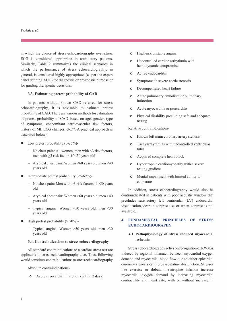
The interpretation of stress echocardiogram should begin with evaluation of the technical adequacy of the test (Table 3). Digital images should be checked to ensure adequate image quality, appropriate triggering and comparability of viewsateachstage.Afterconfirmingtechnicaladequacyofthe images, an assessment should be made of LV size and shape following stress as they often provide useful clues to the presence or absence of ischemia. In normal stress response, end-systolic LV cavity at peak stress is small and looks like a narrow base triangle (Figures 4A, 4B, 5A, 5B, 6A, 6B; Videos 1A, 1B, 2, 3A-E). Focal RWMA may cause outward bulging resulting in distortion of LV end-systolic cavity shape at peak stress (Figures 7A, 7B, 8A, 8B; Videos 4A, 4B, 5). A more extensive RWMA will result in dilatation of LV cavity, manifesting initially as failure to reduce end-systolic volume and eventually as actual increase in end-systolic cavity size (Figures 9A, 9B; Videos 6A-C, 7, 8A, 8B). Thus, regional LV shape change should always alert the interpreter to the possibility of CAD whereas an increase in LV size with stress, in the absence of concomitant valvular or myocardial disease, is usually indicative of multi-vessel ischemia27. These LV cavity shape changes, particularly global LV dilatation, are more common with exercise echocardiography as reduction in afterload with dobutamine often masks these changes27.
Table 3: Important steps for comprehensive interpretation of stress echocardiography
1. First, evaluate the technical adequacy of the images.
2. Perform global function assessment- look for any changes in LV size and shape following stress.
3. Perform segmental wall motion analysis-
a. Focus on wall thickening rather than wall motion.
b. Reviewing only the systole instead of complete cardiac cycle improves the ability to recognize RWMAs.
c. Assess endocardial excursion ‘take off’ in the first half of systole (freeze the image and scroll through early systole frame-by-frame).
d. When looking for ischemic response during DSE, it is best to compare peak-dose images with low-dose images instead of rest-images.
e. It is also important to carefully review recovery images. The presence of even subtle impairment of contractility during recovery in comparison to baseline should be considered as an evidence of ischemia at peak dose.
f. Try to correlate distribution of inducible RWWA with coronary vascular distribution; however, atypical patterns may occur, especially in patients with previous coronary bypass surgery.
g. Isolated RWMA involving basal inferior wall or basal inferoseptum are likely to be false-positive if the adjacent segments supplied by the same vascular territory show normal function.
4. After completing segmental wall motion analysis, review hemodynamic data obtained at peak-stress.
5. Also, correlate with stress induced electrocardiogra-phic changes to avoid missing any subtle RWMA which may have gone unnoticed during initial review of images.
DSE, dobutamine stress echocardiography; LV, left ventricular; RWMA, regional wall motion abnormality
After the global evaluation of the images, segmental wall motion analysis is undertaken (Table 4). In case of exercise stress, dual screen format is used for side-by-side display of ECG-synchronized rest and peak stress images of each view. In case of dobutamine echocardiography, a quad screen format is used for simultaneous comparison of rest, low-dose, peak-dose and recovery images. For segmental wall motion

Burkule et al.
14
Figure 4: An example of normal stress echocardiogram without using ultrasound contrast. A. End-diastolic frame B. End-systolic frame. Upper images are showing apical 4-chamber view and the lower images are showing 2-chamber view. Rest images are on the left side and the immediate post-exercise images are on the right side. There is no left ventricular dilatation or wall motion abnormality in the post-exercise images.
A B
Figure 5: Contrast-enhanced images from a normal stress echocardiogram. A. End-diastolic frame B. End-systolic frame. Upper images are showing apical 2-chamber view and the lower images are showing apical long-axis view. Rest images are on the left side and the immediate post-exercise images are on the right side. There is no left ventricular dilatation or wall motion abnormality in the post-exercise images.
A B
Figure 6: Contrast-enhanced images of apical 4-chamber view in a normal dobutamine stress echocardiogram. A. End-diastolic frame B. End-systolic frame. Upper left- baseline, upper right- 10 mcg/kg/min, lower left- 20 mcg/kg/min, and lower right- 30 mcg/kg/min There is no left ventricular dilatation or wall motion abnormality at any stage.
A B

Indian Academy of Echocardiography Guidelines and Manual for Performance of Stress Echocardiography in Coronary Artery Disease
15
Figure 8: An abnormal exercise stress echocardiogram. A. End-diastolic frame B. End-systolic frame. Upper images are showing apical long-axis view and the lower images are showing apical 2-chamber view. Rest images are on the left side and the immediate post-exercise images are on the right side. There is no visible abnormality in end-diastolic images but end-systolic frames show bulging of apex and distal part of anterospetum post-exercise suggestive of inducible ischemia in the territory of left anterior descending artery.
A B
Figure 7: An abnormal exercise stress echocardiogram. A. End-diastolic frame B. End-systolic frame. Upper images are showing apical 4-chamber view and the lower images are showing apical long-axis view. Rest images are on the left side and the immediate post-exercise images are on the right side. There is no visible abnormality in end-diastolic images but end-systolic frames show bulging of apex and distal part of anterospetum post-exercise suggestive of inducible ischemia in the territory of left anterior descending artery.
analysis, the 16-segment model (not including apical cap) for LV myocardial segmentation is used, as recommended by the American Society of the Echocardiography28. Wall motion of each myocardial segment at rest is carefully analyzed and comparedwith peak stress ‘one segment’ atatime.Foreachsegment,wallmotionshouldbeclassifiedas normal, hypokinesia akinesia or dyskinesia and the same
should be recorded in the reporting template (Appendix B). Segmental wall motion can also be scored semi-quantitatively (1- normal, 2- hypokinetic, 3- akinetic, and 4- dyskinetic or aneurysmal) and wall motion score index (WMSI) can be calculated by dividing total score with the number of segments scored.
Burkule et al.
16
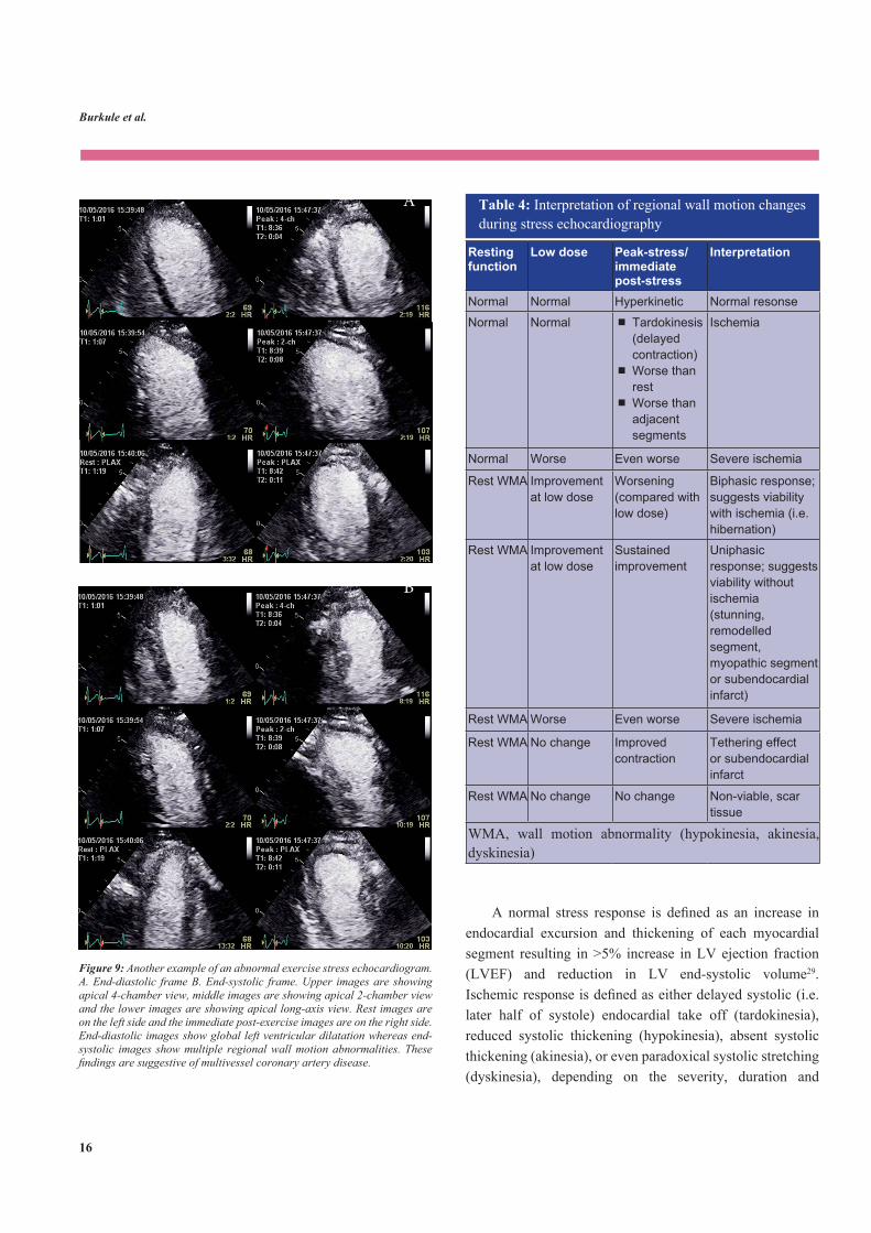
Table 4: Interpretation of regional wall motion changes during stress echocardiography
Resting function
Low dose Peak-stress/immediate post-stress
Interpretation
Normal Normal Hyperkinetic Normal resonse
Normal Normal Tardokinesis (delayed contraction) Worse than rest Worse than adjacent segments
Ischemia
Normal Worse Even worse Severe ischemia
Rest WMA Improvement at low dose
Worsening (compared with low dose)
Biphasic response; suggests viability with ischemia (i.e. hibernation)
Rest WMA Improvement at low dose
Sustained improvement
Uniphasic response; suggests viability without ischemia (stunning, remodelled segment, myopathic segment or subendocardial infarct)
Rest WMA Worse Even worse Severe ischemia
Rest WMA No change Improved contraction
Tethering effect or subendocardial infarct
Rest WMA No change No change Non-viable, scar tissue
WMA, wall motion abnormality (hypokinesia, akinesia, dyskinesia)
A normal stress response is defined as an increase inendocardial excursion and thickening of each myocardial segment resulting in >5% increase in LV ejection fraction (LVEF) and reduction in LV end-systolic volume29. Ischemicresponseisdefinedaseitherdelayedsystolic(i.e.later half of systole) endocardial take off (tardokinesia), reduced systolic thickening (hypokinesia), absent systolic thickening (akinesia), or even paradoxical systolic stretching (dyskinesia), depending on the severity, duration and
Figure 9: Another example of an abnormal exercise stress echocardiogram. A. End-diastolic frame B. End-systolic frame. Upper images are showing apical 4-chamber view, middle images are showing apical 2-chamber view and the lower images are showing apical long-axis view. Rest images are on the left side and the immediate post-exercise images are on the right side. End-diastolic images show global left ventricular dilatation whereas end-systolic images show multiple regional wall motion abnormalities. These findings are suggestive of multivessel coronary artery disease.
A
B

Indian Academy of Echocardiography Guidelines and Manual for Performance of Stress Echocardiography in Coronary Artery Disease
17
transmural extent of ischemia (Videos 9A, 9B, 10-16).
When analyzing RWMA, more emphasis should be put on assessment of wall thickening rather than wall motion (or endocardial excursion). The wall motion may spuriously occur even in an ischemic segment due to following two reasons:
1) Tethering effect of the adjacent hyperkinetic normal segments
2) Translational movement of the heart through the imaging plane in long- or short-axis views. Since LV cavity is tapering on either side of the long-axis imaging plane, a through-plane cardiac motion may give wrong perception of endocardial excursion.
Reviewing only the systole instead of complete cardiac cycle improves the ability to recognize RWMAs (Videos 17a, 17B). It is also advisable to assess endocardial excursion ‘takeoff’inthefirsthalfofsystole,asthisphaseisrelativelyindependent of translational motion and tethering29. Moreover, theoccurrenceofendocardial ‘takeoff’ in laterhalf of systole also helps in recognizing tardokinesia (Figure 10). Freezing the image and scrolling through early systole frame-by-frame is very helpful for this purpose.
When looking for ischemic response during DSE, it is best to compare peak-dose images with low-dose images instead of rest-images. At low-dose, the myocardial segments generally have much better contractility as compared to baseline and therefore any worsening of contractile function, and thus recognition of ischemia, becomes much easier if low-dose images are used as the reference. It is also important to carefully review recovery images. In patients undergoing dobutamine echocardiography, ischemic response is often masked at peak-dose due to reduction in LV cavity size leading to reduction in LV wall stress. Once dobutamine infusion is stopped, rapid increase in LV cavity size increases wall stress and precipitates ischemia (Video 18A, 18B). Additionally, recovery images also help in appreciation of ischemic response when the same cannot be reliably assessed at peak dose due to tachycardia. Since ischemia often persists for a few minutes, contractility may remain compromised during recovery phase also. However, as there is no reason
why a normal myocardial segment should have worse wall motion during recovery as compared to baseline, the presence of even subtle impairment of contractility during recovery in comparison to baseline should be considered as an evidence of ischemia at peak dose.
Ischemic RWMA follow a coronary vascular distribution and this helps in recognizing true wall motion abnormalities. False positive results are common in basal inferoseptal wall and basal inferior wall whereas false negative results are common in basal anterolateral wall. Therefore, isolated abnormalities in basal inferior wall or basal inferoseptum can be disregarded if the adjacent segments supplied by the same vascular territory show normal function30. One should alsocorroborateandconfirmRWMAinaparticularcoronaryperfusion territory in corresponding long and short-axis views. However, atypical patterns (e.g. proximal to mid-septal RWMA with apical sparing) may be seen in patients with patent coronary bypass graft to distal segment of left anterior descending artery with severe disease in proximal segment. Occasionally, a generalized global LV wall hypokinesia (cardiomyopathic response) may be seen in patients with hypertension or diabetes without obstructive CAD. It has been shown that these patients have higher incidence of heart failure,vasculareventsoratrialfibrillationinthefuture.
After completing segmental wall motion analysis, the rest and peak WMSI, WMSI change and rest and peak LV volume and EF ejection fraction can be formally calculated offline (Figure 11). Hemodynamic data obtained at peak-stress imaging is also reviewed. Increase in the severity of mitralregurgitation, rise inLVfillingpressure, increase inpulmonary artery systolic pressure, or RV dilatation, either alone or in combination, may provide useful corroborative evidence of myocardial ischemia.
Finally, it is also important to review stress induced ECGchanges.IftherearesignificantECGchangesbutthereis no apparent RWMA on echocardiography, it would be worthwhile to review the echocardiographic images once again, esp. if the patient also had chest pain during the stress test. This may help recognize any subtle RWMA which may have gone unnoticed during initial review of images.

Burkule et al.
18
Figure 10: An example of tardokinesia. Basal inferoseptum (single arrow) starts to contract early in systole whereas apical lateral wall contracts very late (double arrows).
Figure 11: Estimation of left ventricular volumes and ejection fraction using modified biplane Simpson’s method in post-exercise images.
AP2C- apical two-chamber; AP4C- apical four-chamber; ED- end-diastole; ES- end-systole

Indian Academy of Echocardiography Guidelines and Manual for Performance of Stress Echocardiography in Coronary Artery Disease
19
7. DIAGNOSTIC ACCURACY OF STRESS ECHOCARDIOGRAPHY
StressECGisknowntohavesensitivityandspecificityin the range of 63%-68% and 74%-77% respectively31. Stress echocardiographyhasmuchhighersensitivityandspecificityas compared to stress ECG. In a large meta-analysis, average sensitivity and specificity of exercise echocardiographywere 83% and 84%, respectively, whereas the same for DSE were 80% and 85%32 (Table 5). In women, exercise echocardiographyhashigherspecificity(80%versus64%)and overall diagnostic accuracy (81% versus 64%) for detection of CAD than exercise ECG2.
Table 5: Diagnostic accuracy of stress echocardiography as reported in meta-analyses
For detection of myocardial ischemia*
Stress modality Sensitivity (%)
Specificity (%)
Exercise Dobutamine Adenosine Dipyridamole Atrial acing, transthoracic Atrial acing, transesophageal
82.679.668.471.090.786.2
84.485.180.992.286.191.3
For detection of myocardial viability (i.e. functional recovery following revascularization)**
Imaging modality Sensitivity Specificity
Low dose dobutamine echocardiography Thallium-201 rest-redistribution SPECT Thallium-201 rest-redistribution-reinjection SPECT Technitum-99m-sestamibi SPECT Fluorine-18 fluorodeoxyglucose PET
84
90
86
83
88
81
54
47
69
73
PET- positron emission tomography; SPECT- single-photon emission computed tomography
*Source: Noguchi Y, Nagata-Kobayashi S, Stahl JE, Wong JB. A meta-analytic comparison of echocardiographic stressors. Int J Cardiovasc Imaging.2005;21:189-207;**Source:BaxJJ,WijnsW,CornelJH,VisserFC,BoersmaE,Fioretti PM. Accuracy of currently available techniques for prediction of functional recovery after revascularization in patients with left ventricular dysfunction due to chroniccoronaryarterydisease:comparisonofpooleddata.JAmCollCardiol.1997;30:1451-1460
For dobutamine echocardiography, average sensitivity for one-, two- and three-vessel disease has been reported to be approximately 74%, 86% and 92%, respectively24. The sensitivity is higher for detection of stenosis in the left anterior descending (72%) and right coronary artery (76%), as compared to left circumflex artery (55%)24. The overall sensitivityandspecificityare71%and92%fordipyridamoleechocardiography and 68% and 81% for adenosine stress echocardiography32.
Compared with nuclear imaging (exercise or pharmacological), stress echocardiography (exercise or dobutamine)hasbeenshowntohavebetterspecificity(77-82% versus 36-71%) but lesser sensitivity (80-88% versus 86-98%), resulting in nearly similar overall diagnostic accuracy (80-84% for both)33-36. For detection of multi-vessel CAD, both nuclear imaging and stress echocardiography have similar sensitivity (94%) and specificity (88%)24, 37. Stress echocardiography is better than nuclear stress tests in patients with LBBB, microvascular disease or LVH36 whereas nuclear imaging performs better in presence of single vessel disease, low workload stress test and in patients on beta-blockers38. Stress MRI has higher sensitivity (90%), specificity(81%)anddiagnosticaccuracy(87%)39.
7.1. Factors affecting diagnostic accuracy of stress echocardiography
Operator expertise
Operator expertise in performance and interpretation of stress echocardiography is perhaps the most important determinant of the accuracy of the results40, 41. Operator experience of performing and reading at least 100 studies under expert supervision is necessary to improve the accuracy of the beginner to expert level42.
Workload achieved
WMA develop at a later stage in the ischemic cascade and recover fast. Therefore, stress echocardiography loses itssensitivityifsufficientlyhighrate-pressureproductisnotachievedon treadmilland/or if there isadelay in imagingafter cessation of stress.
During dobutamine echocardiography, one should try to achieve MPHR and maintain it for at least 30-60 sec before

Burkule et al.
20
image acquisition. Addition of atropine helps in achieving target heart rate and improves sensitivity of the test by 5%43. If MPHR cannot be achieved for any reason, every effort should be made to achieved at least 85% of MPHR (also termed as target heart rate).
Delay in imaging
Delay in post-exercise imaging is an important consideration in patients undergoing treadmill stress echocardiography. To avoid any delay, some investigators have also suggested performing scanning at peak stress itself, while the patient is still on treadmill14, 44. On-treadmill peak stress imaging has higher sensitivity (84%) compared to peak supine bicycle imaging (75%) and post-treadmill imaging (60%). The quality of images is similar between on-treadmill versus post-treadmill imaging44.However,‘on-treadmill’scanningistechnicallydifficult.
Beta-blockers and antianginals
On beta-blockers and antianginals, the rate-pressure product achieved is lower and the post exercise heart rate drop occurs faster. Both these factors reduce the sensitivity of the test. Addition of atropine helps improve sensitivity of the test in these situations19.
Hypertension and LVH
Accelerated blood pressure response during exercise causes LV afterload mismatch and can result in RWMA even in absence of obstructive CAD. Further, in presence of LVH hypercontractility with reduced afterload in response to dobutamine infusion can result in dynamicLV outflowtract or mid-cavity obstruction. This may lead to LV apical ballooning, which recovers spontaneously. Conversely, thepresenceof significant concentricLVHwith smallLVcavitysizemaymakeitdifficulttoappreciatetheextentofendocardial excursion and to recognize WMA. However, despite all these challenges, stress echocardiography is still superior to stress ECG or nuclear imaging in patients with LVH36, 45.
Resting RWMA
Detection of inducible ischemia in segments with
resting hypokinesia is challenging due to difficulties inrecognizing subtle deterioration in contractility. Dobutamine echocardiography permits assessment of biphasic response to detect ischemia in hypokinetic or akinetic segments. Resting RWMA can also interfere with assessment of normal wall motion response in adjacent segments due to tethering effects.
Acoustic window
Stress echocardiography is highly dependent on image quality. When acoustic window is not good, the use of contrast for LVO may help improve diagnostic accuracy and reproducibility of stress echocardiography. Rarely when adequate acoustic window is not available and the images are suboptimal even with the use of contrast, the test should be cancelled.
ExtentofCADandfunctionalsignificanceofthestenoticlesion
The ability of stress echocardiography to detect inducible ischemia increases with increasing severity of CAD (severity of coronary stenoses as well as number of vessels involved). However, it should also be recognized that the extent of inducible ischemia depends on the functional significanceand not on anatomic severity of the coronary stenosis. Functional significance of a coronary lesion is determinedby the size of the coronary artery, vasodilatory reserve in the distal perfusion bed and the presence of collateral circulation. While coronary angiography shows anatomic severityofstenosis,fractionalflowreserve(FFR)showsthefunctionalsignificanceofthelesion.ThesensitivityofDSEfor detection of myocardial ischemia increases to 90% for vesselswithadiameterof>2.6mmandFFR<0.7546.
The affected coronary artery also influences thediagnostic accuracy of stress echocardiography. Sensitivity of stress echocardiography is generally lower for ischemia in leftcircumflexarteryterritory,whereasitishighestforleftanterior descending artery lesions.
Table 6 summarizes common causes responsible for false-positive and false-negative stress echocardiography.

Indian Academy of Echocardiography Guidelines and Manual for Performance of Stress Echocardiography in Coronary Artery Disease
21
8. ROLE OF NEWER MODALITIES IN STRESS ECHOCARDIOGRAPHY
As discussed above, stress echocardiography depends primarily on visual assessment of segmental wall motion, which is a subjective process. This leads to significantinterobserver variability in the results. Gaining required expertise in performance of stress echocardiography and following standardized protocol for image acquisition and interpretationsignificantlyimprovethediagnosticaccuracyof the test25, 26 and are strongly recommended. Additionally, several newer techniques have also been used to enhance the diagnostic accuracy of stress echocardiography. However, the role of these modalities at present is only as adjunct to and not as a substitute for visual wall motion analysis.
Table 6: Causes of false negative and false positive stress echocardiograms
False negatives • Inadequate stress • Delayed post-stress imaging• Beta-blocker usage, antianginal therapy• Coronary stenosis in smaller perfusion territory • Left circumflex disease• Ischemia in segments with pre-existing RWMA• Significant left ventricular hypertrophy• Poor image quality
False positives
• Isolated basal inferior septum or basal inferior wall RWMA
• Unmasking of subclinical cardiomyopathy (diabetes, hypothyroidism, or idiopathic)
• Segments with subendocardial infarcts (RWMA despite patent coronary artery)
• Exaggerated hypertensive response during stress • Left ventricular mid cavity dynamic obstruction during
dobutamine echocardiography• Abnormal septal motion (LBBB, previous coronary
bypass surgery)LBBB- left bundle branch block, RWMA- regional wall motion abnormality
8.1. Three-dimensional (3D) echocardiography
The tri-plane simultaneous acquisition mode with a 3D transducer provides good frame rate and image quality comparableto2Dimaging.Tri-planeimagingisbeneficial
during DSE because it reduces time and effort of the operator for acquiring 3 apical images at every stage, without compromising the accuracy of the test47.
Conceptually, single beat 3D volume acquisition at rest and peak can greatly reduce acquisition time, reduce the operator’s effort and can also overcome the problems of off-axis imaging. However, the present generation 3D echocardiography machines lack sufficiently high framerates and line density (in single beat acquisition mode) required for peak-stress imaging.
8.2. Use of contrast
The detection of endocardial excursion and wall thickening at peak heart rate during stress echocardiography requires adequate endocardial visualization. The use of contrast for LVO uniformly improves endocardial visualization in almost every patient48 (Figure 12, Video 19). In addition, the low mechanical index used for contrast imaging also provides clear epicardial visualization, resulting in much better appreciation of segmental wall thickening49. As a result, the number of non-diagnostic studies is reduced considerably. The use of contrast has been demonstrated to significantly improvediagnostic accuracyof the studieswithdifficultacousticwindowandtoimprovethereader’sconfidenceinreportingsuchstudies50. Contrast also improves accuracy of measurement of LVEF and LV volumes at rest and peak stress. The American Society of Echocardiography recommends use of LV cavity contrast in all stress studies in which at least two consecutive LV myocardial segments are not clearly visualized13.
Figure 12: Significant improvement in endocardial visualization with left ventricular cavity contrast in a patient with poor acoustic window.

Burkule et al.
22
Technical aspects of contrast use require understanding of instrumentation and physical dynamics of microbubbles and ultrasound waves. The acquisition mode is set at low mechanical index, a low ultrasound power mode, to prevent destruction of bubbles. However, low mechanical index prevents visualization of regular LV structures due to poor penetration and low intensity backscatter. During the rest images the gain setting is adjusted to visualize only outer shell of the epicardium/pericardium which is a relativelystrongerreflector(Figure13,Video20).
Figure 13: Imaging at low mechanical index. Gain settings are adjusted such that epicardium is clearly visualized.
The only commercially available ultrasound contrast in India is Sonovue®,aSulphurhexafluorideinertgaswithlipidshell. It is available in powder form which is reconstituted by mixing with 5 ml saline. Small aliquot of 0.2-0.3 ml of the prepared solution is injected IV using insulin syringe followed by a constant flush with 5-10 ml saline, till thecontrast appears in right ventricle, when the saline flushshould be stopped. A uniform white LVO should be achieved with crisp endocardial delineation and black myocardial wall thickness in all LV myocardial segments (Figure 14, Video 21). There should not be shadowing at base which would mean too much of contrast bolus volume or too rapid injectionorflushing(Figure15).Inthatcase,oneneedstowait till the ultrasound mediated bubbles destruction leads to uniform opacification. Conversely, visible swirling ofcontrast means less volume of bolus injected too slowly or excess destruction of bubbles due to higher mechanical index. Swirling of contrast in LV cavity should be avoided as it gives false perception of endocardial motion (Figure 16,
Video 22). In this situation, further lowering of mechanical index, administering additional bolus of contrast and quicklyfinishingacquisitionofimagestopreventprolongedinsonation are the strategies used to achieve uniform LVO.
At peak stress during exercise echocardiography, 0.3-0.5 ml contrast bolus is administered with quick saline flushapproximately 10-15 seconds before stopping the treadmill. Due to fast circulation time at peak exercise, the contrast is always seen in the left ventricle with good LVO by the time patient returns to the imaging bed and the post peak stress imaging is started. In case of DSE, a smaller quantity of contrast bolus (0.1-0.2 ml) is required during dobutamine infusion due to accumulation of contrast from previous stages. It is advisable to administer contrast bolus at 2 min 45 sec time point in the stage so that the images for that stage can be acquired in the last 10 seconds of the stage.
Figure 14: Technique for left ventricular contrast opacification. The contrast bolus is followed from initial appearance on the right side to complete left ventricular cavity opacification. Saline flush is stopped as soon as contrast appears in the right ventricle.
Figure 15: Shadowing due to excessive or too rapid contrast administration

Indian Academy of Echocardiography Guidelines and Manual for Performance of Stress Echocardiography in Coronary Artery Disease
23
8.3. Myocardial strain imaging
Given the ability of strain imaging to quantify myocardial contractile function and detect subtle changes in contractility, strain imaging has been used as an adjunct to wall motion analysis during stress echocardiography. The objective is to enhance diagnostic accuracy of the test and to minimize interobserver variability.
There are two methods for myocardial strain imaging- the Doppler-based and the gray-scale based [also known as 2D strain or speckle tracking echocardiography (STE)]. The Doppler-based strain imaging allows sampling at high frame rates suitable for stress echocardiography, but has poor signal-to-noise ratio. With STE, signal-to-noise ratio is robust at rest, but deteriorates considerably at faster heart rates. For Doppler-based strain measurement, velocity encoded images or color TDI images need to be acquired, whereas standard gray-scale images are used for STE. The results of offline analysis are displayed as parametricimagingusingcurvedanatomicMmodeorsegmentalstrain/strain rate waveforms. Dobutamine echocardiography is more suited for strain imaging because it is relatively free of respiratory and motion artifacts.
Since crossover from end systole to early diastole is the phase most vulnerable to ischemia, strain imaging reveals several abnormalities in this phase during ischemic response. These include delayed onset of relaxation, reduced early
diastolic strain rate, increased post-systolic shortening,
delayed peak systolic strain rate, reduction in peak systolic strain rate and reduced end-systolic strain values51-53. In early studies exploring utility of strain imaging during dobutamine echocardiography, Voigt et al demonstrated that magnitude of post-systolic shortening measured using Doppler strain could detect myocardial ischemia with good accuracy51,54. A cut-off value of >35% for the ratio of post-systolic shortening to maximum shortening had a sensitivity of 82% andspecificityof85%fordetectionofCAD.Inasubsequentstudy, Hanekom et al found that Doppler-based peak systolic strain rate measured at peak stress was an excellent predictor of CAD and was superior to post-systolic shortening for this purpose55. This study also compared diagnostic accuracy of Doppler-based strain with STE for detection of inducible ischemia. It was found that STE-based strain had acceptable accuracy for detection of ischemia only in LAD territory but not in other vascular territories. However, despite above studies demonstrating value of strain imaging for detection of myocardial ischemia, segmental strain analysis has considerable variability which limits its routine application inclinicalpractice.Thevariabilityincreasessignificantlyatpeak stress which further affects its clinical utility.
An important observation from animal studies is that the abnormalities of early diastolic relaxation often persist for significantperiodaftercessationofstress56. This phenomenon
Figure 16: Swirling artefact due to too little or too slow contrast administration.
Burkule et al.
24
has been termed as diastolic stunning and may help in detection of transient myocardial ischemia by obviating some of the limitations inherent to peak stress imaging. Gray-scale images can be acquired at ∼10 min after cessation of exercise when the heart rate and hyperventilation have already settled. STE-based strain analysis of these images can reveal markedly impaired diastolic strain recovery in ischemic segments. In a study involving 162 patients with stable angina, segmental transverse strain was obtained at 5 and 10 min after cessation of exercise53. Impairment of segmental transverse strain decay during initial 1/3rd of diastole was almost uniformly seen in ischemic segments and persisted forupto10min.Thiswastermedas‘ischemicmemory’signand was very sensitive for diagnosis of obstructive CAD.
9. PROGNOSTIC VALUE OF STRESS ECHOCARDIOGRAPHY
Stress echocardiography provides a considerable amount of prognostic information that is far greater than just the diagnosis of inducible ischemia conveyed in a dichotomous fashion (i.e. positive or negative stress echocardiogram). This is in line with recent large-scale studies demonstrating that functional tests have superior prognostic power than the anatomical imaging for predicting cardiovascular mortality and major adverse cardiac events (MACE)57. The overall prognostic information provided by stress echocardiography is comparable to nuclear stress imaging using the same stress (exercise or dobutamine) modality58, 59.
Patients with normal exercise stress echocardiogram have better event free survival than age and sex-matched population6. A negative exercise or dobutamine stress echocardiogram predicts low cardiac mortality and MACE rates (less than 1% per year) over next 4-5 years follow-up60-62. This prognostic information is incremental to the Framingham risk score63, Duke treadmill score60 and even coronary angiography findings62. However, the excellent prognostic value of a negative stress echocardiogram is adversely influenced by sub-maximal workload andanti-anginal medications64, 65. Patients with normal stress echocardiography but submaximal peak heart rate have higher risk of cardiac events and incidence of revascularization compared to those who achieve maximal peak heart rate64.
Conversely, the patients with a positive stress echocardiogram have a high risk of MACE even if there is no hemodynamically significantCAD.The risk of cardiac events in patients with so –called ‘false positive’ stressechocardiogram approaches that with true-positive stress echocardiograms66. The pathophysiological mechanisms underlying such false-positive stress echocardiogram include unmasking of underlying subclinical cardiomyopathy, coronary endothelial dysfunction leading to abnormal coronaryflowreserveormicrovasculardysfunction,andinsome cases, transient Takatsubo like stress cardiomyopathy response.
The risk of MACE in patients with true-positive stress echocardiograms is unequivocally high. The prognostic value of a positive stress echocardiogram has been demonstrated in a wide variety of patient subgroups including women67, elderly patients68, diabetics69, patients with or without LV systolic dysfunction70, 71 and those with previous MI72 or previous CABG. This prognostic power of stress echocardiography is incremental to the patient’s baseline clinical data, resting ECG, stress ECG and resting LV systolic function. Among patients with normal stress ECG, those who show stress induced RWMA have twice the annual rates of all-cause mortality and cardiac events than those who have normal stress echocardiogram73. The prognostic value of a positive stress echocardiogram is further enhanced by incorporating data about WMSI, resting WMA and peak-stress LV end-systolic volume and LVEF6, 62, 68, 74, 75. The lack of reduction or an actual increase in LV end-systolic volume at peak stress and failure to increase peak exercise LVEF are independent predictors of cardiac death or non-fatal MI68.
10. ADDITIONAL BENEFITS OF STRESS ECHOCARDIOGRAPHY
The choice between stress echocardiography versus nuclear stress test mainly depends on cost, local availability, and local expertise76. However, stress echocardiography has numerous advantages over nuclear imaging. Stress echocardiography provides additional information, at no extra cost, about LV systolic and diastolic function, valvular hemodynamics, pulmonary pressures etc. at rest and at peak-stress, which is clinically useful in guiding patient management. Furthermore, in many patients without

Indian Academy of Echocardiography Guidelines and Manual for Performance of Stress Echocardiography in Coronary Artery Disease
25
previously known cardiac illness, resting echocardiography performed before initiation of exercise often shows incidental findingofRWMA,significantvalvulardisease,pericardialeffusion, pulmonary hypertension, cardiomyopathies or aortic dissection which itself answers the clinical query. This obviates the need for performing the stress test and prevents serious potential complications. Safety, rapidity, repeatability and lack of exposure to radiation or nephrotoxic drugs are further advantages of stress echocardiography.
11. CLINICAL RELEVANCE OF ISCHEMIA DETECTION WITH STRESS ECHOCARDIOGRAPHY
In stable chronic CAD, several large-scale randomized trials have failed to show incremental benefit of routinerevascularization in reducing cardiac mortality or non-fatal MI in comparison to optimized medical therapy alone77-79. Although revascularization has been shown to more effectively reduce angina, this benefit too is not durablebeyondfirstfewyears.Therefore,thechallengebeforestressimaging is to accurately identify the high-risk subgroup of asymptomatic or minimally symptomatic stable CAD patients in whom revascularization is likely to reduce the risk ofdeath/MIinthepresenteraofoptimizedmedicaltherapy.Traditionally, it is believed that the evidence of moderate or severeischemiaoncardiacstresstestsignifieslargeareaofjeopardized myocardium and high risk of adverse cardiac event rates that justifies coronary revascularization. Thedefinitionofmoderatetosevereischemiaforvariousstresstests is based on their respective thresholds for predicting annual cardiac event rates of 4-6%. This amounts to >10% ischemic myocardium on nuclear imaging, >3/16 newakinetic segments on stress echocardiography and >4/32perfusion defects or >3/16newakineticsegmentsonstresscardiac MRI80.
12. ASSESSMENT OF MYOCARDIAL VIABILITY
12.1. Pathophysiological considerations
A dysfunctional myocardial segment is considered viable if it shows improved contractility over time after restoration of adequate coronary blood flow.Myocardial dysfunctioncan occur due to transient severe ischemia with reperfusion
(myocardial stunning) or due to chronically reduced coronary bloodflowwithseveralultrastructuralchangesinmyocytes(myocardial hibernation)6. Repeated episodes of stunning due to demand supply mismatch resulting from coronary stenosis not severe enough to cause reduction in resting myocardialbloodflow(MBF)canalso lead tomyocardialhibernation without myocardial ultrastructural changes.
In case of ST-elevation myocardial infarction (STEMI), myocardial damage extends from endocardium to epicardium as the advancing wave-front of necrosis with its net extent depending upon the rapidity of reperfusion. The regional myocardial systolic thickening depends upon the extent of preservation of subendocardial helically oriented fibersand the mid-myocardial circumferentially oriented fibers.The transmural extent of myocyte loss and replacement with fibrosis decide the recovery of wall motion and itscontribution to global LV systolic function81.
In patients with stable CAD and chronic LV systolic dysfunction, multiple factors contribute to the regional myocardial dysfunction apart from the acute myocyte loss frompreviousSTEMIornon-ST-elevationMI(NSTEMI).Inearlyphase,transmuralmyocardialbloodflow(TM-MBF)may be near normal or mildly reduced but the coronary flow reserve (CFR) is significantly compromised. Thesubendocardial layerMBF (SE-MBF) is also significantlyreduced, even at rest82. During any episode of acute ischemia, SE-MBF is reduced further. It is noteworthy that reduction in SE-MBF is out of proportion to reduction in TM-MBF with a 25% reduction in TM-MBF resulting in ∼50% reduction in SE-MBF. When there is a 50% to 75% reduction of SE-MBF, segmental akinesia occurs. After relief of an ischemic episode, the hyperemia due to vasodilation is also maldistributed to mid and epicardial layer so that SE-MBF remains reduced for a considerable period of time81. Therefore, demand-supplymismatchinasettingofsignificantcoronarystenosismay cause repetitive stunning and persistent akinesia, even if TM-MBFisnotsignificantlyreducedatrest.
In chronic ischemic dilated cardiomyopathy, advanced regional myocardial remodeling process occurs as a consequence of increased wall stress, exposure to systemic neuro-humoral activation and myocyte adaptation to chronically reduced capillary blood flow.

Burkule et al.
26
The microvasculature significantly shrinks to form sparsecapillary network and reduced capillary blood volume83. The myocytes undergo ultrastructural changes by shutting down energy intensive cellular processes and the sarcomeres, myofilaments and sarcoplasmic reticulum degenerate overtime.Themyofilamentspacegetsfilledwithglycogenandthecytoskeletal proteins and mitochondria also show structural alterations. Eventually, the myocytes undergo apoptosis. The extracellular matrix expands and shows increased amounts of typeIcollagen, typeIIIcollagenandfibronectin.Thesehistopathological changes occur to a variable extent in both ischemic and non-ischemic remote territories in chronic ischemic dilated cardiomyopathy84, 85.
Hence in chronic ischemic LV systolic dysfunction, regional myocardial dysfunction may reflect a spectrumof pathological processes including variable extent of transmural scarring, remodeled myocardium, variable myocardial capillary blood flow and volume and variableamount of interstitial fibrosis. Segments with less fibrosisand with less severe myocytic ultrastructural changes are more likely to improve in function on LDDE and after revascularization. The time to myocardial recovery in such hibernating myocardium post-revascularization would depend on the baseline severity of myocardial ultrastructural changes81.
12.2. Low dose dobutamine echocardiography for assessment of myocardial viability
Low dose dobutamine echocardiography (LDDE) is the primary echocardiography modality for assessment of myocardial viability. It is also the simplest and the least expensive among all the currently available imaging modalities for this purpose [with the exception of end-diastolic wall thickness assessment (EDWT)].
For viability assessment, LDDE relies on the ability of the dysfunctional myocardial segments to improve their contractilityinresponsetolowdose(i.e.2.5–10mcg/kg/min) dobutamine infusion. At this dose, dobutamine produces an inotropic effect without increasing the heart rate. In addition, there is mild systemic and coronary vasodilatation causing reduction in afterload and increase in myocardial perfusion and myocardial blood volume. As a result, there
is no increase in myocardial work load or myocardial oxygen demand. Therefore, a dysfunctional myocardial segment is likely to show augmentation in wall motion and systolic thickening at this dose. The visually discernible improvement in the contractile function, however, depends upon the transmural quantity of the surviving myocytes and the intact contractile apparatus in the myocytes as well as the recruitable residual coronary vasodilatory reserve. For this reason, dobutamine induced augmentation of contractile responseisahighlyspecificpredictorofviability6. However, absence of contractile response can occur in spite of having sufficientviablemyocytesandcanbeexplainedbyabsentrecruitable coronary vasodilatory response, dobutamine induced ischemia in tachycardic patients and complete loss of subendocardial layer (which contributes to systolic thickening). Thus, while a positive LDDE result is useful to ‘rulein’viability,anegativeLDDEresultcannotbeusedto‘ruleout’viabilitywithcertainty.
12.2.1 Performing DSE for myocardial viability
Patient preparation and baseline imaging for myocardial viability assessment are same as for DSE performed for ischemia detection. However, the dobutamine infusion protocol differs13, 81 (Figure 17).
For viability assessment, dobutamine infusion is started atadoseof2.5mcg/kg/minandthedoseisincreasedevery3minto5,7.5,10and20mcg/kg/min.Furtherincreaseinthedoseto30and40mcg/kg/minremainsdebatable.Sincemost of the patients undergoing viability assessment have significantLVsystolicdysfunctionwithmulti-vesselCAD,higher dose is associated with a certain risk of arrhythmias or cardiac decompensation. Moreover, it can be argued that showing augmentation of contractile response at low-dose dobutamine is all that is needed to detect viability and there is really no need to go up to full-dose. However, using full-dose dobutamine protocol permits recognition of biphasic response which is themostspecificpredictorof functional recoveryfollowing revascularization. Additionally, worsening of contractile function at high-dose also makes it easier to appreciate subtle improvements in contractile function at low-dose. Finally, a full-dose protocol also permits detection of inducible myocardial ischemia in normally contracting

Indian Academy of Echocardiography Guidelines and Manual for Performance of Stress Echocardiography in Coronary Artery Disease
27
segments at baseline. Thus, both the protocols have their own advantages and disadvantages. The choice between the twoshoulddependontheoverallpatientprofile,availabilityof the infrastructure to deal with any cardiac emergency if arises and the expectations from the stress test in a given patient. In general, in low resource settings, low-dose dobutamine protocol is preferable, especially if assessment of myocardial viability is the dominant clinical query.
Permorm a baseline echocardiographic study to assess LV systolic function, hemodynamics
Permorm a baseline echocardiographic study to assess LV systolic function, hemodynamics
Acquire the 5 standard views as part of stress protocol. Administer contrast if available. Velocity-encoded gray-scale images or color TDI images may be acquired if contrast is not used
Acquire the 5 standard views as part of stress protocol. Administer contrast if available. Velocity-encoded gray-scale images or color TDI images may be acquired if contrast is not used
Start dobutamine infusion at a dose of 2.5 mcg/kg/min; increase it every 3 min to 5,7.5, 10 mcg/min (or maximum up to 20 mcg/kg/min). Avoid tachycardia
Start dobutamine infusion at a dose of 2.5-5 mcg/kg/min; increase it every 3 min to7.5, 10, 20, 30 and 40 mcg/min
Atropine generally not used
Acquire images during each stage. Administer contrast if available
Acquire images during each stage. Administer contrast if available
Stop dobutamine infusion once maximum dose reached. IV metoprolol may be used if there is tachycardia
Obtain recovery images a few minutes later
Stop dobutamine infusion, IV metoprolol may be used to rapidly reduce heart rate
Recovery images acquired once hear rate is near baseline or at least<100/min
Low-dose protocol Full-dose protocol
Figure 17: Imaging protocols for dobutamine echocardiography performed for myocardial viability assessment.
IV- intravenous; LV- left ventricular; TDI- tissue Doppler imaging.
The imaging principles are almost similar to that for ischemia detection. First, a baseline echocardiogram is performed to assess for LV systolic function and hemodynamics. Good quality images in the five standardviews as described for ischemia protocols should be acquired. In addition, as strain imaging seems to be useful for viability assessment, it is also advisable to obtain velocity-encoded
gray-scale images or color TDI images (depending on the echocardiography equipment) of the three apical views (AP4C, AP2C and APLAX). The stress protocol is then turned on and contrast enhanced images in the 5 standard views are acquired as part of the protocol. Dobutamine infusion is then started. Regardless of the infusion protocol, it is advised that the same set of standard gray-scale images are recorded at the end of each step. Having multiple sets of images enhances the ability to detect viability as improvement in contractile function may sometimes be only transient. The contrast bolus (0.5-1 ml) should be administered at 2 min 45 second time-point during each stage and the images can be acquired 10-15 seconds later. The ability to administer contrast as IV infusion permits uniform LVO and makes it easier to obtain good quality images but the requisite type of infusion pumps are currently not available in India.
It should be noted that contrast interferes with strain imaging and hence if strain imaging at low-dose is required then an appropriate protocol would be to acquire both non-contrast and contrast enhanced images at baseline, followed by only non-contrast images during subsequent stages. The acquisition of contrast-enhanced images at baseline is important as it would allow qualitative assessment of myocardial perfusion also. If contrast is not available, then of course there is no dilemma and only velocity encoded gray-scale images or separate gray-scale and color TDI images need to be acquired at each stage.
Atropine boluses of 0.3-0.6 mg are rarely required in this patient population to achieve target heart rate. IV metoprolol (2-10 mg) is administered at the end of the test to rapidly reverse dobutamine induced tachycardia and arrhythmia.
12.2.2 Interpretation of dobutamine echocardiography for viability
As mentioned above, at low dose (5-10 µg/kg/min),dobutamine augments cardiac contractility without any appreciable increase in myocardial oxygen demand. However, when the dose is increased further, there is progressive increase in heart rate and myocardial oxygen demand. As a result, a hibernating segment will improve at low-dose but worsens again at peak dose (Table 4) (Figure 18, Videos 23A-C).

Burkule et al.
28
Figure 18: Biphasic response during dobutamine stress echocardiography. A dysfunctional apical segment improves at 5 and 20 mcg/kg/min but worsens again at 40 mcg/kg/min dose.
Figure 19: Uniphasic response during dobutamine stress echocardiography. The apical segment (arrow) shows sustained improvement at 10, 20 and 40 mcg/kg/min dose.

Indian Academy of Echocardiography Guidelines and Manual for Performance of Stress Echocardiography in Coronary Artery Disease
29
In contrast, a stunned myocardial segment or non-ischemic, myopathic or remodelled segment will demonstrate a sustained improvement in contractility at low- and peak-dose (Figure 19, Video 24). Tethering may also produce a similar response but more often the augmentation in contractility is visible only at peak-dose with no appreciable increase in contractility at low-dose. When there is no change in contractility at low-dose, it indicates lack of myocardial viability (Figure 20, Video 25). Rarely, a dysfunctional segment may deteriorate at low-dose itself indicating presence of critically reduced MBF at rest and no CFR. Viable segments without contractile reserve on LDDE usually have lower or completely exhausted CFR than viable segments with contractile reserve. Apart from the exhaustion of CFR, other factors, like severity of ultrastructural changes in cardiomyocytes or down-regulation of beta- adrenoreceptors, may also contribute to the lack of contractile reserve in viable segments.
Accordingly, four different types of responses can be seen in dysfunctional myocardial segments during DSE86 (Table 4):
Biphasic: Improvement in contractility at low dose
and worsening at higher dose suggesting viable and ischemic segment (Figure 18, Videos 23A-C). This type of response is the most accurate predictor of functional recovery after revascularization87.
Sustained improvement: Continued improvement in contractility with increasing dobutamine dose (Figure 19, Videos 24). It suggests viable, stunned, remodeled, myopathic or tethered segment or segment with subendocardial infarct but without ischemia. It has lower specificity to predict improvement postrevascularization. However, in practice both biphasic and sustained responses are reported as viability to improve sensitivity. Sustained improvement should be differentiated from improvement occurring only at peak-dose without any improvement at low-dose. This type of response does not indicate myocardial viability.
Deterioration at low dose: Hypokinetic segments becoming akinetic to dyskinetic with low-dose dobutamine itself. These are the segments with critically reduced resting MBF. Such segments have variable chances of recovery with revascularization.
Figure 20: Lack of contractile response to dobutamine suggestive of lack of viability.

Burkule et al.
30
No response: Akinetic segments that show no improvement in contractile function at low-dose (Figure 20, Videos 25). These are nonviable, scarred segments. These segments usually have low myocyte mass and more transmural extent of fibrosis and are unlikely toimprove with revascularization.
Additionally, in ischemic dilated cardiomyopathy, LV end diastolic volumes index >170ml/m2, LV end-systolic volume index >90 ml/m2 and LV end-diastolic diameter index >55 mm/m2 indicate an advanced LV remodeling with very low likelihood of global LVEF recovery post revascularization81.
12.2.3 Sensitivity and specificity of DSE for myocardial viability
LDDE shows average sensitivity of 75% to 80% and a specificity of 80% to 85% for the prediction of functionalrecovery, both early after MI or in the setting of ischemic dilated cardiomyopathy81.Biphasicresponseisthemostspecificformyocardial viability and is the most accurate predictor of functionalrecoveryafterrevascularization(specificity89%and sensitivity 74%)87. Sustained response, though highly sensitive, has a much lower specificity. Generally, bothbiphasic and sustained responses are reported as evidence of viability, which improves sensitivity (86%) but reduces specificity (68%) for prediction of functional recovery88. Combining any response on dobutamine (biphasic, sustained or worsening) further improves sensitivity (88%) but with a significantdropinspecificity(61%)88.
The global LV functional recovery post revascularization depends upon the number of myocardial segments showing viability on baseline LDDE. The improvement in LVEF post-revascularization is expected to be in the range of 5-6 % for 2-5 viable segments at baseline and >10% for 6 or more viable segments89.PatientsshowingsignificantLVEFimprovement with LDDE are likely to show global functional recovery and reverse remodeling after CABG90.
12.2.4 Comparison with other imaging modalities
Preserved EDWT in an infarcted segment indicates
the presence of sufficient mass of recruitable myocytesto effectively cause systolic thickening. Thus, EDWT >6 mm has high sensitivity (>90%) to predict post revascularizationfunctionalrecoverybutspecificityispoor(40-50%)91. Conversely, reduced diastolic wall thickness (<5mm) is highly accurate in identifying resting regionalmyocardial dysfunction that is unlikely to improve after revascularization81.
CombiningEDWTwithLDDEsignificantly improvesthe diagnostic accuracy of the latter by excluding segments showing tethering effect91. A combination of EDWT >6 mm with any improvement on LDDE gives optimum sensitivity (88%)andspecificity (77%)91. Further, the combination of EDWT >5 mm and either dobutamine echocardiography or thallium-201 scintigraphy results in similar high positive predictive value as a combination of dobutamine echocardiography and thallium-201 scintigraphy81.
Contrast LVO permits accurate estimation of EDWT and may be helpful in viability assessment (Figure 21). Additionally, perfusion imaging with myocardial contrast echocardiography (MCE) can also be performed. MCE may be especially useful in evaluation of myocardial viability in dobutamine non-responsive segments. The ten MCE studies in the literature evaluating viability have shown sensitivity rangingfrom62%to92%andspecificityrangingfrom67%to 87% in identifying myocardial viability81.
Compared with nuclear or MRI techniques, LDDE is more specific but less sensitive for prediction of viability.Therefore, in case of negative DSE results, especially in myocardial segments with >5 mm EDWT, it may be prudent to take help of MRI or nuclear method92. Meta-analysis of LDDE trials involving 1090 patients showed sensitivity of 81% and specificity of 80% for prediction of myocardialviability. Similar meta-analysis of thallium scintigraphy trials involving 858 patients showed sensitivity of 86-88%andspecificityof50-60%whereasameta-analysisoffluorodeoxyglucose positron emission tomography (FDG-PET) trials involving 598 patients showed sensitivity of 93%andspecificityof58%.Similarly,thirteenMRIstudiesinvolving 420 patients showed sensitivity of 80-82% and specificityof68-70%forpredictionofmyocardial viability92.

Indian Academy of Echocardiography Guidelines and Manual for Performance of Stress Echocardiography in Coronary Artery Disease
31
12.2.5 Role of strain imaging as adjunct to wall motion analysis during LDDE
Compared with assessment of inducible ischemia, viability assessment is more suited for incorporation of strain imaging as tachycardia is not a limitation here. Doppler-based measurement of segmental longitudinal strain and strain rate has been shown to be useful in enhancing diagnostic accuracy of LDDE for detection of myocardial viability. In a study involving 55 patients undergoing revascularization, a longitudinal strain-rate increment of >0.25/s at low-dose dobutamine predicted functionalrecovery after revascularization with sensitivity 80% and specificity75%93. This diagnostic study was superior to that of wall motion analysis alone. In another study, an increase insystolicstrainratevaluemorethan0.23/sorincreaseinearlydiastolicstrainratemorethan0.8/spredictedviabilityinakineticsegmentswithasensitivity83%andspecificity84%94-96. STE-based strain analysis has also been evaluated forassessmentofviabilitybutithadsufficientaccuracyonlyin anterior circulation and not in the posterior circulation97.
12.3. Clinical algorithm for myocardial viability assessment
Using multiple imaging modalities to assess viability in a single patient is not a cost-effective approach. Such a practice invariably leads to more confusion in clinical decision making due to discordant results. Therefore, in
patients with ischemic dilated cardiomyopathy, myocardial viability assessment should follow a systematic algorithm81. ThefirststepistoaccuratelymeasureEDWToftheakineticsegment on echocardiography. Contrast LVO may help in improving the accuracy of EDWT measurement. In case of poor acoustic window, cardiac MRI may also be used. A preserved segment with EDWT >5-6 mm suggests >50% probability of recovery while a thinned out segment with EDWT <5 mm indicates <5% probability of recovery81. In presence of preserved wall thickness (>5-6 mm), either LDDE or thallium scintigraphy is sufficient to establishviability. However, in the presence of EDWT of <5 mm, more downstream investigations are required. If LDDE results are negative or equivocal in segmentswith EDWT<6mm, acombination of LDDE and late gadolinium enhancement on MRI or LDDE with FDG-PET are required for further assessment.
12.4. Clinical relevance of myocardial viability assessment
The role of myocardial viability assessment in clinical practice is currently quite controversial. A large of number studies, mostly single-center and small, have demonstrated clearbenefitof revascularization inpatientswith evidenceof viable myocardium. However, most of these studies were limited by retrospective and non-randomized nature and wide heterogeneity in the patient populations studied and the modalities used for viability testing. In contrast, the large STICH (Surgical Treatment of IsChemic Heart Failure) trial failed to demonstrate any significant interaction betweenmyocardial viability and medical versus surgical treatment outcome, whether assessed according to treatment assigned (intention to treat) or to the treatment actually received98. Unfortunately, this trial itself had several limitations because of which no definite conclusions can be drawn about thevalue of viability assessment in clinical decision-making.
However, what is well-understood now is that the traditional practice of defining myocardial viability in adichotomous manner as presence or absence of functional recovery after revascularization is too simplistic, considering the complex pathophysiology of chronic ischemic myocardium. Myocardial segments with viable myocytes but with no contractile reserve may still benefit after
Figure 21: Use of ultrasound contrast for left ventricular cavity opacification improves accuracy of end-diastolic wall thickness measurement.

Burkule et al.
32
revascularization by preventing re-infarction, preventing arrhythmias, influencing LV remodeling because of intactepicardial layer and improving diastolic function.
In these circumstances, the practical role of myocardial viability assessment may be envisaged in the following clinical situations: 1) decision about the need to perform PCI of infarct related artery in stable acute myocardial infarction patients presenting after 24 hours of symptoms onset, 2) deciding about CABG in patients with severe triple
vessel CAD and severe heart failure who are at high-risk of perioperative mortality due to co-morbidities, 3) to choose PCI over CABG in a patient with multi-vessel CAD when the left anterior descending artery territory is infarcted and surgical risk appears to be high, and 4) for identifying culprit vessel responsible for angina, for partial revascularization by PCI, in patients of multi-vessel CAD with multiple myocardial infarcts.
13. REFERENCES1. FrakerTD,Jr.,FihnSD,GibbonsRJ,etal.2007chronicangina
focused update of the ACC/AHA 2002 guidelines for themanagement of patients with chronic stable angina: A report of the AmericanCollegeofCardiology/AmericanHeartAssociationtaskforce on practice guidelines writing group to develop the focused update of the 2002 guidelines for the management of patients with chronic stable angina. J Am Coll Cardiol. 2007;50:2264-2274
2. FleisherLA,BeckmanJA,BrownKA,etal.2009ACCF/AHAfocused update on perioperative beta blockade incorporated into theACC/AHA2007 guidelines on perioperative cardiovascularevaluation and care for noncardiac surgery: A report of the American College of Cardiology Foundation/American HeartAssociation task force on practice guidelines. Circulation. 2009;120:e169-e276
3. Douglas PS, Garcia MJ, Haines DE, et al. ACCF/ASE/AHA/ASNC/HFSA/HRS/SCAI/SCCM/SCCT/SCMR 2011 appropriateuse criteria for echocardiography. A report of the American College of Cardiology Foundation appropriate use criteria task force, American Society of Echocardiography, American Heart Association, American Society of Nuclear Cardiology, HeartFailure Society of America, Heart Rhythm Society, Society for Cardiovascular Angiography and Interventions, Society of Critical Care Medicine, Society of Cardiovascular Computed Tomography, and Society for Cardiovascular Magnetic Resonance endorsed by the American College of Chest Physicians. J Am Coll Cardiol. 2011;57:1126-1166
4. McCully RB, Roger VL, Mahoney DW, et al. Outcome after normal exercise echocardiography and predictors of subsequent cardiac events: Follow-up of 1,325 patients. J Am Coll Cardiol. 1998;31:144-149
5. Pryor DB, Harrell FE, Jr., Lee KL, Califf RM, Rosati RA.Estimating the likelihoodof significant coronary artery disease.Am J Med. 1983;75:771-780
6. GibbonsRJ.ACC/AHA2002guidelineupdateforthemanagementof patients with chronic stable angina--summary article: A report oftheAmericancollegeofcardiology/Americanheartassociationtask force on practice guidelines (committee on the management of patients with chronic stable angina). Circulation. 2003;107:149-158
7. Picano E. Anatomical and functional targets of stress testing. Stress echocardiography. Berlin: Springer; 1997:9-17.
8. Picano E. Pathogenetic mechanisms of stress. Stress echocardiography. Berlin: Springer; 1997:62-75.
9. Picano E. Symptoms and signs of myocardial ischemia. Stress echocardiography. Berlin: Springer; 1997:19-25.
10. Burkule N, Bansal M, Mehrotra R, Venkateshvaran A. IAEperformance standards and recommendations for a comprehensive transthoracic echocardiographic study in adults. J Indian Acad Echo. 2015;25:93-125
11. Pellikka PA, Nagueh SF, Elhendy AA, Kuehl CA, SawadaSG. American Society of echocardiography recommendations for performance, interpretation, and application of stress echocardiography. J Am Soc Echocardiogr. 2007;20:1021-1041
12. PeteiroJ,GarridoI,MonserratL,AldamaG,CalvinoR,Castro-Beiras A. Comparison of peak and postexercise treadmill echocardiography with the use of continuous harmonic imaging acquisition. J Am Soc Echocardiogr. 2004;17:1044-1049
13. ParkTH,TayanN,TakedaK,JeonHK,QuinonesMA,ZoghbiWA. Supine bicycle echocardiography improved diagnostic accuracy and physiologic assessment of coronary artery disease with the incorporation of intermediate stages of exercise. J Am Coll Cardiol. 2007;50:1857-1863
14. Klein J, Cheo S, Berman DS, Rozanski A. Pathophysiologicfactors governing the variability of ischemic responses to treadmill and bicycle exercise. Am Heart J. 1994;128:948-955
15. Rallidis L, Cokkinos P, Tousoulis D, NihoyannopoulosP. Comparison of dobutamine and treadmill exercise echocardiography in inducing ischemia in patients with coronary artery disease. J Am Coll Cardiol. 1997;30:1660-1668
16. McNeill AJ, Fioretti PM, el-Said SM, Salustri A, Forster T,Roelandt JR. Enhanced sensitivity for detection of coronaryartery disease by addition of atropine to dobutamine stress echocardiography. Am J Cardiol. 1992;70:41-46
17. Fioretti PM, Poldermans D, Salustri A, et al. Atropine increases the accuracy of dobutamine stress echocardiography in patients taking beta-blockers. Eur Heart J. 1994;15:355-360
18. Picano E, Pingitore A, Conti U, et al. Enhanced sensitivity for detection of coronary artery disease by addition of atropine to dipyridamole echocardiography. Eur Heart J. 1993;14:1216-1222
Indian Academy of Echocardiography Guidelines and Manual for Performance of Stress Echocardiography in Coronary Artery Disease
33
19. Lanzarini L, Fetiveau R, Poli A, Diotallevi P, Barberis P, Previtali M. Results of dipyridamole plus atropine echo stress test for the diagnosis of coronary artery disease. Int J Card Imaging. 1995;11:233-240
20. KaragiannisSE,Bax JJ,ElhendyA, et al.Enhanced sensitivityof dobutamine stress echocardiography by observing wall motion abnormalities during the recovery phase after acute beta-blocker administration. Am J Cardiol. 2006;97:462-465
21. IskandrianAS,VeraniMS,HeoJ.Pharmacologicstresstesting:Mechanism of action, hemodynamic responses, and results in detection of coronary artery disease. J Nucl Cardiol. 1994;1:94-111
22. Geleijnse ML, Fioretti PM, Roelandt JR. Methodology,feasibility, safety and diagnostic accuracy of dobutamine stress echocardiography. J Am Coll Cardiol. 1997;30:595-606
23. Hoffmann R, Lethen H, Marwick T, et al. Standardized guidelines for the interpretation of dobutamine echocardiography reduce interinstitutional variance in interpretation. Am J Cardiol. 1998;82:1520-1524
24. HoffmannR,MarwickTH,PoldermansD,etal.Refinementsinstress echocardiographic techniques improve inter-institutional agreement in interpretation of dobutamine stress echocardiograms. Eur Heart J. 2002;23:821-829
25. Attenhofer CH, Pellikka PA, Oh JK, Roger VL, Sohn DW,SewardJB.Comparisonofischemicresponseduringexerciseanddobutamine echocardiography in patients with left main coronary artery disease. J Am Coll Cardiol. 1996;27:1171-1177
26. Lang RM, Badano LP, Mor-Avi V, et al. Recommendations for cardiac chamber quantification by echocardiography in adults:An update from the American Society of echocardiography and the European association of cardiovascular imaging. J Am Soc Echocardiogr. 2015;28:1-39 e14
27. Marwick TH. Interpretation of stress echocardiography. Stress echocardiography: Its role in the diagnosis and evaluation of coronary artery disease. Massachusetts: Kluwer Academic Publishers; 2003:43-59.
28. BachDS,MullerDW,GrosBJ,ArmstrongWF. False positivedobutamine stress echocardiograms: Characterization of clinical, echocardiographicandangiographicfindings.J Am Coll Cardiol. 1994;24:928-933
29. GarberAM,SolomonNA.Cost-effectivenessofalternative teststrategies for the diagnosis of coronary artery disease. Ann Intern Med. 1999;130:719-728
30. NoguchiY,Nagata-Kobayashi S, Stahl JE,Wong JB.Ameta-analytic comparison of echocardiographic stressors. Int J Cardiovasc Imaging. 2005;21:189-207
31. Marwick TH, Anderson T, Williams MJ, et al. Exerciseechocardiography isanaccurateandcost-efficient technique fordetection of coronary artery disease in women. J Am Coll Cardiol. 1995;26:335-341
32. Marwick T, Willemart B, D’Hondt AM, et al. Selection of the optimal nonexercise stress for the evaluation of ischemic regional myocardial dysfunction and malperfusion. Comparison of dobutamine and adenosine using echocardiography and 99mtc-mibi single photon emission computed tomography. Circulation. 1993;87:345-354
33. KimC,KwokYS,HeagertyP,RedbergR.Pharmacologicstresstesting for coronary disease diagnosis: A meta-analysis. Am. Heart J. 2001;142:934-944
34. Fleischmann KE, Hunink MG, Kuntz KM, Douglas PS. Exercise echocardiography or exercise SPECT imaging? A meta-analysis of diagnostic test performance. JAMA. 1998;280:913-920
35. Fragasso G, Lu C, Dabrowski P, Pagnotta P, Sheiban I, Chierchia SL.Comparisonofstress/restmyocardialperfusiontomography,dipyridamole and dobutamine stress echocardiography for the detection of coronary disease in hypertensive patients with chest pain and positive exercise test. J Am Coll Cardiol. 1999;34:441-447
36. Quinones MA, Verani MS, Haichin RM, Mahmarian JJ,Suarez J, ZoghbiWA. Exercise echocardiography versus 201tlsingle-photon emission computed tomography in evaluation of coronary artery disease. Analysis of 292 patients. Circulation. 1992;85:1026-1031
37. Marwick T, D’Hondt AM, Baudhuin T, et al. Optimal use of dobutamine stress for the detection and evaluation of coronary artery disease: Combination with echocardiography or scintigraphy, or both? J Am Coll Cardiol. 1993;22:159-167
38. Arnold JR, Karamitsos TD, Pegg TJ, et al. Adenosine stressmyocardial contrast echocardiography for the detection of coronary artery disease: A comparison with coronary angiography and cardiac magnetic resonance. JACC Cardiovasc Imaging. 2010;3:934-943
39. Picano E, Lattanzi F, Orlandini A, Marini C, L’Abbate A. Stress echocardiography and the human factor: The importance of being expert. J Am Coll Cardiol. 1991;17:666-669
40. Roger VL, Pellikka PA, Oh JK, Miller FA, Seward JB, TajikAJ.Stressechocardiography.PartI.Exerciseechocardiography:Techniques, implementation, clinical applications, and correlations. Mayo Clin Proc. 1995;70:5-15
41. Varga A, Picano E, Dodi C, Barbieri A, Pratali L, Gaddi O. Madness and method in stress echo reading. Eur Heart J. 1999;20:1271-1275
42. Ling LH, Pellikka PA, Mahoney DW, et al. Atropine augmentation in dobutamine stress echocardiography: Role and incremental value in a clinical practice setting. J Am Coll Cardiol. 1996;28:551-557
43. 43. PeteiroJ,Bouzas-MosqueraA,EstevezR,PazosP,PineiroM, Castro-Beiras A. Head-to-head comparison of peak supine bicycle exercise echocardiography and treadmill exercise echocardiography at peak and at post-exercise for the detection of coronary artery disease. J Am Soc Echocardiogr. 2012;25:319-326
44. MarwickTH,TorelliJ,HarjaiK,etal.Influenceofleftventricularhypertrophy on detection of coronary artery disease using exercise echocardiography. J Am Coll Cardiol. 1995;26:1180-1186
45. Bartunek J, Marwick TH, Rodrigues AC, et al. Dobutamine-induced wall motion abnormalities: Correlations with myocardial fractionalflowreserveandquantitativecoronaryangiography.J Am Coll Cardiol. 1996;27:1429-1436
46. ErogluE,D’HoogeJ,HerbotsL,etal.Comparisonofreal-timetri-plane and conventional 2D dobutamine stress echocardiography

34
for the assessment of coronary artery disease. Eur Heart J. 2006;27:1719-1724
47. Coon PD, Bednarz J, Pearlman AS. Implementing contrastechocardiography in the laboratory. Cardiol Clin. 2004;22:199-210
48. Rafter P, Phillips P, Vannan MA. Imaging technologies and techniques. Cardiol Clin. 2004;22:181-197
49. Plana JC,Mikati IA, Dokainish H, et al. A randomized cross-over study for evaluation of the effect of image optimization with contrast on the diagnostic accuracy of dobutamine echocardiography in coronary artery disease the optimize trial. JACC Cardiovasc Imaging. 2008;1:145-152
50. VoigtJU,ExnerB,SchmiedehausenK,etal.Strain-rateimagingduring dobutamine stress echocardiography provides objective evidence of inducible ischemia. Circulation. 2003;107:2120-2126
51. Abraham TP, Belohlavek M, Thomson HL, et al. Time to onset of regional relaxation: Feasibility, variability and utility of a novel index of regional myocardial function by strain rate imaging. J Am Coll Cardiol. 2002;39:1531-1537
52. Ishii K, Imai M, Suyama T, et al. Exercise-induced post-ischemic left ventricular delayed relaxation or diastolic stunning: Is it a reliable marker in detecting coronary artery disease? J Am Coll Cardiol. 2009;53:698-705
53. VoigtJU,NixdorffU,BogdanR,etal.Comparisonofdeformationimaging and velocity imaging for detecting regional inducible ischaemia during dobutamine stress echocardiography. Eur Heart J. 2004;25:1517-1525
54. Hanekom L, Cho GY, Leano R, Jeffriess L, Marwick TH.Comparison of two-dimensional speckle and tissue Doppler strain measurement during dobutamine stress echocardiography: An angiographic correlation. Eur Heart J. 2007;28:1765-1772
55. AsanumaT,FukutaY,MasudaK,HiokiA,IwasakiM,NakataniS.Assessment of myocardial ischemic memory using speckle tracking echocardiography. JACC Cardiovasc Imaging. 2012;5:1-11
56. Tonino PA, De Bruyne B, Pijls NH, et al. Fractional flowreserve versus angiography for guiding percutaneous coronary intervention. N Engl J Med. 2009;360:213-224
57. Metz LD, Beattie M, Hom R, Redberg RF, Grady D, Fleischmann KE. The prognostic value of normal exercise myocardial perfusion imaging and exercise echocardiography: A meta-analysis. J Am Coll Cardiol. 2007;49:227-237
58. Schinkel AF, Bax JJ, Elhendy A, et al. Long-term prognosticvalue of dobutamine stress echocardiography compared with myocardial perfusion scanning in patients unable to perform exercise tests. Am J Med. 2004;117:1-9
59. MarwickTH,CaseC,VaseyC,AllenS, ShortL,Thomas JD.Prediction of mortality by exercise echocardiography: A strategy for combination with the duke treadmill score. Circulation. 2001;103:2566-2571
60. Poldermans D, Fioretti PM, Boersma E, et al. Long-term prognostic value of dobutamine-atropine stress echocardiography in 1737 patients with known or suspected coronary artery disease: A single-center experience. Circulation. 1999;99:757-762
61. Marwick TH, Case C, Sawada S, et al. Prediction of mortality
using dobutamine echocardiography. J Am Coll Cardiol. 2001;37:754-760
62. Yao S-S, Agarwal V, Chaudhry FA. Risk stratification andprognosis of stress echocardiography across all Framingham risk score groups. J Am Coll Cardiol. 2013;61:E1017
63. Makani H, Bangalore S, Halpern D, Makwana HG, Chaudhry FA. Cardiac outcomes with submaximal normal stress echocardiography: A meta-analysis. J Am Coll Cardiol. 2012;60:1393-1401
64. Sicari R. Risk stratification by stress echocardiography beyondwall motion analysis. JACC Cardiovasc Imaging. 2009;2:260-262
65. From AM, Kane G, Bruce C, Pellikka PA, Scott C, McCully RB. Characteristics and outcomes of patients with abnormal stress echocardiograms and angiographically mild coronary artery disease (<50%stenoses) or normal coronary arteries.J Am Soc Echocardiogr. 2010;23:207-214
66. Heupler S, Mehta R, Lobo A, Leung D, Marwick TH. Prognostic implications of exercise echocardiography in women with known or suspected coronary artery disease. J Am Coll Cardiol. 1997;30:414-420
67. Arruda AM, Das MK, Roger VL, Klarich KW, Mahoney DW, Pellikka PA. Prognostic value of exercise echocardiography in 2,632 patients > or = 65 years of age. J Am Coll Cardiol. 2001;37:1036-1041
68. Elhendy A, Arruda AM, Mahoney DW, Pellikka PA. Prognostic stratificationofdiabeticpatientsbyexerciseechocardiography.J Am Coll Cardiol. 2001;37:1551-1557
69. ElhendyA,SchinkelAF,BaxJJ,VanDomburgRT,PoldermansD. Prognostic value of dobutamine stress echocardiography in patients with normal left ventricular systolic function. J Am Soc Echocardiogr. 2004;17:739-743
70. Smart SC, Dionisopoulos PN, Knickelbine TA, Schuchard T,Sagar KB. Dobutamine-atropine stress echocardiography for risk stratificationinpatientswithchronicleftventriculardysfunction.J Am Coll Cardiol. 1999;33:512-521
71. Bigi R. Incremental prognostic value of stress echocardiography as an adjunct to exercise electrocardiography after uncomplicated myocardial infarction. Heart. 2001;85:417-423
72. Bouzas-MosqueraA,PeteiroJ,Alvarez-GarciaN,etal.Predictionof mortality and major cardiac events by exercise echocardiography in patients with normal exercise electrocardiographic testing. J Am Coll Cardiol. 2009;53:1981-1990
73. ChuahSC,PellikkaPA,RogerVL,McCullyRB,SewardJB.Roleof dobutamine stress echocardiography in predicting outcome in 860 patients with known or suspected coronary artery disease. Circulation. 1998;97:1474-1480
74. Shaw LJ, Vasey C, Sawada S, Rimmerman C, Marwick TH.Impactofgenderonriskstratificationbyexerciseanddobutaminestress echocardiography: Long-term mortality in 4234 women and 6898 men. Eur Heart J. 2005;26:447-456
75. Lee TH, Boucher CA. Clinical practice. Noninvasive tests inpatients with stable coronary artery disease. N Engl J Med. 2001;344:1840-1845
76. Frye RL, August P, Brooks MM, et al. A randomized trial of

35
therapies for type 2 diabetes and coronary artery disease. The New England journal of medicine. 2009;360:2503-2515
77. Boden WE, O’Rourke RA, Teo KK, et al. Optimal medical therapy with or without pci for stable coronary disease. N Engl J Med. 2007;356:1503-1516
78. DeBruyneB,PijlsNH,KalesanB,etal.Fractionalflowreserve-guided pci versus medical therapy in stable coronary disease. N Engl J Med. 2012;367:991-1001
79. ShawLJ,BermanDS,PicardMH,etal.Comparativedefinitionsfor moderate-severe ischemia in stress nuclear, echocardiography, and magnetic resonance imaging. JACC Cardiovasc Imaging. 2014;7:593-604
80. Rahimtoola SH, Dilsizian V, Kramer CM, Marwick TH, VanoverscheldeJL.Chronicischemicleftventriculardysfunction:From pathophysiology to imaging and its integration into clinical practice. JACC Cardiovasc Imaging. 2008;1:536-555
81. Selvanayagam JB, Jerosch-Herold M, Porto I, et al. Restingmyocardialbloodflowisimpairedinhibernatingmyocardium:Amagnetic resonance study of quantitative perfusion assessment. Circulation. 2005;112:3289-3296
82. Shimoni S, FrangogiannisNG,Aggeli CJ, et al.Microvascularstructural correlates of myocardial contrast echocardiography in patients with coronary artery disease and left ventricular dysfunction: Implications for the assessment of myocardial hibernation. Circulation. 2002;106:950-956
83. Heusch G. Hibernating myocardium. Physiol. Rev. 1998;78:1055-1085
84. BorgersM,ThonéF,WoutersL,AusmaJ,ShivalkarB,FlamengW. Structural correlates of regional myocardial dysfunction in patients with critical coronary artery stenosis: Chronic hibernation. Cardiovasc Pathol. 1993;2:237-245
85. ElhendyA,Cornel JH,Roelandt JRTC,et al.Relationbetweencontractile response of akinetic segments during dobutamine stress echocardiography and myocardial ischemia assessed by simultaneous thallium-201 single-photon emission computed tomography. The American Journal of Cardiology. 1996;77:955-959
86. Afridi I, Kleiman NS, Raizner AE, Zoghbi WA. Dobutamineechocardiography in myocardial hibernation. Optimal dose and accuracy in predicting recovery of ventricular function after coronary angioplasty. Circulation. 1995;91:663-670
87. Qureshi U, Nagueh SF, Afridi I, et al. Dobutamineechocardiography and quantitative rest-redistribution 201tl
tomography in myocardial hibernation: Relation of contractile reserve to 201tl uptake and comparative prediction of recovery of function. Circulation. 1997;95:626-635
88. Meluzin J, Cerny J, Frelich M, et al. Prognostic value of theamount of dysfunctional but viable myocardium in revascularized patients with coronary artery disease and left ventricular dysfunction. Investigators of this multicenter study. J Am Coll Cardiol. 1998;32:912-920
89. Pasquet A, Lauer MS, Williams MJ, Secknus MA, Lytle B,Marwick TH. Prediction of global left ventricular function after bypass surgery in patients with severe left ventricular dysfunction. Impact of pre-operative myocardial function, perfusion, and metabolism. Eur Heart J. 2000;21:125-136
90. CwajgJM,CwajgE,NaguehSF,etal.End-diastolicwallthicknessas a predictor of recovery of function in myocardial hibernation: Relation to rest-redistribution t1-201 tomography and dobutamine stress echocardiography. J Am Coll Cardiol. 2000;35:1152-1161
91. Schinkel AF, Bax JJ, Poldermans D, Elhendy A, Ferrari R,Rahimtoola SH. Hibernating myocardium: Diagnosis and patient outcomes. Curr Probl Cardiol. 2007;32:375-410
92. HanekomL,JenkinsC,JeffriesL,etal.Incrementalvalueofstrainrate analysis as an adjunct to wall-motion scoring for assessment of myocardial viability by dobutamine echocardiography: A follow-up study after revascularization. Circulation. 2005;112:3892-3900
93. VoigtJU,ArnoldMF,KarlssonM,etal.Assessmentofregionallongitudinal myocardial strain rate derived from doppler myocardial imaging indexes in normal and infarcted myocardium. J Am Soc Echocardiogr. 2000;13:588-598
94. HoffmannR,AltiokE,NowakB,etal.Strainratemeasurementby Doppler echocardiography allows improved assessment of myocardial viability inpatients with depressed left ventricular function. J Am Coll Cardiol. 2002;39:443-449
95. HoffmannR,AltiokE,NowakB,etal.Strainrateanalysisallowsdetection of differences in diastolic function between viable and nonviable myocardial segments. J Am Soc Echocardiogr. 2005;18:330-335
96. BansalM,JeffriessL,LeanoR,MundyJ,MarwickTH.Assessmentof myocardial viability at dobutamine echocardiography by deformation analysis using tissue velocity and speckle-tracking. JACC Cardiovasc Imaging. 2010;3:121-131
97. Bonow RO, Maurer G, Lee KL, et al. Myocardial viability and survival in ischemic left ventricular dysfunction. N Engl J Med. 2011;364:1617-1625
36
Video legends
Videos provided separately
Video 1: Normal stress echocardiogram. A. Apical4-chamber (upper) and 2-chamber (lower) views. B. Parasternal short-axis (upper) and apical long-axis (lower) views. Rest images are on the left side and the immediate post-exercise images are on the right side.
Video 2: Normal stressechocardiogramwithultrasoundcontrast. Upper images show apical 4-chamber view and the lower images show apical 2-chamber view. Rest images are on the left side and the immediate post-exercise images are on the right side.
Video 3: Normal dobutamine stress echocardiogram.A- Apical 4-chamber view. B- Apical 2-chamber view. C- Apical longa-axis view. D- Parasternal short-axis view at midventricular level. E- Parasternal short-axis view at apical level.
Video 4: An abnormal exercise stress echocardiogram. There is apical dyskinesia in immediate post-exercise images which was not there at rest. A- Apical 4-chamber (upper) and 2-chamber (lower) views. B. Apical 4-chamber (upper) and apical long-axis (lower) views. Rest images are on the left side and the immediate post-exercise images are on the right side.
Video 5: An abnormal dobutamine stress echocardiogram. Apical 4-chamber view is shown. There is apical dyskinesia in peak-dose and recovery images (lower images) which was not there at baseline and low-dose (upper images).
Video 6: An abnormal exercise stress echocardiogram. There is left ventricular cavity dilatation with extensive regional wall motion abnormalities involving left anterior descending artery territory. A- Apical 2-chamber (upper) and apical long-axis (lower) views. B. Apical 4-chamber (upper) and apical long-axis (lower) views. C. Same as 6B but only systole is shown. Rest images are on the left side and the immediate post-exercise images are on the right side.
Video 7: An abnormal exercise stress echocardiogram. There is left ventricular cavity dilatation with extensive
regional wall motion abnormalities suggestive of multivessel disease. Upper images show apical 4-chamber view and the lower images show apical long-axis view. Rest images are on the left side and the immediate post-exercise images are on the right side.
Video 8: An abnormal dobutamine stress echocardiogram. Peak-dose images (lower right) show regional wall motion abnormalities involving left anterior descending artery territory. A- Apical 4-chamber view. B. Apical long-axis view.
Video 9: An abnormal exercise stress echocardiogram. Immediate post-exercise images (right side) show regional wall motion abnormalities involving left anterior descending artery territory. A- Apical 4-chamber (upper) and apical long-axis (lower) views. B. Parasternal short-axis views at midventricular (upper) and apical (lower) level.
Video 10: An abnormal exercise stress echocardiogram. Immediate post-exercise images (right side) show regional wall motion abnormalities involving left anterior descending artery territory. Upper images show apical 4-chamber view and the lower images show apical 2-chamber view.
Video 11: An abnormal exercise stress echocardiogram. There is hypokinesia of distal part of anterolateral and posterolateral walls in immediate post-exercise images (right side). Upper images show apical 4-chamber view and the lower images show apical long-axis view.
Video 12: An abnormal exercise stress echocardiogram involvingleftcircumflexarteryterritory.Thereisakinesiaofposterolateral wall in immediate post-exercise image (right side). Upper images show apical 4-chamber view and the lower images show apical long-axis view.
Video 13: An abnormal exercise stress echocardiogram involving right coronary artery territory. Exercise results in akinesia of basal and mid segments of inferoseptum (upper right) and inferior wall (lower right).
Video 14: An abnormal exercise stress echocardiogram involving right coronary artery territory. Exercise results in akinesia of basal and mid segments of inferoseptum (upper right).

37
Video 15: An abnormal dobutamine stress echocardiogram. Thereisapicalakinesiaatpeak-dosedobutamine(20mcg/kg/mininthiscase,lowerright).
Video 16: An abnormal dobutamine stress echocardiogram. There is akinesia of basal and mid inferior wall at peak-dose dobutamine(40mcg/kg/min,lowerright).
Video 17: Value of reviewing only systole for identifying regional wall motion abnormalities. Apical akinesia is not very well appreciable on full cardiac cycle review (A) but becomes easily apparent when only systole is reviewed (B).
Video 18: Unmasking of inducible ischemia during recovery phase of dobutamine stress echocardiogram. A- Apical 4-chamber view. B- Parasternal long-axis view.
Video 19: Significant improvement in endocardialvisualization with left ventricular cavity contrast in a patient with poor acoustic window.
Video 20: Imaging at low mechanical index. Gain settings are adjusted such that epicardium is clearly visualized.
Video 21: Technique for left ventricular contrast opacification. The contrast bolus is followed from initialappearance on the right side to complete left ventricular
cavity opacification. Saline flush is stopped as soon ascontrast appears in the right ventricle.
Video 22: Swirling artefact due to too little or too slow contrast administration.
Video 23: Dobutamine stress echocardiogram performed for viability assessment showing different types of responses. A- Parasternal long-axis view. B- Apical 4-chamber view. C- Apical 2-chamber view. Mid anterospetum, mid posterolateral segment, mid and apical anterolateral segments and anterior wall are visibly dysfunctional at baseline (upper left), significantlyimproveatlow-dose(upperright)butworseatpeak-dose (lower right). This is classical biphasic response suggestive of viability with ischemia (i.e. hibernation). In comparison, inferior wall remains akinetic throughout the test suggestive of lack of viability.
Video 24: Uniphasic response during dobutamine stress echocardiography. The apical segment shows sustained improvement at low-dose (upper right) and peak-dose (lower right).
Video 25: Lack of contractile response (basal and mid anterolateral wall) to dobutamine suggestive of lack of viability.

38
APPENDIX A
Template for consent form for stress echocardiography
NON-INVASIVE CARDIOLOGY LABORATORY
CONSENT FORM
Patient name: ______________________________________MR/IP No.: _________________
I, ______________________________________resident of _____________________________
(A) Do hereby consent to undergo or (B) Subject my son / daughter / husband /wife / mother /father named _______________________________________________________________
to undergo ______________________________________________procedure / investigation advised by Dr. ___________________________and performed by Dr. ___________________________.
In order to evaluate the ability of my heart to respond to exercise / pharmacological stress, I voluntarily agree to undergo an exercise Stress Test, Pharmacological Stress Echo, Contrast Echo, Contrast Stress Echo.
I understand that the test, like all other procedure in a hospital, may involve an extremely remote possibility of cardiac arrest and also that the test may in very rare cases cause symptoms such as abnormal heart rhythm, fainting, heart attack, chest pain or headache, flushing, nausea, asthma attack, IV site bleeding, contrast adverse reaction, orthopedic injury.
However this test will be conducted by trained expert(s) in a careful manner and will be discontinued well in advance if any significant abnormality is observed.
I have been explained all the risks and benefits involved in this investigation / procedure in a language that is understood by me.
I have read the above and give my consent to proceed with the test and will not hold the hospital or personnel involved responsible if untoward events or injury result.
Patient Details Relative Details Witness DetailsName Name NameSign Sign SignDateTime
DateTime
DateTime
NA NA Relation NA NA
Consultant Name and Signature:______________________________________________________

39
APPENDIX B
Template for reporting findings form stress echocardiography
EXERCISE STRESS ECHOCARDIOGRAPHY REPORT
Name: Age: Sex:
Lab No.: ID No.: Date:
Indication:
Max Predicted Heart Rate (MPHR): 85% MPHR (Target HR):
PROCEDURE
Protocol: Duration:
End point:
Heart Rate Achieved:______bpm (_____% of MPHR) METs achieved:
Stage Time (Min) Heart Rate (bpm) BP (mmHg) SymptomsSupine 0.00 NilStanding 0.00 NilStage I NilStage II NilStage III NilStage IV NilStage V NilRecovery Nil
ECG FINDINGS
Stage ST-T Changes ArrhythmiaBaseline No significant ST-T ChangesStanding No significant ST-T Changes NilExercise No significant ST-T Changes NilRecovery No significant ST-T Changes Nil

40
ECHOCARDIOGRAPHIC FINDINGS
Wall Motion Analysis
LV Segment Rest Immediate Post-exercise InterpretationPLAX
Basal anterior septumMid anterior septum N N NormalBasal posterior N N NormalMild posterior N N NormalApical 4-chamberBasal Septum N A NormalMild Septum N N NormalApical Septum N N NormalBasal Lateral N N NormalMild Lateral N N NormalApical Lateral N N NormalApical 2-chamberBasal inferior N A NormalMild inferior N A NormalApical inferior N N NormalBasal inferior N N NormalMild anterior N N NormalApical anterior N N NormalAbbreviations: N, normal; H, hypokinetic; A, akinetic; D, dyskinetic
41
Other findings:
Parameter Rest Immediate Post-exerciseLV end-diastolic volume (ml)LV end-systolic volume (ml)LV ejection fraction (%)Mitral inflow E/A vel (cm/s)Mitral annular E’ vel (cm/s)Estimated LVEDP (mmHg)Mitral regurgitationTricuspid regurgitationEstimated RVSP (mmHg)
SUMMARY AND INTERPRETATION
1. Normal exercise capacity.2. No chest pain or ECG changes during the stress test.3. Normal heart rate and blood pressure response.4. Normal LV systolic function at rest. No inducible wall motion abnormality.
FINAL IMPRESSION
Stress echocardiography is negative for inducible myocardial ischemia.

42
DOBUTAMINE STRESS ECHOCARDIOGRAPHY REPORT
Name: Age: Sex:
Lab No.: ID No.: Date:
Indication:
Max Predicted Heart Rate (MPHR): 85% MPHR (Target HR):
PROCEDURE
Dobutamine infusion was administered intravenously at a starting dose of 5 μg/kg/min and incremental doses of 5, 10, 20, 30 and 40 μg/kg/min were administered at 3 min intervals. Atropine injection ( mg) was given intravenously in 0.3 mg boluses to achieve the desired heart rate.
Stage Time (Min) Heart Rate (bpm) BP (mmHg) SymptomsBaseline 0.00 Nil
Stage I (5 μg/kg/min) 3.00 Nil
Stage II (10 μg/kg/min) 3.00 Nil
Stage III (20 (μg/kg/min) 3.00 Nil
Stage IV (30 μg/kg/min) 3.00 Nil
Stage V (40 μg/kg/min) 3.00 Nil
Recovery Nil
End point:_______________________________________________
Heart Rate Achieved: __________________bpm ( _______% of MPHR)
ECG FINDINGS
ST-T Changes ArrhythmiaBaseline No significant ST-T Changes NilDobutamine infusion No significant ST-T Changes NilRecovery No significant ST-T Changes Nil
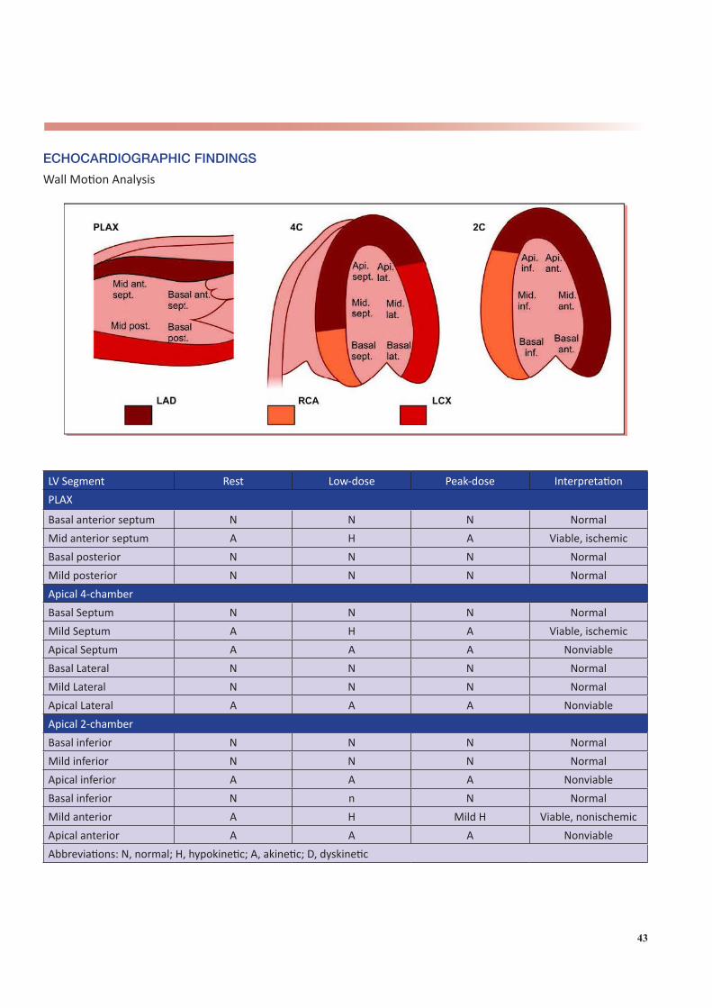
43
ECHOCARDIOGRAPHIC FINDINGS
Wall Motion Analysis
LV Segment Rest Low-dose Peak-dose InterpretationPLAX
Basal anterior septum N N N NormalMid anterior septum A H A Viable, ischemicBasal posterior N N N NormalMild posterior N N N NormalApical 4-chamberBasal Septum N N N NormalMild Septum A H A Viable, ischemicApical Septum A A A NonviableBasal Lateral N N N NormalMild Lateral N N N NormalApical Lateral A A A NonviableApical 2-chamberBasal inferior N N N NormalMild inferior N N N NormalApical inferior A A A NonviableBasal inferior N n N NormalMild anterior A H Mild H Viable, nonischemicApical anterior A A A NonviableAbbreviations: N, normal; H, hypokinetic; A, akinetic; D, dyskinetic

44
Other findings:
Parameter Rest Immediate Post-exerciseLV end-diastolic volume (ml)LV end-systolic volume (ml)LV ejection fraction (%)Mitral inflow E/A vel (cm/s)Mitral annular E’ vel (cm/s)Estimated LVEDP (mmHg)Mitral regurgitationTricuspid regurgitationEstimated RVSP (mmHg)
SUMMARY AND INTERPRETATION
1. No chest pain or ECG changes during the stress test.2. Normal hemodynamics.3. Akinetic apex at baseline, low-dose and peak-dose dobutamine.4. Akinetic mid anterior septum and mid inferoseptum that improve at low-dose but worsen at peak dose dobutamine.5. Akinetic mid anterior wall but shows sustained improvement at low- and peak-dose dobutamine.
FINAL IMPRESSION
Dobutamine stress echocardiogram shows evidence of LAD territory infarct with non-viable apex and areas of viability with or without ischemia in adjacent segments.