IndependentEffects of AldosteroneandPotassium...
9
Independent Effects of Aldosterone and Potassium on Induction of Potassium Adaptation in Rat Kidney Bruce Stanton, Luying Pan, Hans Deetjen, Victoria Guckian, and Gerhard Giebisch Department ofPhysiology, Dartmouth Medical School, Hanover, New Hampshire 03756; and Department ofPhysiology, Yale University School ofMedicine, New Haven, Connecticut 06510 Abstract We examined the independent effects of a high potassium diet and increased aldosterone levels on the development of renal potassium adaptation. This condition is defined by the increased ability of the kidneys to excrete an acute infusion of potassium. Rats were adrenalectomized (ADX) and received aldosterone at basal levels (0.5 Asg/100 g * d) or at high levels (2.0 ;g/100 go d) for 10 d. In each experimental group, animals received either a control diet or a high potassium diet. In ADX animals with basal aldosterone levels, a high potassium intake increased but did not completely restore the ability to excrete potassium and in- duced proliferation of the basolateral membrane of principal cells in the collecting tubule (i.e, morphologic adaptation). In contrast, increased aldosterone did not induce functional adaptation. El- evated aldosterone and dietary potassium intake were required to produce functional potassium adaptation indistinguishable from that in potassium-loaded, adrenal-intact animals. Introduction After a chronic increase in dietary potassium intake, a sequence of adaptive changes takes place in the kidneys that lead to the more effective and enhanced excretion of potassium after an acute potassium load (1-16). This response, called potassium adaptation, is associated with a sharp elevation of plasma al- dosterone levels and with an increase in potassium secretion by the initial collecting, cortical collecting, and medullary collecting tubules (1-3, 11, 12, 17-21). A high potassium intake also stim- ulates Na,K-ATPase (3, 4, 11, 13-15, 22, 23) and induces se- lective amplification of the basolateral membrane of principal cells in the cortical, and medullary collecting tubules (1, 10, 12, 24). The results of several investigations suggest that changes in potassium intake and in aldosterone levels contribute to the de- velopment of the functional, morphologic, and enzymatic changes that characterize potassium adaptation. For example, an increase in plasma aldosterone for 10 d in adrenalectomized rats ingesting a control diet increases the ability of the kidneys Portions of this work have been published in abstract form (1986. Pro- ceedings of the International Union of Physiological Sciences. Vol. 16. 86). Address reprint requests to Dr. Stanton, Department of Physiology, Dartmouth Medical School, Hanover, NH 03756. Receivedfor publication 24 March 1986 and in revisedform 4 August 1986. to excrete an acute infusion of potassium (25) and induces am- plification of the basolateral membrane of principal cells in the initial collecting tubule (26). A chronic high potassium intake also increases Na,K-ATPase in cortical collecting tubules of ad- renalectomized adrenal hormone-deficient animals (13-15). Micropuncture (12, 27), microcatheterization (28), and micro- perfusion ( 1-3, 19-2 1, 29-31) studies in vivo and in vitro have shown that aldosterone and a high potassium diet enhance po- tassium secretion by the initial collecting, cortical collecting, and medullary collecting tubule. Despite the recognition that both a high potassium diet and an increase in aldosterone stimulate potassium secretion by the collecting tubule, the quantitative contribution of each of these factors in the expression of functional and morphologic potas- sium adaptation has not been fully resolved. The present study was conducted to examine the independent effects of a high potassium diet and elevated aldosterone on the development of functional and structural potassium adaptation. We demonstrate that a high potassium intake alone induces functional potassium adaptation, but not to the extent observed in adrenal-intact animals in which both potassium intake and aldosterone levels are high. In addition, a high potassium intake induces significant amplification of the basolateral membrane of principal cells in the collecting tubule. We observed that the effects of aldosterone on potassium excretion depend on the dietary potassium intake. When dietary intake of potassium was normal, an increase in aldosterone did not induce functional potassium adaptation. However, aldoste- rone was effective in stimulating renal potassium excretion when animals received a high potassium diet. Under these conditions aldosterone induced full development of functional and mor- phologic potassium adaptation. We conclude that renal potas- sium adaptation, similar to that observed in adrenal-intact con- trol animals, requires an increase in dietary potassium intake accompanied by appropriately elevated plasma aldosterone levels. Methods Pretreatment of animals. Male Sprague-Dawley rats (Charles River Breedings Laboratories, Inc., Boston, MA), weighing 181-193 g, were divided into six groups (Table I). Animals were either sham-adrenalec- tomized and served as controls (groups 1 and 2) or were adrenalectomized (groups 3-6), under Nembutal anesthesia (35 mg/kg body wt) 10 d before renal function or ultrastructural studies were performed. At the time of adrenalectomy an osmotic minipump (model 2002 Alza Corp., Palo Alto, CA) was inserted subcutaneously in the neck of each animal and served to infuse aldosterone and dexamethasone as described below and in Table I. Group 1: control, control diet (Prolab rat diet, Agway, Syracuse, NY, potassium 1.5% and sodium 0.58%). Group 2: control, high KC1 diet (10 g KCI added/ 100 g food). 198 B. Stanton, L. Pan, H. Deetjen, V. Guckian, and G. Giebisch J. Clin. Invest. © The American Society for Clinical Investigation, Inc. 0021-9738/87/01/0198/09 $ 1.00 Volume 79, January 1987, 198-206
Transcript of IndependentEffects of AldosteroneandPotassium...
Independent Effects of Aldosterone and Potassiumon Induction of Potassium Adaptation in Rat KidneyBruce Stanton, Luying Pan, Hans Deetjen, Victoria Guckian, and Gerhard GiebischDepartment of Physiology, Dartmouth Medical School, Hanover, NewHampshire 03756;and Department of Physiology, Yale University School of Medicine, NewHaven, Connecticut 06510
Abstract
Weexamined the independent effects of a high potassium dietand increased aldosterone levels on the development of renalpotassium adaptation. This condition is defined by the increasedability of the kidneys to excrete an acute infusion of potassium.Rats were adrenalectomized (ADX) and received aldosterone atbasal levels (0.5 Asg/100 g * d) or at high levels (2.0 ;g/100 go d)for 10 d. In each experimental group, animals received either acontrol diet or a high potassium diet. In ADXanimals with basalaldosterone levels, a high potassium intake increased but didnot completely restore the ability to excrete potassium and in-duced proliferation of the basolateral membrane of principal cellsin the collecting tubule (i.e, morphologic adaptation). In contrast,increased aldosterone did not induce functional adaptation. El-evated aldosterone and dietary potassium intake were requiredto produce functional potassium adaptation indistinguishablefrom that in potassium-loaded, adrenal-intact animals.
Introduction
After a chronic increase in dietary potassium intake, a sequenceof adaptive changes takes place in the kidneys that lead to themore effective and enhanced excretion of potassium after anacute potassium load (1-16). This response, called potassiumadaptation, is associated with a sharp elevation of plasma al-dosterone levels and with an increase in potassium secretion bythe initial collecting, cortical collecting, and medullary collectingtubules (1-3, 11, 12, 17-21). A high potassium intake also stim-ulates Na,K-ATPase (3, 4, 11, 13-15, 22, 23) and induces se-lective amplification of the basolateral membrane of principalcells in the cortical, and medullary collecting tubules (1, 10,12, 24).
The results of several investigations suggest that changes inpotassium intake and in aldosterone levels contribute to the de-velopment of the functional, morphologic, and enzymaticchanges that characterize potassium adaptation. For example,an increase in plasma aldosterone for 10 d in adrenalectomizedrats ingesting a control diet increases the ability of the kidneys
Portions of this work have been published in abstract form (1986. Pro-ceedings of the International Union of Physiological Sciences. Vol. 16.86).
Address reprint requests to Dr. Stanton, Department of Physiology,Dartmouth Medical School, Hanover, NH03756.
Receivedfor publication 24 March 1986 and in revisedform 4 August1986.
to excrete an acute infusion of potassium (25) and induces am-plification of the basolateral membrane of principal cells in theinitial collecting tubule (26). A chronic high potassium intakealso increases Na,K-ATPase in cortical collecting tubules of ad-renalectomized adrenal hormone-deficient animals (13-15).Micropuncture (12, 27), microcatheterization (28), and micro-perfusion ( 1-3, 19-2 1, 29-31) studies in vivo and in vitro haveshown that aldosterone and a high potassium diet enhance po-tassium secretion by the initial collecting, cortical collecting,and medullary collecting tubule.
Despite the recognition that both a high potassium diet andan increase in aldosterone stimulate potassium secretion by thecollecting tubule, the quantitative contribution of each of thesefactors in the expression of functional and morphologic potas-sium adaptation has not been fully resolved. The present studywas conducted to examine the independent effects of a highpotassium diet and elevated aldosterone on the development offunctional and structural potassium adaptation.
Wedemonstrate that a high potassium intake alone inducesfunctional potassium adaptation, but not to the extent observedin adrenal-intact animals in which both potassium intake andaldosterone levels are high. In addition, a high potassium intakeinduces significant amplification of the basolateral membraneof principal cells in the collecting tubule.
Weobserved that the effects of aldosterone on potassiumexcretion depend on the dietary potassium intake. Whendietaryintake of potassium was normal, an increase in aldosterone didnot induce functional potassium adaptation. However, aldoste-rone was effective in stimulating renal potassium excretion whenanimals received a high potassium diet. Under these conditionsaldosterone induced full development of functional and mor-phologic potassium adaptation. Weconclude that renal potas-sium adaptation, similar to that observed in adrenal-intact con-trol animals, requires an increase in dietary potassium intakeaccompanied by appropriately elevated plasma aldosteronelevels.
Methods
Pretreatment of animals. Male Sprague-Dawley rats (Charles RiverBreedings Laboratories, Inc., Boston, MA), weighing 181-193 g, weredivided into six groups (Table I). Animals were either sham-adrenalec-tomized and served as controls (groups 1 and 2) or were adrenalectomized(groups 3-6), under Nembutal anesthesia (35 mg/kg body wt) 10 d beforerenal function or ultrastructural studies were performed. At the time ofadrenalectomy an osmotic minipump (model 2002 Alza Corp., PaloAlto, CA) was inserted subcutaneously in the neck of each animal andserved to infuse aldosterone and dexamethasone as described below andin Table I.
Group 1: control, control diet (Prolab rat diet, Agway, Syracuse, NY,potassium 1.5% and sodium 0.58%).
Group 2: control, high KC1 diet (10 g KCI added/ 100 g food).
198 B. Stanton, L. Pan, H. Deetjen, V. Guckian, and G. Giebisch
J. Clin. Invest.© The American Society for Clinical Investigation, Inc.0021-9738/87/01/0198/09 $ 1.00Volume 79, January 1987, 198-206

Table I. Pretreatment ofAnimals
SurgicalGroup treatment Hormone treatment Diet
g1100g/ - d
1. Control None None Control
2. Control None None KCl
3. ADX:basal Aldo ADX Aldosterone 0.5 ControlDexamethasone 1.2
4. ADX:basal Aldo ADX Aldosterone 0.5 KC1Dexamethasone 1.2
5. ADX:high Aldo ADX Aldosterone 2.0 ControlDexamethasone 1.2
6. ADX:high Aldo ADX Aldosterone 2.0 KCIDexamethasone 1.2
Abbreviations: ADX, adrenalectomized; basal Aldo, basal aldosterone;high Aldo, high dose of aldosterone.
Group 3: adrenalectomy plus basal hormone replacement: aldosterone(0.5 Ag/l00 g body wt * d) and dexamethasone (1.2 sgl/I00 gbody wt * d),control diet.
Group 4: adrenalectomy plus basal hormone replacement: aldosterone(0.5 Mg/l00 g body wt * d) and dexamethasone (1.2 AWg/I00 g body wt * d),high KCI diet (10 g KCGadded/100 g food).
Group 5: adrenalectomy plus high aldosterone replacement (2.0 jig/100 g body wt * d) and dexamethasone (1.2 sg/I00 g body wt * d), control
diet.Group 6: adrenalectomy plus high aldosterone replacement (2.0 ,g/
100 g body wt d) and dexamethasone (1.2 Mg100 g body wt d), highKCI diet (10 g KCI added/100 g food).
Wehave shown previously that the aldosterone infusion rate of 0.51Mg/l00 g * d restored plasma levels of aldosterone to control values andthat 2.0 ug/100 g * d increased hormone levels to those measured in ad-renal-intact rats on a high potassium diet (2). The dose of dexamathasonegiven to the adrenalectomized rats was the lowest one that increasedglomerular filtration rate and plasma levels of insulin and glucose tothose measured in control rats (2).
AU animals were pair-fed; food intake varied between 15 and 18 gper day. Animals on the control diet ingested 5.7-6.8 meqof potassium/d and animals on the high potassium diet ingested 25.7-30.8 meq ofpotassium/d. Distilled water was available ad libitum.
Experimental protocol for clearance experiments. Rats were anes-thetized with Inactin (Byk Gluden, Konstanz, Germany, 100 m&/kg bodywt i.p.) and a tracheostomy tube was inserted to ipsure adequate ven-tilation. Throughout the study body temperature was maintained at 37-37.5°C. The left carotid artery and left external jugular vein were can-nulated for the withdrawal of blood samples and infusion of all testsubstances, respectively. Bladder catheterization was performed to allowcomplete collection of urine from both kidneys.
After surgery, all rats received a 2-ml bolus of Ringer's solution (inmillimolar NaCG 115, NaHCO325, KCI 5). This was followed by amaintenance infusion of Ringer's solution containing 3% polyvinylpy-rolidine (PVP)' given at a rate of 4.7 ml/h (Fig. 1). PVPwas given becauseit maintains glomerular filtration rate in adrenalectomized animals givenKCI intravenously (25). After initiation of the maintenance infusion, abolus of tritiated inulin (25 MCi, radiochemical purity 98.5%, NewEngland Nuclear, Boston, MA) was given, and inulin was added to the
1. Abbreviation used in this paper: PVP, polyvinylpyrolidine.
Ringers solution to provide 25 MCi/h. After a 45-min equilibration period,a baseline control urine collection was obtained. Thereafter, KCG wasinfused at a rate of 7 Mmol/min by substituting the Ringer's solution forone containing 90 mMKCG, 30 mMNaCG, 25 NaHCO3, and 3%PVP.After a 30-min equilibration period, two 30-min experimental urine col-lections were obtained (Fig. 1). Blood samples were obtained at the be-ginning and end of each period.
Analytical determinations. Plasma and urine sodium and potassiumconcentrations were measured with a flame photometer using an internallithium standard (Instrumentation Laboratory, Inc., Lexington, MA).Inulin concentrations in plasma and urine were measured by standardtechniques (25). Plasma aldosterone concentration was measured by ra-dioimmunoassay (Diagnostic Products Corp., Los Angeles, CA). Glo-merular filtration rate and urinary excretion rates of sodium and potas-sium were calculated by standard formula (25).
Ultrastructural studies. To examine the effects of a high potassiumdiet on the ultrastructure of the initial collecting tubule, the kidneysfrom four groups of animals were prepared for electron microscopy.Rats in groups 3 and 4, adrenalectomized animals, were studied to ex-amine the effects of a high KCI diet when aldosterone levels were constant.Groups 1 and 2, sham-adrenalectomized animals, were studied to ex-amine the combined effects of an increase in aldosterone and dietaryKCGintake on cell structure. In a previous study we examined the effectsof a high potassium diet on cell structure for 4-6 wk (1).
Kidneys were prepared for electron microscopy as described previ-ously (1, 26). Two to four initial collecting tubules, from each kidney,that were in contact with the renal capsule were selected for study. Foreach animal 13-18 cells, which were cut in cross section and had intactbasement membranes and visible tight junctions, were photographedwith an electron microscope (model lOB; Carl Zeiss, Inc., Thornwood,NY) and enlarged during printing to a magnification of X 10,000. Toeliminate possible observer bias, we coded all sections and micrographs.The code was broken only after all measurements had been made. Theresults of each animal represent a single observation. The following pa-rameters were measured in each cell by methods described in detail pre-viously (1, 26): (a) surface density, which is the ratio of membrane areato cell volume (Sv); (b) boundary length, which is the length of membrane(B); and (c) cell area (A).
Statistics. When appropriate, statistical differences between groupmeans were compared using Student's t test. Otherwise, preliminary in-spection of the data was done by a one-way analysis of variance. If therewas a significant value of Fat P < 0.05, the Least Significant Differencetest was used to identify statistical significance between means (25, 26).All data are expressed as the mean±standard error.
Results
Renal function: Ringer's period. Table II summarizes plasmaand urine electrolyte data, as well as glomerular filtration ratesduring the Ringer's solution infusion period. The most strikingobservation is that potassium excretion in animals on the controldiet, expressed as either total or fractional excretion rates, wassimilar in all experimental groups regardless of hormone levels(groups 1, 3, and 5). Thus, adrenalectomized animals given ap-propriate replacement doses of adrenal corticosteroids excreted
INTRAVENOUS 1< RINGER >1< +KCI vP oINFUSION ~+ 3% PVP + 3% PVP
Equilibration U Equilibration U2 U3
0 45 75 105 135 165TIME (minutes)
Figure 1. Protocol for clearance experiments. Details are presented inthe text.
Renal Potassium Adaptation 199
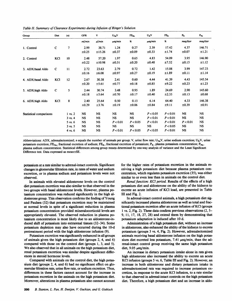
Table 11. Summary of Clearance Experiments during Infusion of Ringer's Solution
Group Diet (n) GFR V UMV FEN. UKV FEK PK PN.
mllmin/m l/min jueq/min % Aeq/min % meqiliter meqiliter
1. Control C 7 2.99 38.71 1.24 0.27 2.39 17.42 4.37 146.71±0.25 ±15.26 ±0.37 ±0.09 ±0.33 ±1.74 ±0.07 ±1.21
2. Control KC1 10 2.48 57.20 1.97 0.65 4.83 54.09 3.95 146.90±0.22 ±10.98 ±0.51 ±0.20 ±0.48 ±7.52 ±0.15 ±1.12
3. ADX:basal Aldo C 11 2.72 23.63 2.79 0.72 1.42 15.08 3.99 147.23±0.16 ±6.08 ±0.97 ±0.27 ±0.19 ±1.89 ±0.11 ±1.14
4. ADX:basal Aldo KC1 12 2.67 30.38 2.41 0.60 4.44 41.39 4.43 145.54±0.20 ±5.61 ±0.77 ±0.18 ±0.85 ±9.22 ±0.23 ±1.23
5. ADX:high Aldo C 5 2.44 30.74 3.48 0.95 1.89 24.69 2.90 145.60±0.18 ±5.64 ±0.70 ±0.17 ±0.40 ±2.33 ±0.13 ±0.68
6. ADX:highAldo KC1 8 2.40 25.64 0.50 0.15 6.14 64.40 4.33 146.38±0.29 ±3.76 ±0.19 ±0.06 ±0.84 ±9.11 ±0.39 ±0.91
Statistical comparisons I vs. 2 NS NS NS NS P< 0.05 P<0.01 NS NS3 vs. 4 NS NS NS NS P<0.01 P<0.01 NS NS5 vs. 6 NS NS P<0.01 P<0.05 P<0.01 P<0.01 P<0.01 NS3 vs. 5 NS NS NS NS NS P< 0.05 NS NS4 vs. 6 NS NS P<0.01 P<0.05 P<0.05 P<0.01 NS NS
Abbreviations: ADX, adrenalectomized; n equals the number of animals per group; V, urine flow rate; UNaV, urine sodium excretion; UKV, urinepotassium excretion; FENa, fractional excretion of sodium; FEK, fractional excretion of potassium; PK, plasma potassium concentration; PN.,plasma sodium concentration. Statistical differences among group means determined by one-way analysis of variance and the Least SignificantDifference test. Data expressed as mean±SE.
potassium at a rate similar to adrenal-intact controls. Significantchanges in glomerular filtration rate, in rates of water and sodiumexcretion, or in plasma sodium and potassium levels were notobserved.
In animals with elevated aldosterone levels on the controldiet potassium excretion was also similar to that observed in thetwo groups with basal aldosterone levels. However, plasma po-tassium concentration was reduced significantly in the high al-dosterone group. This observation confirms the finding of Youngand Paulsen (32) that potassium excretion may be maintainedat normal levels in spite of a significant reduction in plasmapotassium concentration provided mineralocorticoid levels areappropriately elevated. The observed reduction in plasma po-tassium concentration is most likely due to an aldosterone-in-duced shift of potassium into extrarenal tissues (32-34). Somepotassium depletion may also have occurred during the 10-dpretreatment period with the high aldosterone infusion (9).
PQtassium excretion was significantly enhanced in all groupsof animals given the high potassium diet (groups 2, 4, and 6)compared with those on the control diet (groups 1, 3, and 5).Wealso observed that in all animals on the high potassium diet,renal potassium excretion was similar despite significant differ-ences in steroid hormone levels.
Compared with animals on the control diet, the high potas-sium diet (groups 2, 4, and 6) had no significant effect on glo-merular filtration rate, urine flow rate, or sodium excretion. Thus,differences in these factors cannot account for the increase inpotassium excretion in the animals on the high potassium diet.Moreover, alterations in plasma potassium also cannot account
for the higher rates of potassium excretion in the animals re-ceiving a high potassium diet because plasma potassium con-centration, which regulates potassium excretion (35), was eithersimilar to or even less than in animals on the control diet.
Renal function: KCI period. Results of the effects of a highpotassium diet and aldosterone on the ability of the kidneys toexcrete an acute infusion of KC1 load, are presented in TableIII and Fig. 2.
In adrenal-intact control animals, a high potassium diet sig-nificantly increased plasma aldosterone as well as total and frac-tional potassium excretion after an acute infusion of KC1 (groups1 vs. 2, Fig. 2). These data confirm previous observations (2, 5-9, 11, 17, 18, 27, 28) and extend them by demonstrating thatpotassium adaptation is induced after 10 d.
Administration of a high potassium diet, without an increasein aldosterone, also enhanced the ability of the kidneys to excretepotassium (groups 3 vs. 4, Fig. 2). However, adrenalectomizedanimals receiving basal aldosterone infusion on the high potas-sium diet excreted less potassium, 7.41 izeq/min, than the ad-renal-intact control group receiving the same high potassiumdiet, 9.05 ,eq/min.
An increase in dietary potassium intake alone in rats givenhigh aldosterone also increased the ability to excrete an acuteKCGinfusion (groups 5 vs. 6, Table III and Fig. 2). However, anincrease in both aldosterone and dietary potassium intake inadrenalectomized rats was required to increase potassium ex-cretion, in response to the acute KCGinfusion, to a rate similarto that observed in adrenal-intact controls on the high potassiumdiet. Therefore, a high potassium diet and an increase in aldo-
200 B. Stanton, L. Pan, H. Deetjen, V. Guckian, and G. Giebisch
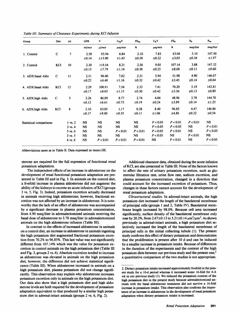
Table III. Summary of Clearance Experiments during KCl Infusion
Group Diet (n) GFR V UNbV FEN. UKV FEK PK PN.
mllmin/ 1/min peq/min % ueqimin % meq/liter meq/liter
1. Control C 7 2.58 93.94 6.84 2.10 7.83 63.04 5.10 147.50±0.14 ±13.90 ±1.43 ±0.58 ±0.22 ±3.03 ±0.34 ±1.97
2. Control KC1 10 2.30 119.54 8.21 2.56 9.05 107.14 3.88 147.32±0.15 ±7.79 ±1.14 ±0.38 ±0.25 ±8.68 ±0.11 ±0.60
3. ADX:basal Aldo C 11 2.51 96.40 7.62 2.31 5.94 51.98 4.90 146.07±0.22 ±6.40 ±1.16 ±0.32 ±0.42 ±3.45 ±0.14 ±0.64
4. ADX:basal Aldo KCI 12 2.29 100.91 7.54 2.33 7.41 70.20 5.19 142.81±0.17 ±8.03 ±1.15 ±0.30 ±0.42 ±5.56 ±0.13 ±0.89
5. ADX:high Aldo C 5 2.26 86.09 8.77 2.74 4.04 48.96 3.78 144.70±0.12 ±6.61 ±0.73 ±0.19 ±0.24 ±3.89 ±0.14 ±1.25
6. ADX:high Aldo KCI 8 2.10 63.05 1.17 0.38 8.40 96.85 4.47 146.86±0.17 ±9.00 ±0.35 ±0.11 ±1.06 ±4.86 ±0.32 ±0.54
Statisticalcomparisons I vs.2 NS NS NS NS P<0.05 P<0.01 P<0.01 NS3 vs. 4 NS NS NS NS P<0.05 P<0.05 NS P<0.015 vs. 6 NS NS P<0.01 P<0.01 P<0.05 P<0.01 NS P<0.053 vs. 5 NS NS NS NS P<0.05 NS P<0.01 NS4 vs. 6 NS P<0.01 P<0.01 P<0.01 NS P<0.01 NS P<0.05
Abbreviations same as in Table II. Data expressed as mean±SE.
sterone are required for the full expression of functional renalpotassium adaptation.
The independent effects of an increase in aldosterone on thedevelopment of renal functional potassium adaptation are pre-sented in Table III and in Fig. 3. In animals on the control diet,a fivefold increase in plasma aldosterone did not augment theability of the kidneys to excrete an acute infusion of KC1 (groups3 vs. 5, Fig. 3). Indeed, potassium excretion actually decreasedin animals receiving high aldosterone; however, fractional ex-cretion was not affected by an increase in aldosterone. It is note-worthy that the lack of an effect of aldosterone was accompaniedby a significant decrease in plasma potassium concentration,from 4.90 meq/liter in adrenalectomized animals receiving thebasal dose of aldosterone to 3.78 meq/liter in adrenalectomizedanimals on the high aldosterone infusion (Table III).
In contrast to the effects of increased aldosterone in animalson a control diet, an increase in aldosterone in animals ingestingthe high potassium diet augmented fractional potassium excre-tion from 70.2% to 96.85%. This last value was not significantlydifferent from 107.14% which was the value for potassium ex-cretion in control animals on the high potassium diet (Table IIIand Fig. 3, groups 2 vs. 6). Absolute excretion tended to increaseas aldosterone was elevated in animals on the high potassiumdiet, however, the difference did not achieve statistical signifi-cance (Table III). When aldosterone increased in animals on ahigh potassium diet, plasma potassium did not change signifi-cantly. This observation may explain why aldosterone increasespotassium excretion only in animals on the high potassium diet.Our data also show that a high potassium diet and high aldo-sterone levels are both required for the development of potassiumadaptation equivalent to that produced by a similar high potas-sium diet in adrenal-intact animals (groups 2 vs. 6, Fig. 2).
Additional clearance data, obtained during the acute infusionof KC1, are also presented in Table Ill. None of the factors knownto affect the rate of urinary potassium excretion, such as glo-merular filtration rate, urine flow rate, sodium excretion, andplasma potassium concentration, changed in a direction thatcould account for the increased excretion of potassium. Thus,changes in these factors cannot account for the development ofrenal potassium adaptation.
Ultrastructural studies. In adrenal-intact animals, the highpotassium diet increased the length of the basolateral membraneof principal cells (groups 1 and 2, Table IV). Basolateral mem-brane length increased by 98.0%. Because cell area increasedsignificantly, surface density of the basolateral membrane onlyrose by 20.2%, from 2.67±0.13 to 3.21±0.14 ,m2/Mm3. As shownpreviously in adrenal-intact animals, a high potassium diet se-lectively increased the length of the basolateral membrane ofprincipal cells in the initial collecting tubule (1). The presentstudy confirms this effect of dietary potassium and demonstratesthat the proliferation is present after 10 d and can be inducedby a smaller increase in potassium intake. Because of differencesin the duration of the experiments and the content of the highpotassium diets between our previous study and the present one,2a quantitative comparison of the two studies is not appropriate.
2. Dietary potassium intake increased approximately fivefold in the pres-ent study for a 10-d period whereas it increased some 16-fold for 4-6wk in our previous study (1). Wereduced the potassium content of thehigh potassium diet in the present study because adrenalectomized an-imals with the basal aldosterone treatment did not survive a 16-foldincrease in potassium intake. This observation also confirms the impor-tance of an increase in aldosterone in the development of renal potassiumadaptation when dietary potassium intake is increased.
Renal Potassium Adaptation 201

10 1
> 6-
ZL 4-
CONTROLCONTROL ADX AOX ADX ADXB ALDO B ALDO H ALDO H ALDO
C KCL C KCL C KCL
1 2 3 4 5 6
0- *a
I . .,,,,,,
TREATMENT ADX ADX ADX ADXB ALDO H ALDO B ALDO H ALDO
DIET C C KCL KCL
GROUP 3 5 4 6
120 -
100
-
LL
80
60
40 -
120 -
100
80
w
20: 1
TREATMENT CONTROL CONTROL ADX ADX ADX ADXB ALDO B ALDO H ALDO H ALDO
DIET C KCL C KCL C KCL
GROUP 1 2 3 4 5 6
Figure 2. Effects of an increase in dietary potassium intake on potas-sium excretion during acute KCG loading. Abbreviations: UKV, totalexcretion; FEK, fractional excretion; C, control diet; KCG, high potas-sium diet; B ALDO, basal aldosterone; H ALDO, high dose of aldoste-rone. (Empty bar) control diet; (solid bar) high K diet. Asterisks indi-cate statistical significance where * P < 0.05 and ** P < 0.01. Dataare expressed as mean±SE.
The effects of an independent increase in dietary potassiumintake on the structure of principal cells in initial collecting tu-bules are presented in Table IV and Fig. 4. Qualitatively, themost striking observation was that a high potassium intake,without an increase in plasma aldosterone levels, led to a sig-nificant increase in the length of the basolateral membrane ofprincipal cells. This observation was confirmed by our quanti-tative morphometric analysis (Table IV). Basolateral membranelength increased by 56.0% after 10 d of a high potassium diet.Because cell area also increased significantly, from 69.0±5.3 to87.3±7.9 um2 per cell, the surface density of the basolateralmembrane, which is the ratio of membrane area divided by cellvolume, increased by only 18.4% from 2.82±0.15 to 3.39±0.18tsm2 gm3. This effect of potassium was limited to the basolateralmembrane; neither luminal membrane length nor surface densityincreased significantly.
Wehave shown previously that administration of aldosteroneat a rate of 2.0 Asg/l00 g body wt * d in adrenalectomized ratsmaintained on a constant potassium intake, increased the lengthof the basolateral membrane of principal cells (26). Treatmentfor d with this dose of aldosterone increased the length of the
basolateral membrane of principal cells from 121.4±7.4 to236.1±26.6 /m per cell (P < 0.01) and increased the surface
60
40
20
0-
TREATMENT ADX ADXALDO H ALDC
DIET C C
ADX ADXD B ALDO H ALDO
KCL KCL
GROUP 3 5 4 6
Figure 3. Effects of an increase in aldosterone levels on potassium ex-cretion during acute KCI infusion. Abbreviations same as Fig. 2.(Empty bar) basal aldosterone; (hatched bar) high aldosterone.
density from 3.31±0.30 to 3.87±0.13 ,um2/,Am3 (P < 0.05). Thus,aldosterone and a high potassium diet are capable of indepen-dently inducing proliferation of the basolateral membrane ofprincipal cells.
Discussion
Many studies have shown in adrenal-intact animals that chronicingestion of a high potassium diet induces a state of potassiumadaptation, characterized by an increased ability to excrete anacute infusion of KCG (1-16) as well as by proliferation of thebasolateral membrane of principal cells in the initial collectingand medullary collecting tubule (1, 10, 24). In such animals thehigh potassium diet elicits a significant, approximate fourfoldincrease in plasma aldosterone levels (1, 2, 12). Thus, it is notpossible to evaluate the independent contribution of the increasein plasma aldosterone vs. a high potassium intake on the de-velopment of renal potassium adaptation. The present experi-ments were designed to permit such an analysis.
The main finding of our study is that an increase in dietarypotassium intake, without a change in plasma aldosterone levels,induces significant but not complete restoration of renal func-tional potassium adaptation. A high potassium diet also leadsto renal morphologic adaptation, defined as an increase in thelength of the basolateral membrane of principal cells in the initialcollecting tubule. Wealso observe that an increase in aldosterone,unaccompanied by a high potassium intake, does not enhancethe ability of the kidneys to excrete an acute potassium load.This should not be taken to mean that aldosterone plays no role
202 B. Stanton, L. Pan, H. Deetjen, V. Guckian, and G. Giebisch
c
d Em 9
6-
4.
OJ L
TREATMENT
DIET
GROUP
10 -

Figure 4. Electron micrographs of principal cells from the initial collecting tubule. (A) Adrenalectomized rats with basal. hormone levels (group 3)on a control diet and (B) adrenalectomized rats with basal hormone levels on a high potassium diet for 10 d (group 4). Bars equal 1 jum. Asterisksmark tubule lumen.
Renal Potassium Adaptation 203

Table IV. Summary of Morphometric Analysis of Principal Cells
Group Diet (n) SvBLM SVLM BBM BIM Area
Lm2A/LM3 pm2/ym3 umAM Asm2
1. Control C 5 2.67 0.44 131.5 20.6 63.3+0.13 ±0.06 ±14.2 ±1.2 ±8.5
2. Control KCl 8 3.21 0.28 260.4 21.8 102.6+0.14 ±0.04 ±23.0 ±1.4 ±6.9
3. ADX:basal Aldo C 6 2.82 0.37 150.3 19.8 69.0+0.15 ±0.03 ±12.2 ±1.4 ±5.3
4. ADX:basal Aldo KCl 8 3.39 0.36 234.5 23.6 87.3+0.18 ±0.04 ±27.6 ±3.5 ±7.9
Statistical comparisons 1 vs. 2 P<0.01 P< 0.05 P<0.01 NS P<0.013 vs. 4 P<0.01 NS P<0.01 NS P<0.01
Abbreviations: ADX, adrenalectomized; n equals number of animals per group; Sv, surface density (membrane area/cell volume); B, boundarylength (length of membrane); area, cell area; BLM, basolateral membrane; LM, luminal membrane. Statistical differences among group meansdetermined by one-way analysis of variance and the Least Significant Difference test. Data expressed as mean±SE.
in the development of renal potassium adaptation because ourstudies show that, for the development of the full renal functionaladaptive response, an increase in potassium intake and plasmaaldosterone are required.
Induction of potassium adaptation by a high potassium dietwas not associated with any significant changes in glomerularfiltration rate, sodium excretion, or plasma potassium, all factorsknown to influence renal potassium excretion. Thus, it is likelythat a high potassium intake, either directly or indirectly, exertsan effect on tubular potassium transport in the kidney.
Micropuncture, microcatheterization, and microperfusionstudies have provided extensive evidence that the increase inurinary potassium excretion in potassium-adapted animals isthe result of increased secretion of potassium by the late distaltubule (initial collecting tubule) and the cortical and medullarycollecting duct (1-3, 12, 17-21, 27, 28). The increase in potas-sium secretion by these segments in potassium-adapted animalsis also associated with proliferation of the basolateral membraneof principal cells and with an increase in the activity ofNa,K-ATPase (1, 3, 4, 10-13, 15, 24).
Several lines of evidence suggest that an independent increasein dietary potassium intake alone, without a rise in plasma al-dosterone, also leads to enhanced potassium secretion by distalnephron segments and thus may account for the increased abilityof the kidney to excrete an acute KCI load. First, Wingo et al.(29) reported that an increase in the potassium content of thediet stimulated potassium secretion in isolated and perfusedcortical collecting tubules from adrenalectomized rabbits. Elec-trophysiological observations in the isolated perfused corticalcollecting tubule of the adrenalectomized rabbit also confirmthat a high potassium intake stimulates potassium secretion bythe cortical collecting tubule (30). Second, Silva and co-workers(13) observed that a high potassium diet increased the activityof Na,K-ATPase in cortical tissue homogenates from adrenal-ectomized animals that were given a fixed dose of mineralocor-ticoids. More recently, Garg and Narang (14) and Chekal et al.( 15) localized this effect of a high potassium diet to the corticalcollecting tubule. Thus, a high potassium intake either directly,
or through a secondary and as yet undefined mechanism, en-hances the activity of the sodium-potassium pump which pro-motes potassium secretion.
Weshow in the present study that a high potassium dietincreases the length of the basolateral membrane of principalcells in the initial collecting tubule. This proliferation of mem-brane correlates with an increased ability to excrete an acuteinfusion of KC1. Although we cannot exclude some effects ofthe high potassium diet on the proximal tubule and the loop ofHenle, taken together, these studies are consistent with the con-clusion that the cortical and medullary collecting ducts are keysites where an increase in dietary potassium intake stimulatesfunctional and structural renal potassium adaptation.
In contrast to the stimulatory effect of potassium on theability of the kidneys to excrete an exogenous potassium load,an independent increase in aldosterone, when dietary potassiumintake was normal, did not lead to functional renal potassiumadaptation. To the contrary, the urinary excretion rate of po-tassium was even less in animals with elevated aldosterone levelsthan in animals with basal aldosterone levels. This finding isconsistent with the observations in dogs by Berliner et al. (9),who found that, as long as dietary potassium intake was heldconstant, an increase in mineralocorticoids did not induce ad-aptation. Weoffer some possible explanations for this obser-vation. It is likely that the high dose of aldosterone, inappropriatefor a normal potassium diet, produces potassium depletion (9,32). As a consequence, kaliuresis after an acute infusion of KCImay be attenuated in that the infused KC1 would be retainedwithin body cells until potassium balance is restored (17, 18). Itis also known that an increase in aldosterone shifts potassiuminto cells (32-34, 36). Accordingly, the infused KC1 may movepreferentially into the intracellular compartment.
The change in plasma potassium concentration after chronicaldosterone administration may also play a decisive role inmodulating potassium excretion during an acute infusion of KC.Our results show that raising aldosterone levels from basal tohigh levels significantly lowers plasma potassium levels in ani-mals on a control diet (from 3.99 to 2.90 meq/liter, Table II).
204 B. Stanton, L. Pan, H. Deetjen, V. Guckian, and G. Giebisch
This might explain why potassium excretion failed to rise duringthe acute KCl infusion in the group with increased aldosteronelevels on the control diet inasmuch as Young et al. (32) havealso shown that urinary potassium excretion is relatively insen-sitive to alterations in plasma potassium when plasma potassiumconcentration is <4 meq/liter. Consistent with this is the obser-vation that, after a rise in aldosterone in animals on the highpotassium diet with a plasma potassium of 4.33 meq/liter, uri-nary potassium excretion was significantly higher compared withanimals having basal aldosterone levels who had plasma potas-sium levels of 4.43 meq/liter.
The modifying role of plasma potassium in determining therenal effects of aldosterone is also demonstrated by the apparentdissociation of the functional and morphologic effects of aldo-sterone. Whereas high aldosterone induces significant amplifi-cation of the basolateral membrane of principal cells, it doesnot, as we now show, increase the ability to excrete an acuteinfusion of KC1. As discussed above, the fall in plasma potassiumlevels after administration of the high aldosterone treatment in-hibits the development of functional potassium adaptation.
Our results suggest that during periods of high potassiumintake, an increase in aldosterone is required for the full devel-opment of functional renal potassium adaptation. This confirmsthe conclusions of Adam and Dawborn (6) and Thatcher andRadike (7) and others (1-16) that aldosterone is important inthe development of potassium adaptation.
Our observation that increased dietary intake of potassiumleads to a proliferation of the basolateral membrane of principalcells in the initial collecting tubule provides structural evidencethat a high potassium diet induces adaptation. Hirsch et al. (12)have also shown that a high potassium diet enhances the surfacedensity of the basolateral membrane of principal cells in theinitial collecting tubule.
Our studies also demonstrate that there is a correlation be-tween the length of the basolateral membrane of principal cellsand the ability to excrete potassium in the urine (Fig. 5). How-ever, two important exceptions are noted. First, as demonstratedin the present study, aldosterone increases membrane length inanimals on a control diet but potassium excretion does not in-crease during infusion of KCI compared with animals havingbasal aldosterone levels. A second exception occurs during therenal response to acute infusion of KCI in adrenalectomized
150-
125 -
I 100-7
I.,75 -
XA 50 -
25 -
,\ _
R2 = 0.86Y = -5.53 + 0.42X 0
00
5o i, .~ . .6 . .6 .
'3o50 100150 200250300B BLM (~m/CELL)
Figure 5. Relationship between fractional potassium excretion (FEK)and the length of the basolateral membrane of principal cells (BBL8) inthe initial collecting tubule of rat. As discussed in the text there is agood correlation between the surface density of the basolateral mem-brane of principal cells and the ability to excrete an acute infusion ofKCL. The effects of an acute infusion of KCI on the ratio of membranearea to cell volume is not plotted. The values represented by the opencircles are from the present study; values represented by the solid cir-cles from Stanton et al. (26). Regression line calculated by leastsquares analysis.
animals with basal aldosterone levels. Acute infusion of KCldoes not increase membrane length, at least in the first 6 h (un-published observations of J. Wade, M. Field, B. Stanton, andG. Giebisch). In this situation potassium excretion increasedduring infusion of KCI in the absence of membrane proliferation.However, this increase in excretion is less than that producedby a similar KCI infusion in animals chronically ingesting thehigh potassium diet (2, 35). This observation underscores theimportance of the structural change in the collecting tubule inthe development of potassium adaptation.
In summary, we have shown that an increase in dietary po-tassium intake, independent of a change in mineralocorticoidlevels, elicits functional and structural renal potassium adapta-tion. However, an increase in both aldosterone and dietary po-tassium intake is required for the full development of functionalrenal potassium adaptation.
Acknowledaments
Wethank Drs. David Young and Bruce Koeppen for helpful discussionsand for a critical review of the manuscript.
This study was supported by the Hitchcock Foundation, Basic Re-search Support Grant funds from Dartmouth Medical School, and byNational Institutes of Health grants DK-34533 and DK-17433. B. Stantonis an Established Investigator of the American Heart Association.
References
1. Stanton, B. A., D. Biemesderfer, J. B. Wade, and G. Giebisch.1981. Structural and functional study of rat distal nephron: Effects ofpotassium adaptation and depletion. Kidney Int. 19:36-48.
2. Stanton, 3. A., and G. H. Giebisch. 1982. Potassium transport bythe renal distal tubule: effects of potassium loading. Am. J. Physiol. 243:F487-F493.
3. Fine, L. B., N. Yanagawa, R. G. Schultze, M. Tuck, and W. Trizna.1979. Functional profile of the isolated uremic nephron: potassium ad-aptation in the rabbit cortical collecting tubule. J. Clin. Invest. 64:1033-1043.
4. Doucet, A., and A. I. Katz. 1980. Renal potassium adaptation:Na-K-ATPase activity along the nephron after chronic potassium loading.Am. J. Physiol. 238:F380-F386.
5. Miller, P. D., C. Waterhouse, R. Owens, and E. Cohen. 1975. Theeffect of potassium loading on the sodium excretion and plasma reninactivity in Addisonian man. J. Clin. Invest. 56;346-353.
6. Adam, W. R., and J. K. Dawborn. 1968. Potassium tolerance inrats. Aust. J. Exp. Biol. 6:757-768.
7. Thatcher, T. S., and A. W. Radike. 1947. Tolerance to potassiumintoxication in the albino rat. Am. J. Physiol. 151:138-146.
8. Adam, W. R., G. J. Goland, and R. M. Wellard. 1984. Renalpotassium adaptation in the rat Role of glucocorticoids and aldosterone.Am. J. Physiol. 246:F300-F308.
9. Berliner, R. W., T. J. Kennedy, and J. G. Hilton. 1950. Renalmechanisms for excretion of potassium. Am. J. Physiol. 162:348-367.
10. Rastegar, A., B. Biemesderfer, M. Kashgaran, and J. Hayslett.1980. Changes in membrane surfaces of collecting duct cells in potassiumadaptation. Kidney Int. 18:293-301.
11. Silva, P., R. S. Brown, and F. H. Epstein. 1977. Adaptation topotassium. Kidney Int. 11:466-475.
12. Hirsch, D., M. Kashgarian, E. L, Boulpaep, and J. P. Hayslett.1984. Role of aldosterone in the mechanism of potassium adaptation inthe initial collecting tubule. Kidney Int. 26:798-807.
13. Silva, P., J. P. Hayslett, and F. H. Epstein. 1973. The role of Na-K-Activated adenosine triphosphatase in potassium adaptation: stimu-lation of enzyme activity by potassium loading. J. Clin. Invest. 52:2265-2271.
Renal Potassium Adaptation 205

14. Garg, L. C., and N. Narang. 1985. Renal adaptation to potassiumin the adrenalectomized rabbit. Role of distal tubular sodium-potassiumadenosine triphosphatase. J. Clin. Invest. 76:1065-1070.
15. Chekal, M., S. K. Mujais, and A. I. Katz. 1985. Time course anddose dependence of the changes in renal Na-K-ATPase during potassiumadaptation. Clin. Res. 33:479A. (Abstr.)
16. Mujais, S. K., M. A. Chekal, W. J. Jones, J. P. Hayslett, andA. I. Katz. 1985. Modulation of renal sodium-potassium-adenosine tri-phosphatase by aldosterone: effect of high physiologic levels on enzymeactivity in isolated rat and rabbit tubules. J. Clin. Invest. 76:170-176.
17. Stanton, B., and G. Giebisch. 1981. Mechanism of urinary po-tassium excretion. Miner. Electrolyte Metab. 5:100-120.
18. Wright, F. S. 1977. Sites and mechanisms of potassium transportalong the renal tubule. Kidney Int. 11:415-432.
19. Mello-Aires, M., G. Giebisch, and G. Malnic. 1973. Kinetics ofpotassium transport across single distal tubules of rat kidney. J. Physiol.(Lond.). 232:47-70.
20. Schwartz, G. J., and M. B. Burg. 1978. Mineralocorticoid effectson cation transport by cortical collecting tubules in vitro. Am. J. Physiol.235:F576-F585.
21. Wade, J. B., R. G. O'Neill, J. L. Pryor, and E. L. Boulpaep. 1979.Modulation of cell membrane area in renal cortical collecting tubulesby corticosteroid hormones. J. Cell Biol. 81:439-445.
22. LeHir, M., B. Kaissling, and U. C. Dubach. 1982. Analysis ofdistal segments in the rabbit tubules after adaptation to altered Na andK intake. II Changes in Na,K-ATPase activity. Cell Tissue Res. 224:493-504.
23. Mujais, S. K., M. A. Chekal, W. J. Jones, J. P. Hayslett, and A.Katz. 1984. Regulation of renal Na-K-ATPase in the rat: role of thenatural mineralo- and glucocorticoid hormones. J. Clin. Invest. 73:13-19.
24. Kaissling, B., and M. LeHir. 1982. Distal tubular segments ofthe rabbit kidney after adaptation to altered Naand K intake. I. Structuralchanges. Cell Tissue Res. 224:469-492.
25. Stanton, B., G. Klein-Robbenhaar, J. Wade, G. Giebisch, andR. DeFronzo. 1985. Effects of adrenalectomy and chronic adrenal cor-
ticosteroid replacement on potassium transport in rat kidney. J. Clin.,Invest. 75:1317-1326.
26. Stanton, B. A., A. Janzen, J. Wade, R. DeFronzo, and G. Giebisch.1985. Ultrastructure of rat initial collecting tubule: effect of adrenal cor-ticosteroid treatment. J. Clin. Invest. 75:1327-1334.
27. Wright, F. S., N. Strieder, N. B. Fowler, and G. Giebisch. 1971.Potassium secretion by distal tubule after potassium adaptation. Am. J.Physiol. 221:437-448.
28. Schon, D. A., K. A. Backman, and J. P. Hayslett. 1981. Role ofthe medullary collecting duct in potassium excretion in potassium adaptedanimals. Kidney Int. 20:655-662, 1981.
29. Wingo, C. S., D. W. Seldin, J. P. Kokko, and H. R. Jacobson.1982. Dietary modulation of active potassium secretion in the corticalcollecting tubule of adrenalectomized rabbits. J. Clin. Invest. 70:579-586.
30. Muto, S., S. C. Sansom, and G. Giebisch. 1986. Effects of highK diet on the transport properties of the isolated cortical collecting duct(CCD) of the adrenalectomized (ADX) rabbit. Kidney Int. 29:403A.(Abstr.)
31. Tomita, K., J. J. Pisano, and M. A. Knepper. 1985. Control ofNa and K transport in the cortical collecting duct of rat: effects of bra-dykinin, vasopressin, and deoxycorticosterone. J. Clin. Invest. 76:132-136.
32. Young, D. B., and A. W. Paulsen. 1983. Interelated effects ofaldosterone and plasma potassium concentration on potassium excretion.Am. J. Physiol. 244:F28-F34.
33. Alexander, E. A., and N. G. Levinsky. 1968. An extrarenal mech-anism of potassium adaptation. J. Clin. Invest. 47:740-748.
34. Bia, M. J., K. A. Tyler, and R. A. DeFronzo. 1982. Regulationof extrarenal potassium homeostasis by adrenal hormones in rats. Am.J. Physiol. 242:F641-F644.
35. Field, M. J., B. A. Stanton, and G. H. Giebisch. 1984. Differentialacute effects of aldosterone, dexamethasone and hyperkalemia on distaltubular potassium secretion in the rat kidney. J. Clin. Invest. 74:1792-1802.
36. Young, D. B., and T. E. Jackson. 1982. Effects of aldosteroneon potassium distribution. Am. J. Physiol. 247:R526-R530.
206 B. Stanton, L. Pan, H. Deetjen, V. Guckjan, and G. Giebisch


![Inositol 1,4,5-Triphosphate-induced Granule Secretion ...dm5migu4zj3pb.cloudfront.net/manuscripts/112000/... · am (U co Co en.01.1 1 10 [U46619] FM Figure2. Secretion in responseto](https://static.fdocuments.us/doc/165x107/5f7b9628f18fdc1987195590/inositol-145-triphosphate-induced-granule-secretion-am-u-co-co-en011-1.jpg)