Increased oxidative stress toxicity and lowered ...
43
Increased oxidative stress toxicity and lowered antioxidant defenses in temporal lobe epilepsy and mesial temporal sclerosis: associations with psychiatric comorbidities. Michael Maes a,b,c , Thitiporn Supasitthumrong a , Chusak Limotai d , Ana Paula Michelin e , Andressa Keiko Matsumoto e , Laura de Oliveira Semão e , João Victor de Lima Pedrão e , Estefânia Gastaldello Moreira e , Andre F. Carvalho f,g , Sunee Sirivichayakul h , Décio Sabbatini Barbosa e , Buranee Kanchanatawan a (a) Department of Psychiatry, Faculty of Medicine, Chulalongkorn University, Bangkok, Thailand (b) Department of Psychiatry, Medical University of Plovdiv, Plovdiv, Bulgaria (c) IMPACT Strategic Research Center, Deakin University, Geelong, Australia (d) Chulalongkorn Comprehensive Epilepsy Center of Excellence (CCEC), The Thai Red Cross Society; Division of Neurology, Department of Medicine, Faculty of Medicine, Chulalongkorn University, Bangkok, Thailand (e) Health Sciences Graduate Program, Health Sciences Center, State University of Londrina, Londrina, PR, Brazil (f) Department of Psychiatry, University of Toronto, Toronto, ON, Canada; (g) Centre for Addiction and Mental Health (CAMH), Toronto, ON, Canada; (h) Faculty of Medicine, Chulalongkorn University, Bangkok, Thailand Corresponding author: Dr. Thitiporn Supasitthumrong, M.D., and Prof. Dr. Michael Maes, M.D., Ph.D. Preprints (www.preprints.org) | NOT PEER-REVIEWED | Posted: 24 January 2020 doi:10.20944/preprints202001.0285.v1 © 2020 by the author(s). Distributed under a Creative Commons CC BY license.
Transcript of Increased oxidative stress toxicity and lowered ...

Increased oxidative stress toxicity and lowered antioxidant defenses in temporal lobe
epilepsy and mesial temporal sclerosis: associations with psychiatric comorbidities.
Michael Maesa,b,c, Thitiporn Supasitthumronga, Chusak Limotaid, Ana Paula Micheline,
Andressa Keiko Matsumotoe, Laura de Oliveira Semãoe, João Victor de Lima Pedrãoe,
Estefânia Gastaldello Moreirae, Andre F. Carvalhof,g, Sunee Sirivichayakulh, Décio Sabbatini
Barbosae, Buranee Kanchanatawana
(a) Department of Psychiatry, Faculty of Medicine, Chulalongkorn University, Bangkok,
Thailand
(b) Department of Psychiatry, Medical University of Plovdiv, Plovdiv, Bulgaria
(c) IMPACT Strategic Research Center, Deakin University, Geelong, Australia
(d) Chulalongkorn Comprehensive Epilepsy Center of Excellence (CCEC), The Thai Red Cross
Society; Division of Neurology, Department of Medicine, Faculty of Medicine, Chulalongkorn
University, Bangkok, Thailand
(e) Health Sciences Graduate Program, Health Sciences Center, State University of Londrina,
Londrina, PR, Brazil
(f) Department of Psychiatry, University of Toronto, Toronto, ON, Canada;
(g) Centre for Addiction and Mental Health (CAMH), Toronto, ON, Canada;
(h) Faculty of Medicine, Chulalongkorn University, Bangkok, Thailand
Corresponding author:
Dr. Thitiporn Supasitthumrong, M.D., and Prof. Dr. Michael Maes, M.D., Ph.D.
Preprints (www.preprints.org) | NOT PEER-REVIEWED | Posted: 24 January 2020 doi:10.20944/preprints202001.0285.v1
© 2020 by the author(s). Distributed under a Creative Commons CC BY license.

Department of Psychiatry
Faculty of Medicine
Chulalongkorn University
Bangkok
Thailand
Michael Maes: [email protected]
Thitiporn Supasitthumrong: [email protected]
Chusak Limotai: [email protected]
Ana Paula Michelin: [email protected]
Andressa Keiko Matsumoto: [email protected]
Laura de Oliveira Semeão: [email protected]
João Victor de Lima Pedrão: [email protected]
Estefania G. Moreira: [email protected]
Andre Carvalho: [email protected]
Sunee Sirivichayakul: [email protected]
Decio Barbosa: [email protected]
Buranee Kanchanatawan: [email protected]
Preprints (www.preprints.org) | NOT PEER-REVIEWED | Posted: 24 January 2020 doi:10.20944/preprints202001.0285.v1

Abstract
Oxidative stress toxicity (OSTOX), as well as lowered antioxidant defenses (ANTIOX), play a
role in temporal lobe epilepsy (TLE). Nevertheless, the associations between OSTOX/ANTIOX
and psychiatric comorbidities in TLE are largely unknown.
Thus, this study examines plasma malondialdehyde (MDA), lipid hydroperoxides (LOOH),
advanced oxidation protein products (AOPP), nitric oxide metabolites (NOx), total radical
trapping antioxidant parameter (TRAP) and sulfhydryl (-SH) groups in Depression due to TLE
(n=25); Anxiety Disorders due to TLE (n=27); Psychotic Disorder due to TLE (n=25); “pure TLE”
(n=27); and healthy controls (n=40).
TLE and mesial temporal sclerosis (MTS) were characterized by significant increases in
OSTOX (MDA, AOPP, LOOH) and lowered ANTIOX (-SH groups, TRAP). The discrimination
of pure TLE from controls yielded a significant area under the ROC curve for MDA (0.999),
AOPP (0.851), -SH groups (0.899) and the OSTOX/ANTIOX ratio (0.996). Seizure frequency is
significantly associated with increased MDA and lowered LOOH and NOx levels. Increased
MDA was associated with the severity of depressive and physiosomatic symptoms, whilst
increased AOPP levels predicted suicidal ideation. Depression and anxiety disorders co-
occurring with TLE showed significantly lower MDA levels than TLE without any
comorbidities. The psychotic and negative symptoms of TLE are associated with increased
MDA levels and excitation with increased LOOH and lowered TRAP levels.
These results indicate that oxidative stress toxicity especially protein oxidation and aldehyde
formation coupled with lowered -SH groups play a key role in the pathophysiology of
Preprints (www.preprints.org) | NOT PEER-REVIEWED | Posted: 24 January 2020 doi:10.20944/preprints202001.0285.v1

TLE/MTS. Increased aldehyde formation also impacts psychopathology, psychosis, as well as
negative and depressive symptoms.
Key words: oxidative stress, neuroimmunomodulation, major depression, inflammation,
neurotoxicity, schizophrenia
Preprints (www.preprints.org) | NOT PEER-REVIEWED | Posted: 24 January 2020 doi:10.20944/preprints202001.0285.v1

Introduction
Temporal lobe epilepsy (TLE) is the most common type of epilepsy with an incidence
rate of 10.4 per 100,000 (1945 – 1964) [1] and is characterized by recurrent focal seizures that
originate in the temporal lobes [2-4]. Mesial temporal sclerosis (MTS) or hippocampal sclerosis,
which consists of neuronal cell loss, gliosis and sclerosis in the dentate gyrus, CA1, 3 and CA4
regions, is the most common etiology that accounts for 43%-73% of TLE cases [5-7]. Moreover,
TLE is characterized by a high prevalence of comorbid neuropsychiatric syndromes (around
54.1%) with a high prevalence of depression (42.9%) and anxiety (18.4%), especially generalized
anxiety disorder (GAD), while psychosis shows a lower prevalence [8,9]. In another study [10],
the lifetime prevalence of psychiatric disorders in TLE was as high as 70.0% with mood
disorders showing a prevalence of 49.3%, anxiety disorders 42.5% and psychosis 5.5%. Those
comorbidities between TLE and psychiatric disorders have a negative impact on health-related
quality of life (HR-QoL) [11], although no significant associations were observed between those
psychiatric comorbidities and TLE features including age at onset and response to treatment
with antiepileptic drugs (AEDs) [8].
There is now evidence that increased production of reactive oxygen (ROS) and nitrogen
(RNS) species, lowered antioxidant defenses and increased oxidative stress toxicity play a role
in epilepsy and TLE [12-14]. Increased nitro-oxidative stress may be the consequence of
epileptic seizures, especially recurrent seizures, but may also contribute to epileptogenesis and
treatment resistance [12-15]. In fact, epileptogenesis is accompanied by increased ROS
production and lipid peroxidation, as well as hippocampal neurodegeneration and neuronal
network reorganization with reactive gliosis, which together increase vulnerability to new
Preprints (www.preprints.org) | NOT PEER-REVIEWED | Posted: 24 January 2020 doi:10.20944/preprints202001.0285.v1

seizures [15]. Following induction of experimental TL status epilepticus, increased production
of superoxide, nitric oxide (NO) and, consequently, peroxynitrite may contribute to apoptotic
cell death in hippocampal neuronal cells through activation of the caspase-3 signaling pathway
[16,17]. Indicants of oxidative stress toxicity in epilepsy are increased levels of
malondialdehyde (MDA) and 4-hydroxynonenal (4-HNE) (indicating lipid peroxidation with
consequent aldehyde formation), protein carbonyls (indicating protein oxidation), nitro-tyrosine
(indicating increased nitration of proteins with production of immunogenic neoantigens) and 8-
hydroxy-2-deoxyguanosine (indicating oxidative damage to DNA) [18-21]. Lowered levels of
superoxide dismutase [22], catalase and glutathione peroxidase [19], vitamin E, and sulfhydryl
(-SH) groups [23] are observed in patients with epilepsy. On the other hand, some papers did not
detect alterations in advanced oxidation protein products (AOPP) and sulfhydryl or thiol (-SH)
groups in epilepsy while NO levels were significantly lowered [21,24]. Interestingly, in a small-
n study, some of the redox variables were associated with epilepsy characteristics including
frequency of seizures, age at onset of seizures and number of drugs [21]. Nevertheless, despite
the changes in ROS/RNS and indices of oxidative stress toxicity in epilepsy, it remains largely
unknown whether comorbid psychiatric syndromes could contribute to changes in peripheral
redox parameters in patients with MTS.
Recently, it was shown that psychiatric disorders including major depression and GAD
are accompanied by increased ROS/RNS as well as oxidative stress toxicity as indicated by
increased levels of MDA and AOPP, and lowered levels of -SH groups and total radical
trapping antioxidant parameter (TRAP) [25-27]. The association between redox status and
psychosis is more complex, with no or minimal changes being observed in first-episode
Preprints (www.preprints.org) | NOT PEER-REVIEWED | Posted: 24 January 2020 doi:10.20944/preprints202001.0285.v1
psychosis [28,29] and in chronic schizophrenia [30], although deficit, but not non-deficit
schizophrenia, is accompanied by increased levels of AOPP and lipid hydroperoxides (LOOH),
but not MDA, and lowered levels of -SH groups and TRAP (Maes et al., in preparation). A recent
study showed that MDA levels were significantly higher in TLE patients with depression than
in those without [31], suggesting that this comorbidity may increase oxidative stress toxicity.
Thus, the aims of the present study were to examine a) whether TLE and MTS are
characterized by increased levels of LOOH, MDA, AOPP, and NOx (NO metabolites), and
lowered levels of TRAP and -SH groups; and b) whether these redox parameters are associated
with comorbid depressive, anxiety and psychotic symptoms. The a priori hypotheses are that
TLE and MTS and especially TLE with comorbid psychiatric disorders are accompanied by
increased MDA, AOPP, LOOH and NOx and lowered TRAP and sulfhydryl levels.
Subjects and methods
Participants
In this case-control study, we recruited 104 patients with TLE and 40 healthy controls.
The TLE outpatients were admitted to the Comprehensive Epilepsy Unit of the King
Chulalongkorn Memorial Hospital, Bangkok, Thailand from December 2013 – December 2014.
All patients were diagnosed as suffering from TLE by a senior neurologist specialized in
epilepsy based on a history of seizure clinical characteristics, EEG record and magnetic
resonance imaging (MRI). Moreover, the study group of patients with TLE was divided into 4
subgroups based on the presence of psychiatric comorbidities diagnosed using DSM-IV-TR
criteria, namely a) Mood Disorders Due to TLE with depressed features (n=25); b) Anxiety
Disorder Due to TLE with panic attacks, GAD or obsessive-compulsive symptoms (n=27); c)
Preprints (www.preprints.org) | NOT PEER-REVIEWED | Posted: 24 January 2020 doi:10.20944/preprints202001.0285.v1

Psychotic Disorder Due to TLE with delusions or hallucinations (n=25); and d) “pure TLE” when
those (or other) psychiatric comorbidities were absent (n=27).
Exclusion criteria for healthy controls were a diagnosis of epilepsy including febrile
seizures in children or any axis-1 diagnosis of psychiatric disorders and a family history of
epilepsy, mood or psychotic disorders. We excluded TLE patients when a) they presented axis
I disorders other than Mood, Anxiety or Psychotic Disorders Due to TLE; b) suffered from inter-
ictal dysphoric disorder (IDD) according to Blumer’s criteria [32], and c) suffered from a recent
seizure including aura (last week prior to the study). Additional exclusion criteria for Mood
Disorders due to TLE were: the presence of anxiety and psychosis; for Anxiety Disorders due
to TLE: the presence of mood disorders or psychosis; for Psychotic Disorder due to TLE: the
presence of mood disorders or anxiety; and for “pure TLE” patients: the presence of any
psychiatric comorbidity. Exclusion criteria for both patients and controls were: a)
neurodegenerative/neuroinflammatory disorders including multiple sclerosis, stroke, and
Parkinson’s, Huntington’s or Alzheimer’s disease; b) (auto)immune disorders including diabetes,
psoriasis, rheumatoid arthritis, inflammatory bowel disease, and chronic obstructive pulmonary
disease; c) an immune, inflammatory or allergic response three months before the study; d) a
lifetime history of treatment with immunomodulatory drugs including glucocorticoids; e) use
of therapeutic doses of antioxidants or ω3-polyunsaturated fatty acid supplements three months
before inclusion in the study; and f) pregnant or lactating women.
All participants provided written informed consent to take part in the study. Approval
for the study was obtained from the Institutional Review Board of the Faculty of Medicine,
Chulalongkorn University, Bangkok, Thailand (IRB number 305/56), which is in compliance
Preprints (www.preprints.org) | NOT PEER-REVIEWED | Posted: 24 January 2020 doi:10.20944/preprints202001.0285.v1

with the International Guideline for Human Research protection as required by the Declaration
of Helsinki, The Belmont Report, CIOMS Guideline and International Conference on
Harmonization on Good Clinical Practice (ICH-GCP).
Measurements
Semi-structured interviews were conducted by the senior neurologist and a senior
psychiatrist specialized in epilepsy comorbidities. The neurologist collected socio-demographic
data and rated epilepsy-related characteristics including age at onset of TLE, lesion location,
type of epilepsy, presence of aura and type of aura, frequency and type of seizures, family
history of epilepsy, post-ictal confusion, precipitating factors, and use of AEDs. Epilepsy
semiology was performed and the diagnosis TLE was made based on a history of partial
seizures and registration of epileptiform activity over one or both temporal regions. MRI results
were used to make the diagnosis of TLE with MTS, other/undefined TLE, and TLE with
tumoral origin and the radiologist and senior neurologist verified the diagnosis of MTS. The
senior psychiatrist assessed patients and controls for depression, anxiety and psychotic
symptoms using DSM-IV-TR criteria of a) Mood Disorders Due to TLE with depressed features,
which included patients with ictus-related depression and major or minor depression who were
in remission, partial remission or acute episode. b) Anxiety Disorder Due to TLE with panic
attacks, generalized anxiety or obsessive-compulsive symptoms, which includes ictus-related
anxiety (fear, horror, déjà-vu, and panic). c) Psychotic Disorder Due to TLE with delusions (being
possessed, persecutory, ideas of reference, paranoid) or hallucinations (visual, auditory, tactile,
gustatory, and olfactory) with or without severe disorganized behaviors. Patients allocated to
this diagnosis also may show ictus-related psychoses as defined by Kanchanatawan et al. [33]
Preprints (www.preprints.org) | NOT PEER-REVIEWED | Posted: 24 January 2020 doi:10.20944/preprints202001.0285.v1
including pre-ictal psychosis, post-ictal psychosis, peri-ictal psychoses, psychotic aura, ictal
psychosis, and inter-ictal psychosis or schizophrenia-like psychosis. It should be underscored
that the following symptoms were not regarded as psychosis: horror, fear, deja-vu, deja-vecu,
going mad, forced thinking, autoscopic phenomena, and out-of-body experiences. In addition,
the senior psychiatrist scored the Hamilton Depression (HAM-D) and Anxiety (HAM-A) rating
scale and the Brief Psychiatric Rating Scale (BPRS) [34-36] in patients and controls. Smoking
behavior was assessed using the Fagerstrom rating scale [37]. Body mass index was computed
as body weight (in kg) divided by length (meter)2.
Assays
Blood for the assay of the nitro-oxidative stress biomarkers was sampled at 8.00 a.m.
after an overnight fast. Serum was aliquoted and stored at −80 °C until thawed for assay. The
biomarkers measured include MDA, AOPP, LOOH, NOx, TRAP, -SH groups. The methods
were described previously [26,27]. “MDA levels were measured through complexation with two
molecules of thiobarbituric acid using MDA estimation through high-performance liquid
chromatography (HPLC Alliance e2695, Waters’, Barueri, SP, Brasil) [38]. Experimental
conditions included the use of a column Eclipse XDB-C18 (Agilent, USA); mobile phase
consisting of 65% potassium phosphate buffer (50 mM pH 7.0) and 35% HPLC grade methanol;
flow rate of 1.0 mL/minute; temperature of 30 øC; wavelength of 532 nm. MDA concentration
in the samples was quantified based on a calibration curve and are expressed in mmol of
MDA/mg proteins.” AOPP was quantified in a microplate reader (EnSpire, Perkin Elmer, USA)
at a wavelength of 340 nm [39,40] and is expressed in mM of equivalent chloramine T. LOOH
Preprints (www.preprints.org) | NOT PEER-REVIEWED | Posted: 24 January 2020 doi:10.20944/preprints202001.0285.v1

was quantified by chemiluminescence in a Glomax Luminometer (TD 20/20), in the dark, at 30
°C for 60 min [41,42] and the results are expressed in relative light units (RLU). NOx was
assessed in a microplate reader (EnSpire®, Perkin Elmer, USA) at a wavelength of 540 nm by
measuring the concentration of nitrite and nitrate [43] and results are expressed as μM. TRAP
was evaluated in a microplate reader (Victor X-3, Perkin Elmer, USA) and results are expressed
in µM Trolox [44]. -SH groups were evaluated in a microplate reader (EnSpire®, Perkin Elmer,
USA) at a wavelength of 412 nm and results are expressed in μM”. [45,46]
Statistics
Analysis of variance was employed to check differences in continuous variables among
scale variables while analysis of contingency tables (χ2-tests) was employed to check
relationships between nominal variables. Binary regression analysis (automatic, step-up) was
used to assess the best biomarker prediction of TLE (or subgroups) as dependent variables and
controls as a reference group. Multivariate general linear model (GLM) analysis was used to
delineate the associations between biomarkers and diagnosis while adjusting for possible
intervening variables including age, sex, BMI, smoking and the drug state. Tests for between-
subject effects were used to delineate the relationships between diagnosis and each of the
biomarkers and, subsequently, we computed model-generated estimated marginal mean (SE)
values and carried out protected pair-wise comparisons among group means. We employed p-
corrections for false discovery rate (FDR) to adjust for multiple statistical tests [47]. Multiple
regression analysis (automatic, stepwise) was employed to delineate the significant biomarkers
which are associated with the BPRS, HAM-D, HAM-A and MMSE scores. All regression
Preprints (www.preprints.org) | NOT PEER-REVIEWED | Posted: 24 January 2020 doi:10.20944/preprints202001.0285.v1

analyses were checked for multicollinearity using VIF and tolerance values. All results are
additionally bootstrapped using 5000 samples and the bootstrapped results are shown in case
of discrepant results. All tests were two-tailed and a p-value of 0.05 was used for statistical
significance. IBM SPSS25 for windows was used to analyze the data.
Using the scores of the BPRS, HAM-D, and HAM-A we have computed different
symptom domain scores: psychosis was computed as the sum of 4 BPRS items, namely item 4
(conceptual disorganization), item 11 (suspiciousness), item 12 (hallucinations) and item 15
(unusual thought content); excitation was computed as sum of 2 BPRS items, namely item 8
(grandiosity) + item 17 (excitation), and negative symptoms as the sum of 2 BPRS items, namely
item 3 (emotional withdrawal) + item 16 (blunted affect). The HAM-D physiosomatic subdomain
score was computed as the sum of 5 HAM-D items, namely item 11 (somatic anxiety), item 12
(somatic symptoms, gastro-intestinal), item 13 (somatic symptoms general), item 14 (genital
symptoms) and item 15 (hypochondriasis). We used item 3 of the HAM-D to assess suicidal
ideation. An index of general psychopathology was assessed as the sum of the z values of the
BPRS (z BPRS), + z HAM-D + z HAM-A.
Results.
Demographic and clinical data
Table 1 displays the demographic data of the patients and controls who participated in
the present study. There were no significant differences in age, BMI, marital status, and
smoking among the 5 diagnostic classes. There were somewhat more women in the TLE study
groups with depression and anxiety as compared with the TLE group with psychotic features.
Preprints (www.preprints.org) | NOT PEER-REVIEWED | Posted: 24 January 2020 doi:10.20944/preprints202001.0285.v1

Patients with TLE were somewhat less well educated than healthy controls. Therefore, we have
statistically adjusted the results of multiple regression analyses for the putative effects of
education (e.g. the MMSE and neurocognitive test results). This table also shows that there are
no significant differences among the 4 TLE subgroups in the frequency of seizures, the age of
onset of epilepsy, a history of aura and status epilepticus. Definite MTS was diagnosed in 55
of the TLE patients.
The HAM-D score was significantly higher in patients with TLE with depression than
in the other 4 diagnostic categories. Table 1 also shows that the HAM-A and BPRS scores were
significantly different between the 5 diagnostic categories with the lowest levels in healthy
controls and the highest values in TLE + anxiety and TLE + psychosis diagnostic categories,
respectively.
Associations between ONS biomarkers and TLE with and without comorbidities
Table 2 shows the results of multivariate GLM analysis, which examined the
associations between biomarkers and diagnosis while adjusting for sex, age, BMI and smoking.
We found a highly significant association between diagnosis and the biomarkers with an effect
size of 0.321 and there were also significant effects of sex and BMI but not age or smoking.
Tests for-between-subject effects showed significant associations between diagnosis and
TRAP, -SH groups, MDA and AOPP with a very strong association between MDA and
diagnosis (effect size of 0.689). The second GLM analysis in Table 2 shows that there were
strong associations between diagnosis and the OSTOX, ANTIOX and OSTOX/ANTIOX
indices. Table 3 shows that TRAP, -SH groups and the ANTIOX index were significantly lower
in the 4 TLE groups than in controls, whereas MDA, AOPP, OSTOX, and OSTOX/ANTIOX
Preprints (www.preprints.org) | NOT PEER-REVIEWED | Posted: 24 January 2020 doi:10.20944/preprints202001.0285.v1

index were significantly increased in all 4 TLE groups as compared with controls. Moreover,
MDA was higher in “pure TLE” than in Mood Disorder due to TLE with depressive features or
Anxiety Disorder due to TLE; and significantly higher in Psychotic Disorder due to TLE than
in Anxiety Disorder due to TLE. LOOH was significantly higher in “pure TLE”, Psychotic
Disorder due to TLE and Mood Disorder due to TLE than in normal controls. The OSTOX and
OSTOX/ANTIOX indices were significantly higher in all TLE subgroups than in controls and
higher in “pure TLE” than in Anxiety Disorder due to TLE.
Figure 1 shows the 6 O&NS biomarkers as well as the three indices in healthy controls
versus TLE patients. TRAP (F=31.97, p<0.001, effect size: 0.189), -SH groups (F=47.13, p<0.001,
effect size: 0.256), and the ANTIOX index (F=78.43, p<0.001, effect size: 0.364), were
significantly lowered in TLE as compared with controls, while MDA (F=233.31, p<0.001, effect
size: 0.630), AOPP (F=51.06, p<0.001, effect size: 0.271), LOOH (F=8.70, p=0.004, effect size:
0.060), and the OSOX (F=160.50, p<0.001, effect size: 0.540), and OSTOX/ANTIOX (F=214.15,
p<0.001, effect size: 0.610) indices were significantly higher in TLE than in controls (all results
of GLM analyses with age, sex, BMI and TUD as covariates). Moreover, MTS and pure TLE
with MTS showed the same pattern of disorders in the biomarkers. ROC analysis showed a
highly significant separation of pure TLE versus controls for MDA (AUC ROC=0.999,
SE=0.002, p<0.001), AOPP (AUC ROC=0.851, SE=0.046, p<0.001), -SH groups (AUC
ROC=0.899, SE=0.039, p<0.001), TRAP (AUC ROC=0.743, SE=0.063, p=0.001), OSTOX (AUC
ROC=0.981, SE=0.013, p<0.001), ANTIOX (AUC ROC=0.920, SE=0.031, p<0.001),
OSTOX/ANTIOX (AUC ROC=0.996, SE=0.004, p<0.001) whereas LOOH was less significant
(AUC ROC=0.665, SE=0.067, p=0.024).
Preprints (www.preprints.org) | NOT PEER-REVIEWED | Posted: 24 January 2020 doi:10.20944/preprints202001.0285.v1

Effects of putative confounding variables.
As shown in Table 2 there were significant effects of sex and BMI on the biomarkers.
Tests for between-subject effects showed significant effects of sex on TRAP (F=12.92, df=1/134,
p<0.001), and LOOH (F=6.85, df=1/134, p=0.010) with higher TRAP and LOOH values in men
than in women. Analysis of parameter estimates showed that BMI was associated with AOPP
only (t=+4.47, p<0.001). The effects of antiepileptic drugs (AEDs) and other treatments were
examined using the GLM analyses shown in Table 2 which considered the effects of phenytoin
(n=38), valproate (n=34), phenobarbital (n=26), carbamazepine (n=61), lamotrigine (n=27),
levetiracetam (n=38), topiramate (n=12), clonazepam (n=10), gabapentin (n=8), clobazam (n=58),
antipsychotics (n=9), antidepressants (n=16), anxiolytics (n=10), CaCo3 (n=13) and folic acid
(n=27). Multivariate and univariate GLM analyses (even without p correction for FDR) showed
no significant effects of AEDs or other drugs. There were no significant associations (Spearman
rank-order correlations) between the number of AEDs the patients were taking and any of the
biomarker data even without p correction for FDR.
Best prediction of TLE and subtypes using biomarkers
Table 4 shows the results of automatic binary logistic regression analyses with TLE or
TLE subtypes as dependent variables and biomarkers as explanatory variables. MDA was the
single best biomarker predictor of TLE (χ2=148.34, df=1, p<0.001) with a Nagelkerke value of
0.935, an Odd’s ratio of 154.75 and a sensitivity of 99.1% and specificity of 94.7%. MDA
combined with the ANTIOX index were the best predictors of MTS (χ2=102.09, df=2, p<0.001;
Preprints (www.preprints.org) | NOT PEER-REVIEWED | Posted: 24 January 2020 doi:10.20944/preprints202001.0285.v1

Nagelkerke = 0.852, sensitivity of 98.5% and specificity of 97.1%) and “pure TLE” (without
comorbidities) + MTS (χ2=48.33, df=2, p<0.001; Nagelkerke = 0.835, sensitivity of 93.8% and
specificity of 97.4%). A history of post-ictal confusion was also associated with MDA and the
ANTIOX index (χ2=45.51, df=1, p<0.001; Nagelkerke = 0.366, sensitivity of 71.4% and
specificity of 69.7%). There was a significant albeit weak association between TRAP levels and
a history of status epilepticus with a Nagelkerke value of 0.068 (χ2=6.00, df=1, p<0.014). MDA
was also significantly associated with a history of aura (χ2=56.57, df=1, p<0.001; Nagelkerke =
0.435, sensitivity of 76.3% and specificity of 70.3%).
Biomarker predictors of seizure frequency and psychiatric rating scale scores.
In order to examine the associations between biomarkers and seizure frequency, we
have performed automatic multiple regression analyses with seizure frequency as dependent
variable and biomarkers are explanatory variables while allowing for the effects of age and sex
(Table 5). We found that 30.0% of the variance in seizure frequency was explained by MDA
(positively) and TRAP, NOx and age (inversely). In the restricted study sample of TLE patients,
we found that 18.2% of the variance in TLE seizure frequency was explained by age at onset,
LOOH and NOx (all inversely). We found that 28.8% of the variance in the BPRS score and
10.6% in psychosis was explained by MDA (positively) and education (negatively). 9.1% of the
variance in excitation was explained by LOOH (positively) and TRAP (negatively). A large part
of the variance in negative symptoms (30.1%) was predicted by MDA (positively) and AOPP
(inversely), male sex, and lower education and age. MDA was also the best predictor of the
HAM-D score together with education (inversely associated) and female sex. Furthermore,
Preprints (www.preprints.org) | NOT PEER-REVIEWED | Posted: 24 January 2020 doi:10.20944/preprints202001.0285.v1

MDA was the single best predictor of physiosomatic symptoms explaining 14.0% of the
variance in this symptom domain. Suicidal ideation was positively associated with AOPP
levels, while the HAM-A score was predicted by MDA (positively) and TRAP (negatively)
which together explained 14.6% of the variance in anxiety levels. Elevated MDA levels and
lowered education together explained 14.6% of the variance in the psychopathology index.
Prediction of MDA and AOPP levels
Table 6 shows the results of automatic regression analyses with MDA and AOPP as
dependent variables. We found that 34.5% of the variance in MDA could be explained by LOOH
(positively) and -SH groups, TRAP, and NOx (all negatively), while 9.1% of the variance in
AOPP is explained by lowered -SH groups.
Discussion
The first major finding of this study is that TLE (with or without comorbidities) and
MTS are characterized by increased oxidative stress toxicity as assessed with MDA, LOOH
and AOPP and lowered antioxidant defenses as assessed with TRAP and -SH groups. The
discrimination of TLE (without comorbidities) from controls is highly significant with an AUC
ROC curve for MDA of 0.999 with a sensitivity of 99.1% and specificity of 94.7% for TLE. In
fact, TLE is accompanied by a huge increase in MDA of 260% and a difference of 2.2 SDs in
MDA levels between TLE and controls. Moreover, also the AUC ROC curves for AOPP (AUC
ROC=0.851), -SH groups (AUC ROC=0.899) and the oxidative stress toxicity / antioxidant ratio
(AUC ROC=0.996) were all highly significant. These results indicate that TLE and MTS are
Preprints (www.preprints.org) | NOT PEER-REVIEWED | Posted: 24 January 2020 doi:10.20944/preprints202001.0285.v1

characterized by highly specific peripheral changes in aldehyde formation (increased MDA
levels) as well as protein oxidation (increased AOPP) and lowered antioxidant defenses
(especially reduced sulfhydryl groups). As such, MDA alone or the oxidative stress toxicity /
antioxidant ratio may be used as external validating criterion for the diagnosis of TLE and MTS
versus healthy controls. These findings extend those of a previous paper reporting increased
levels of MDA in patients with TLE and MTS [21]. Animal models of TLE also show indicants
of lipid peroxidation and aldehyde formation and lowered levels of antioxidants including
vitamin E, although other antioxidants may be increased including GSH, SOD, and catalase
[48-54]. In patients with epilepsy, increased levels of MDA and 4-HNE (both indicating
increased lipid peroxidation and aldehyde formation), protein oxidation (as assessed with
protein carbonyls) and lowered antioxidant defenses including sulfhydryl groups, superoxide
dismutase, catalase, GSH and vitamin E and C were frequently observed [21,18-20,22,23,55].
Only a few papers examined AOPP levels in epilepsy and reported negative findings in drug-
resistant partial complex seizures and idiopathic epilepsy syndrome [21,56]. There are also some
negative findings on sulfhydryl groups in epilepsy [57]. We could not find any changes in NOx
levels between TLE patients and controls, findings that are in agreement with the negative
report by [58] while other authors reported lower NO levels in epilepsy [21].
It should be stressed that our results were controlled for possible effects of background
variables including age, sex, smoking, and BMI, which all may affect oxidative and antioxidant
biomarkers [26,27]. Some studies reported significant effects of AEDs on MDA, NO levels and
-SH groups [59-65]. Therefore, we controlled our results for possible effects of AEDs. No
significant effects of AEDs on the biomarkers were found while there were no associations
between the number of AEDs taking by the patients and the biomarkers. These results extend
Preprints (www.preprints.org) | NOT PEER-REVIEWED | Posted: 24 January 2020 doi:10.20944/preprints202001.0285.v1

the findings of [58] who reported no significant differences in MDA, protein carbonyls and NO
levels between subjects on AED-monotherapy and polytherapy. Menon et al. [58] also reported
higher levels of MDA and protein carbonyls (and no changes in NO) in untreated patients with
epilepsy than in controls, suggesting that the increased oxidative stress toxicity is not induced
by AEDs. Moreover, there were no significant differences in MDA, protein carbonyls and NO
levels between both AED-treated and untreated patient groups [58], suggesting that AEDs do
not affect these three biomarkers. Finally, as in our study, Menon et al. [58] were unable to find
any effects of individual AEDs namely carbamazepine, valproate and phenytoin on the
biomarkers. Other studies were also unable to find differences in oxidative biomarkers
including carbonyls, lipid peroxidation, and antioxidant enzymes between treated and untreated
patients [62,66].
The second major finding of this study is that not only TLE and MTS are characterized
by highly increased oxidative biomarkers, but also that some features of TLE are associated
with those biomarkers. Thus, seizure frequency was significantly associated with increased
MDA but lowered LOOH, indicating that seizure frequency increases with aldehyde formation
but not lipid peroxidation per se. Animal models of epilepsy show that reducing ROS
production through the administration of corilagin is associated with lowered seizure frequency
[67]. Moreover, activation of Nrf2 following administration of RTA 408 attenuates ROS
production in association with a reduction in late spontaneous seizures [68] while sub-acute
treatment with a cannabinoid agonist (WIN 55,212-2) attenuates recurrent seizures while
normalizing the thiol redox state. On the other hand, the administration of vitamin E, which
reduced oxidative damage (protein carbonyl levels), had no significant effect on seizure
frequency [69]. Our results that NOx levels are inversely associated with seizure frequency
Preprints (www.preprints.org) | NOT PEER-REVIEWED | Posted: 24 January 2020 doi:10.20944/preprints202001.0285.v1
suggest a causal association with increased NO use whereby NO may be consumed by
increased nitration (e.g. the formation of 3-nitrotyrosine) and nitrosylation, which both may
induce neurodegenerative processes [25,70-72]. As such, our findings extend the results of a
previous report that in epilepsy patients, seizure frequency is significantly associated with the
expression of 3-nitrotyrosine, a consequence of enhanced nitration processes [21]. All in all, it
may appear that aldehyde formation (rather than protein oxidation or lipid peroxidation) and
increased NO consumption, through nitration/nitrosylation processes, are associated with
seizure frequency. Moreover, we also observed that increased aldehyde formation is associated
with aura, while lowered TRAP is associated with a history of status epilepticus.
The third major finding of this study is that in the study sample of patients and controls
combined, increased MDA predicts the severity of depressive and physiosomatic symptoms
whilst increased AOPP levels predict suicidal ideation. These results are in agreement with the
knowledge that depression and physiosomatic symptoms are accompanied by increased
oxidative stress including lipid peroxidation and aldehyde formation and lowered antioxidant
defenses as well [73-75]. Previously, a significant association between suicidal behaviors and
oxidative toxicity including elevated AOPP levels was reported [76]. Moreover, de Araujo Filho
et al. [31] observed that MDA levels were significantly increased in comorbid TLE patients and
depression. Nevertheless, in our study, mood disorders due to TLE with depressive symptoms
and anxiety disorders due to TLE are accompanied by somewhat lowered MDA levels, while
anxiety disorders due to TLE also show a lower lowered oxidative stress toxicity index than
patients with TLE without these comorbidities. All in all, while increased aldehyde formation
is associated with depression and anxiety disorder due to TLE, it appears that, in patients with
those comorbidities, MDA levels are somewhat lower than in pure TLE, suggesting that
Preprints (www.preprints.org) | NOT PEER-REVIEWED | Posted: 24 January 2020 doi:10.20944/preprints202001.0285.v1

oxidative stress toxicity is somewhat lower when those two comorbidities are present. These
findings contrast our a priori hypothesis that both comorbidities are accompanied by increased
oxidative stress toxicity which would reflect cumulative effects of increased levels in both TLE
and affective disorders. Moreover, comorbidities between depression and neuroinflammatory
disorders including multiple sclerosis and stroke are associated not only with increased
morbidity and mortality but also with increased inflammatory and oxidative stress biomarkers
[25,77-79]. Therefore, our findings could indicate that depression and anxiety disorder due to
TLE have a different pathophysiology than major depression and anxiety disorders such as
GAD [26,27].
The current study also shows that psychotic disorder due to TLE is associated with
significantly higher MDA levels as compared with healthy controls and anxiety disorders due
to TLE. Moreover, psychotic and negative symptoms are associated with increased MDA
levels, while excitation is associated with increased LOOH and lowered TRAP levels.
Likewise, indices of general psychopathology were strongly associated with increased MDA
levels. Previously, it was detected that first episode psychosis and chronic schizophrenia are
not associated with increased nitro-oxidative stress [28-30]. Nevertheless, previous studies
showed that hallucinations-delusions and excitation, assessed over a broader range of
schizophrenia syndromes, are associated with increased LOOH and lower TRAP/-SH groups,
while negative symptoms are associated with increased AOPP and lowered TRAP/-SH groups
(Maes et al., in preparation). Thus, the oxidative pathophysiology of psychotic disorder due to
TLE appears to be different from the oxidative stress profile observed in schizophrenia,
suggesting that psychosis and negative symptoms in both conditions are not the same
nosological entities.
Preprints (www.preprints.org) | NOT PEER-REVIEWED | Posted: 24 January 2020 doi:10.20944/preprints202001.0285.v1

In our study, increased aldehyde formation in TLE was strongly predicted by increased
LOOH, and lowered antioxidant levels (-SH groups and TRAP) and NOx. These findings reflect
that MDA formation is a consequence of lipid peroxidation and peroxyl radical propagation
and that formation of peroxynitrite may aggravate lipid peroxidation and MDA formation,
explaining the inverse association between NOx levels and MDA [26]. Moreover, lowered
antioxidant defenses increase vulnerability to oxidative stress toxicity and, therefore, the direct
toxicity exerted by MDA including signaling pathway alterations, ATP depletion,
mitochondrial dysfunctions, oxidative DNA damage and mutagenicity, disruption of cellular
homeostasis, apoptosis and cell death, immune activation, and neurodegenerative processes
[25]. The formation of AOPP may further aggravate this direct MDA-induced toxicity for
example by causing new ROS/RNS formation, conformational changes in proteins, loss of
functional activity of proteins, modulation of gene expression and intracellular signaling,
induction of apoptosis and necrosis [25]. Moreover, lowered -SH groups may indicate formation
of sulfide bonds with alterations in secondary and tertiary protein structure, which may lead to
increased susceptibility to proteolysis [26]. There is now evidence that increased oxidative stress
including in the hippocampus increases susceptibility to seizures and that peripheral activation
of immune-inflammatory responses may induce oxidative stress in the hippocampus [20]. There
is also some evidence that inflammatory responses and associated redox mechanisms may
initiate or augment seizures and play a role in disease progression [80,81]. In this respect, it is
interesting to note that the administration of pilocarpine and kainic acid, two models of TLE
[82,83] may induce increases in hippocampal thiobarbituric acid reactive substances (TBARS),
an assay used to assess MDA production [84]. Therefore, it may be posited that the highly
specific increase in peripheral MDA and AOPP formation may contribute to epileptogenesis
and MTS.
Preprints (www.preprints.org) | NOT PEER-REVIEWED | Posted: 24 January 2020 doi:10.20944/preprints202001.0285.v1

The results of this study should be discussed with regard to its limitations. First, this is
a case-control study and therefore no causal inferences can be made. Secondly, it would have
been more interesting if we also had measured myeloperoxidase and xanthine oxidase as well
as antioxidant enzymes including catalase and superoxide dismutase. Future research should
focus on possible differences or similarities between psychosis/depression/anxiety due to TLE
and the same symptom domains in schizophrenia, major depression or anxiety disorders. In
addition, future research should examine disabilities and HR-QoL in depression and anxiety
disorder due to TLE versus “pure TLE”. Indeed, based on our findings that pure TLE is
accompanied by higher MDA levels and the knowledge that MDA is associated with a lower
HR-QoL [85] one could predict (albeit counterintuitively) a worse HR-QoL in pure TLE when
no comorbidities with affective symptoms are present.
All in all, TLE and MTS are associated with increased LOOH, MDA, and AOPP levels
and lowered TRAP and -SH groups. Increased MDA coupled with lowered LOOH and NOx
levels are associated with seizure frequency, indicating a key role of aldehyde formation and
NO-related mechanisms. Increased MDA formation is also associated with the severity of
depressive and physiosomatic symptoms while increased AOPP is associated with suicidal
ideation. Psychotic and negative symptoms in TLE are associated with increased MDA levels
and excitation with increased LOOH coupled with lowered TRAP levels. Oxidative stress
toxicity and lowered antioxidant defenses play a key role in the pathophysiology of TLE and
MTS and comorbid psychopathologies.
Funding
Preprints (www.preprints.org) | NOT PEER-REVIEWED | Posted: 24 January 2020 doi:10.20944/preprints202001.0285.v1

The study was supported by the Ratchadapisek Research Funds, Faculty of Medicine,
Chulalongkorn University (Grant No. RA 57/024).
Conflict of interest
The authors have no conflict of interest with any commercial or other association in connection
with the submitted article.
Author’s contributions
All the contributing authors have participated in the manuscript. BK and MM designed the
study. BK and CL recruited patients and completed diagnostic interviews and rating scale
measurements. MM carried out the statistical analyses. All authors contributed to interpretation
of the data and writing of the manuscript. All authors approved the final version of the
manuscript.
Preprints (www.preprints.org) | NOT PEER-REVIEWED | Posted: 24 January 2020 doi:10.20944/preprints202001.0285.v1

References
1. Téllez-Zenteno JF, Hernández-Ronquillo L (2012) A review of the epidemiology of temporal
lobe epilepsy. Epilepsy Res Treat 2012:630853-630853. doi:10.1155/2012/630853
2. Hauser WA, Kurland LT (1975) The epidemiology of epilepsy in Rochester, Minnesota, 1935
through 1967. Epilepsia 16 (1):1-66. doi:10.1111/j.1528-1157.1975.tb04721.x
3. Brodie MJ, Zuberi SM, Scheffer IE, Fisher RS (2018) The 2017 ILAE classification of seizure
types and the epilepsies: what do people with epilepsy and their caregivers need to know?
Epileptic disorders : international epilepsy journal with videotape 20 (2):77-87. doi:10.1684/epd.2018.0957
4. (NINDS) NIoNDaS (2016) The Epilepsies and Seizures: Hope Through Research. U.S. National Institutes of Health (NIH). Accessed 18 January 2020
5. Tassi L, Meroni A, Deleo F, Villani F, Mai R, Russo GL, Colombo N, Avanzini G, Falcone
C, Bramerio M, Citterio A, Garbelli R, Spreafico R (2009) Temporal lobe epilepsy: neuropathological and clinical correlations in 243 surgically treated patients. Epileptic
disorders : international epilepsy journal with videotape 11 (4):281-292. doi:10.1684/epd.2009.0279
6. Blumcke I, Thom M, Wiestler OD (2002) Ammon's horn sclerosis: a maldevelopmental
disorder associated with temporal lobe epilepsy. Brain pathology (Zurich, Switzerland) 12
(2):199-211
7. Blümcke I, Thom M, Aronica E, Armstrong DD, Vinters HV, Palmini A, Jacques TS,
Avanzini G, Barkovich AJ, Battaglia G, Becker A, Cepeda C, Cendes F, Colombo N, Crino P,
Cross JH, Delalande O, Dubeau F, Duncan J, Guerrini R, Kahane P, Mathern G, Najm I, Ozkara
C, Raybaud C, Represa A, Roper SN, Salamon N, Schulze-Bonhage A, Tassi L, Vezzani A,
Spreafico R (2011) The clinicopathologic spectrum of focal cortical dysplasias: a consensus
classification proposed by an ad hoc Task Force of the ILAE Diagnostic Methods Commission. Epilepsia 52 (1):158-174. doi:10.1111/j.1528-1167.2010.02777.x
8. Bragatti JA, Torres CM, Londero RG, Martin KC, Souza AC, Hidalgo MP, Chaves ML,
Bianchin MM (2011) Prevalence of psychiatric comorbidities in temporal lobe epilepsy in a
Southern Brazilian population. Arquivos de neuro-psiquiatria 69 (2a):159-165. doi:10.1590/s0004-282x2011000200003
9. Beletsky V, Mirsattari SM (2012) Epilepsy, mental health disorder, or both? Epilepsy Res
Treat 2012:163731. doi:10.1155/2012/163731
10. de Oliveira GN, Kummer A, Salgado JV, Portela EJ, Sousa-Pereira SR, David AS, Teixeira
AL (2010) Psychiatric disorders in temporal lobe epilepsy: an overview from a tertiary service
in Brazil. Seizure 19 (8):479-484. doi:10.1016/j.seizure.2010.07.004
11. Johnson EK, Jones JE, Seidenberg M, Hermann BP (2004) The relative impact of anxiety,
depression, and clinical seizure features on health-related quality of life in epilepsy. Epilepsia
45 (5):544-550. doi:10.1111/j.0013-9580.2004.47003.x
12. Aguiar CC, Almeida AB, Araujo PV, de Abreu RN, Chaves EM, do Vale OC, Macedo DS,
Woods DJ, Fonteles MM, Vasconcelos SM (2012) Oxidative stress and epilepsy: literature
review. Oxidative medicine and cellular longevity 2012:795259. doi:10.1155/2012/795259
Preprints (www.preprints.org) | NOT PEER-REVIEWED | Posted: 24 January 2020 doi:10.20944/preprints202001.0285.v1

13. Sudha K, Rao AV, Rao A (2001) Oxidative stress and antioxidants in epilepsy. Clinica
chimica acta; international journal of clinical chemistry 303 (1-2):19-24. doi:10.1016/s0009-8981(00)00337-5
14. Waldbaum S, Patel M (2010) Mitochondria, oxidative stress, and temporal lobe epilepsy. Epilepsy research 88 (1):23-45. doi:10.1016/j.eplepsyres.2009.09.020
15. Puttachary S, Sharma S, Stark S, Thippeswamy T (2015) Seizure-induced oxidative stress in
temporal lobe epilepsy. Biomed Res Int 2015:745613-745613. doi:10.1155/2015/745613
16. Chuang YC, Chen S, Lin T-K, Liou C, Chang W, Chan SHH, Chang A (2007) Upregulation
of nitric oxide synthase II contributes to apoptotic cell death in the hippocampal CA3 subfield
via a cytochrome c/caspase-3 signaling cascade following induction of experimental temporal
lobe status epilepticus in the rat. Neuropharmacology 52:1263-1273. doi:10.1016/j.neuropharm.2007.01.010
17. Chuang Y-C, Chen S-D, Liou C-W, Lin T-K, Chang W-N, Chan SHH, Chang A (2008) Contribution of nitric oxide, superoxide anion, and peroxynitrite to activation of mitochondrial
apoptotic signaling in hippocampal CA3 subfield following experimental temporal lobe status
epilepticus. Epilepsia 50:731-746. doi:10.1111/j.1528-1167.2008.01778.x
18. Pecorelli A, Natrella F, Belmonte G, Miracco C, Cervellati F, Ciccoli L, Mariottini A,
Rocchi R, Vatti G, Bua A, Canitano R, Hayek J, Forman HJ, Valacchi G (2015) NADPH
oxidase activation and 4-hydroxy-2-nonenal/aquaporin-4 adducts as possible new players in
oxidative neuronal damage presents in drug-resistant epilepsy. Biochimica et biophysica acta
1852 (3):507-519. doi:10.1016/j.bbadis.2014.11.016
19. Lopez J, Gonzalez ME, Lorigados L, Morales L, Riveron G, Bauza JY (2007) Oxidative
stress markers in surgically treated patients with refractory epilepsy. Clinical biochemistry 40
(5-6):292-298. doi:10.1016/j.clinbiochem.2006.11.019
20. Ho YH, Lin YT, Wu CW, Chao YM, Chang AY, Chan JY (2015) Peripheral inflammation
increases seizure susceptibility via the induction of neuroinflammation and oxidative stress in
the hippocampus. Journal of biomedical science 22:46. doi:10.1186/s12929-015-0157-8
21. Lorigados Pedre L, Gallardo JM, Morales Chacón LM, Vega García A, Flores-Mendoza M,
Neri-Gómez T, Estupiñán Díaz B, Cruz-Xenes RM, Pavón Fuentes N, Orozco-Suárez S (2018) Oxidative Stress in Patients with Drug Resistant Partial Complex Seizure. Behav Sci (Basel) 8
(6):59. doi:10.3390/bs8060059
22. Ben-Menachem E, Kyllerman M, Marklund S (2000) Superoxide dismutase and glutathione
peroxidase function in progressive myoclonus epilepsies. Epilepsy research 40 (1):33-39. doi:10.1016/s0920-1211(00)00096-6
23. Menon B, Ramalingam K, Kumar RV (2014) Low plasma antioxidant status in patients with
epilepsy and the role of antiepileptic drugs on oxidative stress. Ann Indian Acad Neurol 17
(4):398-404. doi:10.4103/0972-2327.144008
24. Arhan E, Serdaroglu A, Ozturk B, Ozturk HS, Ozcelik A, Kurt N, Kutsal E, Sevinc N (2011) Effects of epilepsy and antiepileptic drugs on nitric oxide, lipid peroxidation and xanthine
oxidase system in children with idiopathic epilepsy. Seizure 20 (2):138-142. doi:https://doi.org/10.1016/j.seizure.2010.11.003
25. Maes M, Galecki P, Chang YS, Berk M (2011) A review on the oxidative and nitrosative
stress (O&NS) pathways in major depression and their possible contribution to the
Preprints (www.preprints.org) | NOT PEER-REVIEWED | Posted: 24 January 2020 doi:10.20944/preprints202001.0285.v1
(neuro)degenerative processes in that illness. Progress in neuro-psychopharmacology &
biological psychiatry 35 (3):676-692. doi:10.1016/j.pnpbp.2010.05.004
26. Maes M, Landucci Bonifacio K, Morelli NR, Vargas HO, Barbosa DS, Carvalho AF, Nunes
SOV (2019) Major Differences in Neurooxidative and Neuronitrosative Stress Pathways
Between Major Depressive Disorder and Types I and II Bipolar Disorder. Molecular
neurobiology 56 (1):141-156. doi:10.1007/s12035-018-1051-7
27. Maes M, Bonifacio KL, Morelli NR, Vargas HO, Moreira EG, St Stoyanov D, Barbosa DS,
Carvalho AF, Nunes SOV (2018) Generalized Anxiety Disorder (GAD) and Comorbid Major
Depression with GAD Are Characterized by Enhanced Nitro-oxidative Stress, Increased Lipid
Peroxidation, and Lowered Lipid-Associated Antioxidant Defenses. Neurotoxicity research 34
(3):489-510. doi:10.1007/s12640-018-9906-2
28. Boll KM, Noto C, Bonifácio KL, Bortolasci CC, Gadelha A, Bressan RA, Barbosa DS,
Maes M, Moreira EG (2017) Oxidative and nitrosative stress biomarkers in chronic
schizophrenia. Psychiatry Research 253:43-48. doi:https://doi.org/10.1016/j.psychres.2017.03.038
29. Fraguas D, Díaz-Caneja CM, Rodríguez-Quiroga A, Arango C (2017) Oxidative Stress and
Inflammation in Early Onset First Episode Psychosis: A Systematic Review and Meta-Analysis. Int J Neuropsychopharmacol 20 (6):435-444. doi:10.1093/ijnp/pyx015
30. Noto C, Ota VK, Santoro ML, Ortiz BB, Rizzo LB, Higuchi CH, Cordeiro Q, Belangero
SI, Bressan RA, Gadelha A, Maes M, Brietzke E (2015) Effects of depression on the cytokine
profile in drug naive first-episode psychosis. Schizophrenia research 164 (1-3):53-58. doi:10.1016/j.schres.2015.01.026
31. Filho G, Martins D, Lopes A, Brait B, Furlan A, Oliveira C, Marques L, Souza D, Almeida
E (2018) Oxidative stress in patients with refractory temporal lobe epilepsy and mesial temporal
sclerosis: Possible association with major depressive disorder? Epilepsy & behavior : E&B
80:191-196. doi:10.1016/j.yebeh.2017.12.025
32. Blumer D, Montouris G, Davies K (2004) The interictal dysphoric disorder: recognition,
pathogenesis, and treatment of the major psychiatric disorder of epilepsy. Epilepsy Behav 5
(6):826-840. doi:10.1016/j.yebeh.2004.08.003
33. Kanchanatawan B, Limothai C, Srikijvilaikul T, Maes M (2014) Clinical predictors of 2-year outcome of resective epilepsy surgery in adults with refractory epilepsy: a cohort study. BMJ Open 4 (4):e004852-e004852. doi:10.1136/bmjopen-2014-004852
34. Hamilton M (1959) The assessment of anxiety states by rating. The British journal of medical
psychology 32 (1):50-55. doi:10.1111/j.2044-8341.1959.tb00467.x
35. Hamilton M (1960) A rating scale for depression. Journal of neurology, neurosurgery, and
psychiatry 23:56-62. doi:10.1136/jnnp.23.1.56
36. Overall JE, Gorham DR (1962) The Brief Psychiatric Rating Scale. Psychological Reports
10 (3):799-812. doi:10.2466/pr0.1962.10.3.799
37. Heatherton TF, Kozlowski LT, Frecker RC, Fagerstrom KO (1991) The Fagerstrom Test for
Nicotine Dependence: a revision of the Fagerstrom Tolerance Questionnaire. British journal of
addiction 86 (9):1119-1127. doi:10.1111/j.1360-0443.1991.tb01879.x
38. Bastos AS, Loureiro AP, de Oliveira TF, Corbi SC, Caminaga RM, Junior CR, Orrico SR
(2012) Quantitation of malondialdehyde in gingival crevicular fluid by a high-performance
Preprints (www.preprints.org) | NOT PEER-REVIEWED | Posted: 24 January 2020 doi:10.20944/preprints202001.0285.v1

liquid chromatography-based method. Analytical biochemistry 423 (1):141-146. doi:10.1016/j.ab.2012.01.016
39. Hanasand M, Omdal R, Norheim KB, Gøransson LG, Brede C, Jonsson G (2012) Improved
detection of advanced oxidation protein products in plasma. Clinica chimica acta; international
journal of clinical chemistry 413 (9):901-906
40. Witko-Sarsat V, Friedlander M, Capeillere-Blandin C, Nguyen-Khoa T, Nguyen AT,
Zingraff J, Jungers P, Descamps-Latscha B (1996) Advanced oxidation protein products as a
novel marker of oxidative stress in uremia. Kidney international 49 (5):1304-1313. doi:10.1038/ki.1996.186
41. Gonzalez Flecha B, Llesuy S, Boveris A (1991) Hydroperoxide-initiated
chemiluminescence: an assay for oxidative stress in biopsies of heart, liver, and muscle. Free
radical biology & medicine 10 (2):93-100
42. Panis C, Herrera ACSA, Victorino VJ, Campos FC, Freitas LF, De Rossi T, Colado Simão
AN, Cecchini AL, Cecchini R (2012) Oxidative stress and hematological profiles of advanced
breast cancer patients subjected to paclitaxel or doxorubicin chemotherapy. Breast Cancer Res
Treat 133 (1):89-97
43. Navarro-Gonzálvez JA, García-Benayas C, Arenas J (1998) Semiautomated measurement of
nitrate in biological fluids. Clin Chem 44 (3):679-681
44. Repetto M, Reides C, Carretero MLG, Costa M, Griemberg G, Llesuy S (1996) Oxidative
stress in blood of HIV infected patients. Clinica chimica acta; international journal of clinical
chemistry 255 (2):107-117
45. Hu ML (1994) Measurement of protein thiol groups and glutathione in plasma. Methods
Enzymol 233:380-385
46. Taylan E, Resmi H (2010) The analytical performance of a microplatemethod for total
sulfhydryl measurement in biological samples. Turkish Journal of Biochemistry 35:275–278
47. Benjamini Y, Hochberg Y (1995) Controlling The False Discovery Rate - A Practical And
Powerful Approach To Multiple Testing. J Royal Statist Soc, Series B 57:289-300. doi:10.2307/2346101
48. Shakeel S, Rehman MU, Tabassum N, Amin U, Mir MUR (2017) Effect of Naringenin (A
naturally occurring flavanone) Against Pilocarpine-induced Status Epilepticus and Oxidative
Stress in Mice. Pharmacogn Mag 13 (Suppl 1):S154-S160. doi:10.4103/0973-1296.203977
49. Hussein AM, Ghalwash M, Magdy K, Abulseoud OA (2016) Beta Lactams Antibiotic
Ceftriaxone Modulates Seizures, Oxidative Stress and Connexin 43 Expression in
Hippocampus of Pentylenetetrazole Kindled Rats. Journal of epilepsy research 6 (1):8-15. doi:10.14581/jer.16002
50. Kiasalari Z, Khalili M, Shafiee S, Roghani M (2016) The effect of Vitamin E on learning
and memory deficits in intrahippocampal kainate-induced temporal lobe epilepsy in rats. Indian
journal of pharmacology 48 (1):11-14. doi:10.4103/0253-7613.174394
51. Khamse S, Sadr SS, Roghani M, Hasanzadeh G, Mohammadian M (2015) Rosmarinic acid
exerts a neuroprotective effect in the kainate rat model of temporal lobe epilepsy: Underlying
mechanisms. Pharmaceutical biology 53 (12):1818-1825. doi:10.3109/13880209.2015.1010738
52. Dariani S, Baluchnejadmojarad T, Roghani M (2013) Thymoquinone attenuates astrogliosis,
neurodegeneration, mossy fiber sprouting, and oxidative stress in a model of temporal lobe
Preprints (www.preprints.org) | NOT PEER-REVIEWED | Posted: 24 January 2020 doi:10.20944/preprints202001.0285.v1

epilepsy. Journal of molecular neuroscience : MN 51 (3):679-686. doi:10.1007/s12031-013-0043-3
53. Peternel S, Pilipović K, Zupan G (2009) Seizure susceptibility and the brain regional
sensitivity to oxidative stress in male and female rats in the lithium-pilocarpine model of
temporal lobe epilepsy. Progress in neuro-psychopharmacology & biological psychiatry 33:456-462. doi:10.1016/j.pnpbp.2009.01.005
54. Tejada J, Costa K, Bertti P, Garcia-Cairasco N (2012) The epilepsies - Complex challenges
needing complex solutions - Tejada et al 2012. 55. Leutner S, Eckert A, Muller WE (2001) ROS generation, lipid peroxidation and antioxidant
enzyme activities in the aging brain. Journal of neural transmission (Vienna, Austria : 1996) 108
(8-9):955-967. doi:10.1007/s007020170015
56. Grosso S, Longini M, Rodriguez A, Proietti F, Piccini B, Balestri P, Buonocore G (2011) Oxidative stress in children affected by epileptic encephalopathies. Journal of the neurological
sciences 300 (1-2):103-106. doi:10.1016/j.jns.2010.09.017
57. Ercegovac M, Jovic N, Simic T, Beslac-Bumbasirevic L, Sokic D, Djukic T, Savic-Radojevic A, Matic M, Mimic-Oka J, Pljesa-Ercegovac M (2010) Byproducts of protein, lipid
and DNA oxidative damage and antioxidant enzyme activities in seizure. Seizure : the journal
of the British Epilepsy Association 19:205-210. doi:10.1016/j.seizure.2010.02.002
58. Menon B, Ramalingam K, Kumar RV (2012) Oxidative stress in patients with epilepsy is
independent of antiepileptic drugs. Seizure 21 (10):780-784. doi:10.1016/j.seizure.2012.09.003
59. Hamed SA, Abdellah MM, El-Melegy N (2004) Blood levels of trace elements, electrolytes,
and oxidative stress/antioxidant systems in epileptic patients. Journal of pharmacological
sciences 96 (4):465-473. doi:10.1254/jphs.fpj04032x
60. Liu X, Zou H, Slaughter C, Wang X (1997) DFF, a heterodimeric protein that functions
downstream of caspase-3 to trigger DNA fragmentation during apoptosis. Cell 89 (2):175-184. doi:10.1016/s0092-8674(00)80197-x
61. Verrotti A, Cerminara C, Domizio S, Mohn A, Franzoni E, Coppola G, Zamponi N, Parisi
P, Iannetti P, Curatolo P (2008) Levetiracetam in absence epilepsy. Developmental medicine
and child neurology 50 (11):850-853. doi:10.1111/j.1469-8749.2008.03099.x
62. Peker E, Oktar S, Ari M, Kozan R, Doğan M, Cagan E, Söğüt S (2009) Nitric oxide, lipid
peroxidation, and antioxidant enzyme levels in epileptic children using valproic acid. Brain
research 1297:194-197. doi:10.1016/j.brainres.2009.08.048
63. Karabiber H, Yakinci C, Durmaz Y, Temel I, Mehmet N (2004) Serum nitrite and nitrate
levels in epileptic children using valproic acid or carbamazepine. Brain & development 26:15-18. doi:10.1016/S0387-7604(03)00076-7
64. Arhan E, Kurt ANC, Neselioglu S, Yerel O, Ucar HK, Aydin K, Serdaroglu A (2019) Effects
of antiepileptic drugs on dynamic thiol/disulphide homeostasis in children with idiopathic
epilepsy. Seizure 65:89-93. doi:10.1016/j.seizure.2018.12.019
65. Liu CS, Wu HM, Kao SH, Wei YH (1997) Phenytoin-mediated oxidative stress in serum of
female epileptics: a possible pathogenesis in the fetal hydantoin syndrome. Human &
experimental toxicology 16 (3):177-181. doi:10.1177/096032719701600308
Preprints (www.preprints.org) | NOT PEER-REVIEWED | Posted: 24 January 2020 doi:10.20944/preprints202001.0285.v1

66. Levine RL, Williams JA, Stadtman ER, Shacter E (1994) Carbonyl assays for determination
of oxidatively modified proteins. Methods in enzymology 233:346-357. doi:10.1016/s0076-6879(94)33040-9
67. Yu X, Zhou T, Yu H, Chang L-Y, Wei L-L (2018) Corilagin Reduces the Frequency of
Seizures and Improves Cognitive Function in a Rat Model of Chronic Epilepsy. Med Sci Monit
24:2832-2840. doi:10.12659/MSM.906509
68. Shekh-Ahmad T, Eckel R, Dayalan Naidu S, Higgins M, Yamamoto M, Dinkova-Kostova
AT, Kovac S, Abramov AY, Walker MC (2018) KEAP1 inhibition is neuroprotective and
suppresses the development of epilepsy. Brain 141 (5):1390-1403. doi:10.1093/brain/awy071
69. Pansani A, Colugnati D, Schoorlemmer G, Sonoda E, Cavalheiro E, Arida R, Scorza F,
Cravo S (2011) Repeated amygdala-kindled seizures induce ictal rebound tachycardia in rats. Epilepsy & behavior : E&B 22:442-449. doi:10.1016/j.yebeh.2011.07.034
70. Morris JM, Roberts CL, Bowen JR, Patterson JA, Bond DM, Algert CS, Thornton JG,
Crowther CA (2016) Immediate delivery compared with expectant management after preterm
pre-labour rupture of the membranes close to term (PPROMT trial): a randomised controlled
trial. Lancet (London, England) 387 (10017):444-452. doi:10.1016/s0140-6736(15)00724-2
71. Morris G, Berk M, Klein H, Walder K, Galecki P, Maes M (2017) Nitrosative Stress,
Hypernitrosylation, and Autoimmune Responses to Nitrosylated Proteins: New Pathways in
Neuroprogressive Disorders Including Depression and Chronic Fatigue Syndrome. Molecular
neurobiology 54 (6):4271-4291. doi:10.1007/s12035-016-9975-2
72. Morris G, Walder K, Carvalho AF, Tye SJ, Lucas K, Berk M, Maes M (2018) The role of
hypernitrosylation in the pathogenesis and pathophysiology of neuroprogressive diseases. Neuroscience and biobehavioral reviews 84:453-469. doi:10.1016/j.neubiorev.2017.07.017
73. Maes M, Mihaylova I, Kubera M, Leunis JC, Twisk FN, Geffard M (2012) IgM-mediated
autoimmune responses directed against anchorage epitopes are greater in Myalgic
Encephalomyelitis/Chronic Fatigue Syndrome (ME/CFS) than in major depression. Metabolic
brain disease 27 (4):415-423. doi:10.1007/s11011-012-9316-8
74. Maes M, Twisk FN, Ringel K (2012) Inflammatory and cell-mediated immune biomarkers
in myalgic encephalomyelitis/chronic fatigue syndrome and depression: inflammatory markers
are higher in myalgic encephalomyelitis/chronic fatigue syndrome than in depression. Psychotherapy and psychosomatics 81 (5):286-295. doi:10.1159/000336803
75. Anderson G, Berk M, Maes M (2014) Biological phenotypes underpin the physio-somatic
symptoms of somatization, depression, and chronic fatigue syndrome. Acta psychiatrica
Scandinavica 129 (2):83-97. doi:10.1111/acps.12182
76. Vargas H, Nunes S, Castro M, Bortolasci C, Barbosa D, Morimoto H, Venugopal K, Dodd
S, Maes M, Berk M (2013) Oxidative stress and lowered total antioxidant status are associated
with history of suicide attempts. Journal of affective disorders 150. doi:10.1016/j.jad.2013.05.016
77. Kallaur AP, Lopes J, Oliveira SR, Simao AN, Reiche EM, de Almeida ER, Morimoto HK,
de Pereira WL, Alfieri DF, Borelli SD, Kaimen-Maciel DR, Maes M (2016) Immune-Inflammatory and Oxidative and Nitrosative Stress Biomarkers of Depression Symptoms in
Subjects with Multiple Sclerosis: Increased Peripheral Inflammation but Less Acute
Neuroinflammation. Molecular neurobiology 53 (8):5191-5202. doi:10.1007/s12035-015-9443-4
Preprints (www.preprints.org) | NOT PEER-REVIEWED | Posted: 24 January 2020 doi:10.20944/preprints202001.0285.v1

78. Vesic K, Toncev G, Drakulic S, Borovcanin M (2018) Oxidative stress and
neuroinflammation should be both considered in the occurrence of fatigue and depression in
multiple sclerosis. Acta Neurologica Belgica. doi:10.1007/s13760-018-1015-8
79. Fang M, Zhong L, Jin X, Cui R, Yang W, Gao S, Lv J, Li B, Liu T (2019) Effect of
Inflammation on the Process of Stroke Rehabilitation and Poststroke Depression. Front
Psychiatry 10:184-184. doi:10.3389/fpsyt.2019.00184
80. Thom M (2014) Review: Hippocampal sclerosis in epilepsy: a neuropathology review. Neuropathol Appl Neurobiol 40 (5):520-543. doi:10.1111/nan.12150
81. Rowley S, Liang L-P, Fulton R, Shimizu T, Day B, Patel M (2015) Mitochondrial respiration
deficits driven by reactive oxygen species in experimental temporal lobe epilepsy. Neurobiol
Dis 75:151-158. doi:10.1016/j.nbd.2014.12.025
82. Curia G, Longo D, Biagini G, Jones RS, Avoli M (2008) The pilocarpine model of temporal
lobe epilepsy. Journal of neuroscience methods 172 (2):143-157. doi:10.1016/j.jneumeth.2008.04.019
83. Lévesque M, Avoli M, Bernard C (2016) Animal models of temporal lobe epilepsy following
systemic chemoconvulsant administration. Journal of neuroscience methods 260:45-52. doi:10.1016/j.jneumeth.2015.03.009
84. Dal-Pizzol F, Klamt F, Vianna M, Schröder N, Quevedo J, Benfato M, Moreira JC, Walz R
(2000) Lipid peroxidation in hippocampus early and late after status epilepticus induced by
pilocarpine or kainic acid in Wistar rats. Neuroscience letters 291:179-182. doi:10.1016/S0304-3940(00)01409-9
85. Nunes CS, Maes M, Roomruangwong C, Moraes JB, Bonifacio KL, Vargas HO, Barbosa
DS, Anderson G, de Melo LGP, Drozdstoj S, Moreira E, Carvalho AF, Nunes SOV (2018) Lowered quality of life in mood disorders is associated with increased neuro-oxidative stress
and basal thyroid-stimulating hormone levels and use of anticonvulsant mood stabilizers. Journal of evaluation in clinical practice 24 (4):869-878. doi:10.1111/jep.12918
Preprints (www.preprints.org) | NOT PEER-REVIEWED | Posted: 24 January 2020 doi:10.20944/preprints202001.0285.v1

Figure 1. Nitro-oxidative stress biomarkers in patients with temporal lobe epilepsy (TLE) and
healthy controls (HC).
TRAP: total radical-trapping antioxidant parameter; -SH: sulfhydryl groups; MDA: malondialdehyde; AOPP: advanced oxidation protein products; LOOH: lipid hydroperoxides;
NOx: nitric oxide metabolites. OSTOX: index of oxidative stress toxicity; ANTIOX: index of
antioxidant activity; OSTOX/ANTIOX: ratio of oxidative stress toxicity / antioxidant activity.
Preprints (www.preprints.org) | NOT PEER-REVIEWED | Posted: 24 January 2020 doi:10.20944/preprints202001.0285.v1
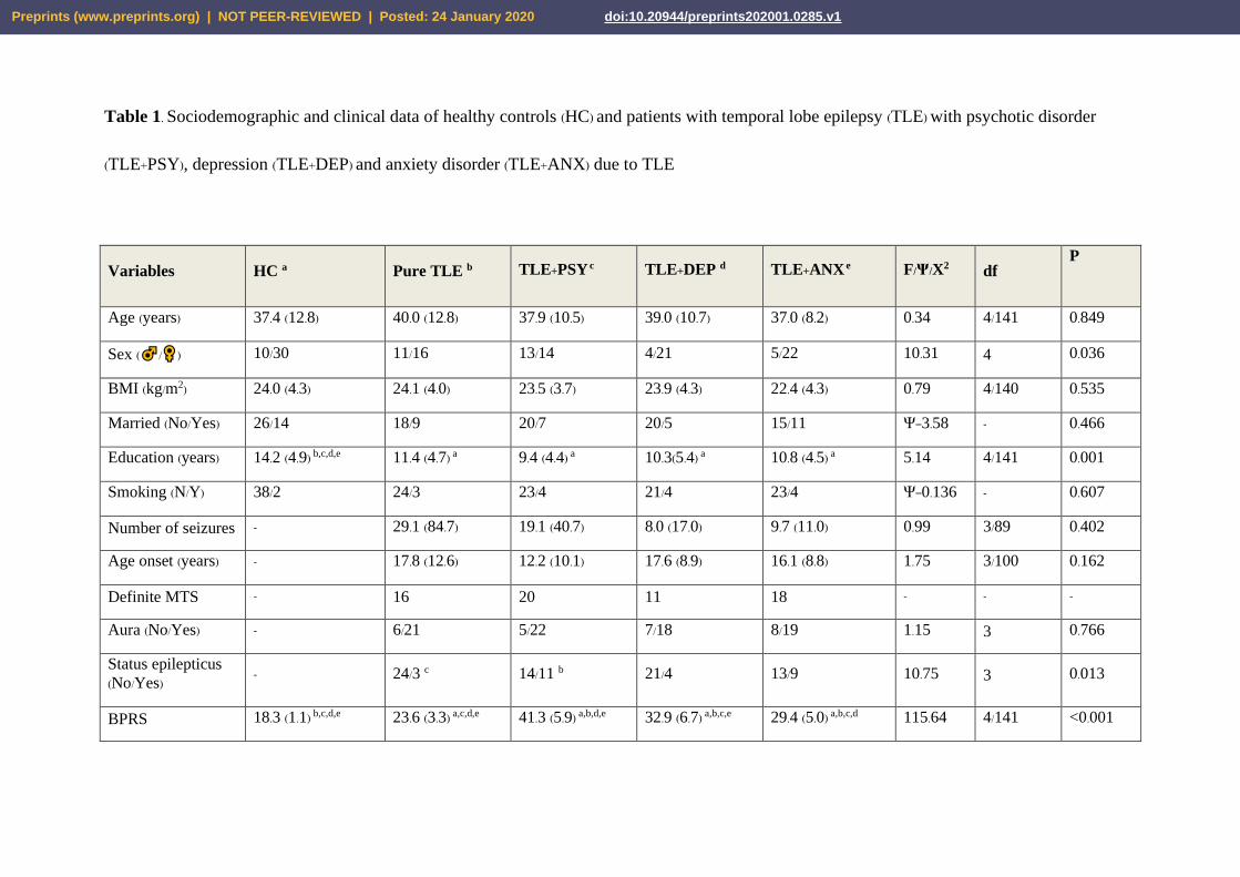
Table 1. Sociodemographic and clinical data of healthy controls (HC) and patients with temporal lobe epilepsy (TLE) with psychotic disorder
(TLE+PSY), depression (TLE+DEP) and anxiety disorder (TLE+ANX) due to TLE
Variables HC a Pure TLE b TLE+PSY c TLE+DEP d TLE+ANX e F/Ψ/X2 df P
Age (years) 37.4 (12.8) 40.0 (12.8) 37.9 (10.5) 39.0 (10.7) 37.0 (8.2) 0.34 4/141 0.849
Sex ( / ) 10/30 11/16 13/14 4/21 5/22 10.31 4 0.036
BMI (kg/m2) 24.0 (4.3) 24.1 (4.0) 23.5 (3.7) 23.9 (4.3) 22.4 (4.3) 0.79 4/140 0.535
Married (No/Yes) 26/14 18/9 20/7 20/5 15/11 Ψ=3.58 - 0.466
Education (years) 14.2 (4.9) b,c,d,e 11.4 (4.7) a 9.4 (4.4) a 10.3(5.4) a 10.8 (4.5) a 5.14 4/141 0.001
Smoking (N/Y) 38/2 24/3 23/4 21/4 23/4 Ψ=0.136 - 0.607
Number of seizures - 29.1 (84.7) 19.1 (40.7) 8.0 (17.0) 9.7 (11.0) 0.99 3/89 0.402
Age onset (years) - 17.8 (12.6) 12.2 (10.1) 17.6 (8.9) 16.1 (8.8) 1.75 3/100 0.162
Definite MTS - 16 20 11 18 - - -
Aura (No/Yes) - 6/21 5/22 7/18 8/19 1.15 3 0.766
Status epilepticus
(No/Yes) - 24/3 c 14/11 b 21/4 13/9 10.75 3 0.013
BPRS 18.3 (1.1) b,c,d,e 23.6 (3.3) a,c,d,e 41.3 (5.9) a,b,d,e 32.9 (6.7) a,b,c,e 29.4 (5.0) a,b,c,d 115.64 4/141 <0.001
Preprints (www.preprints.org) | NOT PEER-REVIEWED | Posted: 24 January 2020 doi:10.20944/preprints202001.0285.v1

HAM-D 0.6 (2.0) b,c,d,e
4.8 (2.5) a,d,e
5.8 (2.9) a,e
19.8 (4.9) a,b,c,e
10.3 (3.8) a,b,c,d 145.21 4/140 <0.001
HAM-A 2.6 (5.4) b,c,d,e
7.8 (3.9) a,c,d,e
11.6 (6.7) a,b,c,e
18.9 (8.8) a,b,c,e 23.8 (5.4) a,b,c,d 59.69 4/141 <0.001
All values are shown as mean (SD); BMI: body mass index; MTS: mesial temporal sclerosis.
BPRS: Brief Psychiatric Rating Scale; HAM-D: Hamilton Depression Rating Scale; HAM-A: Hamilton Anxiety Rating Scale; Pure TLE: TLE without any psychiatric comorbidities
Preprints (www.preprints.org) | NOT PEER-REVIEWED | Posted: 24 January 2020 doi:10.20944/preprints202001.0285.v1

Table 2. Results of multivariate GLM analysis examining the differences between diagnostic groups (diagnosis), namely healthy controls, temporal
lobe epilepsy with and without depression, psychosis, or anxiety.
Tests Dependent variables Exploratory variables F df p Partial Eta
Squared
Multivariate All 6 biomarkers:
TRAP, -SH, LOOH,
MDA, AOPP, Nox
Diagnosis
Sex
Age
BMI
Smoking
10.48
3.85
1.37
4.30
1.37
24/528
6/129
6/129
6/129
6/129
<0.001
0.001
0.231
0.001
0.233
0.321
0.152
0.060
0.167
0.060
Between-subject
effects
TRAP
-SH
MDA
AOPP
LOOH
NOx
Diagnosis
Diagnosis
Diagnosis
Diagnosis
Diagnosis
Diagnosis
8.56
13.44
74.05
13.07
2.27
2.05
1/134
1/134
1/134
1/134
1/134
1/134
<0.001
<0.001
<0.001
<0.001
0.065
0.091
0.204
0.286
0.689
0.290
0.063
0.058
Multivariate All 3 composite scores:
Diagnosis
Sex
Age
23.19
6.91
8/266
2/133
<0.001
0.001
0.411
0.094
Preprints (www.preprints.org) | NOT PEER-REVIEWED | Posted: 24 January 2020 doi:10.20944/preprints202001.0285.v1

BMI
Smoking
2.00
1.51
0.29
2/133
2/133
2/133
0.139
0.224
0.7456
0.029
0.022
0.004
Between-subject
effects
OSTOX
ANTIOX
OSTOX/ANTIOX
Diagnosis
Diagnosis
Diagnosis
43.60
19.95
57.06
1/134
1/134
1/134
<0.001
<0.001
<0.001
0.565
0.373
0.630
Diagnosis: five diagnostic groups, namely Psychotic Disorder due to temporal lobe epilepsy (TLE), Mood Disorder due to TLE with
depressive features, Anxiety Disorder due to TLE, Pure TLE (that is no comorbidities) and healthy controls
TRAP: total radical-trapping antioxidant parameter; -SH: sulfhydryl groups; MDA: malondialdehyde; AOPP: advanced oxidation protein
products; LOOH: lipid hydroperoxides; NOx: nitric oxide metabolites.
OSTOX: index of oxidative stress toxicity; ANTIOX: index of antioxidant activity; OSTOX/ANTIOX: ratio of oxidative stress toxicity /
antioxidant activity.
Preprints (www.preprints.org) | NOT PEER-REVIEWED | Posted: 24 January 2020 doi:10.20944/preprints202001.0285.v1

Table 3. Model-generated estimated marginal means (SE) of nitro-oxidative and antioxidant biomarkers in healthy controls (HC) and patients with
temporal lobe epilepsy (TLE) with and without psychosis, depression or anxiety.
Variables HC a Pure TLE b TLE+PSY c TLE+DEP d TLE+ANX e
TRAP (µmol Trolox) 995.2 (23.7) b-e 835.6 (27.1) a 852.5 (26.9) a 874.9 (29.6) a 816.5 (29.0) a
-SH (µmol/L) 321.8 (9.4) b-e 233.0 (10.8) a,e 250.7 (10.7) a 241.7 (11.8) a 268.4 (11.5) a,b
MDA (µM/mg protein) 2.32 (0.16) b-e 6.06 (0.19) a,d,e 5.54 (0.19) a,e 5.03 (0.21) a,b 4.79 0.20 a,b,c
AOPP (µmol/L/eq. cloramin T) 208.9 (30.0) b-e 409.3 (31.9) a 351.7 (31.7) a 427.7 (34.9) a 337.7 (34.2) a
LOOH (RLU) 1175 (53) b,c,d 1329 (60) a 1369 (60) a 1329 (66) a 1298 (65)
NOx (µmol/L) 7.67 (0.99) 4.49 (1.13) 6.03 (1.13) 7.69 (1.24) 6.91 (1.22)
OSTOX -1.123 (0.111) b-e 0.717 (0.126) a,e 0.535 (0.126) a 0.466 (0.138) a 0.203 (0.135) a,b
ANTIOX 1.097 (0.136) b-e -0.438 (0.155) a -0.193 (0.154) a -0.189 (0.169) a -0.170 (0.166) a
OSTOX/ANTIOX -1.304 (0.104) b-e 0.681 (0.119) a,e 0.430 (0.118) a 0.387 (0.130) a 0.221 (0.127) a,b
TLE+PSY: Psychotic Disorder due to TLE; TLE+DEP: Mood Disorder due to TLE with depressive features; TLE+ANX: Anxiety
Disorder due to TLE; Pure TLE: TLE without ant psychiatric comorbidities
TRAP: total radical-trapping antioxidant parameter; -SH: sulfhydryl groups; MDA: malondialdehyde; AOPP: advanced oxidation protein
products; LOOH: lipid hydroperoxides; NOx: nitric oxide metabolites.
Preprints (www.preprints.org) | NOT PEER-REVIEWED | Posted: 24 January 2020 doi:10.20944/preprints202001.0285.v1

OSTOX: index of oxidative stress toxicity; ANTIOX: index of antioxidant activity; OSTOX/ANTIOX: ratio of oxidative stress toxicity /
antioxidant activity.
Preprints (www.preprints.org) | NOT PEER-REVIEWED | Posted: 24 January 2020 doi:10.20944/preprints202001.0285.v1

Table 4. Results of automatic binary logistic regression analyses with temporal lobe epilepsy (TLE) and phenotypes as dependent variables.
Dependent variables Explanatory variables B SE Wald
df P Odd’s ratio 95% CI
interval
TLE MDA 5.04 1.441 12.83 1 <0.001 154.75 9.18-2609
MTS MDA
ANTIOX index
3.32
-1.83
0.704
0.727
22.19
6.35
1
1
<0.001
0.012
27.56
0.160
6.93-109.52
0.038-0.666
Pure TLE + MTS MDA
ANTIOX index
1.85
-2.84
0.974
0.392
7.52
4.16
1
1
0.006
0.041
6.34
0.06
1.69-23.74
0.00-0.89
Hx of post-ictal
confusion
MDA
ANTIOX index
0.856
0.917
0.318
0.332
7.25
7.62
1
1
0.007
0.006
2.35
2.50
1.26-4.39
1.31-4.80
Hx of status epilepticus TRAP -0.562 0.242 5.39 1 0.020 0.57 0.35-0.92
Hx of aura MDA 1.62 0.273 35.15 1 <0.001 5.04 2.95-8.59
TLE: temporal lobe epilepsy; MTS: mesial temporal sclerosis; Pure TLE: TLE without any psychiatric comorbidities
Hx: a history of
MDA: malondialdehyde; TRAP: total radical trapping antioxidant parameter; ANTIOX index: index of antioxidant activities computed as z value
of TRAP + z value of thiol (-SH) groups
Preprints (www.preprints.org) | NOT PEER-REVIEWED | Posted: 24 January 2020 doi:10.20944/preprints202001.0285.v1
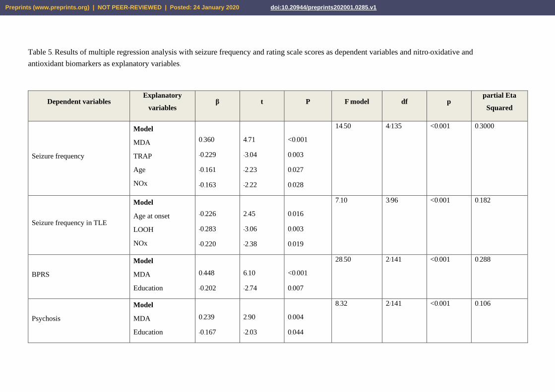
Table 5. Results of multiple regression analysis with seizure frequency and rating scale scores as dependent variables and nitro-oxidative and
antioxidant biomarkers as explanatory variables.
Dependent variables Explanatory
variables β t P F model df p
partial Eta
Squared
Seizure frequency
Model
MDA
TRAP
Age
NOx
0.360
-0.229
-0.161
-0.163
4.71
-3.04
-2.23
-2.22
<0.001
0.003
0.027
0.028
14.50 4/135 <0.001 0.3000
Seizure frequency in TLE
Model
Age at onset
LOOH
NOx
-0.226
-0.283
-0.220
2.45
-3.06
-2.38
0.016
0.003
0.019
7.10 3/96 <0.001 0.182
BPRS
Model
MDA
Education
0.448
-0.202
6.10
-2.74
<0.001
0.007
28.50 2/141 <0.001 0.288
Psychosis
Model
MDA
Education
0.239
-0.167
2.90
-2.03
0.004
0.044
8.32 2/141 <0.001 0.106
Preprints (www.preprints.org) | NOT PEER-REVIEWED | Posted: 24 January 2020 doi:10.20944/preprints202001.0285.v1

Excitation-grandiosity
Model
TRAP
LOOH
-0.225
0.187
-2.80
2.32
0.006
0,022
7.06 2/141 0.001 0.091
Negative symptoms
Model
Female sex
Education
Age
MDA
AOPP
-0.359
-0.331
-0.149
0.254
-0.219
-4.95
-4.29
-1.98
3.18
-2.75
<0.001
<0.001
0.049
0.002
0.007
11.86 5/138 <0.001 0.301
HAM-D
Model
MDA
Education
Female sex
0.317
-0.205
0.161
4.04
-2.59
2.10
<0.001
0.011
0.037
11.42 3/139 <0.001 0.198
Physiosomatic symptoms of
HAM-D
Model
MDA
0.374
4.81
<0.001
23.09 1/142 <0.001 0.140
Suicidal ideation Model
AOPP
0.261
3.22
0.002
10.36 1/142 0.002 0.068
HAM-A
Model
MDA
TRAP
0.271
-0.201
3.33
-.247
0.001
0.015
12.05 2/141 <0.001 0.146
Preprints (www.preprints.org) | NOT PEER-REVIEWED | Posted: 24 January 2020 doi:10.20944/preprints202001.0285.v1

Psychopathology index
Model
MDA
Education
0.415
-0.219
5.54
-2.92
<0.001
0.004
25.42 2/140 <0.001 0.146
BPRS: Brief Psychiatric Rating Scale; Psychosis: sum of 4 BPRS items, namely item 4 (conceptual disorganization), item 11 (suspiciousness), item 12
(hallucinations) and item 15 (unusual thought content); Excitation-grandiosity: sum of 2 BPRS items, namely item 8 (grandiosity) and item 17
(excitement); Negative symptoms: sum of 2 BPRS items, namely item 3 (emotional withdrawal) and item 16 (blunted affect)
HAM-D: Hamilton Depression Rating Scale; Physiosomatic symptoms of the HAM-D: sum of 5 HAM-D items, namely item 11 (anxiety somatic), item
12 (somatic symptoms), item 13 (somatic symptoms general), item 14 (genital symptoms) and item 15 (hypochondriasis); Suicidal ideation: item 3 of the
HAM-D; HAM-A: Hamilton Anxiety Rating Scale; Psychopathology index: index of overall severity of psychopathology computed as z value BPRS (z
BPRS) + z HAM-D + z HAM-A.
MDA: malondialdehyde; TRAP: total radical-trapping antioxidant parameter; LOOH: lipid hydroperoxides; AOPP: advanced oxidation protein products
Preprints (www.preprints.org) | NOT PEER-REVIEWED | Posted: 24 January 2020 doi:10.20944/preprints202001.0285.v1

Table 6. Results of multiple regression analyses with oxidative stress toxicity biomarkers as dependent variables.
Dependent variables Explanatory variables β t p F model df P partial Eta
Squared
MDA
Model
-SH
NOx
TRAP
LOOH
-0.450
-0.203
-0.193
0.174
-6.32
-2.92
-2.77
2.48
<0.001
0.004
0.006
0.014
18.34 4/139 <0.001 0.345
AOPP Model
-SH
-0.302
-3.77
<0.001
14.24 1/141 <0.001 0.091
MDA: malondialdehyde; AOPP: advanced oxidation protein products; -SH: sulfhydryl groups; NOx: nitric oxide metabolites; TRAP: total radical-
trapping antioxidant parameter; LOOH: lipid hydroperoxides
Preprints (www.preprints.org) | NOT PEER-REVIEWED | Posted: 24 January 2020 doi:10.20944/preprints202001.0285.v1