Includes transport of: 1. oxygen 2. carbon dioxide 3 nutrients 4. water 5. ions 6. hormones 7....
42
Includes transport of : 1. oxygen 2. carbon dioxide 3 nutrients 4. water 5. ions 6. hormones 7. antibodies 8. metabolic wastes Functions of Human Circulatory System Chp. 8
-
date post
21-Dec-2015 -
Category
Documents
-
view
217 -
download
0
Transcript of Includes transport of: 1. oxygen 2. carbon dioxide 3 nutrients 4. water 5. ions 6. hormones 7....
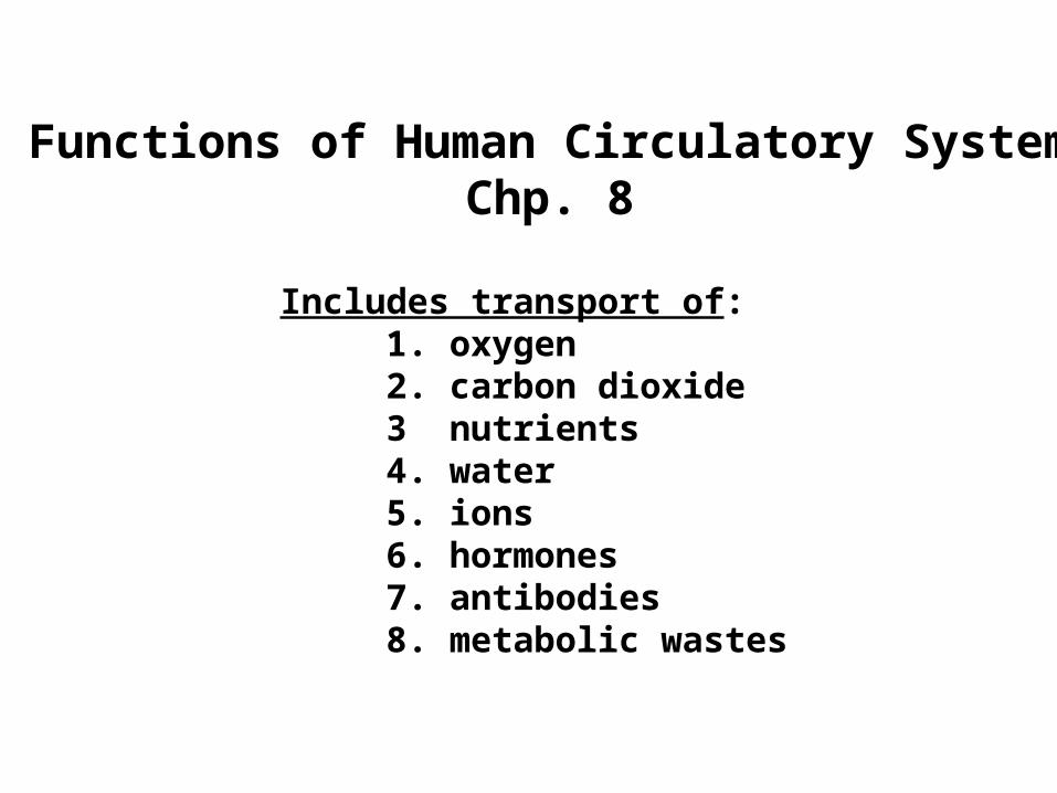
Includes transport of:1. oxygen2. carbon dioxide 3 nutrients 4. water 5. ions 6. hormones 7. antibodies 8. metabolic wastes
Functions of Human Circulatory SystemChp. 8
The Human Circulatory System
• 4 chambered heart• Arteries• Capillaries• Veins
Arteries
• muscular vessels carrying blood away from heart
• carry oxygenated blood– Exception- pulmonary artery (to lungs)
Capillaries
• thin walled (one cell layer) vessels • arise from arterioles (tiny arteries) • form capillary beds• all exchange between blood & cells occurs here
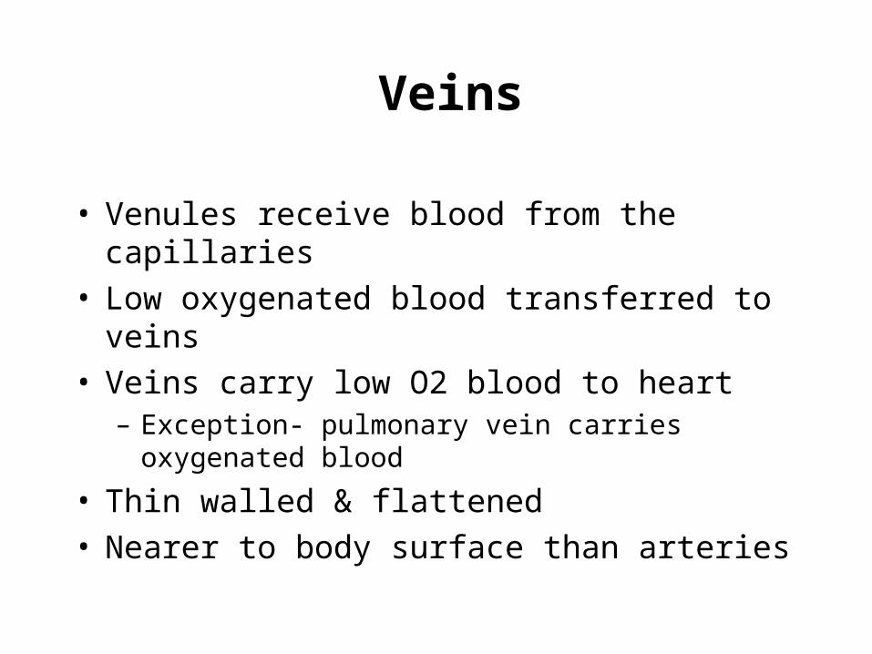
Veins
• Venules receive blood from the capillaries• Low oxygenated blood transferred to veins• Veins carry low O2 blood to heart
– Exception- pulmonary vein carries oxygenated blood
• Thin walled & flattened• Nearer to body surface than arteries
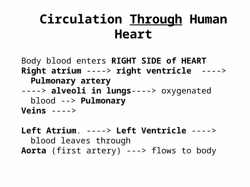
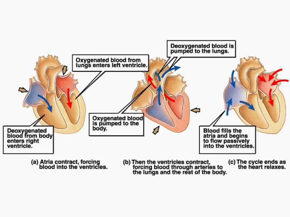
Circulation Through Human Heart
Body blood enters RIGHT SIDE of HEART Right atrium ----> right ventricle ----> Pulmonary artery ----> alveoli in lungs----> oxygenated blood --> PulmonaryVeins ---->
Left Atrium. ----> Left Ventricle ----> blood leaves throughAorta (first artery) ---> flows to body
Control of the Heart
1. Extrinsic (outside)
2. Intrinsic (within)
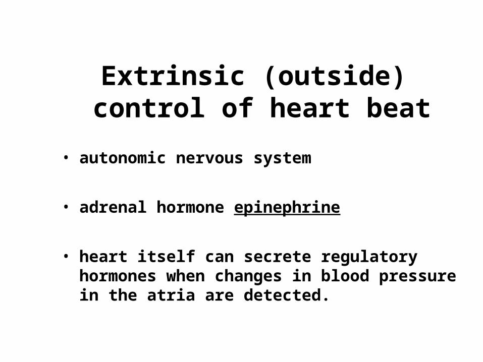
Extrinsic (outside) control of heart beat
• autonomic nervous system
• adrenal hormone epinephrine
• heart itself can secrete regulatory hormones when changes in blood pressure in the atria are detected.
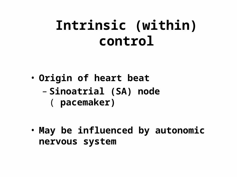
Intrinsic (within) control
• Origin of heart beat – Sinoatrial (SA) node ( pacemaker)
• May be influenced by autonomic nervous system
Human Circulatory System Circuits
1. Hepatic Portal Circuit
2. Renal Circuit
3. Cardiac Circuit
4. Systemic Circuit
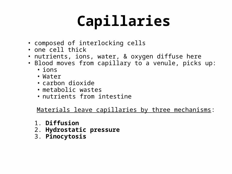
Capillaries• composed of interlocking cells • one cell thick• nutrients, ions, water, & oxygen diffuse here • Blood moves from capillary to a venule, picks up:
• ions• Water• carbon dioxide• metabolic wastes• nutrients from intestine
Materials leave capillaries by three mechanisms:
1. Diffusion2. Hydrostatic pressure3. Pinocytosis
Veins
• Entering blood volume equals that leaving arteries
• blood pressure is much lower than in arteries
Movement through veins assisted by:
1) one way flap-like valves allow blood to move in one direction (toward heart)
2) some smooth muscle around larger veins that contracts and moves blood
3) limb and breathing movements literally massages veins and squeezes blood along
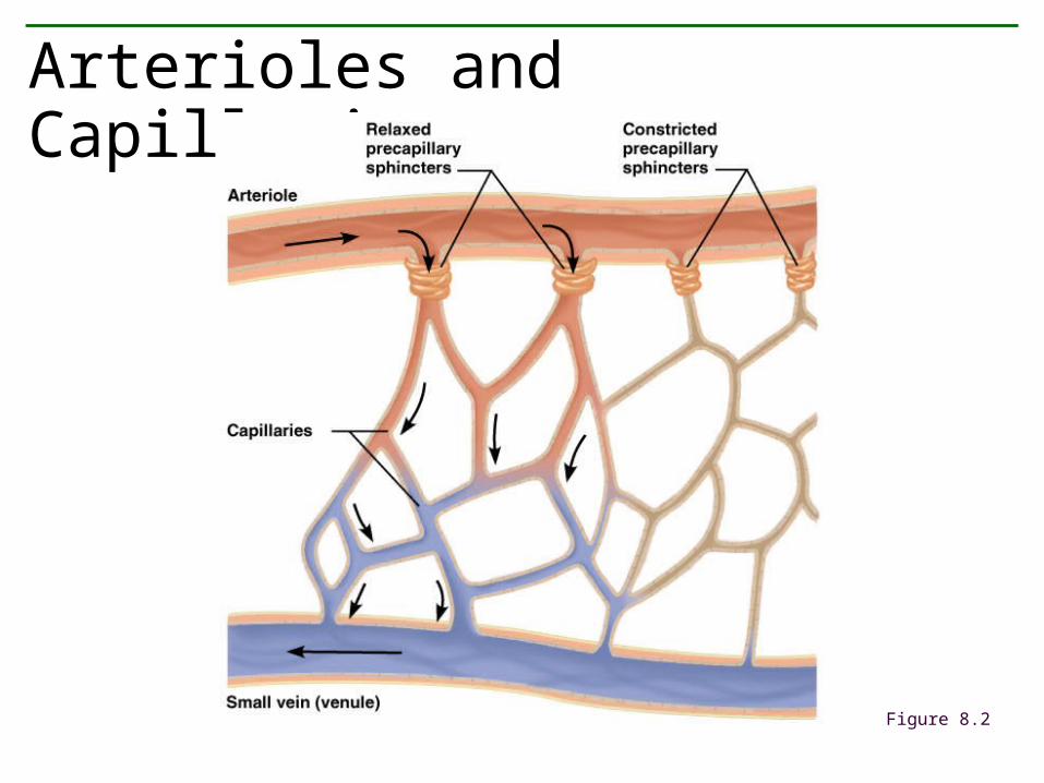
Arterioles and Capillaries
Figure 8.2
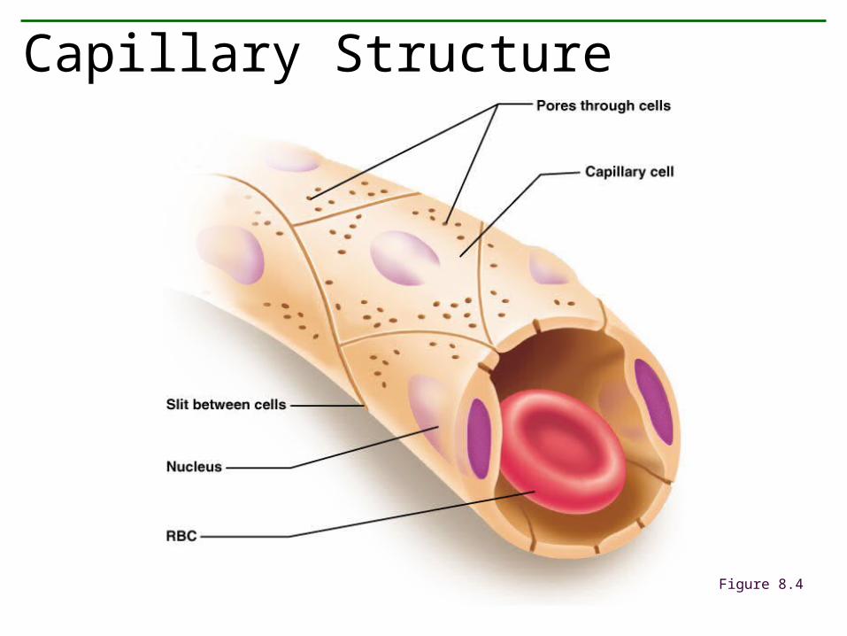
Capillary Structure
Figure 8.4
• Function: maintain blood volume; also functions in immune system
• Structure– Blind-ended capillaries– Lymphatic vessels– Lymph
Lymphatic System
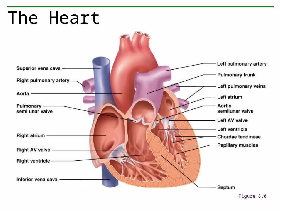
The Heart
Figure 8.8
• Structure
• Layers; epicardium, myocardium, endocardium
• Chambers: two atrias, two ventricles
• Valves– Two atrioventricular valves: tricuspid and
bicuspid (mitral)– Two semilunar valves: pulmonary and aortic
The Heart
– Deoxygenated blood through the vena cava to the right atrium
– Deoxygenated blood through the right atrioventricular valve to the right ventricle
– Deoxygenated blood through the pulmonary semilunar valve to the pulmonary trunk and the lungs
– Oxygenated blood through the pulmonary veins to the left atrium
– Oxygenated blood through the left atrioventricular valve to the left ventricle
Pulmonary Circuit: Oxygenation of Blood
– Oxygenated blood through the aortic semilunar valve to the aorta
– Oxygenated blood through branching arteries and arterioles to the tissues
– Oxygenated blood through the arterioles to capillaries
– Deoxygenated blood from capillaries into venules and veins
– Ultimately to the vena cava and into the right atrium
Systemic Circuit: Delivery of Oxygenated Blood to Tissues and Return of Blood to the Heart
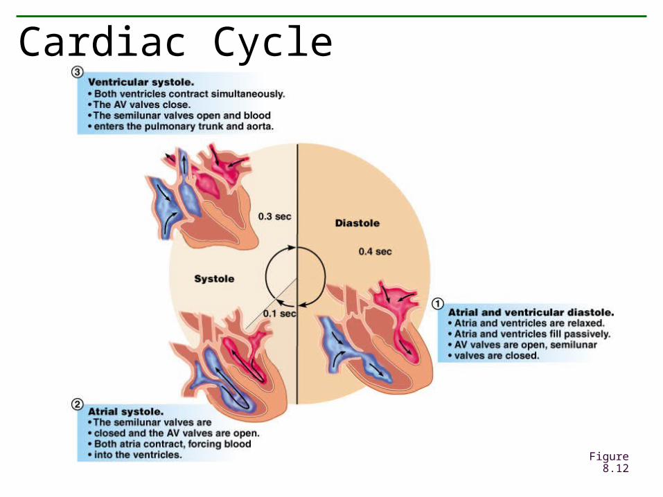
Cardiac Cycle
Figure 8.12
• Lub-dub
• Heart murmurs
Heart Sounds and Heart Valves
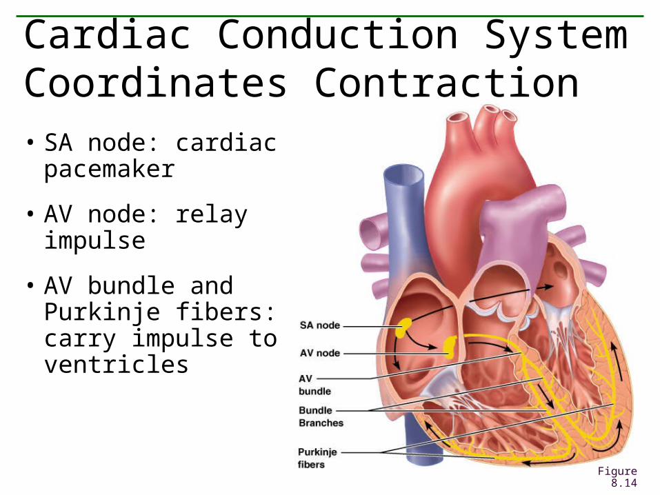
Cardiac Conduction System Coordinates Contraction
• SA node: cardiac pacemaker
• AV node: relay impulse
• AV bundle and Purkinje fibers: carry impulse to ventricles
Figure 8.14
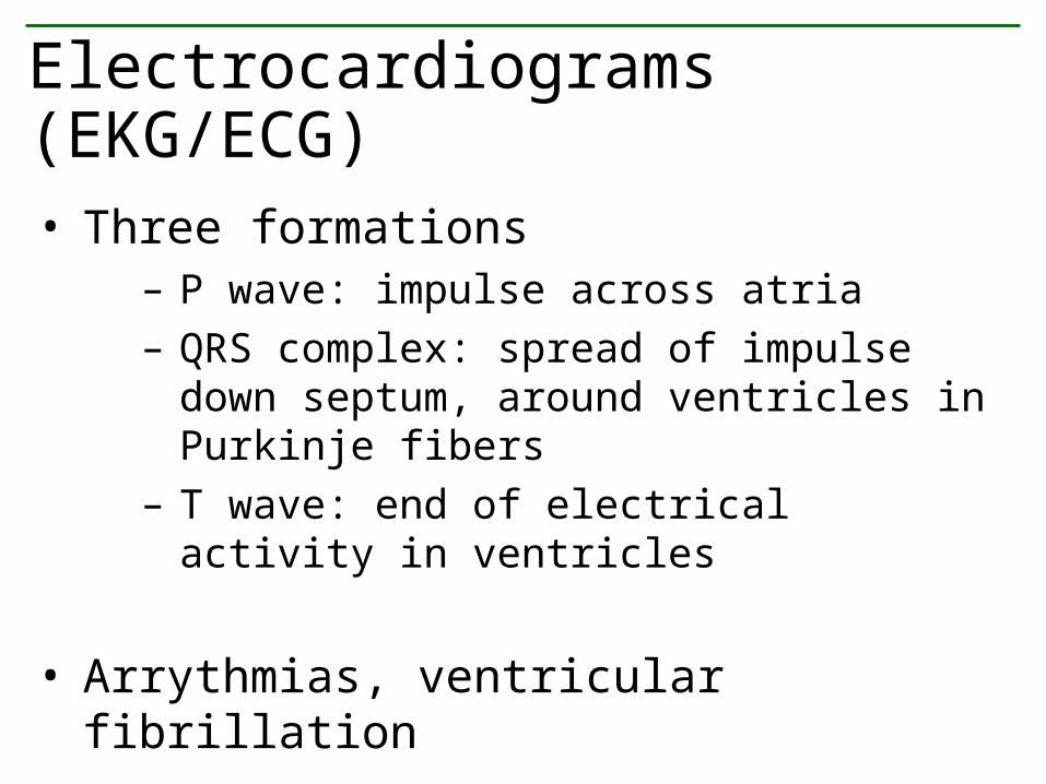
• Three formations– P wave: impulse across atria– QRS complex: spread of impulse down septum,
around ventricles in Purkinje fibers– T wave: end of electrical activity in ventricles
• Arrythmias, ventricular fibrillation
Electrocardiograms (EKG/ECG)
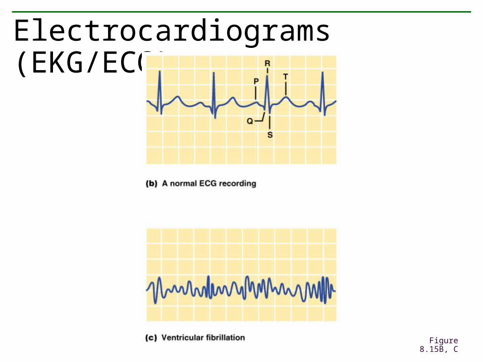
Electrocardiograms (EKG/ECG) (cont.)
Figure 8.15B, C
• Definitions: “normal”– Systolic pressure– Diastolic pressure
• Measurement: sphygmomanometer
Blood Pressure
• Hypertension: high blood pressure– Definition– The silent killer– Risk factors
• Hypertension: blood pressure too low– Clinical signs: dizziness, fainting– Causes: orthostatic, severe burns, blood loss
Blood Pressure
• Baroreceptors: pressure receptors in aorta andcarotid arteries
• Steps in mechanism– Blood pressure rises, vessels stretched– Signals sent to brain in the cardiovascular center– Heart signaled to lower heart rate and force of
contraction– Arterioles vasodilate, increasing blood flow to tissues– Combined effect lowers blood pressure
Regulation of the Cardiovascular System: Baroreceptors
• Medulla oblongata signals– Sympathetic nerves: constrict blood vessels,
raising blood pressure– Parasympathetic nerves: dilate blood vessels,
lowering blood pressure
• Hormones: epinephrine (adrenaline)
• Local requirements dictate local blood flow
Regulation of the Cardiovascular System: Nervous and Endocrine Factors
• Angina pectoris: A warning
• Myocardial infarction/heart attack: permanent cardiac damage
• Congestive heart failure: decrease in pumping efficiency
• Embolism: blockage of blood vessels
• Stroke: impaired blood flow to the brain
Cardiovascular Disorders
• Smoking: Don’t
• Blood lipids: monitor cholesterol levels
• Exercise: regular and moderate
• Blood pressure: treat hypertension
• Weight: being overweight increases risk of heart attack and stroke
• Control of Diabetes Mellitus: early diagnosis and treatment delays onset of related problems
• Stress: avoid chronic stress
Reducing the Risk of Cardiovascular Disease
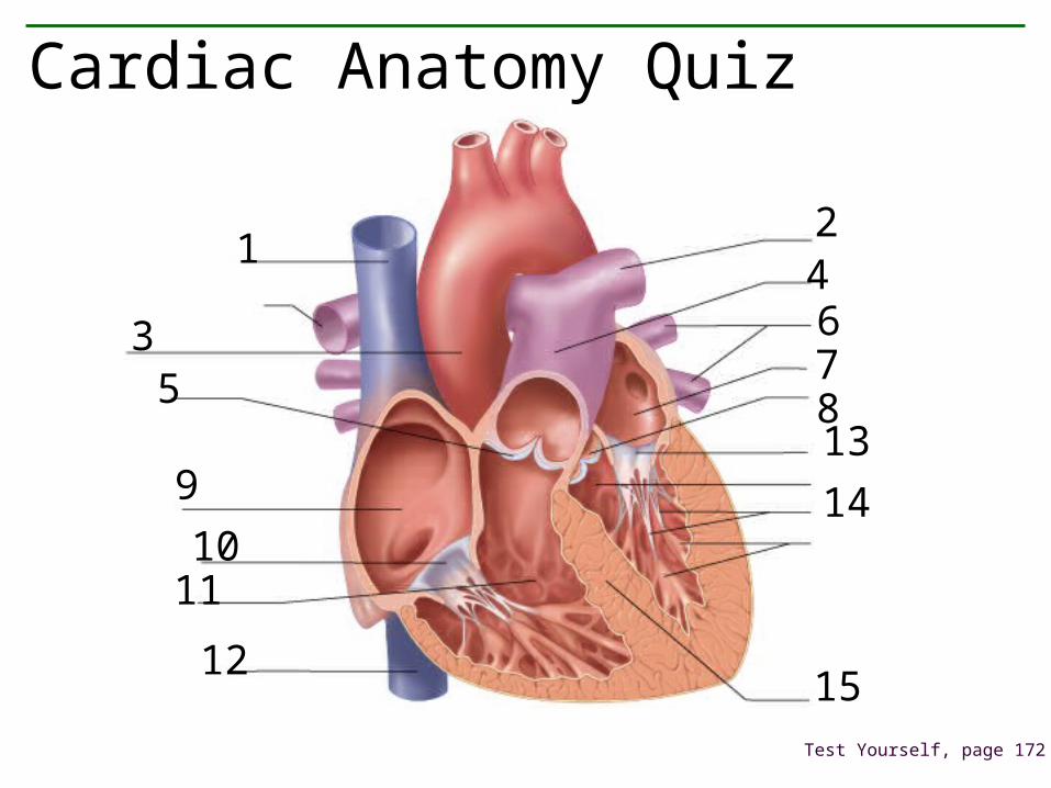
Cardiac Anatomy Quiz
Test Yourself, page 172
12
3
4
5
678
9
1011
12
13
14
15

Blood
• Connective tissue • plasma matrix + 3 types cells
Plasma = 90% water + 10% plasma solids.Solids include:
ureaamino acidsglucosehormones
ionsfatsproteins
The 3 Major Blood Proteins
albumins - large proteins that bind impurities & some toxins, aid in transport of
hormones, fatty acids and ions, help maintain osmotic balance.
globins - include antibodies (immunoglobins)
fibrinogen - important in blood clotting
Blood Cells
A) Erythrocytes
B) Leukocytes
C) Platelets
Erythrocytes (red blood cells)
1. small, disk-like shape
2. no nucleus
3. cannot reproduce
4. last 4 months then rupture
5. produced by red bone marrow
6. contain hemoglobin
7. carry oxygen
Leukocytes (white blood cells)
• Nucleus present
• Active in immune system• most are neutrophils that engulf microorganisms• Basophils• Eosinophils• lymphocytes
Platelets (thrombocytes)
• tiny • numerous• cell fragments• aid blood clotting
Circulatory system + lymphatic system = Proper Osmotic Conditions
Three Fluid Regions Of Body
1) fluid of blood and lymph 2) interstitial fluid - watery fluid between and among cells
3) intracellular fluid