Incidental os odontoideum: current management strategies
6
Neurosurg Focus / Volume 31 / December 2011 Neurosurg Focus 31 (6):E10, 2011 1 O S ODONTOIDEUM is an anatomical abnormality in which the tip of the odontoid process lacks conti- nuity with the body of C-2. It appears as a smooth- margined, apical osseous segment separated from the base of the odontoid process by an obvious gap. Os odontoi- deum may be discovered as part of a workup for neck pain and/or neurological symptoms, but it is also often found incidentally. Most spine surgeons agree that patients with signs or symptoms of neurological dysfunction should un- dergo stabilization, but the role for surgical stabilization in asymptomatic patients and those with neck pain alone remains controversial. In an article in the “Guidelines for the Manage- ment of Acute Cervical Spine and Spinal Cord Injuries” supplement published in 2002 jointly by the American Association of Neurological Surgeons and the Congress of Neurological Surgeons, the authors reviewed the avail- able literature on os odontoideum and found no Class I or Class II data to generate treatment standards or guide- lines. 1 The following management option was given for patients with incidental os odontoideum based on the available Class III data: “Patients with os odontoideum, either with or without C1–2 instability, who have neither symptoms nor neurological signs may be managed with clinical and radiographic surveillance.” Even so, the au- thors acknowledged that patients with C1–2 instability are at risk for future spinal cord damage and that surgical stabilization and fusion of C1–2 is “meritorious.” In fact, the literature regarding the management of os odontoi- deum remains limited to Class III data. Here we present the case of a patient with an incidentally discovered os odontoideum and review the embryological aspects and relevant upper cervical spinal anatomy and literature. We conclude by presenting a management strategy that we believe is rational given our experience, along with data from the available literature. Illustrative Case Clinical Presentation This 14-year-old girl was admitted to the hospital after having possible seizures, intermittent headaches, and dizziness. She underwent MR imaging of her brain, which showed a mild Chiari I malformation with approx- imately 6 mm of tonsillar herniation, but also a dysplastic odontoid. On her cervical spine MR images there was no spinal cord signal change or syringohydromyelia, and the VA anatomy was normal (Fig. 1A). A CT scan confirmed the presence of an os odontoideum. The os fragment was Incidental os odontoideum: current management strategies P AUL KLIMO JR., M.D., M.P.H., 1 V ALERIE COON, M.D., 2 AND DOUGLAS BROCKMEYER, M.D. 2 1 Semmes-Murphey Neurologic & Spine Institute, Memphis, Tennessee; and 2 Department of Neurosurgery, Primary Children’s Medical Center, University of Utah, Salt Lake City, Utah Os odontoideum was first described in the late 1880s and still remains a mystery in many respects. The genesis of os odontoideum is thought to be prior bone injury to the odontoid, but a developmental cause probably also exists. The spectrum of presentation is striking and ranges from patients who are asymptomatic or have only neck pain to those with acute quadriplegia, chronic myelopathy, or even sudden death. By definition, the presence of an os odon- toideum renders the C1–2 region unstable, even under physiological loads in some patients. The consequences of this instability are exemplified by numerous cases in the literature in which a patient with os odontoideum has suffered a spinal cord injury after minor trauma. Although there is little debate that patients with os odontoideum and clini- cal or radiographic evidence of neurological injury or spinal cord compression should undergo surgery, the dispute continues regarding the care of asymptomatic patients whose os odontoideum is discovered incidentally. The authors’ clinical experience leads them to believe that certain subgroups of asymptomatic patients should be strongly consid- ered for surgery. These subgroups include those who are young, have anatomy favorable for surgical intervention, and show evidence of instability on flexion-extension cervical spine x-rays. This recommendation is bolstered by the fact that surgical fusion of the C1–2 region has evolved greatly and can now be done with considerable safety and success. When atlantoaxial instrumentation is used, fusion rates for os odontoideum should approach 100%. (DOI: 10.3171/2011.9.FOCUS11227) KEY WORDS • incidental os odontoideum • atlantoaxial instability • atlantoaxial fusion • screw fixation 1 Abbreviation used in this paper: VA = vertebral artery. Unauthenticated | Downloaded 10/02/21 04:20 PM UTC
Transcript of Incidental os odontoideum: current management strategies

Neurosurg Focus / Volume 31 / December 2011
Neurosurg Focus 31 (6):E10, 2011
1
Os OdOntOideum is an anatomical abnormality in which the tip of the odontoid process lacks conti-nuity with the body of C-2. It appears as a smooth-
margined, apical osseous segment separated from the base of the odontoid process by an obvious gap. Os odontoi-deum may be discovered as part of a workup for neck pain and/or neurological symptoms, but it is also often found incidentally. Most spine surgeons agree that patients with signs or symptoms of neurological dysfunction should un-dergo stabilization, but the role for surgical stabilization in asymptomatic patients and those with neck pain alone remains controversial.
In an article in the “Guidelines for the Manage-ment of Acute Cervical Spine and Spinal Cord Injuries” supplement published in 2002 jointly by the American Association of Neurological Surgeons and the Congress of Neurological Surgeons, the authors reviewed the avail-able literature on os odontoideum and found no Class I or Class II data to generate treatment standards or guide-lines.1 The following management option was given for patients with incidental os odontoideum based on the available Class III data: “Patients with os odontoideum, either with or without C1–2 instability, who have neither symptoms nor neurological signs may be managed with
clinical and radiographic surveillance.” Even so, the au-thors acknowledged that patients with C1–2 instability are at risk for future spinal cord damage and that surgical stabilization and fusion of C1–2 is “meritorious.” In fact, the literature regarding the management of os odontoi-deum remains limited to Class III data. Here we present the case of a patient with an incidentally discovered os odontoideum and review the embryological aspects and relevant upper cervical spinal anatomy and literature. We conclude by presenting a management strategy that we believe is rational given our experience, along with data from the available literature.
Illustrative CaseClinical Presentation
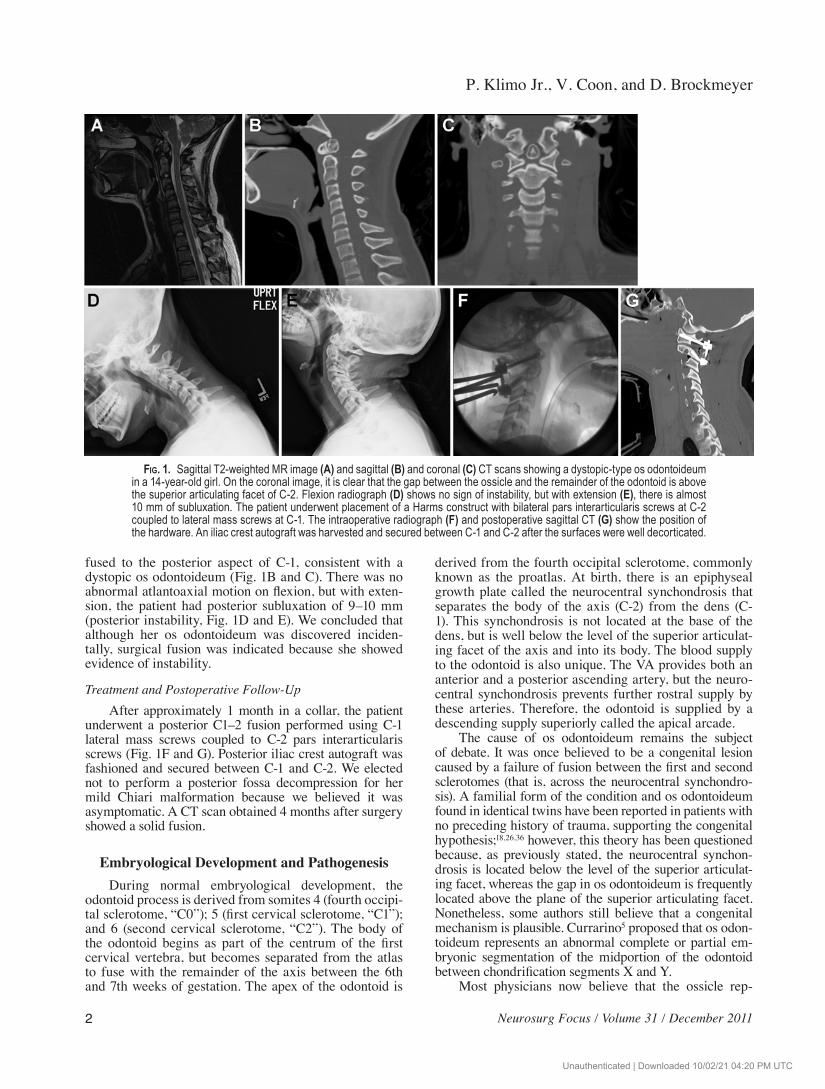
This 14-year-old girl was admitted to the hospital after having possible seizures, intermittent headaches, and dizziness. She underwent MR imaging of her brain, which showed a mild Chiari I malformation with approx-imately 6 mm of tonsillar herniation, but also a dysplastic odontoid. On her cervical spine MR images there was no spinal cord signal change or syringohydromyelia, and the VA anatomy was normal (Fig. 1A). A CT scan confirmed the presence of an os odontoideum. The os fragment was
Incidental os odontoideum: current management strategies
Paul Klimo Jr., m.D., m.P.H.,1 Valerie Coon, m.D.,2 anD Douglas BroCKmeyer, m.D.2
1Semmes-Murphey Neurologic & Spine Institute, Memphis, Tennessee; and 2Department of Neurosurgery, Primary Children’s Medical Center, University of Utah, Salt Lake City, Utah
Os odontoideum was first described in the late 1880s and still remains a mystery in many respects. The genesis of os odontoideum is thought to be prior bone injury to the odontoid, but a developmental cause probably also exists. The spectrum of presentation is striking and ranges from patients who are asymptomatic or have only neck pain to those with acute quadriplegia, chronic myelopathy, or even sudden death. By definition, the presence of an os odon-toideum renders the C1–2 region unstable, even under physiological loads in some patients. The consequences of this instability are exemplified by numerous cases in the literature in which a patient with os odontoideum has suffered a spinal cord injury after minor trauma. Although there is little debate that patients with os odontoideum and clini-cal or radiographic evidence of neurological injury or spinal cord compression should undergo surgery, the dispute continues regarding the care of asymptomatic patients whose os odontoideum is discovered incidentally. The authors’ clinical experience leads them to believe that certain subgroups of asymptomatic patients should be strongly consid-ered for surgery. These subgroups include those who are young, have anatomy favorable for surgical intervention, and show evidence of instability on flexion-extension cervical spine x-rays. This recommendation is bolstered by the fact that surgical fusion of the C1–2 region has evolved greatly and can now be done with considerable safety and success. When atlantoaxial instrumentation is used, fusion rates for os odontoideum should approach 100%. (DOI: 10.3171/2011.9.FOCUS11227)
Key WorDs • incidental os odontoideum • atlantoaxial instability • atlantoaxial fusion • screw fixation
1
Abbreviation used in this paper: VA = vertebral artery.
Unauthenticated | Downloaded 10/02/21 04:20 PM UTC
P. Klimo Jr., V. Coon, and D. Brockmeyer
2 Neurosurg Focus / Volume 31 / December 2011
fused to the posterior aspect of C-1, consistent with a dystopic os odontoideum (Fig. 1B and C). There was no abnormal atlantoaxial motion on flexion, but with exten-sion, the patient had posterior subluxation of 9–10 mm (posterior instability, Fig. 1D and E). We concluded that although her os odontoideum was discovered inciden-tally, surgical fusion was indicated because she showed evidence of instability.
Treatment and Postoperative Follow-UpAfter approximately 1 month in a collar, the patient
underwent a posterior C1–2 fusion performed using C-1 lateral mass screws coupled to C-2 pars interarticularis screws (Fig. 1F and G). Posterior iliac crest autograft was fashioned and secured between C-1 and C-2. We elected not to perform a posterior fossa decompression for her mild Chiari malformation because we believed it was asymp tomatic. A CT scan obtained 4 months after surgery showed a solid fusion.
Embryological Development and PathogenesisDuring normal embryological development, the
odontoid process is derived from somites 4 (fourth occipi-tal sclerotome, “C0”); 5 (first cervical sclerotome, “C1”); and 6 (second cervical sclerotome, “C2”). The body of the odontoid begins as part of the centrum of the first cervical vertebra, but becomes separated from the atlas to fuse with the remainder of the axis between the 6th and 7th weeks of gestation. The apex of the odontoid is
derived from the fourth occipital sclerotome, commonly known as the proatlas. At birth, there is an epiphyseal growth plate called the neurocentral synchondrosis that separates the body of the axis (C-2) from the dens (C-1). This synchondrosis is not located at the base of the dens, but is well below the level of the superior articulat-ing facet of the axis and into its body. The blood supply to the odontoid is also unique. The VA provides both an anterior and a posterior ascending artery, but the neuro-central synchondrosis prevents further rostral supply by these arteries. Therefore, the odontoid is supplied by a descending supply superiorly called the apical arcade.
The cause of os odontoideum remains the subject of debate. It was once believed to be a congenital lesion caused by a failure of fusion between the first and second sclerotomes (that is, across the neurocentral synchondro-sis). A familial form of the condition and os odontoideum found in identical twins have been reported in patients with no preceding history of trauma, supporting the congenital hypothesis;18,26,36 however, this theory has been questioned because, as previously stated, the neurocentral synchon-drosis is located below the level of the superior articulat-ing facet, whereas the gap in os odontoideum is frequently located above the plane of the superior articulating facet. Nonetheless, some authors still believe that a congenital mechanism is plausible. Currarino5 proposed that os odon-toideum represents an abnormal complete or partial em-bryonic segmentation of the midportion of the odontoid between chondrification segments X and Y.
Most physicians now believe that the ossicle rep-
Fig. 1. Sagittal T2-weighted MR image (A) and sagittal (B) and coronal (C) CT scans showing a dystopic-type os odontoideum in a 14-year-old girl. On the coronal image, it is clear that the gap between the ossicle and the remainder of the odontoid is above the superior articulating facet of C-2. Flexion radiograph (D) shows no sign of instability, but with extension (E), there is almost 10 mm of subluxation. The patient underwent placement of a Harms construct with bilateral pars interarticularis screws at C-2 coupled to lateral mass screws at C-1. The intraoperative radiograph (F) and postoperative sagittal CT (G) show the position of the hardware. An iliac crest autograft was harvested and secured between C-1 and C-2 after the surfaces were well decorticated.
Unauthenticated | Downloaded 10/02/21 04:20 PM UTC

Neurosurg Focus / Volume 31 / December 2011
Incidental os odontoideum
3
resents a posttraumatic phenomenon resulting from a nonhealed odontoid synchondrosis fracture. In essence, the odontoid fragment fails to remodel and unite with the body of C-2. This scenario is supported by the ob-servation that many patients with os odontoideum have a history of remote trauma, particularly in early child-hood.23 The most commonly cited theory was proposed by Fielding et al.,9 who suggested that with a fracture or disruption through the neurocentral synchondrosis, the alar ligaments that attach to the apex of the odontoid may gradually distract the fragment away from the base. The apex and base of the odontoid continue to have adequate perfusion, but the midportion suffers from lack of blood supply and thus contributes to poor healing.
The “traumatic cause” hypothesis is supported by case reports of patients with a previously documented in-tact C-2 who later were found to have os odontoideum after remote trauma. Schuler et al.31 reported on a 2-year-old patient who fell out of her crib and complained of neck pain; her initial cervical x-ray was normal. After continued neck pain, repeat cervical x-rays were obtained 13 months after her injury, which demonstrated os odon-toideum with atlantoaxial instability. A recent case report by Zygourakis et al.41 supports the delayed formation of an os odontoideum through a combination of trauma and vascular compromise. Their patient was a 2-year-old girl who sustained a C1–2 ligamentous injury but no bone in-jury. Four years later an os odontoideum was found, and 10 years after trauma the child had developed mild at-lantoaxial hypermobility that continued to be managed nonoperatively. It is likely that os odontoideum most commonly arises as a result of remote trauma, but some cases are also probably due to faulty embryogenesis. Os odontoideum needs to be differentiated from a persistent ossiculum terminale. The latter represents congenital nonunion of the odontoid body from a terminal ossicle located above the transverse atlantal ligament. Thus, at-lantoaxial instability is usually not associated with an os-siculum terminale.
Presentation and Imaging FeaturesRowland et al.29 categorized symptoms of os odon-
toideum into 4 groups: Group 1, local symptoms only (for example, neck pain, headaches) and no signs of myelopa-thy; Group 2, posttraumatic transient myelopathy; Group 3, persistent myelopathy; and Group 4, cerebral symptoms suggestive of posterior circulation ischemia. Patients with symptoms in Group 2 classically have transient complete tetraplegia immediately after a traumatic event, which is often minor in nature. Those with symptoms in Group 3 have chronic, often progressive myelopathy because of repeated trauma to the spinal cord as a result of excessive motion of the ring of C-1 and the ossicle. Patients with posterior circulation symptoms are quite rare, but have been reported.10,34 Asymptomatic patients, by definition, have incidentally discovered os odontoideum. This group comprised 15% of patients in our recent series.19
Os odontoideum is a rare radiographic diagnosis. Two types of os odontoideum have been defined based on the position of the dens tip: orthotopic and dystopic.8 In
the orthotopic type, the dens is in an anatomical position. In dystopic os odontoideum, the dens tip is in any other position. Most commonly, the fragment is located near the foramen magnum, where it may fuse with the clivus, or it may be fixed to the anterior ring of the atlas. Plain cervi-cal x-rays are often sufficient to suggest the diagnosis of an os odontoideum, but a high-resolution multiplanar CT scan is necessary to provide the detailed anatomy of the os odontoideum and any associated cervical or skull base anomalies for surgical planning. Dynamic flexion and ex-tension lateral radiographs may be obtained to determine whether there is any abnormal atlantoaxial motion and, if so, in which direction is the subluxation: anterior, poste-rior, or both.
In our series, a subset of 60 patients with os odontoi-deum who underwent dynamic imaging had predominant-ly anterior instability (70%), with almost equal posterior instability (10%) or combined anterior/posterior instability (13%). No motion was seen in 7% of the patients. Although many spine surgeons believe that a patient with normal mentation can undergo volitional flexion and extension im-aging without risking neurological injury, particularly in those who are without symptoms, these studies should be ordered with caution in those who have had recent neu-rological injury or chronic myelopathy. An MR imaging study should also be done to evaluate for any intrinsic spinal cord signal change at the level of os odontoideum that would indicate prior or continuing spinal cord trauma. Rarely, synovial cysts may develop as a result of the abnor-mal motion, and these are best evaluated using MR imag-ing.17,27 Careful evaluation of the regional vascular anatomy is necessary because anomalies have been reported in as-sociation with an os odontoideum, particularly the pres-ence of an anomalous VA38,39 or persistent fetal anterior-posterior circulation connection.21
Biomechanics and StabilityThe stability of the C1–2 joint is determined by the
integrity of the odontoid process, the transverse ligament, and to a lesser extent, the alar ligaments. Thus, the biome-chanical principles of C1–2 stability are inherently violat-ed with os odontoideum. With an incompetent odontoid process, the spinal cord may be compressed posteriorly during flexion by the posterior ring of C-1 or on exten-sion by posterior translation of the os itself (Fig. 2). In the absence of an intact odontoid process, we believe that the existing stabilizing structures—the thinner vertical por-tion of the cruciate ligament (an extension of the posterior longitudinal ligament), the thin anterior longitudinal liga-ment, and the lax capsular ligaments—are insufficient to provide the necessary spinal stability and protection of the spinal cord. An os odontoideum is biomechanically analogous and in some cases difficult to distinguish from a chronic nonunited Type 2 odontoid fracture. Although it is well accepted among most spine surgeons that Type 2 fractures, whether acute or chronic but not healed, are unstable and require treatment, there continues to be a significant difference of opinion among surgeons regard-ing the treatment of an incidental os odontoideum.
Unauthenticated | Downloaded 10/02/21 04:20 PM UTC

P. Klimo Jr., V. Coon, and D. Brockmeyer
4 Neurosurg Focus / Volume 31 / December 2011
Natural History and Risk of InjuryDescriptions of the natural history of os odontoi-
deum are very limited and dated. Several reports have presented evidence of patients who were treated conser-vatively without further incident. Fielding et al.9 reported a series of 35 patients, 8 of whom had no radiographic evidence of C1–2 instability and underwent conservative management. Each of them remained without symptoms at follow-up evaluation. Similarly, all 5 of the asymptom-atic patients with os odontoideum in whom the condition was managed without surgical intervention by Dai et al.6 remained stable at follow-up. Spierings and Braakman32 analyzed 37 patients with os odontoideum, in 20 of whom the condition was managed conservatively. The authors found that patients with a minimal sagittal diameter (de-fined as the distance between the posterior border of the body of C-2 and the posterior atlantal arch on flexion) of < 13 mm had the greatest risk of developing permanent or progressive cord signs. This suggests that incidental os odontoideum may be managed without surgical interven-tion unless the minimum sagittal diameter is < 13 mm or there is progressive neurological decline. Although these studies suggest that in some patients with incidental os odontoideum the condition can be safely managed con-servatively, in sum the studies provide less than defini-tive evidence as to the natural history and risks associated with untreated os odontoideum.
Sudden death7,25,30 and significant neurological mor-bidity20,24,33 with minor trauma have both been reported as the initial presentation of patients with a previously undiagnosed os odontoideum. Choit et al.4 reported on 2 children who each had an os odontoideum that had not been appreciated on initial imaging and that caused sub-sequent morbidity. One of the children sustained a severe high spinal cord injury after a minor accident. We previ-ously presented 3 case examples of patients in whom an os odontoideum was initially diagnosed but who never
received treatment and who subsequently developed a spinal cord injury.19 More recently, Zhang et al.40 reported on 10 patients with atlantoaxial instability from os odon-toideum, including 3 who were asymptomatic, who later suffered a spinal cord injury. The mechanism was a fall in 6 patients, a minor motor vehicle accident in 3, and assault in 1. Based on this experience, the authors ad-vocated fusion and instrumentation for all patients with radiographically unstable os odontoideum, whether they were symptomatic or not.
White and Panjabi37 defined clinical instability as “the loss of the ability of the spine under physiological loads to maintain its pattern of displacement so that there is no initial or additional neurological deficit, no major deformity, and no incapacitating pain.” For some patients with os odontoideum, everyday physiological loads are well tolerated and supported by the accessory structures described previously. Given the numerous reports of pa-tients suffering neurological injury from minor trauma, however, we believe that the ability to tolerate physiologi-cal loads or minor trauma cannot be assumed, because the potential loss of that gamble could be catastrophic.
For some patients, particularly young children and teenagers, it is difficult or even impossible to limit day-to-day activity to “physiological” loading, without severely restricting their activity and thus negatively impacting the quality of their life. This includes the normal day-to-day episodes of ground-level falls in toddlers or, for example, falls from playground equipment in older children. For teenagers, giving up contact or collision sports may be relatively easy, but the pushing and shoving that goes on in school hallways, as another example, may not be so easy to avoid. Conversely, it is relatively easy for most young adults with os odontoideum to modify their activi-ties to reduce the risk of spinal cord injury. Their greatest risk obviously lies in the possibility of a motor vehicle accident, which is a risk shared by almost everyone. That
Fig. 2. Diagrams depicting an os odontoideum in neutral (A), flexed (B), and extended (C) positions. With an os odontoideum (A), abnormal motion may occur anteriorly, posteriorly, or both. Flexion (B) can cause translation of the C-1 ring and ossicle com-plex to the point that it may impinge on the dorsal aspect of the cord. On extension (C), the anterior ring with the ossicle can strike the ventral aspect of the cord. Reproduced with permission from Klimo P Jr, Kan P, Rao G, et al: Os odontoideum: presentation, diagnosis, and treatment in a series of 78 patients. Clinical article. J Neurosurg Spine 9:332–342, 2008.
Unauthenticated | Downloaded 10/02/21 04:20 PM UTC

Neurosurg Focus / Volume 31 / December 2011
Incidental os odontoideum
5
is why we choose to use the patient’s age as a surgical selection criterion for an incidentally discovered os odon-toideum; we believe that young patients are more likely to need protection than older ones.
Management of Incidental Os OdontoideumWe believe that all patients with neurological symp-
toms or significant neck pain due to os odontoideum should undergo surgical stabilization. Patients with inci-dentally discovered os odontoideum should be considered for surgery on a case-by-case basis. Those patients who show evidence of radiographic instability at the atlan-toaxial level, who are relatively young (< 20 years old), and who have bone anatomy favorable for screw fixation should be strongly considered for surgery. Asymptomatic patients who do not meet the criteria given above may be monitored with serial radiographs and clinical evaluations to detect whether instability or significant symptoms are developing. Arvin et al.2 presented similar recommenda-tions, arguing that patients with a stable os odontoideum without evidence of spinal cord compression should be evaluated annually with dynamic plain x-rays, repeat MR imaging in 5 years, and avoidance of all contact sports.
SurgeryExcellent clinical results and high rates of fusion (>
90%) for both adults and children who have undergone in-strumentation of the C1–2 level have been demonstrated in numerous series.12,14,15,22,28,35 Surgical techniques and fu-sion technology have evolved tremendously over the last several decades. Screw fixation techniques have largely replaced the stand-alone graft/wiring procedures such as the Gallie and Brooks techniques of the past, leading to ex-cellent results.3,11 Placement of transarticular C1–2 screws was the main method of screw fixation of the C1–2 joint, but this technique involved a risk of injuring the VA.13 The surgeon now has available other types of screws, including C-1 lateral mass screws, C-2 pedicle or pars screws, C-1 hooks, and C-2 laminar screws, to minimize the chance of injuring the VA. Consequently, the use of transarticu-lar screws has become less popular. Hardware can now be placed with minimal risk of neurological or vascular injury. Although harvested autograft, typically from the patient’s posterior iliac crest, is still the main grafting sub-strate, there is now evidence that allograft may work just as well, thus eliminating donor site-related issues, particularly pain.16 Although there is a loss of approximately 40%–50% of rotation with fusion of C1–2, we have found that older adults seem to adapt well to this loss of motion. Younger adults and children often have little, if any, limitation to motion after atlantoaxial fusion, because other spinal lev-els assume more rotatory function. It is our opinion that the loss of rotation after a C1–2 fusion seems to be a small price to pay for securing spinal stability and avoiding po-tentially catastrophic cord compression.
ConclusionsThe incomplete odontoid process found in os odon-
toideum results in weakness of the C1–2 joint. It is poten-tially dangerous to assume that the remaining support-ing structures are strong enough to sustain physiological loads and forces from even minor trauma. Patients with an incidentally discovered os odontoideum should be considered carefully for surgery, taking into account their age, activity level, and radiographic findings, including evidence of atlantoaxial instability and anatomy favor-able for surgical instrumentation. Surgery can be done safely, with minimal risk of complications, and with an excellent chance of achieving optimal clinical results.
Disclosure
The authors report no conflict of interest concerning the mate-rials or methods used in this study or the findings specified in this paper.
Author contributions to the study and manuscript prepara-tion include the following. Conception and design: Brockmeyer. Ac quisition of data: Coon. Drafting the article: Klimo, Coon. Re -viewed submitted version of manuscript: all authors. Approved the final version of the manuscript on behalf of all authors: Klimo. Study supervision: Brockmeyer.
Acknowledgment
The authors thank Kristin Kraus, M.Sc., for editorial assistance with the preparation of this paper.
References
1. Anonymous: Os odontoideum. Neurosurgery 50 (3 Suppl): S148–S155, 2002
2. Arvin B, Fournier-Gosselin MP, Fehlings MG: Os odontoi-deum: etiology and surgical management. Neurosurgery 66 (3 Suppl):22–31, 2010
3. Brooks AL, Jenkins EB: Atlanto-axial arthrodesis by the wedge compression method. J Bone Joint Surg Am 60:279–284, 1978
4. Choit RL, Jamieson DH, Reilly CW: Os odontoideum: a sig-nificant radiographic finding. Pediatr Radiol 35:803–807, 2005
5. Currarino G: Segmentation defect in the midodontoid process and its possible relationship to the congenital type of os odon-toideum. Pediatr Radiol 32:34–40, 2002
6. Dai L, Yuan W, Ni B, Jia L: Os odontoideum: etiology, diagno-sis, and management. Surg Neurol 53:106–109, 2000
7. Dempster AG, Heap SW: Fatal high cervical spinal cord injury in an automobile accident complicating os odontoideum. Am J Forensic Med Pathol 11:252–256, 1990
8. Fielding JW, Griffin PP: Os odontoideum: an acquired lesion. J Bone Joint Surg Am 56:187–190, 1974
9. Fielding JW, Hensinger RN, Hawkins RJ: Os odontoideum. J Bone Joint Surg Am 62:376–383, 1980
10. Fukuda M, Aiba T, Akiyama K, Nishiyama K, Ozawa T: Cer-ebellar infarction secondary to os odontoideum. J Clin Neu-rosci 10:625–626, 2003
11. Gallie WE: Fractures and dislocations of the cervical spine. Am J Surg 46:495–499, 1939
12. Gluf WM, Brockmeyer DL: Atlantoaxial transarticular screw fixation: a review of surgical indications, fusion rate, compli-cations, and lessons learned in 67 pediatric patients. J Neuro-surg Spine 2:164–169, 2005
13. Gluf WM, Schmidt MH, Apfelbaum RI: Atlantoaxial transar-ticular screw fixation: a review of surgical indications, fusion rate, complications, and lessons learned in 191 adult patients. J Neurosurg Spine 2:155–163, 2005
14. Goel A, Desai KI, Muzumdar DP: Atlantoaxial fixation using
Unauthenticated | Downloaded 10/02/21 04:20 PM UTC

P. Klimo Jr., V. Coon, and D. Brockmeyer
6 Neurosurg Focus / Volume 31 / December 2011
plate and screw method: a report of 160 treated patients. Neu-rosurgery 51:1351–1357, 2002
15. Harms J, Melcher RP: Posterior C1-C2 fusion with polyaxial screw and rod fixation. Spine (Phila Pa 1976) 26:2467–2471, 2001
16. Hillard VH, Fassett DR, Finn MA, Apfelbaum RI: Use of al-lograft bone for posterior C1-2 fusion. Clinical article. J Neu-rosurg Spine 11:396–401, 2009
17. Kirk HJ, Pik JH: A novel operative technique to manage a symptomatic synovial cyst associated with an os odontoide-um. J Clin Neurosci 16:822–824, 2009
18. Kirlew KA, Hathout GM, Reiter SD, Gold RH: Os odontoi-deum in identical twins: perspectives on etiology. Skeletal Radiol 22:525–527, 1993
19. Klimo P Jr, Kan P, Rao G, Apfelbaum R, Brockmeyer D: Os odontoideum: presentation, diagnosis, and treatment in a se-ries of 78 patients. Clinical article. J Neurosurg Spine 9:332–342, 2008
20. Kuhns LR, Loder RT, Farley FA, Hensinger RN: Nuchal cord changes in children with os odontoideum: evidence for associ-ated trauma. J Pediatr Orthop 18:815–819, 1998
21. Lee SH, Kim ES, Eoh W: Posterior C1-2 fusion using a poly-axial screw/rod system for os odontoideum with bilateral persistence of the first intersegmental artery. Case report. J Neurosurg Spine 14:10–13, 2011
22. Leonard JR, Wright NM: Pediatric atlantoaxial fixation with bilateral, crossing C-2 translaminar screws. Technical note. J Neurosurg 104 (1 Suppl):59–63, 2006
23. Menezes AH: Pathogenesis, dynamics, and management of os odontoideum. Neurosurg Focus 6(6):e2, 1999
24. Minderhoud JM, Braakman R, Penning L: Os odontoideum, clinical, radiological and therapeutic aspects. J Neurol Sci 8:521–544, 1969
25. Mixter SJ, Osgood RB: IV. Traumatic lesions of the atlas and axis. Ann Surg 51:193–207, 1910
26. Morgan MK, Onofrio BM, Bender CE: Familial os odontoi-deum. Case report. J Neurosurg 70:636–639, 1989
27. Ogata T, Kawatani Y, Morino T, Yamamoto H: Resolution of intraspinal retro-odontoid cyst associated with os odontoide-um after posterior fixation. J Spinal Disord Tech 22:58–61, 2009
28. Reilly CW, Choit RL: Transarticular screws in the manage-ment of C1-C2 instability in children. J Pediatr Orthop 26: 582–588, 2006
29. Rowland LP, Shapiro JH, Jacobson HG: Neurological syn-dromes associated with congenital absence of the odontoid process. AMA Arch Neurol Psychiatry 80:286–291, 1958
30. Schiller F, Nieda I: Malformation of the odontoid process—
report of a case and clinical survey. Calif Med 86:394–398, 1957
31. Schuler TC, Kurz L, Thompson DE, Zemenick G, Hensinger RN, Herkowitz HN: Natural history of os odontoideum. J Pe-diatr Orthop 11:222–225, 1991
32. Spierings EL, Braakman R: The management of os odontoide-um. Analysis of 37 cases. J Bone Joint Surg Br 64:422–428, 1982
33. Stevens JM, Chong WK, Barber C, Kendall BE, Crockard HA: A new appraisal of abnormalities of the odontoid process associated with atlanto-axial subluxation and neurological disability. Brain 117:133–148, 1994
34. Takakuwa T, Hiroi S, Hasegawa H, Hurukawa K, Endo S, Shi-mamura T: Os odontoideum with vertebral artery occlusion. Spine (Phila Pa 1976) 19:460–462, 1994
35. Wang MY: Transarticular screws and C1 hooks fixation for os odontoideum with atlantoaxial dislocation. World Neuro-surg 75:440, 2011
36. Wang S, Wang C: Familial dystopic os odontoideum: a report of three cases. J Bone Joint Surg Am 93:e44, 2011
37. White AA III, Panjabi MM: The problem of clinical insta-bility in the human spine: a systematic approach, in Clinical Biomechanics of the Spine, ed 2. Philadelphia: JB Lippin-cott, 1990, pp 277–378
38. Yamazaki M, Koda M, Aramomi MA, Hashimoto M, Masaki Y, Okawa A: Anomalous vertebral artery at the extraosse-ous and intraosseous regions of the craniovertebral junction: analysis by three-dimensional computed tomography angiog-raphy. Spine (Phila Pa 1976) 30:2452–2457, 2005
39. Yamazaki M, Koda M, Yoneda M, Aiba A, Moriya H: Anoma-lous vertebral artery at the craniovertebral junction in a pa-tient with Down syndrome. Case report. J Neurosurg Spine 1:338–341, 2004
40. Zhang Z, Zhou Y, Wang J, Chu T, Li C, Ren X, et al: Acute traumatic cervical cord injury in patients with os odontoide-um. J Clin Neurosci 17:1289–1293, 2010
41. Zygourakis CC, Cahill KS, Proctor MR: Delayed development of os odontoideum after traumatic cervical injury: support for a vascular etiology. Case report. J Neurosurg Pediatr 7:201–204, 2011
Manuscript submitted August 18, 2011.Accepted September 22, 2011.Address correspondence to: Paul Klimo Jr., M.D., M.P.H.,
Semmes-Murphey Neurologic & Spine Institute, 6325 Humphreys Boulevard, Memphis, Tennessee 38120. email: [email protected].
Unauthenticated | Downloaded 10/02/21 04:20 PM UTC