Inactivation of Capicua in adult mice causes T-cell...
14
Inactivation of Capicua in adult mice causes T-cell lymphoblastic lymphoma Lucía Simón-Carrasco, 1 Osvaldo Graña, 2 Marina Salmón, 1 Harrys K.C. Jacob, 1 Alejandro Gutierrez, 3 Gerardo Jiménez, 4,5 Matthias Drosten, 1 and Mariano Barbacid 1 1 Molecular Oncology Programme, Centro Nacional de Investigaciones Oncológicas (CNIO), 28029 Madrid, Spain; 2 Bioinformatics Unit, Structural Biology and Biocomputing Programme, Centro Nacional de Investigaciones Oncológicas (CNIO), 28029 Madrid, Spain; 3 Division of Hematology/Oncology, Boston Children’s Hospital, Dana-Farber Cancer Institute, Harvard Medical School, Boston, Massachusetts 02115, USA; 4 Institut de Biologia Molecular de Barcelona-Consejo Superior de Investigaciones Científicas (CSIC), Parc Cientifíc de Barcelona, 08028 Barcelona, Spain; 5 Institució Catalana de Recerca i Estudis Avançats (ICREA), 08028 Barcelona, Spain CIC (also known as Capicua) is a transcriptional repressor negatively regulated by RAS/MAPK signaling. Whereas the functions of Cic have been well characterized in Drosophila, little is known about its role in mammals. CIC is inactivated in a variety of human tumors and has been implicated recently in the promotion of lung metastases. Here, we describe a mouse model in which we inactivated Cic by selectively disabling its DNA-binding activity, a mutation that causes derepression of its target genes. Germline Cic inactivation causes perinatal lethality due to lung differentiation defects. However, its systemic inactivation in adult mice induces T-cell acute lymphoblastic lymphoma (T-ALL), a tumor type known to carry CIC mutations, albeit with low incidence. Cic inactivation in mice induces T-ALL by a mechanism involving derepression of its well-known target, Etv4. Importantly, human T-ALL also relies on ETV4 expression for maintaining its oncogenic phenotype. Moreover, Cic inactivation renders T- ALL insensitive to MEK inhibitors in both mouse and human cell lines. Finally, we show that Ras-induced mouse T- ALL as well as human T-ALL carrying mutations in the RAS/MAPK pathway display a genetic signature indicative of Cic inactivation. These observations illustrate that CIC inactivation plays a key role in this human malignancy. [Keywords: CIC; Ras signaling; T-ALL; mouse models; Etv4] Supplemental material is available for this article. Received April 10, 2017; revised version accepted July 24, 2017. Ras proteins have been the subject of intense investiga- tion due to their key role in mitogenic signaling (Malum- bres and Barbacid 2003). Genetic studies have provided evidence that Ras signaling is essential for mammalian cell proliferation (Drosten et al. 2010). However, constitu- tive activation of individual members of the downstream mitogen-activated protein kinase (MAPK) pathway, in- cluding the Raf, Mek, and Erk kinases, can bypass the re- quirement for Ras proteins (Drosten et al. 2010). However, little is known regarding those events that take place be- yond MAPK signaling, in part due to the complexity of the events triggered by this kinase cascade in which just the Erk proteins alone are known to phosphorylate >150 substrates (Roskoski 2012). While searching for potential downstream effectors of the MAPK pathway, the Drosophila protein Capicua (Cic) (Jiménez et al. 2000) came to our attention. Cic pro- teins are evolutionarily conserved transcriptional repres- sors that directly bind DNA via their HMG-box with the assistance of a C-terminal motif (Forés et al. 2017). Both the Drosophila and mammalian Cic loci express short (Cic-S) and long (Cic-L) protein isoforms that are tran- scribed from two independent promoters (Lam et al. 2006; Jiménez et al. 2012; Forés et al. 2015). Moreover, Drosophila Cic proteins have a docking site that allows di- rect binding of Rolled, the Drosophila counterpart of the mammalian Erk kinases (Astigarraga et al. 2007). Cic re- pressor activity is down-regulated by receptor tyrosine ki- nase-mediated MAPK signaling (Jiménez et al. 2012). In Drosophila, the Cic locus has been implicated in a variety of developmental processes such as antero–posterior or dorso–ventral patterning of the embryo (Jiménez et al. 2000), specification of wing veins (Roch et al. 2002), and tissue growth in various contexts. Indeed, during cell pro- liferation of both larval tissues and adult intestinal stem cells, the mitogenic signals initiated by Ras signaling Corresponding authors: [email protected], [email protected] Article published online ahead of print. Article and publication date are online at http://www.genesdev.org/cgi/doi/10.1101/gad.300244.117. © 2017 Simón-Carrasco et al. This article is distributed exclusively by Cold Spring Harbor Laboratory Press for the first six months after the full-issue publication date (see http://genesdev.cshlp.org/site/misc/ terms.xhtml). After six months, it is available under a Creative Commons License (Attribution-NonCommercial 4.0 International), as described at http://creativecommons.org/licenses/by-nc/4.0/. GENES & DEVELOPMENT 31:1–13 Published by Cold Spring Harbor Laboratory Press; ISSN 0890-9369/17; www.genesdev.org 1 Cold Spring Harbor Laboratory Press on May 30, 2019 - Published by genesdev.cshlp.org Downloaded from
Transcript of Inactivation of Capicua in adult mice causes T-cell...

Inactivation of Capicua in adult micecauses T-cell lymphoblastic lymphomaLucía Simón-Carrasco,1 Osvaldo Graña,2 Marina Salmón,1 Harrys K.C. Jacob,1 Alejandro Gutierrez,3
Gerardo Jiménez,4,5 Matthias Drosten,1 and Mariano Barbacid1
1Molecular Oncology Programme, Centro Nacional de Investigaciones Oncológicas (CNIO), 28029Madrid, Spain; 2BioinformaticsUnit, Structural Biology and Biocomputing Programme, Centro Nacional de Investigaciones Oncológicas (CNIO), 28029 Madrid,Spain; 3Division of Hematology/Oncology, Boston Children’s Hospital, Dana-Farber Cancer Institute, Harvard MedicalSchool, Boston, Massachusetts 02115, USA; 4Institut de Biologia Molecular de Barcelona-Consejo Superior de InvestigacionesCientíficas (CSIC), ParcCientifíc de Barcelona, 08028 Barcelona, Spain; 5InstitucióCatalana deRecerca i Estudis Avançats (ICREA),08028 Barcelona, Spain
CIC (also known as Capicua) is a transcriptional repressor negatively regulated by RAS/MAPK signaling. Whereasthe functions of Cic have been well characterized in Drosophila, little is known about its role in mammals. CIC isinactivated in a variety of human tumors and has been implicated recently in the promotion of lung metastases.Here, we describe a mouse model in which we inactivated Cic by selectively disabling its DNA-binding activity, amutation that causes derepression of its target genes. Germline Cic inactivation causes perinatal lethality due tolung differentiation defects. However, its systemic inactivation in adult mice induces T-cell acute lymphoblasticlymphoma (T-ALL), a tumor type known to carryCICmutations, albeit with low incidence.Cic inactivation inmiceinduces T-ALL by a mechanism involving derepression of its well-known target, Etv4. Importantly, human T-ALLalso relies on ETV4 expression for maintaining its oncogenic phenotype. Moreover, Cic inactivation renders T-ALL insensitive toMEK inhibitors in bothmouse and human cell lines. Finally, we show that Ras-inducedmouse T-ALL as well as human T-ALL carrying mutations in the RAS/MAPK pathway display a genetic signature indicativeof Cic inactivation. These observations illustrate that CIC inactivation plays a key role in this human malignancy.
[Keywords: CIC; Ras signaling; T-ALL; mouse models; Etv4]
Supplemental material is available for this article.
Received April 10, 2017; revised version accepted July 24, 2017.
Ras proteins have been the subject of intense investiga-tion due to their key role in mitogenic signaling (Malum-bres and Barbacid 2003). Genetic studies have providedevidence that Ras signaling is essential for mammaliancell proliferation (Drosten et al. 2010). However, constitu-tive activation of individual members of the downstreammitogen-activated protein kinase (MAPK) pathway, in-cluding the Raf, Mek, and Erk kinases, can bypass the re-quirement for Ras proteins (Drosten et al. 2010). However,little is known regarding those events that take place be-yond MAPK signaling, in part due to the complexity ofthe events triggered by this kinase cascade in which justthe Erk proteins alone are known to phosphorylate >150substrates (Roskoski 2012).While searching for potential downstream effectors of
the MAPK pathway, the Drosophila protein Capicua(Cic) (Jiménez et al. 2000) came to our attention. Cic pro-teins are evolutionarily conserved transcriptional repres-sors that directly bind DNA via their HMG-box with the
assistance of a C-terminal motif (Forés et al. 2017). Boththe Drosophila and mammalian Cic loci express short(Cic-S) and long (Cic-L) protein isoforms that are tran-scribed from two independent promoters (Lam et al.2006; Jiménez et al. 2012; Forés et al. 2015). Moreover,DrosophilaCic proteins have a docking site that allows di-rect binding of Rolled, the Drosophila counterpart of themammalian Erk kinases (Astigarraga et al. 2007). Cic re-pressor activity is down-regulated by receptor tyrosine ki-nase-mediated MAPK signaling (Jiménez et al. 2012). InDrosophila, theCic locus has been implicated in a varietyof developmental processes such as antero–posterior ordorso–ventral patterning of the embryo (Jiménez et al.2000), specification of wing veins (Roch et al. 2002), andtissue growth in various contexts. Indeed, during cell pro-liferation of both larval tissues and adult intestinal stemcells, the mitogenic signals initiated by Ras signaling
Corresponding authors: [email protected], [email protected] published online ahead of print. Article and publication date areonline at http://www.genesdev.org/cgi/doi/10.1101/gad.300244.117.
© 2017 Simón-Carrasco et al. This article is distributed exclusively byCold Spring Harbor Laboratory Press for the first six months after thefull-issue publication date (see http://genesdev.cshlp.org/site/misc/terms.xhtml). After six months, it is available under a Creative CommonsLicense (Attribution-NonCommercial 4.0 International), as described athttp://creativecommons.org/licenses/by-nc/4.0/.
GENES & DEVELOPMENT 31:1–13 Published by Cold Spring Harbor Laboratory Press; ISSN 0890-9369/17; www.genesdev.org 1
Cold Spring Harbor Laboratory Press on May 30, 2019 - Published by genesdev.cshlp.orgDownloaded from

become dispensable when Cic is mutated (Tseng et al.2007; Jin et al. 2015).
In contrast, the role of mammalian Cic has been lesswell characterized. However, it has been shown thatMAPK signaling also regulates the repressor activity ofhuman CIC proteins. Likewise, ERK and its downstreamsubstrate, p90RSK, can phosphorylate human CIC, there-by interfering with its nuclear import and DNA-bindingcapabilities, respectively (Dissanayake et al. 2011). Fur-thermore, CIC has been implicated in several human pa-thologies. For instance, CIC is known to form nuclearcomplexes with Ataxin 1 (ATXN1) and Ataxin 1-like(ATXN1L, also known as BOAT [brother of Ataxin 1])(Lam et al. 2006). ATXN1 is a glutamine-rich protein im-plicated in spinocerebellar ataxia type 1 (SCA1), and ex-pansion of its polyglutamine tract, which is known tocause SCA1, alters the repressive properties of CIC(Crespo-Barreto et al. 2010). These results suggest thatATXN1 and ATXN1L act as Cic corepressors in mam-mals. Indeed, expression of polyglutamine-expandedATXN1 causes deregulation of a subset of CIC targetgenes. Moreover, mice lacking both Atxn1 and Atxn1Lshow destabilization of Cic proteins accompanied by up-regulation of Cic target genes such as the Pea3 familymembers Etv4 or Etv5 (Lee et al. 2011). As a consequence,these mice display a series of developmental defects suchas omphalocele and lung alveolarization deficiencies.
Mutations in CIC have been identified recently in sev-eral human cancers. The highest incidence of mutatedCIC is found in oligodendrogliomas (53%–69%), whereone allele is usually lost upon loss of heterozygosity(LOH) of chromosomes 1p and 19q, and the other is affect-ed by missense or nonsense mutations (Bettegowda et al.2011; Yip et al. 2012). Likewise, CIC mutations havealso been identified, albeit at lower frequency, in severalother malignancies, such as stomach adenocarcinomas(12.9%), endometrial carcinomas (6.9%), colorectal carci-nomas (6.1%), or melanomas (5.2%) (Cerami et al. 2012;Gao et al. 2013). Thesemutations appear to be a late eventduring tumorigenesis and have been found to promotemetastases (Okimoto et al. 2017). Most missense muta-tions in CIC cluster into codons encoding the HMG-boxor the C1motif, thus interfering with CIC’s DNA-bindingabilities and causing derepression of its target genes (Foréset al. 2017). Other mutations produce premature stopcodons, altered splice sites, and frameshift insertions ordeletions that are also thought to inactivate CIC’s repres-sor function.
To better understand the role of Cic in mammals, wegenerated a genetically engineered mouse model express-ing defective Cic proteins that cannot bind DNA andhence repress expression of its target genes. Our datashow that Cic is an essential gene in mouse developmentand that systemic disruption of Cic DNA binding in adultmice causes T-cell acute lymphoblastic lymphoma (T-ALL). We also show that human T-ALL carrying muta-tions in the RAS/MAPK pathway displays a genetic sig-nature indicative of CIC inactivation, thus suggestingthat CIC inactivation plays a key role in this humanmalignancy.
Results
Generation of mice carrying inactive Cic alleles
To genetically analyze the role ofCic in mammals, we de-cided to generate amouse strain carrying conditional loss-of-function (LOF) Cic alleles. To this end, we introducedloxP sequences flanking exons 2–6 by homologous recom-bination (Fig. 1A; Supplemental Fig. S1A). These exons en-code the highly conserved HMG-box (Supplemental Fig.S1B), a domain required for DNA binding and hence re-pressor activity (Jiménez et al. 2012; Forés et al. 2017).Elimination of exons 2–6 maintains the Cic ORF, direct-ing the synthesis of mutant Cic-L and Cic-S isoformsthat lack 288 amino acids, a region that includes theHMG-box (Fig. 1B). In this study, the conditional alleleis designated as Ciclox, whereas the recombined allele isdesignated asCicΔ2–6, and its gene products are designatedas CicΔ2–6. To confirm that deletion of exons 2–6 elimi-nates Cic activity, we engineered a human CIC-S cDNAlacking those sequences corresponding to exons 2–6 fusedto a GFP tag (Fig. 1C,D). The resulting GFP-CICΔ2–6 pro-tein completely lacks the ability to bind to the promotersof well-known CIC target genes such as CCND1 (CyclinD1) and the PEA3 family of transcription factors ETV1,ETV4, and ETV5, as demonstrated by chromatin immuno-precipitation (ChIP) assays using full-lengthGFP-CIC-S asa positive control (Fig. 1E).
Cic activity is essential for late embryonic development
To assess the role of Cic in development, we generatedgermline Cic+/Δ2–6 mice by mating Cic+/lox animals (seethe Supplemental Material) with EIIa-Cre transgenicmice that express the bacterial Cre recombinase in theirzygotes (Lakso et al. 1996). Heterozygous Cic+/Δ2–6 ani-mals were viable and did not show obvious defects forup to 1 yr of age. Homozygous CicΔ2–6/Δ2–6 embryoswere present at Mendelian ratios at embryonic day 18.5(E18.5). However, E18.5 CicΔ2–6/Δ2–6 embryos were signif-icantly smaller than their wild-type or heterozygouscounterparts and died immediately after birth (Fig. 2A).Most of these embryos (70%) presented an omphalocele,an abdominal wall closure defect (Fig. 2B). Omphalocelesoccur naturally during mammalian development whenthe gut transiently herniates the umbilical space due tolimited space in the peritoneal cavity (Doyonnas et al.2001). This structure is usually resolved by E16 in mice,thus indicating that Cic function may be required for re-traction of the gut from the umbilical cord. Interestingly,a similar phenotype was observed in embryos lacking theCic corepressors Atxn1 andAtxn1L, an observation attrib-uted to the decrease of Cic protein levels in Atxn1−/−;Atxn1L−/− mice (Lee et al. 2011). CicΔ2–6/Δ2–6 embryosdid not display hydrocephali, another phenotype frequent-ly detected in Atxn1−/−;Atxn1L−/− mice (Lee et al. 2011).In addition, we could not analyze whether lung alveolari-zation was affected, as shown previously in mice lackingthe Cic-L isoform (Lee et al. 2011), since CicΔ2–6/Δ2–6
embryos died right after birth. However, expression of
Simón-Carrasco et al.
2 GENES & DEVELOPMENT
Cold Spring Harbor Laboratory Press on May 30, 2019 - Published by genesdev.cshlp.orgDownloaded from

matrix metalloproteases known to be responsible for de-fective lung alveolarization was not affected at E18.5, al-though Cic target genes were markedly derepressed(Supplemental Fig. S2A,B).
Further characterization of E18.5 CicΔ2–6/Δ2–6 embryosrevealed a dramatic increase in proliferating (Ki67+) lungcells, suggesting a defect in terminal differentiation ofthe respiratory epithelium (Fig. 2C; Supplemental Fig.S2C). Immunohistochemistry (IHC) studies involvingγH2AX and active Caspase 3 failed to reveal signs ofDNA damage or increased apoptosis (Supplemental Fig.S2D). To determine whether terminal differentiation ofthe lung is affected in CicΔ2–6/Δ2–6 embryos, we analyzedlung differentiation markers by IHC. E18.5 CicΔ2–6/Δ2–6
embryos displayed persistent TTF-1 (also known asNkx2-1) expression levels in a high proportion of epitheli-al cells, a phenotype reminiscent of lungs in the canalicu-lar stage (Naltner et al. 2000;Moreno-Barriuso et al. 2006),which suggests delayed or altered alveolar maturation(Fig. 2C). Consistent with this defect, we detected reducedexpression levels of SP-C, amarker of type II alveolar cells,and increased periodic acid Schiff (PAS) staining, thusdemonstrating that the lungs ofCicΔ2–6/Δ2–6 embryos can-not produce sufficient amounts of surfactant for postnatallife (Fig. 2C).
Cic inactivation in mouse embryonic fibroblasts (MEFs)
MEFs obtained from E13.5CicΔ2–6/Δ2–6 embryos displayedthe same proliferative properties as wild-type MEFs(Supplemental Fig. S3A). As expected, homozygous ex-pression of CicΔ2–6 proteins resulted in derepression ofCic target genes, such as those encoding the Pea3 familyof transcription factors (Supplemental Fig. S3B,C). Like-wise, we did not detect significant differences in the acti-vation of the MAPK or PI3K signaling pathways(Supplemental Fig. S3D). In addition, CicΔ2–6/Δ2–6 MEFsare as efficiently transformed as wild-type MEFs by H-RasG12V and E1A but not by E1A alone, thus suggestingthat Cic inactivation does not phenocopy Ras activationin MEFs (Supplemental Fig. S3E,F). These results indicatethat Cic proteins do not play an important role in normalcell proliferation, at least in MEFs.Previous studies have demonstrated that ablation of Cic
activity in the Drosophila eye imaginal disc as well as inintestinal stem cells sustains proliferation in the absenceof Ras (Tseng et al. 2007; Jin et al. 2015). To test whetherinactivation of Cic could sustain cell proliferation inMEFs devoid of Ras proteins, we generated H-Ras−/−;N-Ras−/−;K-Raslox/lox and H-Ras−/−;N-Ras−/−;K-Raslox/lox;Ciclox/lox MEFs. These cells were infected with adenoviralparticles expressing the Cre recombinase in order to ab-late K-Ras expression and generate “Rasless” cells. TheseRasless cells failed to proliferate regardless of whetherthey retained Cic repressor activity (Supplemental Fig.S3G). These results indicate that Ras signaling must bemediated by additional effectors, at least in MEFs.
Cic inactivation in the brain is not sufficient to initiateoligodendroglioma development
Human oligodendrogliomas often display losses of chro-mosomes 1p and 19q. Recently, the CIC locus, locatedin 19q, has been found to be mutated in these tumors
Figure 1. CicΔ2–6 proteins cannot bind toCic target genepromot-ers. (A) Schematic representation of the targetedmouseCic locus.Exons corresponding to Cic-L and Cic-S proteins are indicated.Coding exons are indicated by filled boxes, and noncoding exonsare indicated by open boxes. LoxP sites are depicted as red trian-gles. (B) Representation of intact Cic-L and Cic-S proteins (top)and corresponding mutant products after Cre-mediated recombi-nation (bottom). Exon 1A (unique to Cic-L) is indicated by blueboxes, and exon 1B (present only in Cic-S) is indicated by red box-es. (C ) Schematic representation of human CIC-S and CIC-SΔ2–6
proteins fused to GFP. Sequences encoded by exon 1B (red boxes)and theGFP tag (green boxes) are shown. (D)Western blot analysisofGFP-CIC-S andGFP-CIC-SΔ2–6 (Δ2–6) proteins expressed in Flp-In T-REx 293 cells. Protein expression was detected using anti-GFPantibodies.As a control, cellswere transfectedwith an emptyvector (vector). GAPDHexpression served as a loading control. (E)ChIP assay using GFP antibodies in Flp-In T-REx 293 cells stablyexpressing GFP-CIC-S (blue bars) or GFP-CIC-SΔ2–6 (black bars).Flp-In T-REx 293 cells stably transfected with an empty vectorwere used as a control (red bars). Specificity of GFP immunopre-cipitations was validated using an unrelated antibody (mock). As-sociation with the CIC-binding elements in the ETV1, ETV4,ETV5, andCCND1 promoters aswell as binding to theCDK1 pro-moter lacking known CIC-binding sites (negative control) wereanalyzed by quantitative RT–PCR and normalized to the amountof input DNA. Data represent mean ± SD.
Capicua is a tumor suppressor in mice
GENES & DEVELOPMENT 3
Cold Spring Harbor Laboratory Press on May 30, 2019 - Published by genesdev.cshlp.orgDownloaded from
following a mutational pattern indicative of LOF, thussuggesting that CIC may act as a tumor suppressor(Bettegowda et al. 2011; Yip et al. 2012). To determinewhether inactivation of CIC is sufficient to initiateoligodendroglioma development, we crossed Ciclox/lox
mice with a strain expressing the Cre recombinase underthe control of the human glial fibrillary acidic proteinpromoter (hGFAP-Cre) (Zhuo et al. 2001). This strainexpresses Cre in the central nervous system from E13.5onward, resulting in efficient recombination in cells de-rived from GFAP-positive progenitors, such as neurons,astrocytes, and oligodendrocytes (Casper and McCarthy2006). Indeed, crossing the hGFAP-Cre mice with theRosa26LSLlacZ reporter strain (Mao et al. 1999) revealedefficient recombination in most if not all cells of thebrain (Supplemental Fig. S4A). hGFAP-Cre-mediated re-combination of Ciclox alleles did not result in tumor for-mation even at 1 yr of age. Indeed, we could not detectany significant alterations in the brain at the histopatho-logical level despite efficient recombination of Ciclox
alleles followed by significant derepression of Cic targetgenes such as Etv4 and Etv5 (Supplemental Fig. S4B,C).These observations indicate that Cic inactivation in thebrain does not initiate oligodendroglioma formation,thus suggesting the need for other cooperating events inthis tumor type.
Cic inactivation in adult mice causes T-celllymphoblastic lymphoma
CIC mutations have also been identified in a variety ofother human tumors (Cerami et al. 2012; Gao et al.2013). Hence, we decided to systemically inactivate Cicin adult mice. To this end, we crossed Ciclox/lox animalswith mice carrying a transgene encoding a ubiquitouslyexpressed CreERT2 recombinase (hUBC-CreERT2)(Ruzankina et al. 2007). Continuous exposure of thesecompound mice to a tamoxifen (Tmx) diet from 4 wk of
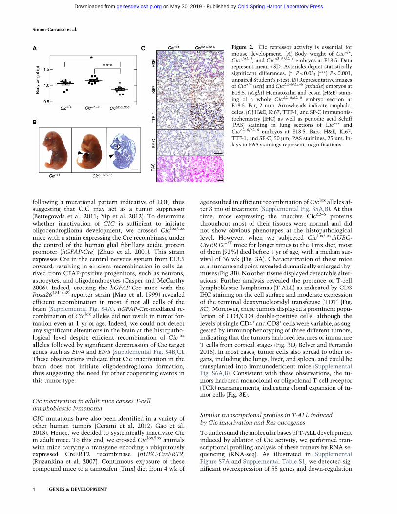
age resulted in efficient recombination ofCiclox alleles af-ter 3 mo of treatment (Supplemental Fig. S5A,B). At thistime, mice expressing the inactive CicΔ2–6 proteinsthroughout most of their tissues were normal and didnot show obvious phenotypes at the histopathologicallevel. However, when we subjected Ciclox/lox;hUBC-CreERT2+/T mice for longer times to the Tmx diet, mostof them (92%) died before 1 yr of age, with a median sur-vival of 36 wk (Fig. 3A). Characterization of these miceat a humane end point revealed dramatically enlarged thy-muses (Fig. 3B). No other tissue displayed detectable alter-ations. Further analysis revealed the presence of T-celllymphoblastic lymphomas (T-ALL) as indicated by CD3IHC staining on the cell surface and moderate expressionof the terminal deoxynucleotidyl transferase (TDT) (Fig.3C). Moreover, these tumors displayed a prominent popu-lation of CD4/CD8 double-positive cells, although thelevels of single CD4+ and CD8+ cells were variable, as sug-gested by immunophenotyping of three different tumors,indicating that the tumors harbored features of immatureT cells from cortical stages (Fig. 3D; Belver and Ferrando2016). In most cases, tumor cells also spread to other or-gans, including the lungs, liver, and spleen, and could betransplanted into immunodeficient mice (SupplementalFig. S6A,B). Consistent with these observations, the tu-mors harbored monoclonal or oligoclonal T-cell receptor(TCR) rearrangements, indicating clonal expansion of tu-mor cells (Fig. 3E).
Similar transcriptional profiles in T-ALL inducedby Cic inactivation and Ras oncogenes
Tounderstand themolecular bases of T-ALL developmentinduced by ablation of Cic activity, we performed tran-scriptional profiling analysis of these tumors by RNA se-quencing (RNA-seq). As illustrated in SupplementalFigure S7A and Supplemental Table S1, we detected sig-nificant overexpression of 55 genes and down-regulation
Figure 2. Cic repressor activity is essential formouse development. (A) Body weight of Cic+/+,Cic+/Δ2–6, and CicΔ2–6/Δ2–6 embryos at E18.5. Datarepresent mean ± SD. Asterisks depict statisticallysignificant differences. (∗) P < 0.05; (∗∗∗) P < 0.001,unpaired Student’s t-test. (B) Representative imagesof Cic+/+ (left) and CicΔ2–6/Δ2–6 (middle) embryos atE18.5. (Right) Hematoxilin and eosin (H&E) stain-ing of a whole CicΔ2–6/Δ2–6 embryo section atE18.5. Bar, 2 mm. Arrowheads indicate omphalo-celes. (C ) H&E, Ki67, TTF-1, and SP-C immunohis-tochemistry (IHC) as well as periodic acid Schiff(PAS) staining in lung sections of Cic+/+ andCicΔ2–6/Δ2–6 embryos at E18.5. Bars: H&E, Ki67,TTF-1, and SP-C, 50 µm; PAS stainings, 25 µm. In-lays in PAS stainings represent magnifications.
Simón-Carrasco et al.
4 GENES & DEVELOPMENT
Cold Spring Harbor Laboratory Press on May 30, 2019 - Published by genesdev.cshlp.orgDownloaded from
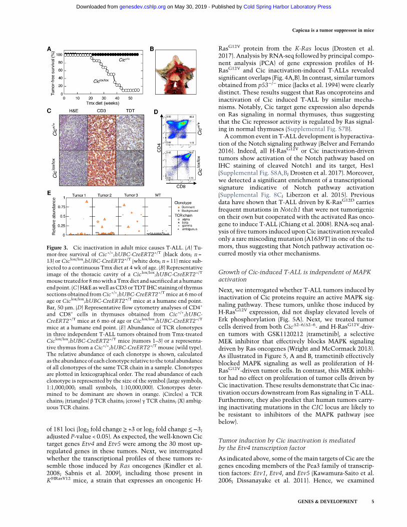
of 181 loci (log2 fold change≥ +3 or log2 fold change≤−3;adjusted P-value < 0.05). As expected, the well-known Cictarget genes Etv4 and Etv5 were among the 30 most up-regulated genes in these tumors. Next, we interrogatedwhether the transcriptional profiles of these tumors re-semble those induced by Ras oncogenes (Kindler et al.2008; Sabnis et al. 2009), including those present inKHRasV12 mice, a strain that expresses an oncogenic H-
RasG12V protein from the K-Ras locus (Drosten et al.2017). Analysis by RNA-seq followed by principal compo-nent analysis (PCA) of gene expression profiles of H-RasG12V and Cic inactivation-induced T-ALLs revealedsignificant overlaps (Fig. 4A,B). In contrast, similar tumorsobtained from p53−/− mice (Jacks et al. 1994) were clearlydistinct. These results suggest that Ras oncoproteins andinactivation of Cic induced T-ALL by similar mecha-nisms. Notably, Cic target gene expression also dependson Ras signaling in normal thymuses, thus suggestingthat the Cic repressor activity is regulated by Ras signal-ing in normal thymuses (Supplemental Fig. S7B).A common event in T-ALL development is hyperactiva-
tion of the Notch signaling pathway (Belver and Ferrando2016). Indeed, all H-RasG12V or Cic inactivation-driventumors show activation of the Notch pathway based onIHC staining of cleaved Notch1 and its target, Hes1(Supplemental Fig. S8A,B; Drosten et al. 2017). Moreover,we detected a significant enrichment of a transcriptionalsignature indicative of Notch pathway activation(Supplemental Fig. 8C; Liberzon et al. 2015). Previousdata have shown that T-ALL driven by K-RasG12D carriesfrequent mutations in Notch1 that were not tumorigenicon their own but cooperated with the activated Ras onco-gene to induce T-ALL (Chiang et al. 2008). RNA-seq anal-ysis of five tumors induced uponCic inactivation revealedonly a rare miscoding mutation (A1639T) in one of the tu-mors, thus suggesting that Notch pathway activation oc-curred mostly via other mechanisms.
Growth of Cic-induced T-ALL is independent of MAPKactivation
Next, we interrogated whether T-ALL tumors induced byinactivation of Cic proteins require an active MAPK sig-naling pathway. These tumors, unlike those induced byH-RasG12V expression, did not display elevated levels ofErk phosphorylation (Fig. 5A). Next, we treated tumorcells derived from both CicΔ2–6/Δ2–6- and H-RasG12V-driv-en tumors with GSK1120212 (trametinib), a selectiveMEK inhibitor that effectively blocks MAPK signalingdriven by Ras oncogenes (Wright and McCormack 2013).As illustrated in Figure 5, A and B, trametinib effectivelyblocked MAPK signaling as well as proliferation of H-RasG12V-driven tumor cells. In contrast, this MEK inhibi-tor had no effect on proliferation of tumor cells driven byCic inactivation. These results demonstrate that Cic inac-tivation occurs downstream from Ras signaling in T-ALL.Furthermore, they also predict that human tumors carry-ing inactivating mutations in the CIC locus are likely tobe resistant to inhibitors of the MAPK pathway (seebelow).
Tumor induction by Cic inactivation is mediatedby the Etv4 transcription factor
As indicated above, some of themain targets of Cic are thegenes encoding members of the Pea3 family of transcrip-tion factors: Etv1, Etv4, and Etv5 (Kawamura-Saito et al.2006; Dissanayake et al. 2011). Hence, we examined
Figure 3. Cic inactivation in adult mice causes T-ALL. (A) Tu-mor-free survival of Cic+/+;hUBC-CreERT2+/T (black dots; n =13) or Ciclox/lox;hUBC-CreERT2+/T (white dots; n = 11) mice sub-jected to a continuous Tmx diet at 4 wk of age. (B) Representativeimage of the thoracic cavity of a Ciclox/lox;hUBC-CreERT2+/T
mouse treated for 8mowith aTmxdiet and sacrificed at ahumaneendpoint. (C ) H&E aswell asCD3orTDT IHCstaining of thymussections obtained fromCic+/+;hUBC-CreERT2+/Tmice at 6mo ofage or Ciclox/lox;hUBC-CreERT2+/T mice at a humane end point.Bar, 50 µm. (D) Representative flow cytometry analyses of CD4+
and CD8+ cells in thymuses obtained from Cic+/+;hUBC-CreERT2+/T mice at 6 mo of age or Ciclox/lox;hUBC-CreERT2+/T
mice at a humane end point. (E) Abundance of TCR clonotypesin three independent T-ALL tumors obtained from Tmx-treatedCiclox/lox;hUBC-CreERT2+/T mice (tumors 1–3) or a representa-tive thymus from a Cic+/+;hUBC-CreERT2+/T mouse (wild type).The relative abundance of each clonotype is shown, calculatedas theabundanceof each clonotype relative to the total abundanceof all clonotypes of the same TCR chain in a sample. Clonotypesare plotted in lexicographical order. The read abundance of eachclonotype is represented by the size of the symbol (large symbols,1:1,000,000; small symbols, 1:10,000,000). Clonotypes deter-mined to be dominant are shown in orange. (Circles) α TCRchains; (triangles) β TCR chains; (cross) γ TCR chains; (X) ambig-uous TCR chains.
Capicua is a tumor suppressor in mice
GENES & DEVELOPMENT 5
Cold Spring Harbor Laboratory Press on May 30, 2019 - Published by genesdev.cshlp.orgDownloaded from

whether any of these proteins might be responsible formediating tumor induction upon inactivation of Cic.Since Etv4was the genemost up-regulated in cells lackingactiveCic repression, we introduced Etv4-deficient alleles(Etv4NLZ/NLZ) (Livet et al. 2002) into Ciclox/lox;UBC-CreERT2+/T mice. As illustrated in Figure 5C, reduced ex-pression of Etv4 in Ciclox/lox;Etv4+/NLZ;UBC-CreERT2+/T
animals had no effect on tumor development, since all ofthemice developed T-ALL with a median survival similarto that of animals carrying wild-type Etv4 alleles. Howev-er, complete ablation of Etv4 dramatically prevented theinduction of T-ALL upon inactivation of Cic. Indeed,four of five Ciclox/lox;Etv4NLZ/NLZ;UBC-CreERT2+/T
mice had no signs of thymic disease by 1 yr of age, atime when all Ciclox/lox;Etv4+/+;UBC-CreERT2+/T ani-mals had already succumbed to the disease. Only oneout of these five mice displayed limited tumor formationby 1 yr of age, indicating that tumor growth was dramati-cally delayed. These results strongly indicate that Etv4 is akey mediator of the tumorigenic consequences of Cic in-activation. Notably, absence of Etv4 did not per se affectnormal thymocyte development (Fig. 5D,E). Interestingly,CicΔ2–6/Δ2–6;Etv4NLZ/NLZ tumor cells displayed increasedlevels of Etv5 and, to a lesser extent, Etv1 expression, in-dicating that in the absence of Etv4, Etv5 is the primarymember of the Pea3 family to respond to Cic inactivation(Fig. 5F). However, this increased expression is not suffi-cient to induce T-ALL, at least in most mice. Whetherconcomitant ablation of Etv4 and Etv5 expression willcompletely block T-ALL development or whether overex-pression of Etv4 alone is sufficient to cause T-ALL re-mains to be determined.
Identification of a gene signature indicative of CICinactivation in mouse and human T-ALL
Human T-ALL has been classified based on transcription-al profiling. Work by Ferrando et al. (2002) identified genesignatures that correspond to specific stages of T-cell de-
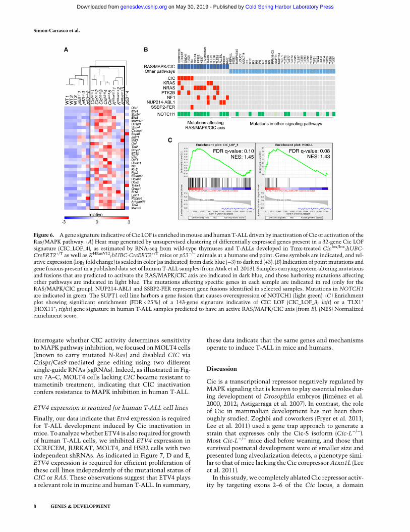
velopment. We compared these human T-ALL signatureswith expression data obtained from T-ALL tumors in-duced by either H-RasG12V expression or Cic inactivation.As illustrated in Supplemental Figure S8D, tumors in-duced by Cic inactivation were highly enriched for theTLX1+ signature (also known asHOX11+), thus suggestingthat these tumors share features of human T-ALL arrestedat the early cortical stage (Belver and Ferrando 2016). Inagreement with the PCA data, H-RasG12V-driven tumorsalso displayed enrichment of this signature but to a slight-ly lesser degree. These observations suggest that tumorsdriven by Ras oncogenes should display a transcriptionalsignature indicative of Cic inactivation. To this end, wefirst established a Cic LOF gene signature based on genesthat carried evolutionarily conserved Cic-binding sitesand became derepressed in tumors induced by Cic inacti-vation. Specifically, we selected a 32-gene signature (CicLOF signature CIC_LOF_4) based on genes that were up-regulated at least a log2 fold change of 1.5 and harboredat least one Cic-binding sequence (CBS) (Kawamura-Saito et al. 2006) with a conservation score of >0.875in their promoters (see the Supplemental Material;Supplemental Fig. S9A; Supplemental Table S2). Next,we performed unsupervised clustering of the independentT-ALL tumor samples using this Cic LOF signature. As il-lustrated in Figure 6A, Cic LOF- and H-RasG12V-inducedtumors formed a common cluster. Although they didnot cluster with the aforementioned tumors, p53-null tu-mors showed a limited enrichment of the Cic LOF signa-ture (Supplemental Fig. S9B). These observations indicatethat Cic target genes are derepressed in H-RasG12V-driventumors, suggesting that hyperactive Ras signaling causesinactivation of Cic to promote T-ALL.
We next wanted to interrogate whether the Cic LOFgene signature derived from ourmurine T-ALL expressiondata is conserved in humanT-ALL samples harboring acti-vation of the RAS/MAPK/CIC axis. To this end, we tookadvantage of a published T-ALL data set that includesgeneexpressionandmutationdata from31T-ALLpatients
Figure 4. Similar transcriptional profiles inT-ALL driven by Cic inactivation or Rashyperactivation. (A) PCA of gene expressionprofiles obtained from wild-type thymuses(black dots) and T-ALLs developed in Tmx-treated Ciclox/lox;hUBC-CreERT2+/T (reddots), KHRasV12;hUBC-CreERT2+/T (greendots), or p53−/− mice (blue dots) at a humaneend point. (B) Heat map displaying differen-tially expressed genes (log2 fold change≥ +3or log2 fold change≤−3; adjusted P-value <0.05; false discovery rate [FDR] 0.05) as esti-mated by RNA-seq from samples shown inA. Relative expression (log2 fold change) isscaled in color (as indicated) from dark blue(−3) to dark red (+3). Gene symbols are listedin Supplemental Table S1.
Simón-Carrasco et al.
6 GENES & DEVELOPMENT
Cold Spring Harbor Laboratory Press on May 30, 2019 - Published by genesdev.cshlp.orgDownloaded from

and 18 T-ALL cell lines (Atak et al. 2013). We selectedthose samples that carried either a mutation in CIC orany other alteration that predicts CIC inactivation via ac-tivation of the MAPK pathway, such as mutations inNRAS, KRAS, NF1, or PTK2B. In addition, we includedsamples carrying the NUP214-ABL1 or SSBP2-FER fu-sions, since their activated tyrosine kinases are known toinduce MAPK activation. Following this strategy, we as-sembled 16 out of the 49 samples into a group that was ex-pected to harbor inactive CIC via one of the mechanismsdescribed above (Fig. 6B). Since our Cic LOF signature de-rived frommouse tumors was composed of only 32 genes,we did not find enrichment of this restricted signature inthe RAS/MAPK/CIC sample group. However, when weused a more relaxed signature composed of 143 genesthat were significantly up-regulated in Cic LOF-driven T-ALL and contained at least one CBS (CIC_LOF_3)(SupplementalTable S3; Supplemental Fig. S9A),we foundthis signature to be significantly enriched in the RAS/MAPK/CIC sample group (Fig. 6C). In addition, we foundtheTLX1+ signature to be enriched in this group of 16 sam-ples, providing further evidence for the similarity between
human andmouse tumors (Fig. 6C). Taken together, thesedata indicate that human T-ALL samples driven by theRAS/MAPK/CIC axis and mouse tumors driven by H-RasG12V expression or Cic inactivation share a gene signa-ture characteristic of CIC inactivation.
CIC inactivation in human T-ALL causes resistance toMAPK inhibition
Sequencing analysis of primary T-ALL clinical specimensrevealed the presence of CIC mutations in 10% (n = 7 of69) of cases analyzed (Supplemental Fig. S10A–C). Otherstudies also identifiedCICmutations, albeit at a lower fre-quency (Supplemental Fig. S10D; Atak et al. 2013;Kataoka et al. 2015). These mutations are predicted tocause resistance toMAPK inhibition, similar to our obser-vation in murine T-ALL cells. To test this hypothesis, wefirst determined the sensitivity of a variety of human T-ALL cell lines to trametinib. Notably, exposure to trame-tinib had no effect in CCRFCEM and JURKAT cells(Supplemental Fig. S11A,B). In contrast, MOLT4 andHSB2 cells were sensitive to this MEK inhibitor. To
Figure 5. MurineT-ALL driven byCic inactivation is resistant toMEK inhibition and requires Etv4. (A)Western blot analysis of p-Erk1/2and total Erk1/2 in T-ALL cells obtained from tumors developed in Tmx-treated KHRasV12;hUBC-CreERT2+/T (left) or Ciclox/lox;hUBC-CreERT2+/T (right) mice treated with the indicated concentrations of trametinib for 24 h. GAPDH expression served as a loading control.(B) Growth curve of T-ALL cells obtained from tumors developed in Tmx-treated KHRasV12;hUBC-CreERT2+/T (left) or Ciclox/lox;hUBC-CreERT2+/T (right) mice treated with the indicated concentrations of trametinib for the indicated time. (C ) Tumor-free survivalof Ciclox/lox;Etv4+/NLZ;hUBC-CreERT2+/T (open dots; n = 11) or Ciclox/lox;Etv4NLZ/NLZ;hUBC-CreERT2+/T (closed dots; n = 5) mice sub-jected to a continuous Tmx diet starting at 4 wk of age. (D) Percentage of thymocyte populations in 1-mo-old wild-type (open bars; n = 3)or Etv4NLZ/NLZ (closed bars; n = 3) mice. CD4/CD8-double-negative, CD4/CD8-double-positive, CD4-single-positive, and CD8-single-positive populations are shown. Data represent mean ± SD. (E) Percentage of CD4/CD8 double-negative (DN) thymocyte subpopula-tions in 1-mo-old wild-type (open bars; n = 3) or Etv4NLZ/NLZ (closed bars; n = 3) mice. DN1 (CD44+/CD25−), DN2 (CD44+/CD25+),DN3 (CD44−/CD25+), and DN4 (CD44−/CD25−) subpopulations are shown. Data represent mean ± SD. (F ) Relative expression levels ofEtv1, Etv4, and Etv5 mRNAs in wild-type thymuses (open bars; n = 2); T-ALLs developed in Tmx-treated Ciclox/lox;hUBC-CreERT2+/T
mice (CicΔ2–6/Δ2–6; red bars; n = 2); or the only tumor that developed in Tmx-treated Ciclox/lox;Etv4NLZ/NLZ;hUBC-CreERT2+/T mice(CicΔ2–6/Δ2–6;Etv4NLZ/NLZ; closed bars). β-Actin expression levels were used for normalization. Data represent mean ± SD.
Capicua is a tumor suppressor in mice
GENES & DEVELOPMENT 7
Cold Spring Harbor Laboratory Press on May 30, 2019 - Published by genesdev.cshlp.orgDownloaded from
interrogate whether CIC activity determines sensitivitytoMAPK pathway inhibition, we focused onMOLT4 cells(known to carry mutated N-Ras) and disabled CIC viaCrispr/Cas9-mediated gene editing using two differentsingle-guide RNAs (sgRNAs). Indeed, as illustrated in Fig-ure 7A–C, MOLT4 cells lacking CIC became resistant totrametinib treatment, indicating that CIC inactivationconfers resistance to MAPK inhibition in human T-ALL.
ETV4 expression is required for human T-ALL cell lines
Finally, our data indicate that Etv4 expression is requiredfor T-ALL development induced by Cic inactivation inmice. To analyzewhether ETV4 is also required for growthof human T-ALL cells, we inhibited ETV4 expression inCCRFCEM, JURKAT, MOLT4, and HSB2 cells with twoindependent shRNAs. As indicated in Figure 7, D and E,ETV4 expression is required for efficient proliferation ofthese cell lines independently of the mutational status ofCIC or RAS. These observations suggest that ETV4 playsa relevant role in murine and human T-ALL. In summary,
these data indicate that the same genes and mechanismsoperate to induce T-ALL in mice and humans.
Discussion
Cic is a transcriptional repressor negatively regulated byMAPK signaling that is known to play essential roles dur-ing development of Drosophila embryos (Jiménez et al.2000, 2012; Astigarraga et al. 2007). In contrast, the roleof Cic in mammalian development has not been thor-oughly studied. Zoghbi and coworkers (Fryer et al. 2011;Lee et al. 2011) used a gene trap approach to generate astrain that expresses only the Cic-S isoform (Cic-L−/−).Most Cic-L−/− mice died before weaning, and those thatsurvived postnatal development were of smaller size andpresented lung alveolarization defects, a phenotype simi-lar to that ofmice lacking the Cic corepressorAtxn1L (Leeet al. 2011).
In this study, we completely ablated Cic repressor activ-ity by targeting exons 2–6 of the Cic locus, a domain
Figure 6. A gene signature indicative of Cic LOF is enriched inmouse and humanT-ALL driven by inactivation of Cic or activation of theRas/MAPK pathway. (A) Heat map generated by unsupervised clustering of differentially expressed genes present in a 32-gene Cic LOFsignature (CIC_LOF_4), as estimated by RNA-seq from wild-type thymuses and T-ALLs developed in Tmx-treated Ciclox/lox;hUBC-CreERT2+/T as well as KHRasV12;hUBC-CreERT2+/T mice or p53−/− animals at a humane end point. Gene symbols are indicated, and rel-ative expression (log2 fold change) is scaled in color (as indicated) from dark blue (−3) to dark red (+3). (B) Indication of pointmutations andgene fusions present in a published data set of humanT-ALL samples (fromAtak et al. 2013). Samples carrying protein-alteringmutationsand fusions that are predicted to activate the RAS/MAPK/CIC axis are indicated in dark blue, and those harboring mutations affectingother pathways are indicated in light blue. The mutations affecting specific genes in each sample are indicated in red (only for theRAS/MAPK/CIC group). NUP214-ABL1 and SSBP2-FER represent gene fusions identified in selected samples. Mutations in NOTCH1are indicated in green. The SUPT1 cell line harbors a gene fusion that causes overexpression of NOTCH1 (light green). (C ) Enrichmentplot showing significant enrichment (FDR < 25%) of a 143-gene signature indicative of CIC LOF (CIC_LOF_3; left) or a TLX1+
(HOX11+; right) gene signature in human T-ALL samples predicted to have an active RAS/MAPK/CIC axis (from B). (NES) Normalizedenrichment score.
Simón-Carrasco et al.
8 GENES & DEVELOPMENT
Cold Spring Harbor Laboratory Press on May 30, 2019 - Published by genesdev.cshlp.orgDownloaded from

encoding the HMG-box required for DNA binding and re-pressor activity of both Cic isoforms, Cic-L and Cic-S. Asexpected, the truncated CicΔ2–6 proteins did not bind tothe promoters of their target genes. When Cic activitywas inactivated in the germline, none of the CicΔ2–6/Δ2–6
mice survived after birth despite being present at Mende-lian ratios at E18.5. We detected dramatic defects in lungmaturation, thus suggesting that these immature lungswere primarily responsible for the perinatal lethality inCicΔ2–6/Δ2–6 mice. However, we could not assess the pres-ence of lung alveolarization defects such as those ob-served in surviving Cic-L−/− and Atxn1L−/− animals
because lung alveolarization occurs at later stages of post-natal life.The majority of E18.5 CicΔ2–6/Δ2–6 embryos (70%) also
presented omphaloceles, a phenotype observed inAtxn1−/−;Atxn1L−/− mice (Lee et al. 2011), thus suggest-ing that the Cic/Atxn1 or Cic/Atxn1L complex is essen-tial for the regulation of abdominal wall closure.However, our CicΔ2–6/Δ2–6 embryos did not recapitulateall of the defects present in Atxn1−/−;Atxn1L−/− mice.For instance, we did not observe hydrocephali, suggestingthat Atxn1/Atxn1L proteins may possess Cic-indepen-dent functions. Alternatively, the different phenotypesmay be explained by low residual levels of Cic proteinsin Atxn1−/−;Atxn1L−/− mice (Lee et al. 2011).Previous studies have shown that Cic inactivation in
the eye imaginal disc and adult intestinal stem cells inDrosophila rescues the proliferation defects caused by ab-lation of the single Ras gene (Tseng et al. 2007; Jin et al.2015). This indicates that Cic is a key mediator that con-nects MAPK signaling with the transcriptional programrequired to process mitogenic signals. However, ablationof Cic activity in cells lacking the three Ras isoformsfailed to restore their proliferative properties, indicatingthat Ras mitogenic signals must be mediated by addition-al components of the transcriptional machinery, at leastin these cells.CIC is recurrently mutated in human oligodendroglio-
mas. These tumors display mutations in one CIC allele,whereas the other is lost as part of larger 19q chromosom-al deletions, suggesting that CIC functions as a tumor sup-pressor (Bettegowda et al. 2011; Yip et al. 2012). However,selective inactivation of Cic proteins in the mouse brainwas not sufficient to drive oligodendroglioma formation.As illustrated here, selective expression of inactive Cicproteins in most brain cells, including neuronal and glialprecursors derived from hGFAP-Cre-expressing progeni-tors, failed to induce any detectable alterations in 1-yr-old mice. In human oligodendrogliomas, CIC is alwayscomutatedwith IDH1 or IDH2 (Yip et al. 2012) or, less fre-quently, FUBP1, thus raising the possibility that tumorformation requires cooperation of these cancer geneswithCIC (Bettegowda et al. 2011). Interestingly, Idh1mu-tant mice also fail to develop brain tumors (Sasaki et al.2012). Finally, it should also be noted that CICmutationsare not always maintained in recurrent oligodendroglio-mas, arguing that these mutations may be subclonal sec-ondary events that do not necessarily provide a growthadvantage for tumor cells (Aihara et al. 2017).Conditional inactivation of Cic proteins in adult mice
resulted in the development of T-ALL after 6–9 mo withalmost complete penetrance (>90%), thus demonstratingthat Cic can act as a tumor suppressor in mice. These tu-mors were significantly enriched in the TLX1+ signature,thus suggesting that Cic inactivation causes thymocytearrest at the early cortical stage (Belver and Ferrando2016). Interestingly, Cic is likely a target of the Tlx1 re-pressor, a fact that raises the possibility that Tlx1-mediat-ed repression of Cic plays a role in T-ALL developmentinduced by this transcriptional repressor (De Keers-maecker et al. 2010; Della Gatta et al. 2012). However,
Figure 7. Resistance to MEK inhibition and requirement forETV4 expression in human T-ALL cell lines. (A) Western blotanalysis of p-ERK1/2 and total ERK1/2 expression in MOLT4cells treated with the indicated concentrations of trametinib for24 h. GAPDH expression served as a loading control. (B) Westernblot analysis of CIC expression levels in MOLT4 cells 2 wk afterinfection with lentiviruses expressing Cas9 and the indicatedsgRNAs. GAPDH expression served as a loading control. (C ) Pro-liferation of MOLT4 cells 2 wk after infection with lentivirusesexpressing Cas9 and the indicated sgRNAs and treated with theindicated concentrations of trametinib. (D) Proliferation ofCCRFCEM, JURKAT, HSB2, andMOLT4 cells infected with len-tiviruses expressing two independent shRNAs targeting ETV4 ora nontargeting control. (E) Relative ETV4 mRNA expression lev-els in 293T cells infected with lentiviruses expressing a controlshRNA or two different shRNAs against ETV4.
Capicua is a tumor suppressor in mice
GENES & DEVELOPMENT 9
Cold Spring Harbor Laboratory Press on May 30, 2019 - Published by genesdev.cshlp.orgDownloaded from
the functional relevance of this pathway for T-ALL devel-opment remains to be determined.
T-ALL development via activation of Ras oncogeneshas also been described in mouse tumor models (Kindleret al. 2008; Sabnis et al. 2009). More recently, we observedT-ALL in mice that express the H-RasG12V oncoproteindriven from the K-Ras promoter (Drosten et al. 2017).These tumors are indistinguishable from those inducedupon Cic inactivation. Indeed, the gene expression pro-files of tumors induced byH-RasG12V expression or Cic in-activation are highly similar. Hence, we propose that asignificant proportion of gene expression changes in Ras-driven T-ALL occurs via inactivation of Cic. Likewise, agene signature indicative of Cic inactivation was highlyenriched in Ras-driven T-ALL.More importantly, this sig-nature is also significantly enriched in human T-ALLsamples harboring mutations that are predicted to causeaberrant signaling through the RAS/MAPK/CIC axis.These observations indicate that CIC acts downstreamfrom the RAS/MAPK pathway in T-ALL. Although RASmutations are not frequent in these cancers (∼5%), ithas been suggested that up to 50% of all human T-ALLcases have aberrant RAS signaling (von Lintig et al.2000). In addition, RAS/MAPK-activating mutations aremuch more prevalent in relapsed T-ALL (Oshima et al.2016), thus suggesting that CIC inactivation through hy-peractive Ras signalingmay play a relevant role in a signif-icant number of T-ALL cases.
However, these T-ALL tumors may present differentresponses to pharmacological inhibition of the MAPKpathway. For instance, tumors that developed as a conse-quence of K-RasG12D activation in mice are sensitive toMek inhibition (Dail et al. 2010). Likewise, as demonstrat-ed here, tumor cells derived from lymphomas driven byH-RasG12V expression could be efficiently inhibited uponexposure to the MEK inhibitor trametinib. However, T-ALLs that arose upon Cic inactivation were completelyresistant to this inhibitor, indicating that mutations inthis locus activate mitogenic signaling downstreamfrom the MAPK cascade. Indeed, CIC inactivation in hu-man T-ALL cell lines also caused resistance to MEK inhi-bition. Hence, CIC mutations may develop as a potentialmechanism of resistance to MAPK inhibitors. Of note,mutations in CIC have been identified in human T-ALL,albeit at a low frequency (Atak et al. 2013; Kataoka et al.2015; this study). Consistent with our observations, CIChas been identified recently in a screen to detect novelmechanisms that cause resistance to trametinib in othertumor models (Wang et al. 2017). Similarly, CIC has alsobeen identified in a screen for genes whose absence pro-motes resistance to EGFR inhibition (Liao et al. 2017).Finally, Bivona and colleagues (Okimoto et al. 2017) haveshown that CIC inactivation is associated with advancedtumor stages andmetastasis formation in lung and gastrictumors, thus suggesting that these tumors may be intrin-sically resistant to MEK or EGFR inhibition.
The emerging role of CIC mutations in human cancersmakes it necessary to understand the mechanisms thatmediate its repressor activity. As indicated above, thebest-known CIC targets are the members of the PEA3
family of transcription factors: ETV1, ETV4, and ETV5.However, the functional consequences of inhibiting CICrepression in human tumors are unknown. Here, we pro-vide genetic evidence that in T-ALL induced by inactiva-tion of Cic proteins, the main Cic effector is thetranscription factor Etv4. Indeed, ablation of Etv4 expres-sion in Cic inactivation-induced T-ALL completely pre-vented the development of this disease in four out offive mice and significantly delayed tumor formation inthe remaining animal. Notably, thymocyte developmentwas not altered in Etv4-deficient mice, indicating thatEtv4 (and possibly other Pea3 family members such asEtv5) is selectively required for tumor formation.WhetherEtv4 is also required for Ras-driven T-ALL remains to bedetermined. However, similar observations were madeupon depletion of ETV4 in human T-ALL cell lines, al-though growth inhibition appears to be independent ofthe mutational status of CIC or RAS. Thus, a betterunderstanding of the molecular events triggered by CICinactivation should providemore effective therapeutic ap-proaches to treat those human tumors driven by eitherRAS hyperactivation or CIC mutations.
Materials and methods
Mice
ThehUBC-CreERT2 (Ruzankinaet al. 2007),EIIa-Cre (Laksoetal.1996), p53−/− (Jacks et al. 1994), hGFAP-Cre (Zhuo et al. 2001),Etv4NLZ/NLZ (Livet et al. 2002), H-Ras−/−;N-Ras−/−;K-Raslox/lox
(Drosten et al. 2010), and KHRasV12 (Drosten et al. 2017) strainshave been described previously. Activation of the inducibleCreERT2 recombinase was achieved by feeding the mice with aTmx-containing diet (Harlan Laboratories). All mice were main-tained in a mixed 129/Sv-C57BL/6 background. For tumor celltransplantation assays, single-cell suspensions (106 cells per 200µL of PBS) were injected into the tail veins of immunodeficientmice. All animal experiments were approved by the Ethical Com-mittees of the SpanishNational Cancer Research Centre (CNIO),the Carlos III Health Institute, and the Autonomous Communityof Madrid and were performed in accordance with the guidelinesstated in the International Guiding Principles for Biomedical Re-search Involving Animals, developed by the Council for Interna-tional Organizations of Medical Sciences (CIOMS).
Cell lines
MEFs were extracted from E13.5 embryos and cultured in DMEMsupplemented with 10% FBS. T-ALL cell lines established fromtumor-bearing mice and human T-ALL cell lines were propagatedin RPMI1640 supplemented with 10% FBS. For growth inhibitionassays, murine cells were seeded at a density of 2 × 10⁵ cells permilliliter with various concentrations of trametinib. Human celllines were seeded in 96-well plates at a density of 10,000 cellsper well, and proliferationwas assessed using the (4,5-dimethylth-iazol-2-yl)-2,5-diphenyltetrazoliumbromide (MTT) assay (Roche).
Plasmids
The GFP-tagged human CIC-S construct in pcDNA5/FRT/TO(Dissanayake et al. 2011) was a kind gift of Dr. Carol MacKintosh(University of Dundee, UK). The deletion of exons 2–6 (spanningresidues 24–311) was introduced using the QuikChange site-
Simón-Carrasco et al.
10 GENES & DEVELOPMENT
Cold Spring Harbor Laboratory Press on May 30, 2019 - Published by genesdev.cshlp.orgDownloaded from

directed mutagenesis kit (Agilent) following the manufacturer’sguidelines. All plasmids were stably introduced into Flp-In T-REx 293 cells (Invitrogen) following instructions from the manu-facturer. Human CIC targeting sgRNAs (#1, CCCCTCCGTGCAGCCGAGCG; #2, GCCTCGCTCGGCTGCACGGA) or a non-targeting control sgRNA (CTCGTGAACAAGATCCGAC) werecloned into lentiCRISPRv2 (Addgene 52961). Sigma MISSIONshRNA lentiviral vectors were used to knock down ETV4 expres-sion in human cell lines (#232, TRCN0000013937; and #736,TRCN0000013934). A nontargeting shRNA vector (SHC002)was used as control.
Histopathology and IHC
For histological analyses, tissues were fixed in 10% buffered for-malin and embedded in paraffin. Hematoxilin and eosin (H&E)staining and IHC analyses were performed on 3-µm paraffin sec-tions. For IHC, the following antibodies were used: CD3 (Phar-mingen, 553057), cleaved Notch1 (Abcam, ab8925), Hes1(CNIO), TDT (DAKO, A3524), Ki67 (Master Diagnostica,0003110QD), γH2AX (Millipore, 05-636), and active Caspase 3(Cell Signaling, 9661). PAS staining was performed by incubationfor 5 min with periodic acid followed by 20 min of incubationwith Schiff reagent and hematoxylin counterstaining.
Western blot analysis
Western blot analysis of protein extracts obtained from tissues,cell lines, or MEFs was performed as described (Drosten et al.2010). To specifically probe for Cic expression levels, protein ex-tracts were prepared in TST buffer as described (Lam et al. 2006).Primary antibodies used included polyclonal Cic antisera for themouse proteins (Lam et al. 2006; Kim et al. 2015), human CIC(Abcam, ab123822), pErk1/2 (Cell Signaling Technology, 9101),pAkt (Cell Signaling Technology, 9271), Akt (Cell SignalingTechnology, 9272), Erk1 (Santa Cruz Biotechnology, C16), andGAPDH (Sigma-Aldrich, G8795).
Flow cytometry analysis
Thymocytes were obtained by mechanical dissociation of thethymus in RPMI1640. Before analysis, cells were preincubatedwith purified anti-mouse CD16/32 antibodies (1:200; BD Phar-mingen) for 15 min on ice to block nonspecific Fc receptor-medi-ated binding. Aliquots of 5 × 10⁶ cells were stained for 20 min atroom temperature with the following monoclonal antibodies:PECy7 α-Gr1 (1:200), BUV737 α-CD19 (1:400), APCCy7 α-Ter119 (1:200), PE α-Nk1.1/α-DX5 (1:200; to exclude non T cells),FITC α-CD4 (1:800), BUV395 α-CD8 (1:400), APC α-CD44 (1:200),and PerCPCy5.5 α-CD25 (1:200). Samples were processed on aLSRFortessa flow cytometer (BDPharmingen) and analyzed usingFlowJo (Tree Star).
Statistical analysis
Data are mean ± SD for Figures 1E, 5D–F, and 7C–E. Comparisonbetween different genotypes in Figure 2B was performed using anunpaired Student’s t-test. P-values of <0.05 were considered to bestatistically significant (P < 0.05 [∗] and P < 0.001 [∗∗∗]).
Acknowledgments
We are grateful to Carol MacKintosh (University of Dundee, UK)for the pcDNA5/FRT/TO-GFP-CIC plasmid, and Huda Zoghbi
(Baylor College ofMedicine, Houston, TX) and Yoontae Lee (Uni-versity of Pohang, Korea) for Cic antisera. We thank Scott Brownand Robert Holt (University of Vancouver, Canada) for theirhelp with TCR abundance calculations. We also thank CarmenG. Lechuga, Marta San Roman, Raquel Villar, Beatriz Jiménez,and Nuria Cabrera for excellent technical assistance. We valuethe support of Sagrario Ortega (Transgenic Mice Core Unit,CNIO) for help in generating the Cic mutant mice, OrlandoDominguez (Genomics Core Unit, CNIO) for the RNA-seq analy-sis, and the Histopathology Core Unit. This work was supportedby grants from the Fundació La Marató de TV3 (20131730/1)to G.J. and M.B., and the European Research Council (ERC-AG/250297-RAS AHEAD), the EU-Framework Programme(HEALTH-F2-2010-259770/LUNGTARGET and HEALTH-2010-260791/EUROCANPLATFORM), the Spanish Ministry ofEconomy and Competitiveness (SAF2014-59864-R), the Autono-mous Community of Madrid (S2011/BDM-2470/ONCOCYCLE),and the Asociación Española contra el Cáncer (AECC)(GC16173694BARB) to M.B. M.B. is the recipient of an EndowedChair fromtheAXAResearchFund. L.S.-C.was supportedbya fel-lowship from thePrograma de Formación de Personal Investigator(FPI) of the Spanish Ministry of Economy and Competitiveness.M.D. and M.B. conceived and designed the study. L.S.-C., O.G.,G.J., M.D., and M.B. developed the methodology. L.S.-C., O.G.,M.S., and M.D. acquired the data. L.S.-C., O.G., M.S., H.K.C.J.,G.J., M.D., and M.B. analyzed and interpreted the data. L.S-C.,O.G., G.J., M.D., and M.B. wrote, reviewed, and/or revised themanuscript. G.J. provided material support. A.G. analyzed theT-ALL sequencing. M.D. and M.B. supervised the study.
References
Aihara K,MukasaA,NagaeG,NomuraM, Yamamoto S, UedaH,Tatsuno K, Shibahara J, Takahashi M, Momose T, et al. 2017.Genetic and epigenetic stability of oligodendrogliomas at re-currence. Acta Neuropathol Commun 5: 18.
Astigarraga S, Grossman R, Díaz-Delfín J, Caelles C, Paroush Z,Jimenéz G. 2007. A MAPK docking site is critical for down-regulation of Capicua by Torso and EGFR RTK signaling.EMBO J 26: 668–677.
Atak ZK,Gianfelici V, HulselmansG, De Keersmaecker K, Deva-sia AG, Geerdens E, Mentens N, Chiaretti S, Durinck K, Uyt-tebroeck A, et al. 2013. Comprehensive analysis oftranscriptome variation uncovers known and novel driverevents in T-cell acute lymphoblastic leukemia. PLoS Genet9: 1003997.
Belver L, Ferrando AA. 2016. The genetics and mechanisms of Tcell acute lymphoblastic leukemia. Nat Rev Cancer 16:494–507.
Bettegowda C, Agrawal N, Jiao Y, Sausen M, Wood LD, HrubanRH, Rodriguez FJ, Cahill DP, McLendon R, Riggins G, et al.2011. Mutations in CIC and FUBP1 contribute to human oli-godendroglioma. Science 333: 1453–1455.
Casper KB, McCarthy KD. 2006. GFAP-positive progenitor cellsproduce neurons and oligodendrocytes throughout the CNS.Mol Cell Neurosci 31: 676–684.
Cerami E, Gao J, Dogrusoz U, Gross BE, Sumer SO, Aksoy BA,Jacobsen A, Byrne CJ, Heuer ML, Larsson E, et al. 2012. ThecBio Cancer Genomics Portal: an open platform for exploringmultidimensional cancer genomics data. Cancer Discov 2:401–404.
ChiangMY,XuL, ShestovaO,HistenG, L’Heureux S, RomanyC,Childs ME, Gimotty PA, Aster JC, Pear WS. 2008. Leukemia-associated NOTCH1 alleles are weak tumor initiators but
Capicua is a tumor suppressor in mice
GENES & DEVELOPMENT 11
Cold Spring Harbor Laboratory Press on May 30, 2019 - Published by genesdev.cshlp.orgDownloaded from

accelerate K-ras-initiated leukemia. J Clin Invest 118:3181–3194.
Crespo-Barreto J, Fryer JD, Shaw CA, Orr HT, Zoghbi HY. 2010.Partial loss of ataxin-1 function contributes to transcriptionaldysregulation in spinocerebellar ataxia type 1 pathogenesis.PLoS Genet 6: e1001021.
Dail M, Li Q, McDaniel A, Wong J, Akagi K, Huang B, Kang HC,Kogan SC, Shokat K, Wolff L, et al. 2010. Mutant Ikzf1,KrasG12D, and Notch1 cooperate in T lineage leukemogene-sis andmodulate responses to targeted agents. Proc Natl AcadSci 107: 5106–5111.
De Keersmaecker K, Real PJ, Della Gatta G, Palomero T, SulisML, Tosello V, Van Vlierberghe P, Barnes K, Castillo M, SoleX, et al. 2010. The TLX1 oncogene drives aneuploidy in Tcell transformation. Nat Med 16: 1321–1328.
Della Gatta G, Palomero T, Perez-Garcia A, Ambesi-ImpiombatoA, BansalM, Carpenter ZW,DeKeersmaecker K, Sole X, Xu L,Paietta E, et al. 2012. Reverse engineering of TLX oncogenictranscriptional networks identifies RUNX1 as a tumor sup-pressor in T-ALL. Nat Med 18: 436–441.
Dissanayake K, Toth R, Blakey J, Olsson O, Campbell DG, Pres-cott AR,MacKintoshC. 2011. ERK/p90(RSK)/14-3-3 signalinghas an impact on expression of PEA3 Ets transcription factorsvia the transcriptional repressor capicua. Biochem J 433:515–525.
Doyonnas R, Kershaw DB, Duhme C, Merkens H, Chelliah S,Graf T,McGagnyKM. 2001. Anuria, omphalocele, and perina-tal lethality in mice lacking the Cd34-related protein podoca-lyxin. J Exp Med 194: 13–27.
Drosten M, Dhawahir A, Sum EY, Urosevic J, Lechuga CG, Este-ban LM, Castellano E, Guerra C, Santos E, Barbacid M. 2010.Genetic analysis of Ras signaling pathways in cell prolifera-tion, migration and survival. EMBO J 29: 1091–1104.
Drosten M, Simón-Carrasco L, Hernández-Porras I, Lechuga CG,Blasco MT, Jacob HK, Fabbiano S, Potenza N, Bustelo XR,Guerra C, et al. 2017. H-Ras and K-Ras oncoproteins inducedifferent tumor spectra when driven by the same regulatorysequences. Cancer Res 77: 707–718.
Ferrando AA, Neuberg DS, Staunton J, Loh ML, Huard C, Rai-mondi SC, Behm FG, Pui CH, Downing JR, Gilliland DG,et al. 2002.Gene expression signatures define novel oncogenicpathways inT cell acute lymphoblastic leukemia.CancerCell1: 75–87.
Forés M, Ajuria L, Samper N, Astigarraga S, Nieva C, GrossmanR, González-Crespo S, Paroush Z, Jiménez G. 2015. Originsof context-dependent gene repression byCapicua. PLoSGenet11: e1004902.
Forés M, Simón-Carrasco L, Ajuria L, Samper N, González-Crespo S, Drosten M, Barbacid M, Jiménez G. 2017. A newmode of DNA binding distinguishes Capicua from otherHMG-box factors and explains its recurrentmutation patternsin cancer. PLoS Genet 13: e1006622.
Fryer JD, Yu P, KangH,Mandel-BrehmC, Carter AN,Crespo-Bar-reto J, Gao Y, Flora A, Shaw C, Orr HT, et al. 2011. Exerciseand genetic rescue of SCA1 vie the transcriptional repressorCapicua. Science 334: 690–693.
Gao J, Aksoy BA, Dogrusoz U, Dresdner G, Gross B, Sumer SO,Sun Y, Jacobsen A, Sinha R, Larsson E, et al. 2013. Integrativeanalysis of complex cancer genomics and clinical profiles us-ing the cBioPortal. Sci Signal 6: pl1.
Jacks T, Remington L, Williams BO, Schmitt EM, Halachmi S,Bronson RT, Weinberg RA. 1994. Tumor spectrum analysisin p53-mutant mice. Curr Biol 4: 1–7.
Jiménez G, Guichet A, Ephrussi A, Casanova J. 2000. Relief ofgene repression by torso RTK signaling: role of capicua inDro-
sophila terminal and dorsoventral patterning. Genes Dev 14:224–231.
JiménezG, Shvartsman SY, Paroush Z. 2012. The Capicua repres-sor—a general sensor of RTK signaling in development anddisease. J Cell Sci 125: 1383–1391.
Jin Y, Ha N, Forés M, Xiang J, Glässer C, Maldera J, Jiménez G,Edgar BA. 2015. EGFR/Ras signaling controls Drosophila in-testinal stem cell proliferation via Capicua-regulated genes.PLoS Genet 11: e1005634.
Kataoka K, Nagata Y, Kitanaka A, Shiraishi Y, Shimamura T,Yasunaga JI, Totoki Y, Chiba K, Sato-Otsubo A, Nagae G,et al. 2015. Integratedmolecular analysis of adult T cell leuke-mia/lymphoma. Nat Genet 47: 1304–1315.
Kawamura-Saito M, Yamazaki Y, Kaneko K, Kawaguchi N, Kan-da H,Mukai H, GotohT,Motoi T, FukayamaM, AburataniH,et al. 2006. Fusion between CIC andDUX4 up-regulates PEA3family genes in Ewing-like sarcomas with t(4;19)(q35;q13)translocation. Hum Mol Genet 15: 2125–2137.
KimE, Park S, ChoiN, Lee J, Yoe J, KimS, JungHY, KimKT, KangH, Fryer JD, et al. 2015. Deficiency of Capicua disrupts bileacid homeostasis. Sci Rep 5: 8272.
Kindler T, Cornejo MG, Scholl C, Liu J, Leeman DS, Haydu JE,Fröhling S, Lee BH, Gilliland DG. 2008. K-RasG12D-inducedT-cell lymphoblastic lymphoma/leukemias harbor Notch1mutations and are sensitive to γ-secretase inhibitors. Blood112: 3373–3382.
Lakso M, Pichel JG, Gorman JR, Sauer B, Okamoto Y, Lee E, AltFW, Westphal H. 1996. Efficient in vivo manipulation ofmouse genomic sequences at the zygote stage. Proc NatlAcad Sci 93: 5860–5865.
Lam YC, Bowman AB, Jafar-Nejad P, Lim J, Richman R, Fryer JD,Hyun JD, Duvick LA, Orr HT, Botas J, et al. 2006. ATAXIN-1interacts with the repressor Capicua in its native complex tocause SCA1 neuropathology. Cell 127: 1335–1347.
Lee Y, Fryer JD, Kang H, Crespo-Barreto J, Bowman AB, Gao Y,Kahle JJ, Hong JS, Kheradmand F, Orr HT, et al. 2011.ATXN1 protein family and CIC regulate extracellular matrixremodeling and lung alveolarization. Dev Cell 21: 746–757.
Liao S, Davoli T, Leng Y, Li MZ, XuQ, Elledge SJ. 2017. A geneticinteraction analysis identifies cancer drivers that modifyEGFR dependency. Genes Dev 31: 1–13.
Liberzon A, Birger C, Thorvaldsdóttir H, Ghandi M, Mesirov JP,Tamayo P. 2015. The molecular signature database hallmarkgene set collection. Cell Syst 1: 417–425.
Livet J, SigristM, Stroebel S, De Paolo V, Price SR, HendersonCE,Jessell TM, Arber S. 2002. ETS gene Pea3 controls the centralposition and terminal arborization of specific motor neuronpools. Neuron 35: 877–892.
Malumbres M, Barbacid M. 2003. RAS oncogenes: the first 30years. Nat Rev Cancer 3: 459–465.
Mao X, Fujiwara Y, Orkin SH. 1999. Improved reporter strain formonitoring Cre recombinase-mediated DNA excisions inmice. Proc Natl Acad Sci 96: 5037–5042.
Moreno-Barriuso N, López-Malpartida AV, de Pablo F, Pichel JG.2006. Alterations in alveolar epithelium differentiation andvasculogenesis in lungs of LIF/IGF-I double deficient embryos.Dev Dyn 235: 2040–2050.
Naltner A, Wert S, Whitsett JA, Yan C. 2000. Temporal/spatialexpression of nuclear receptor coactivators in the mouselung. Am J Physiol Lung Cell Mol Physiol 279: L1066–L1074.
Okimoto RA, Breitenbuecher F, Olivas VR,WuW, Gini B, HofreeM, Asthana S, Hrustanovic G, Flanagan J, Tulpule A, et al.2017. Inactivation of Capicua drives cancer metastases. NatGenet 49: 87–96.
Simón-Carrasco et al.
12 GENES & DEVELOPMENT
Cold Spring Harbor Laboratory Press on May 30, 2019 - Published by genesdev.cshlp.orgDownloaded from

Oshima K, Khiaganian H, da Silva-Almeida AC, Tzoneva G,Abate F, Ambesi-Impiombato A, Sanchez-Martin M, Carpen-ter Z, Penson A, Perez-Garcia A, et al. 2016. Mutational land-scape, clonal evolution patterns, and role of RASmutations inrelapsed acute lymphoblastic leukemia. Proc Natl Acad Sci113: 11306–11311.
Roch F, Jiménez G, Casanova J. 2002. EGFR signaling inhibitsCapicua-dependent repression during specification of Droso-phila wing veins. Development 129: 993–1002.
Roskoski R Jr. 2012. ERK1/2 MAP kinases: structure, function,and regulation. Pharmacol Res 66: 105–143.
Ruzankina Y, Pinzon-Guzman C, Asare A, Ong T, Pontano L,Cotsarelis G, Zediak VP, Velez M, Bhandoola A, Brown EJ.2007. Deletion of the developmentally essential gene ATRin adult mice leads to age-related phenotypes and stem cellloss. Cell Stem Cell 1: 113–126.
Sabnis AJ, Cheung LS, Dail M, Kang HC, Santaguida M, Hermis-ton ML, Passagué E, Shannon K, Braun BS. 2009. OncogenicKras initiates leukemia in hematopoietic stem cells. PLoSBiol 7: e1000059.
SasakiM, KnobbeCB, ItsumiM, Elia AJ, Harris IS, Chio II, CairnsRA, McCracken S, Wakeham A, Haight J, et al. 2012. D-2-hydroxyglutarate produced bymutant IDH1 perturbs collagen
maturation and basementmembrane function.GenesDev 26:2038–2049.
Tseng AS, Tapon N, Kanda H, Cigizoglu S, Edelmann L, PellockB, White K, Hariharan IK. 2007. Capicua regulates cell prolif-eration downstream of the receptor tyrosine kinase/ras signal-ing pathway. Curr Biol 17: 728–733.
von Lintig FC, Huvar I, Law P, Diccianni MB, Yu AL, Boss GR.2000. Ras activation in normal white blood cells and child-hood acute lymphoblastic leukemia. Clin Cancer Res 6:1804–1810.
Wang B, Krall EB, Aguirre AJ, Kim M, Widlund HG, Doshi MB,Sicinska E, Sulahian R, Goodale A, Cowley GS, et al. 2017.ATXN1L, CIC, and ETS transcription factors modulate sensi-tivity to MAPK pathway inhibition. Cell Rep 18: 1543–1557.
Wright CJ,McCormack PL. 2013. Trametinib: first global approv-al. Drugs 73: 1245–1254.
Yip S, Butterfield YS, Morozova O, Chittaranjan S, Blough MD,An J, Birol I, Chesnelong C, Chiu R, Chuah E, et al. 2012. Con-current CIC mutations, IDH mutations, and 1p/19q loss dis-tinguish oligodendrogliomas from other cancers. J Pathol226: 7–16.
Zhuo L, Theis M, Alvarez-Maya I, Brenner M, Willecke K, Mess-ing A. 2001. hGFAP-cre transgenic mice for manipulation ofglial and neuronal function in vivo. Genesis 31: 85–94.
Capicua is a tumor suppressor in mice
GENES & DEVELOPMENT 13
Cold Spring Harbor Laboratory Press on May 30, 2019 - Published by genesdev.cshlp.orgDownloaded from

10.1101/gad.300244.117Access the most recent version at doi: published online August 21, 2017Genes Dev.
Lucía Simón-Carrasco, Osvaldo Graña, Marina Salmón, et al. lymphomaInactivation of Capicua in adult mice causes T-cell lymphoblastic
Material
Supplemental
http://genesdev.cshlp.org/content/suppl/2017/08/21/gad.300244.117.DC1
Published online August 21, 2017 in advance of the full issue.
License
Commons Creative
.http://creativecommons.org/licenses/by-nc/4.0/at Creative Commons License (Attribution-NonCommercial 4.0 International), as described
). After six months, it is available under ahttp://genesdev.cshlp.org/site/misc/terms.xhtmlsix months after the full-issue publication date (see This article is distributed exclusively by Cold Spring Harbor Laboratory Press for the first
ServiceEmail Alerting
click here.right corner of the article or
Receive free email alerts when new articles cite this article - sign up in the box at the top
Published by © 2017 Simón-Carrasco et al.; Published by Cold Spring Harbor Laboratory Press
Cold Spring Harbor Laboratory Press on May 30, 2019 - Published by genesdev.cshlp.orgDownloaded from