In vivo performance of bilayer hydroxyapatite … vivo performance of bilayer hydroxyapatite...
12
In vivo performance of bilayer hydroxyapatite scaffolds for bone tissue regeneration in the rabbit radius Teja Guda • John A. Walker • Beth E. Pollot • Mark R. Appleford • Sunho Oh • Joo L. Ong • Joseph C. Wenke Received: 20 September 2010 / Accepted: 14 January 2011 / Published online: 2 February 2011 Ó Springer Science+Business Media, LLC 2011 Abstract The objective of this study was to investigate the in vivo biomechanical performance of bone defects implanted with novel bilayer hydroxyapatite (HAp) scaf- folds that mimic the cortical and cancellous organization of bone. The scaffolds maintained architectural continuity in a rabbit radius segmental defect model and were compared to an untreated defect group (negative control) and autolo- gous bone grafts (positive control). Micro-CT evaluations indicated total bone and scaffold volume in the experi- mental group was significantly greater than the defect group but lesser than the autologous bone graft treatment. The flexural toughness of the scaffold and the autograft groups was significantly greater than the flexural toughness of the defect group. Interestingly, the absolute density of the bone mineral as well as calcium to phosphorus (Ca/P) ratio in that mineral for the scaffold and autograft contra- lateral bones was significantly higher than those for the defect contralaterals suggesting that the scaffolds contrib- uted to calcium homeostasis. It was concluded from this study that new bone regenerated in the bilayer HAp scaffolds was comparable to the empty defects and while the HAp scaffolds provided significant increase in modulus when compared to empty defect and their flexural toughness was comparable to autografts after 8 weeks of implantation. 1 Introduction An estimated 500,000 bone grafting procedures are per- formed annually in the US, with more than half of these procedures related to spinal fusion [1]. However, conven- tional use of autograft and allograft materials for these procedures carry the associated risk of donor site morbidity as well as disease transmission from allogeneic bone ori- gin. Autologous grafts from the iliac crest remain the gold standard for small bone defects [2] but limited availability restricts their therapeutic use as bone fillers for treating non-unions, delayed unions, ostectomies and large seg- mental defects from bony loss due to neoplasia or cysts [3, 4]. This has led to a strong clinical need to develop syn- thetic material-based scaffolds as alternatives to autografts and allografts. Parameters that must be addressed during scaffold fabrication include material selection for promot- ing tissue regeneration and integration, graft architecture design for sustaining tissue in-growth as well as nutrient perfusion and transport, and scaffold strength for providing mechanical stability and functionality [5, 6]. Hydroxyapatite (HAp), a crystalline phase of calcium phosphate found naturally in bone minerals, has shown tremendous promise as a graft material. It exhibits initial mechanical rigidity and structure, and demonstrates osteoconductive as well as angiogenic properties in vivo [7–9]. Additionally, fabricated porous HAp scaffolds were reported to promote a strong mechanical interlock with T. Guda (&) J. A. Walker J. C. Wenke Extremity Trauma and Regenerative Medicine Task Area, United States Army Institute of Surgical Research, 3400 Rawley E. Chambers Ave., Fort Sam Houston, TX 78234, USA e-mail: [email protected] T. Guda B. E. Pollot M. R. Appleford S. Oh J. L. Ong Department of Biomedical Engineering, The University of Texas at San Antonio, One UTSA Circle, San Antonio, TX 78249, USA T. Guda Wake Forest Institute of Regenerative Medicine, Wake Forest University Health Sciences, Richard H. Dean Biomedical Building, 391 Technology Way, Winston-Salem, NC 27157, USA 123 J Mater Sci: Mater Med (2011) 22:647–656 DOI 10.1007/s10856-011-4241-7
Transcript of In vivo performance of bilayer hydroxyapatite … vivo performance of bilayer hydroxyapatite...

In vivo performance of bilayer hydroxyapatite scaffolds for bonetissue regeneration in the rabbit radius
Teja Guda • John A. Walker • Beth E. Pollot •
Mark R. Appleford • Sunho Oh • Joo L. Ong •
Joseph C. Wenke
Received: 20 September 2010 / Accepted: 14 January 2011 / Published online: 2 February 2011
� Springer Science+Business Media, LLC 2011
Abstract The objective of this study was to investigate
the in vivo biomechanical performance of bone defects
implanted with novel bilayer hydroxyapatite (HAp) scaf-
folds that mimic the cortical and cancellous organization of
bone. The scaffolds maintained architectural continuity in a
rabbit radius segmental defect model and were compared to
an untreated defect group (negative control) and autolo-
gous bone grafts (positive control). Micro-CT evaluations
indicated total bone and scaffold volume in the experi-
mental group was significantly greater than the defect
group but lesser than the autologous bone graft treatment.
The flexural toughness of the scaffold and the autograft
groups was significantly greater than the flexural toughness
of the defect group. Interestingly, the absolute density of
the bone mineral as well as calcium to phosphorus (Ca/P)
ratio in that mineral for the scaffold and autograft contra-
lateral bones was significantly higher than those for the
defect contralaterals suggesting that the scaffolds contrib-
uted to calcium homeostasis. It was concluded from this
study that new bone regenerated in the bilayer HAp scaffolds
was comparable to the empty defects and while the HAp
scaffolds provided significant increase in modulus when
compared to empty defect and their flexural toughness was
comparable to autografts after 8 weeks of implantation.
1 Introduction
An estimated 500,000 bone grafting procedures are per-
formed annually in the US, with more than half of these
procedures related to spinal fusion [1]. However, conven-
tional use of autograft and allograft materials for these
procedures carry the associated risk of donor site morbidity
as well as disease transmission from allogeneic bone ori-
gin. Autologous grafts from the iliac crest remain the gold
standard for small bone defects [2] but limited availability
restricts their therapeutic use as bone fillers for treating
non-unions, delayed unions, ostectomies and large seg-
mental defects from bony loss due to neoplasia or cysts [3,
4]. This has led to a strong clinical need to develop syn-
thetic material-based scaffolds as alternatives to autografts
and allografts. Parameters that must be addressed during
scaffold fabrication include material selection for promot-
ing tissue regeneration and integration, graft architecture
design for sustaining tissue in-growth as well as nutrient
perfusion and transport, and scaffold strength for providing
mechanical stability and functionality [5, 6].
Hydroxyapatite (HAp), a crystalline phase of calcium
phosphate found naturally in bone minerals, has shown
tremendous promise as a graft material. It exhibits initial
mechanical rigidity and structure, and demonstrates
osteoconductive as well as angiogenic properties in vivo
[7–9]. Additionally, fabricated porous HAp scaffolds were
reported to promote a strong mechanical interlock with
T. Guda (&) � J. A. Walker � J. C. Wenke
Extremity Trauma and Regenerative Medicine Task Area,
United States Army Institute of Surgical Research,
3400 Rawley E. Chambers Ave., Fort Sam Houston,
TX 78234, USA
e-mail: [email protected]
T. Guda � B. E. Pollot � M. R. Appleford � S. Oh � J. L. Ong
Department of Biomedical Engineering, The University of Texas
at San Antonio, One UTSA Circle, San Antonio,
TX 78249, USA
T. Guda
Wake Forest Institute of Regenerative Medicine,
Wake Forest University Health Sciences, Richard H. Dean
Biomedical Building, 391 Technology Way, Winston-Salem,
NC 27157, USA
123
J Mater Sci: Mater Med (2011) 22:647–656
DOI 10.1007/s10856-011-4241-7
Report Documentation Page Form ApprovedOMB No. 0704-0188
Public reporting burden for the collection of information is estimated to average 1 hour per response, including the time for reviewing instructions, searching existing data sources, gathering andmaintaining the data needed, and completing and reviewing the collection of information. Send comments regarding this burden estimate or any other aspect of this collection of information,including suggestions for reducing this burden, to Washington Headquarters Services, Directorate for Information Operations and Reports, 1215 Jefferson Davis Highway, Suite 1204, ArlingtonVA 22202-4302. Respondents should be aware that notwithstanding any other provision of law, no person shall be subject to a penalty for failing to comply with a collection of information if itdoes not display a currently valid OMB control number.
1. REPORT DATE 01 MAR 2011
2. REPORT TYPE N/A
3. DATES COVERED -
4. TITLE AND SUBTITLE In vivo performance of bilayer hydroxyapatite scaffolds for bone tissueregeneration in the rabbit radius
5a. CONTRACT NUMBER
5b. GRANT NUMBER
5c. PROGRAM ELEMENT NUMBER
6. AUTHOR(S) Guda T., Walker J. A., Pollot B. E., Appleford M. R., Oh S., Ong J. L.,Wenke J. C.,
5d. PROJECT NUMBER
5e. TASK NUMBER
5f. WORK UNIT NUMBER
7. PERFORMING ORGANIZATION NAME(S) AND ADDRESS(ES) United States Army Institute of Surgical Research, JBSA Fort SamHouston, TX
8. PERFORMING ORGANIZATIONREPORT NUMBER
9. SPONSORING/MONITORING AGENCY NAME(S) AND ADDRESS(ES) 10. SPONSOR/MONITOR’S ACRONYM(S)
11. SPONSOR/MONITOR’S REPORT NUMBER(S)
12. DISTRIBUTION/AVAILABILITY STATEMENT Approved for public release, distribution unlimited
13. SUPPLEMENTARY NOTES
14. ABSTRACT
15. SUBJECT TERMS
16. SECURITY CLASSIFICATION OF: 17. LIMITATION OF ABSTRACT
UU
18. NUMBEROF PAGES
11
19a. NAME OFRESPONSIBLE PERSON
a. REPORT unclassified
b. ABSTRACT unclassified
c. THIS PAGE unclassified
Standard Form 298 (Rev. 8-98) Prescribed by ANSI Std Z39-18
host bone tissue [7, 10, 11]. Since the extent of bony
in-growth within the scaffold, the functionality of newly
regenerative bone tissue, and the development of a vas-
cularized network within the scaffolds are dictated by the
porous scaffold architecture, extensive studies have been
performed to optimize pore configurations needed for
maximal bone tissue integration [12–14].
Greater in vitro cell proliferation was observed in smaller
pore grafts as a result of higher surface area, whereas rapid
in vivo bone infiltration was reported for scaffolds with high
porosity and interconnectivity [14]. Scaffolds with pore
sizes in the range of 300–500 lm were suggested to show
greater bone forming efficiency in vivo, with new bone
formation occurring directly into the pore spaces [12, 15–
17]. However, the main disadvantage of having highly
macroporous scaffolds is the reduced mechanical strength.
Since an optimal range of both pore size and interconnec-
tivity are essential to achieve both initial stabilization and
long term bony integration, it has been recommended that
scaffolds should contain gradients in pore sizes [18].
Previously, we demonstrated that open pore HAp scaf-
folds could be prepared by template coating to produce an
open porous highly interconnected structure with a mean
porosity of 77% [7]. In a canine mandible model in vivo,
these scaffolds regenerated 7% of the defect at 3 weeks and
59% at 12 weeks [7]. It is hypothesized that increasing
surface area of the scaffold would increase initial and
progressive in-growth of bone.
Mimicking natural long bones, where there is an outer
cortical and an inner trabecular layer, the in vivo biome-
chanical performance of bilayer HAp scaffolds was eval-
uated in this study. The fabricated bilayer HAp scaffolds
consisted of a trabecular-like inner core with large inter-
connecting ([450 lm) pores to promote fluid transport and
an outer cortical-like shell to provide mechanical stability
through a denser design while maintaining interconnecting
(*200 lm) pores. This design would also significantly
increase scaffold surface area by incorporating smaller
pore volumes on the periphery. Biomechanical stability,
histological analyses and micro-computed tomography
(Micro-CT) were performed on these scaffolds after
8 weeks implantation in a rabbit radius segmental defect
model to test the hypothesis of better interfacial bone in
growth for functional stabilization of a large defect as well
as their overall bone regeneration capacity.
2 Materials and methods
2.1 Scaffold preparation
Using a previously described template coating process [7],
bilayer HAp scaffolds were prepared. Briefly, a poly-
urethane sponge template (EN Murray, Denver, CO) for the
trabecular cores was snuggly fit into a pipe-like polyure-
thane sponge template for the outer cortical shell, resulting
in a one piece bilayer scaffold template. Mean template
pore size for the outer dense shell (73% total volume) and
inner porous core (27% total volume) was 200 and 450 lm,
respectively. The templates were designed to mimic a
10 mm segmental defect in the rabbit radius model and had
an elliptical cross-section to match the bone with a 5 mm
major axis and a 3 mm minor axis. The one piece bilayer
templates were then twice coated in distilled water-based
HAp slurry. Binders used with the slurry to improve sintering
and to stabilize the scaffold structure included 3% high
molecular weight polyvinyl alcohol, 1% v/v carboxymeth-
ylcellulose, 1% v/v ammonium polyacrylate dispersant, and
3% v/v N,N-dimethylformamide drying agent. Coated
sponges were then vacuum-dried overnight before sintering
to 1,230�C for 3 h in a high temperature furnace (Thermo-
lyne, Dubuque, Iowa). All scaffolds were sterilized using
ethylene oxide gas sterilization prior to implantation.
2.2 Porosity measurement
Prior to animal study, the porosity of bilayer scaffolds was
characterized using helium pycnometry (Accupyc 1340,
Norcross, GA) to measure the true solid volume of the
scaffold (Vsolid). The length and diameter of each scaffold
was determined by averaging three independent measure-
ments per scaffold to calculate the volume of the bounding
cylinder (Vcyl). This allowed computation of porosity as the
void volume fraction using the equation
Porosity %ð Þ ¼ 1� Vsolid=Vcyl
� �� �� 100:
2.3 Animal surgery
A unilateral 10 mm segmental defect was created in the left
radial diaphysis of 36 skeletally matured New Zealand White
rabbits (Myrtles Rabbitry, Inc., Thompson Station, TN), of a
minimum age of 1 year. This study has been conducted in
compliance with the Animal Welfare Act, the implementing
Animal Welfare Regulations and in accordance with the
principles of the Guide for the Care and Use of Laboratory
Animals. A 20 mm incision was made over the middle third
of the radius. The overlying tissues were then dissected to
expose the radial diaphysis where a 10 mm segmental defect
was created with an oscillating saw, under copious irrigation
with sterile normal saline. The defects either had no treat-
ments (negative controls: Defect Group), were implanted
with 10 mm scaffolds (experimental treatment: Scaffold
Group), or implanted with autologous bone grafts (positive
controls: Autograft Group) harvested from the iliac crest of
648 J Mater Sci: Mater Med (2011) 22:647–656
123

the same rabbit [19]. After scaffold or autologous bone graft
implantation, internal fixation was not necessary because of
the fibro-osseous syndesmosis between the ulna and radius.
Immediately after implantation, the soft tissues were
approximated with a continuous 2-0 Vicryl� (Ethicon Inc.,
Somerville, NJ) and the skin was closed with deep dermal
stitches using a 3-0 Vicryl� (Ethicon Inc., Somerville, NJ).
All animals were kept alive for 8 weeks post surgery.
2.4 Radiographic evaluation
Immediately following surgery, standardized radiographs
were made of the defect limbs. Standardized radiographs
were again made of the experimental limbs at 2, 4 and
6 weeks and at 8 weeks immediately after sacrifice.
2.5 Micro-CT evaluation
Following sacrifice, micro-CT analyses of all samples were
performed prior to histology or mechanical testing. All
samples were either wrapped in gauze hydrated with phos-
phate buffered saline (PBS) and preserved frozen post sac-
rifice or placed in formalin, depending on the type of testing
after micro-CT evaluation. Micro-CT analysis was per-
formed using Skyscan 1076 (Skyscan, Kontich, Belgium) at
a 8.77 lm pixel resolution while hydrated with formalin or
PBS. The images were reconstructed using NRecon software
(Skyscan, Kontich, Belgium) to generate grayscale images
ranging from 0 to 255 which was equivalent to the density
range 0.81–3.34 gm/cm3. The micro-CT reconstructed axial
slices were then evaluated using CTAn software (Skyscan,
Kontich, Belgium) to determine the bone regeneration pat-
terns in vivo in terms of density, growth profiles and overall
bone volume. The density of the new bone formed in the
defect and the average density of the bone in the autograft
group were evaluated. This was compared to the density of
the regenerated bone within the scaffold, while excluding the
HAp scaffold itself. New bone evaluation was based on
differences in density between the scaffold (2.5 gm/cm3
mean) and the newly forming osteoid or remodeling native
bone (1.2–1.7 gm/cm3). While the HAp scaffold could be
separated in micro-CT evaluation from the regenerated bone,
this was not possible for the autograft group and all the data
reported for that group included both the regenerated bone as
well as the remodeled autograft. The region of interest was a
3-D volume that extended over the 10 mm defect space
created at the time of surgery. The bone area in each 8.77 mm
section of this 10 mm defect space was computed for all
three treatments to observe the trends in bone regeneration
along the length of the defect: from the proximal to the distal
interface. Total bone formed within the defect spaces was
also measured, which included the calcified interosseous
syndesmosis, but excluded the ulna.
2.6 Histological evaluation
Immediately following sacrifice, four excised radius and
ulna per group were placed in formalin for histological
evaluation. Bone-scaffold blocks were embedded in one-
component photo-curing resin (Exakt 7200 VLC, Oklahoma
City, OK), and thin sections of the bone-scaffold blocks
were prepared using a precision microsaw (Buehler, Lake
Bluff, IL). Sections were progressively polished to 1,200
grit paper and adhered to glass slides using a methyl
methacrylate resin (Surgipath Medical Ind., Richmond,
IL). The sections were stained for connective tissues with
Paragon (toluidine blue and basic fuchsin) and calcified
bone tissue with Alizarin Red, followed by imaging at 409
magnification with a digital camera (QImaging, Burnaby,
Canada) on a Nikon TE300 microscope (Leica Microsys-
tems GmbH, Wetzlar, Germany). Mineralized bone was
stained red with Alizarin Red, whereas fibrous tissues were
stained purple with Paragon, and the HAp scaffolds were
opaque and appear blue/black.
2.7 Biomechanical evaluation
Immediately following sacrifice, eight excised radius and
ulna per group were wrapped in gauze hydrated with PBS
and preserved frozen for mechanical testing. The radius
and ulna were cut to a 26 mm length centered about the
defect while preserving the interosseous membrane since
micro-CT analysis showed significant bone ingrowth into
the scaffolds from the fibro-osseous syndesmosis. The
corresponding sites on the intact contralateral limbs were
excised to serve as controls for the biomechanical evalua-
tion. The specimens were tested to flexural failure in a four
point bending configuration with a 10 mm spacing between
the loading supports and a 20 mm spacing between the
base supports. The specimens were loaded in dorsal–ven-
tral orientation at a constant strain rate of 0.5 mm/min on
an Insight 5 uniaxial test frame (MTS Systems Corp., Eden
Prairie, MN). Flexural modulus and peak flexural strength
were measured, and flexural toughness was computed as
the total energy observed. The moment of inertia of the
specimen was calculated from the micro-CT images using
the CTAn software package.
2.8 Bone mineral evaluation
Following mechanical testing, 6 mm sections from the
mid-diaphysis of the contralateral forearms from all three
groups were sectioned and ashed in a Thermolyne 48000
muffle furnace (Barnstead/Thermolyne Corporation,
Dubuque, IA) at 600�C for 24 h. The ashed samples were
then weighed and their volume measured using helium
pycnometry, as described previously to measure absolute
J Mater Sci: Mater Med (2011) 22:647–656 649
123

mineral density (specific gravity of the mineral). Since the
true volume of the ashed bone is used in this calculation, it
is comparable to the absolute density of the mineral phase.
Chemically pure hydroxyapatite on this scale had a value
of 3.08 gm/cm3. The ashed samples were then cut in half
along the long axis of the ulna. Under a JCM-5700 scan-
ning electron microscope (Jeol Ltd., Tokyo, Japan), energy
dispersive spectroscopy was performed on the outer corti-
cal and intramedullary surfaces and calcium to phosphorus
(Ca/P) ratios were calculated.
2.9 Statistical analysis
All data is reported as mean ± standard error of the mean.
Significance in micro-CT and mineral density measures
reported was determined using one way ANOVA and Tu-
key’s test for post hoc evaluation. Significance in biome-
chanical evaluations and Ca/P ratios was determined using
a two way ANOVA and Tukey’s test for post hoc evalu-
ation. Paired T-test analysis was performed between defect
and contralateral limbs from the same animal, with sig-
nificance levels set at P \ 0.05 for all statistical measures
reported.
3 Results
3.1 Porosity measurement
With specific gravity of HAp computed at 3.04 g/cm3,
helium pycnometry indicated a mean scaffold porosity of
65.32 ± 3.99% prior to animal implantation.
3.2 Radiographic evaluation
Representative radiographs showing surgically cut bone
interfaces are seen for defects receiving no treatments,
defects implanted with scaffolds and defects implanted
with autologous bone grafts immediately after surgery
(week 0 in Fig. 1). In the defect group, it is easy to observe
in the weeks post-surgery, that the representative radio-
graphs show new bone formation, with no bridging at
defect sites. Evidence of new bone formation at the scaf-
fold bone interface was observed for defects implanted
with scaffolds and good integration was observed at the
interfaces in the scaffold treatment group at 8 weeks
(Fig. 1). For defects implanted with autologous bone
grafts, bridging of the defects was observed with woven
and mature bone spanning the defect site and the newly
formed bone was indistinguishable from the implanted
autologous graft (Fig. 1). Bone formation in all defects was
observed from three distinct fronts of bone formation, that
is, from the proximal interface, from the distal interface,
and from the interosseous syndesmosis that spans from the
ulna to the radius. However, these three distinct fronts of
bone formation were more clearly exhibited in defects with
no treatment.
3.3 Micro-CT evaluation
Micro-CT analysis indicated new bone formation and bone
in-growth within the scaffolds, including regions in the
interior of the scaffolds away from the interfaces after
8 weeks post surgery (Fig. 2). Micro-CT sections showed
conical growth fronts from the interfaces in defects with no
Fig. 1 Defect with no scaffold
as negative control (row 1),
defect with the HAp bilayer
scaffold (row 2), and defect with
autologous bone graft as the
clinical control (row 3) at
immediately after surgery, and
weeks 2, 4 and 6 while animals
were in veterinary care and at
8 weeks immediately after
sacrifice
650 J Mater Sci: Mater Med (2011) 22:647–656
123
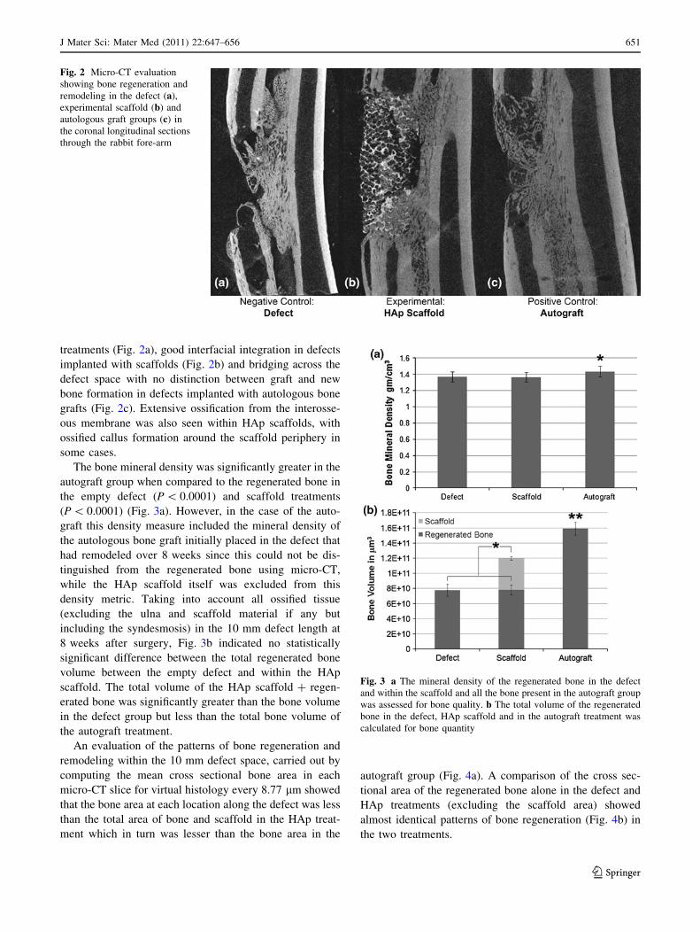
treatments (Fig. 2a), good interfacial integration in defects
implanted with scaffolds (Fig. 2b) and bridging across the
defect space with no distinction between graft and new
bone formation in defects implanted with autologous bone
grafts (Fig. 2c). Extensive ossification from the interosse-
ous membrane was also seen within HAp scaffolds, with
ossified callus formation around the scaffold periphery in
some cases.
The bone mineral density was significantly greater in the
autograft group when compared to the regenerated bone in
the empty defect (P \ 0.0001) and scaffold treatments
(P \ 0.0001) (Fig. 3a). However, in the case of the auto-
graft this density measure included the mineral density of
the autologous bone graft initially placed in the defect that
had remodeled over 8 weeks since this could not be dis-
tinguished from the regenerated bone using micro-CT,
while the HAp scaffold itself was excluded from this
density metric. Taking into account all ossified tissue
(excluding the ulna and scaffold material if any but
including the syndesmosis) in the 10 mm defect length at
8 weeks after surgery, Fig. 3b indicated no statistically
significant difference between the total regenerated bone
volume between the empty defect and within the HAp
scaffold. The total volume of the HAp scaffold ? regen-
erated bone was significantly greater than the bone volume
in the defect group but less than the total bone volume of
the autograft treatment.
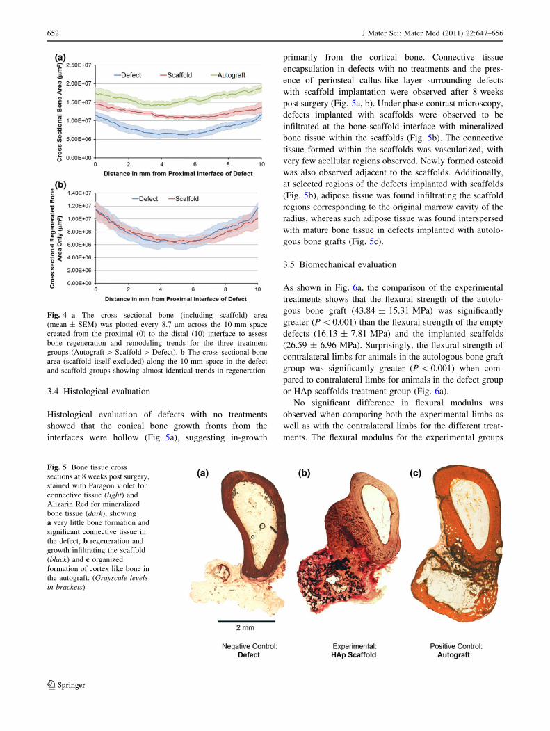
An evaluation of the patterns of bone regeneration and
remodeling within the 10 mm defect space, carried out by
computing the mean cross sectional bone area in each
micro-CT slice for virtual histology every 8.77 lm showed
that the bone area at each location along the defect was less
than the total area of bone and scaffold in the HAp treat-
ment which in turn was lesser than the bone area in the
autograft group (Fig. 4a). A comparison of the cross sec-
tional area of the regenerated bone alone in the defect and
HAp treatments (excluding the scaffold area) showed
almost identical patterns of bone regeneration (Fig. 4b) in
the two treatments.
Fig. 2 Micro-CT evaluation
showing bone regeneration and
remodeling in the defect (a),
experimental scaffold (b) and
autologous graft groups (c) in
the coronal longitudinal sections
through the rabbit fore-arm
Fig. 3 a The mineral density of the regenerated bone in the defect
and within the scaffold and all the bone present in the autograft group
was assessed for bone quality. b The total volume of the regenerated
bone in the defect, HAp scaffold and in the autograft treatment was
calculated for bone quantity
J Mater Sci: Mater Med (2011) 22:647–656 651
123
3.4 Histological evaluation
Histological evaluation of defects with no treatments
showed that the conical bone growth fronts from the
interfaces were hollow (Fig. 5a), suggesting in-growth
primarily from the cortical bone. Connective tissue
encapsulation in defects with no treatments and the pres-
ence of periosteal callus-like layer surrounding defects
with scaffold implantation were observed after 8 weeks
post surgery (Fig. 5a, b). Under phase contrast microscopy,
defects implanted with scaffolds were observed to be
infiltrated at the bone-scaffold interface with mineralized
bone tissue within the scaffolds (Fig. 5b). The connective
tissue formed within the scaffolds was vascularized, with
very few acellular regions observed. Newly formed osteoid
was also observed adjacent to the scaffolds. Additionally,
at selected regions of the defects implanted with scaffolds
(Fig. 5b), adipose tissue was found infiltrating the scaffold
regions corresponding to the original marrow cavity of the
radius, whereas such adipose tissue was found interspersed
with mature bone tissue in defects implanted with autolo-
gous bone grafts (Fig. 5c).
3.5 Biomechanical evaluation
As shown in Fig. 6a, the comparison of the experimental
treatments shows that the flexural strength of the autolo-
gous bone graft (43.84 ± 15.31 MPa) was significantly
greater (P \ 0.001) than the flexural strength of the empty
defects (16.13 ± 7.81 MPa) and the implanted scaffolds
(26.59 ± 6.96 MPa). Surprisingly, the flexural strength of
contralateral limbs for animals in the autologous bone graft
group was significantly greater (P \ 0.001) when com-
pared to contralateral limbs for animals in the defect group
or HAp scaffolds treatment group (Fig. 6a).
No significant difference in flexural modulus was
observed when comparing both the experimental limbs as
well as with the contralateral limbs for the different treat-
ments. The flexural modulus for the experimental groups
Fig. 4 a The cross sectional bone (including scaffold) area
(mean ± SEM) was plotted every 8.7 lm across the 10 mm space
created from the proximal (0) to the distal (10) interface to assess
bone regeneration and remodeling trends for the three treatment
groups (Autograft [ Scaffold [ Defect). b The cross sectional bone
area (scaffold itself excluded) along the 10 mm space in the defect
and scaffold groups showing almost identical trends in regeneration
Fig. 5 Bone tissue cross
sections at 8 weeks post surgery,
stained with Paragon violet for
connective tissue (light) and
Alizarin Red for mineralized
bone tissue (dark), showing
a very little bone formation and
significant connective tissue in
the defect, b regeneration and
growth infiltrating the scaffold
(black) and c organized
formation of cortex like bone in
the autograft. (Grayscale levelsin brackets)
652 J Mater Sci: Mater Med (2011) 22:647–656
123
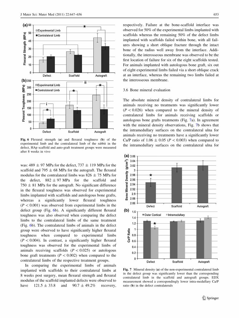
was: 489 ± 97 MPa for the defect, 737 ± 119 MPa for the
scaffold and 795 ± 68 MPa for the autograft. The flexural
modulus for the contralateral limbs was 826 ± 75 MPa for
the defect, 882 ± 97 MPa for the scaffold and
750 ± 81 MPa for the autograft. No significant difference
in the flexural toughness was observed for experimental
limbs implanted with scaffolds and autologous bone grafts,
whereas a significantly lower flexural toughness
(P \ 0.001) was observed from experimental limbs in the
defect group (Fig. 6b). A significantly different flexural
toughness was also observed when comparing the defect
limbs to the contralateral limbs of the same treatment
(Fig. 6b). The contralateral limbs of animals in the defect
group were observed to have significantly higher flexural
toughness when compared to experimental limbs
(P \ 0.004). In contrast, a significantly higher flexural
toughness was observed for the experimental limbs of
animals receiving scaffolds (P \ 0.025) or autologous
bone graft treatments (P \ 0.002) when compared to the
contralateral limbs of the respective treatment groups.
In comparing the experimental limbs of animals
implanted with scaffolds to their contralateral limbs at
8 weeks post surgery, mean flexural strength and flexural
modulus of the scaffold implanted defects were observed to
have 121.5 ± 33.8 and 90.7 ± 49.2% recovery,
respectively. Failure at the bone-scaffold interface was
observed for 50% of the experimental limbs implanted with
scaffolds whereas the remaining 50% of the defect limbs
implanted with scaffolds failed within bone, with all fail-
ures showing a short oblique fracture through the intact
bone of the radius well away from the interface. Addi-
tionally, the interosseous membrane was observed to be the
first location of failure for six of the eight scaffolds tested.
For animals implanted with autologous bone graft, six out
of eight experimental limbs failed via a short oblique crack
at an interface, whereas the remaining two limbs failed at
the interosseous membrane.
3.6 Bone mineral evaluation
The absolute mineral density of contralateral limbs for
animals receiving no treatments was significantly lower
(P \ 0.026) when compared to the mineral density of
contralateral limbs for animals receiving scaffolds or
autologous bone grafts treatments (Fig. 7a). In agreement
with the mineral density observations, Fig. 7b shows that
the intramedullary surfaces on the contralateral ulna for
animals receiving no treatments have a significantly lower
Ca/P ratio of 1.06 ± 0.05 (P \ 0.003) when compared to
the intramedullary surfaces on the contralateral ulna for
Fig. 6 Flexural strength (a) and flexural toughness (b) of the
experimental limb and the contralateral limb of the rabbit in the
defect, HAp scaffold and auto-graft treatment groups were measured
after 8 weeks in vivo
Fig. 7 Mineral density (a) of the non-experimental contralateral limb
in the defect group was significantly lower than the corresponding
contralateral limb in the scaffold and autograft groups. EDX
measurement showed a correspondingly lower intra-medullary Ca/P
ratio (b) in the defect contralaterals
J Mater Sci: Mater Med (2011) 22:647–656 653
123
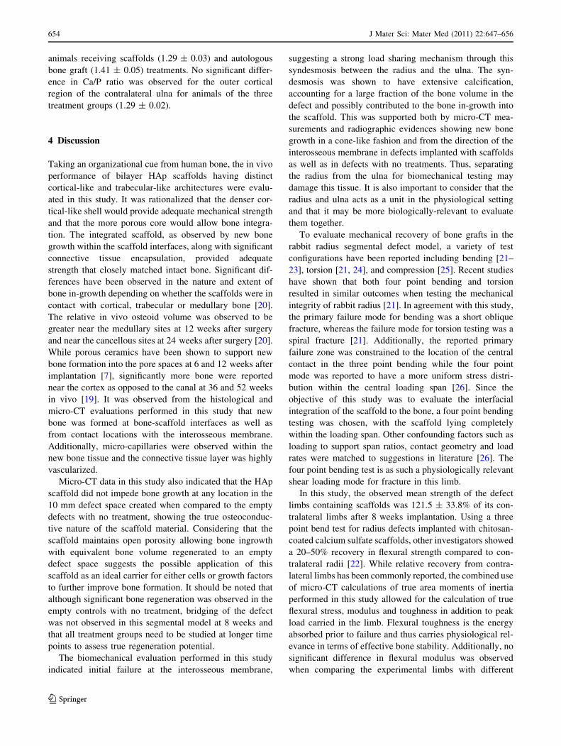
animals receiving scaffolds (1.29 ± 0.03) and autologous
bone graft (1.41 ± 0.05) treatments. No significant differ-
ence in Ca/P ratio was observed for the outer cortical
region of the contralateral ulna for animals of the three
treatment groups (1.29 ± 0.02).
4 Discussion
Taking an organizational cue from human bone, the in vivo
performance of bilayer HAp scaffolds having distinct
cortical-like and trabecular-like architectures were evalu-
ated in this study. It was rationalized that the denser cor-
tical-like shell would provide adequate mechanical strength
and that the more porous core would allow bone integra-
tion. The integrated scaffold, as observed by new bone
growth within the scaffold interfaces, along with significant
connective tissue encapsulation, provided adequate
strength that closely matched intact bone. Significant dif-
ferences have been observed in the nature and extent of
bone in-growth depending on whether the scaffolds were in
contact with cortical, trabecular or medullary bone [20].
The relative in vivo osteoid volume was observed to be
greater near the medullary sites at 12 weeks after surgery
and near the cancellous sites at 24 weeks after surgery [20].
While porous ceramics have been shown to support new
bone formation into the pore spaces at 6 and 12 weeks after
implantation [7], significantly more bone were reported
near the cortex as opposed to the canal at 36 and 52 weeks
in vivo [19]. It was observed from the histological and
micro-CT evaluations performed in this study that new
bone was formed at bone-scaffold interfaces as well as
from contact locations with the interosseous membrane.
Additionally, micro-capillaries were observed within the
new bone tissue and the connective tissue layer was highly
vascularized.
Micro-CT data in this study also indicated that the HAp
scaffold did not impede bone growth at any location in the
10 mm defect space created when compared to the empty
defects with no treatment, showing the true osteoconduc-
tive nature of the scaffold material. Considering that the
scaffold maintains open porosity allowing bone ingrowth
with equivalent bone volume regenerated to an empty
defect space suggests the possible application of this
scaffold as an ideal carrier for either cells or growth factors
to further improve bone formation. It should be noted that
although significant bone regeneration was observed in the
empty controls with no treatment, bridging of the defect
was not observed in this segmental model at 8 weeks and
that all treatment groups need to be studied at longer time
points to assess true regeneration potential.
The biomechanical evaluation performed in this study
indicated initial failure at the interosseous membrane,
suggesting a strong load sharing mechanism through this
syndesmosis between the radius and the ulna. The syn-
desmosis was shown to have extensive calcification,
accounting for a large fraction of the bone volume in the
defect and possibly contributed to the bone in-growth into
the scaffold. This was supported both by micro-CT mea-
surements and radiographic evidences showing new bone
growth in a cone-like fashion and from the direction of the
interosseous membrane in defects implanted with scaffolds
as well as in defects with no treatments. Thus, separating
the radius from the ulna for biomechanical testing may
damage this tissue. It is also important to consider that the
radius and ulna acts as a unit in the physiological setting
and that it may be more biologically-relevant to evaluate
them together.
To evaluate mechanical recovery of bone grafts in the
rabbit radius segmental defect model, a variety of test
configurations have been reported including bending [21–
23], torsion [21, 24], and compression [25]. Recent studies
have shown that both four point bending and torsion
resulted in similar outcomes when testing the mechanical
integrity of rabbit radius [21]. In agreement with this study,
the primary failure mode for bending was a short oblique
fracture, whereas the failure mode for torsion testing was a
spiral fracture [21]. Additionally, the reported primary
failure zone was constrained to the location of the central
contact in the three point bending while the four point
mode was reported to have a more uniform stress distri-
bution within the central loading span [26]. Since the
objective of this study was to evaluate the interfacial
integration of the scaffold to the bone, a four point bending
testing was chosen, with the scaffold lying completely
within the loading span. Other confounding factors such as
loading to support span ratios, contact geometry and load
rates were matched to suggestions in literature [26]. The
four point bending test is as such a physiologically relevant
shear loading mode for fracture in this limb.
In this study, the observed mean strength of the defect
limbs containing scaffolds was 121.5 ± 33.8% of its con-
tralateral limbs after 8 weeks implantation. Using a three
point bend test for radius defects implanted with chitosan-
coated calcium sulfate scaffolds, other investigators showed
a 20–50% recovery in flexural strength compared to con-
tralateral radii [22]. While relative recovery from contra-
lateral limbs has been commonly reported, the combined use
of micro-CT calculations of true area moments of inertia
performed in this study allowed for the calculation of true
flexural stress, modulus and toughness in addition to peak
load carried in the limb. Flexural toughness is the energy
absorbed prior to failure and thus carries physiological rel-
evance in terms of effective bone stability. Additionally, no
significant difference in flexural modulus was observed
when comparing the experimental limbs with different
654 J Mater Sci: Mater Med (2011) 22:647–656
123
treatments as well as comparing the contralateral limbs of
animals receiving different treatments.
Significant differences in flexural toughness were
observed, with the contralateral limbs of animals with no
treatments were observed to have significantly lower flex-
ural toughness when compared to the contralateral limbs of
animals receiving scaffolds or autologous bone graft
treatments. Similarly, the absolute mineral density and the
Ca/P ratio of intermedullary surfaces on the contralateral
limbs for animals receiving no treatments were signifi-
cantly lower when compared to the absolute mineral den-
sity and the Ca/P ratio of intermedullary surfaces on the
contralateral limbs for animals receiving scaffolds or
autologous bone grafts treatments. Although the rabbit
radius defect is a widely used animal model to test bone
regeneration, and bone mineral density was often reported
from radiographic data as a measure of tissue healing [27,
28], this is the first study to report changes in bone mineral
composition at other anatomical sites during the fracture
healing process. However, the effect of the HAp scaffolds
and autologous bone grafts to maintain absolute mineral
density and Ca/P ratios at distant bone sites, as observed in
the diaphyses of the contralateral bones in this study, bears
further investigation in the futures since maintaining cal-
cium homeostasis [29] is also a function of the normal
skeleton.
Additionally, it is also possible that connective tissue
callus may have contributed to the mechanical stabilization
of the scaffold and the extent of load carried through the
radius. From histological and micro-CT evaluations per-
formed in this study, the relatively dense encapsulation was
observed to be a highly organized structure containing
densely packed strand-like collagen bundles. Since pure
ceramics have low energy absorption to failure and high
stiffness, the observation of relatively higher flexural
toughness in the scaffolds as compared to intact bone
which was also comparable to the toughness of autologous
graft material also supported the role played by the con-
nective tissues in enhancing the stability of scaffolds
implanted in the defect sites.
5 Conclusions
This study suggests that the HAp scaffolds can serve as an
ideal scaffold carrier platform for bone regeneration because
they do not impede bone formation, add to the strength and
toughness of the defect site, and may reduce deleterious
effects on the skeleton during the healing process.
New bone in-growth in defects was promoted with the
implantation of bilayer HAp scaffolds, with total regener-
ated bone volume being comparable to empty defects and
flexural toughness being comparable to defects implanted
with autologous bone grafts. The scaffolds also promoted
the presence of connective tissue encapsulation around the
scaffolds, thereby possibly contributing to the stability of
the implanted scaffolds. Additionally, it was also con-
cluded that the therapeutic effect of HAp scaffold
implantation was comparable to autologous bone graft
implantation in defects with respect to the maintenance of
mineral density and intramedullary Ca/P ratios at distant
bone sites.
Acknowledgments This study was supported in part by the
Department of Defense funds and the Orthopaedic Extremity Trauma
Research Program grants (USAMRMC # W81XWH-08-1-0393 and
W81XWH-07-1-0717). The opinions or assertions contained herein
are the private views of the author and are not to be construed as
official or as reflecting the views of the Department of the Army or
the Department of Defense.
References
1. Greenwald AS, Boden SD, Goldberg VM, Khan Y, Laurencin
CT, Rosier RN. Bone-graft substitutes: facts, fictions, and
applications. J Bone Joint Surg Am. 2001;83-A:98–103.
2. Giannoudis PV, Dinopoulos H, Tsiridis E. Bone substitutes: an
update. Injury. 2005;36:S20–7.
3. Glancy GL, Brugioni DJ, Eilert RE, Chang FM. Autograft versus
allograft for benign lesions in children. Clin Orthop Relat Res.
1991;262:28–33.
4. Laurie SW, Kaban LB, Mulliken JB, Murray JE. Donor-site
morbidity after harvesting rib and iliac bone. Plast Reconstr Surg.
1984;73(6):933–8.
5. Guda T, Appleford M, Oh S, Ong JL. A cellular perspective to
bioceramic scaffolds for bone tissue engineering: the state of the
art. Curr Top Med Chem. 2008;8(4):290–9.
6. Oh S, Oh N, Appleford M, Ong JL. Bioceramics for tissue
engineering applications—a review. Am J Biochem Biotechnol.
2006;2(2):49–56.
7. Appleford MR, Oh S, Oh N, Ong JL. In vivo study on
hydroxyapatite scaffolds with trabecular architecture for bone
repair. J Biomed Mater Res A. 2009;89(4):1019–27.
8. Kilian O, Wenisch S, Karnati S, Baumgart-Vogt E, Hild A, Fu-
hrmann R, et al. Observations on the microvasculature of bone
defects filled with biodegradable nanoparticulate hydroxyapatite.
Biomaterials. 2008;29(24–25):3429–37.
9. Yoshikawa T, Ohgushi H, Nakajima H, Yamada E, Ichijima K,
Tamai S, et al. In vivo osteogenic durability of cultured bone in
porous ceramics: a novel method for autogenous bone graft
substitution. Transplantation. 2000;69(1):128–34.
10. Costantino PD, Friedman CD, Jones K, Chow LC, Pelzer HJ,
Sisson GA Sr. Hydroxyapatite cement. I. Basic chemistry and
histologic properties. Arch Otolaryngol Head Neck Surg.
1991;17(4):379–84.
11. Ohgushi H, Dohi Y, Tamai S, Tabata S. Osteogenic differentia-
tion of marrow stromal stem cells in porous hydroxyapatite
ceramics. J Biomed Mater Res. 1993;27(11):1401–7.
12. Kuboki Y, Jin Q, Takita H. Geometry of carriers controlling
phenotypic expression in BMP-induced osteogenesis and chon-
drogenesis. J Bone Joint Surg Am. 2001;83-A:S105–15.
13. Martin RB, Chapman MW, Sharkey NA, Zissimos SL, Bay B,
Shors EC. Bone ingrowth and mechanical properties of coralline
hydroxyapatite 1 yr after implantation. Biomaterials. 1993;
14(5):341–8.
J Mater Sci: Mater Med (2011) 22:647–656 655
123

14. Mastrogiacomo M, Scaglione S, Martinetti R, Dolcini L, Bel-
trame F, Cancedda R, et al. Role of scaffold internal structure on
in vivo bone formation in macroporous calcium phosphate bi-
oceramics. Biomaterials. 2006;27(17):3230–7.
15. Daculsi G, Passuti N. Effect of the macroporosity for osseous
substitution of calcium phosphate ceramics. Biomaterials.
1990;11:86–7.
16. Gauthier O, Bouler JM, Aguado E, Pilet P, Daculsi G. Macro-
porous biphasic calcium phosphate ceramics: influence of mac-
ropore diameter and macroporosity percentage on bone ingrowth.
Biomaterials. 1998;19(1–3):133–9.
17. Shimazaki K, Mooney V. Comparative study of porous
hydroxyapatite and tricalcium phosphate as bone substitute.
J Orthop Res. 1985;3(3):301–10.
18. Karageorgiou V, Kaplan D. Porosity of 3D biomaterial scaffolds
and osteogenesis. Biomaterials. 2005;26(27):5474–91.
19. Stubbs D, Deakin M, Chapman-Sheath P, Bruce W, Debes J,
Gillies RM, et al. In vivo evaluation of resorbable bone graft
substitutes in a rabbit tibial defect model. Biomaterials. 2004;
25(20):5037–44.
20. Lu JX, Gallur A, Flautre B, Anselme K, Descamps M, Thierry B,
et al. Comparative study of tissue reactions to calcium phosphate
ceramics among cancellous, cortical, and medullar bone sites in
rabbits. J Biomed Mater Res. 1998;42(3):357–67.
21. Bowers KW, Edmonds JL, Girod DA, Jayaraman G, Chua CP,
Toby EB. Osteocutaneous radial forearm free flaps. The necessity
of internal fixation of the donor-site defect to prevent patholog-
ical fracture. J Bone Joint Surg Am. 2000;82(5):694–704.
22. Cui X, Zhang B, Wang Y, Gao Y. Effects of chitosan-coated
pressed calcium sulfate pellet combined with recombinant human
bone morphogenetic protein 2 on restoration of segmental bone
defect. J Craniofac Surg. 2008;19(2):459–65.
23. Shafiei Z, Bigham AS, Dehghani SN, Nezhad ST. Fresh cortical
autograft versus fresh cortical allograft effects on experimental
bone healing in rabbits: radiological, histopathological and bio-
mechanical evaluation. Cell Tissue Bank. 2009;10(1):19–26.
24. Mori M, Isobe M, Yamazaki Y, Ishihara K, Nakabayashi N.
Restoration of segmental bone defects in rabbit radius by bio-
degradable capsules containing recombinant human bone mor-
phogenetic protein-2. J Biomed Mater Res. 2000;50(2):191–8.
25. Kaito T, Myoui A, Takaoka K, Saito N, Nishikawa M, Tamai N,
et al. Potentiation of the activity of bone morphogenetic protein-2
in bone regeneration by a pla-peg/hydroxyapatite composite.
Biomaterials. 2005;26(1):73–9.
26. Draper ER, Goodship AE. A novel technique for four-point
bending of small bone samples with semi-automatic analysis.
J Biomech. 2003;36(10):1497–502.
27. Liu Y, Lu Y, Tian X, Cui G, Zhao Y, Yang Q, et al. Segmental
bone regeneration using an rhBMP-2-loaded gelatin/nanohy-
droxyapatite/fibrin scaffold in a rabbit model. Biomaterials.
2009;30(31):6276–85.
28. Wu T, Nan K, Chen J, Jin D, Jiang S, Zhao P, et al. A new bone
repair scaffold combined with chitosan/hydroxyapatite and sus-
tained releasing icariin. Chin Sci Bull. 2009;54(17):2953–61.
29. Issa JPM, Bentley MVLB, Iyomasa MM, Sebald W, Albuquerque
RFD. Sustained release carriers used to delivery bone morpho-
genetic proteins in the bone healing process. Anat Histol
Embryol. 2008;37(3):181–7.
656 J Mater Sci: Mater Med (2011) 22:647–656
123

Copyright of Journal of Materials Science: Materials in Medicine is the property of Springer Science &
Business Media B.V. and its content may not be copied or emailed to multiple sites or posted to a listserv
without the copyright holder's express written permission. However, users may print, download, or email
articles for individual use.