In vitro proliferation and differentiation of hepatic oval ...
8
In vitro proliferation and differentiation of hepatic oval cells and their potential capacity for intrahepatic transplantation Z. Li 1 *, J. Chen 1 *, L. Li 2 , J.H. Ran 2 , J. Liu 2 , T.X. Gao 3 , B.Y. Guo 3 , X.H. Li 1 , Z.H. Liu 1 , G.J. Liu 1 , Y.C. Gao 1 and X.L. Zhang 1 1 Department of Hepatobiliary Surgery, Liaocheng People’s Hospital, Liaocheng, Shandong, China 2 The First People’s Hospital of Kunming, Kunming, Yunnan, China 3 Dongchangfu Hospital of Women and Child Health Care, Liaocheng, Shandong, China Abstract Hepatic oval cells (HOCs) are recognized as facultative liver progenitor cells that play a role in liver regeneration after acute liver injury. Here, we investigated the in vitro proliferation and differentiation characteristics of HOCs in order to explore their potential capacity for intrahepatic transplantation. Clusters or scattered HOCs were detected in the portal area and interlobular bile duct in the liver of rats subjected to the modified 2-acetylaminofluorene and partial hepatectomy method. Isolated HOCs were positive for c-kit and CD90 staining (99.8% and 88.8%, respectively), and negative for CD34 staining (3.6%) as shown by immunostaining and flow cytometric analysis. In addition, HOCs could be differentiated into hepatocytes and bile duct epithelial cells after leukemia inhibitory factor deprivation. A two-cuff technique was used for orthotopic liver transplantation, and HOCs were subsequently transplanted into recipients. Biochemical indicators of liver function were assessed 4 weeks after transplantation. HOC transplantation significantly prolonged the median survival time and improved the liver function of rats receiving HOCs compared to controls (P=0.003, Student t-test). Administration of HOCs to rats also receiving liver transplantation significantly reduced acute allograft rejection compared to control liver transplant rats 3 weeks following transplantation (rejection activity index score: control=6.3±0.9; HOC=3.5±1.5; P=0.005). These results indicate that HOCs may be useful in therapeutic liver regeneration after orthotopic liver transplantation. Key words: Hepatic oval cells; Proliferation; Differentiation; Liver transplantation Introduction Liver transplantation offers an efficient and efficacious approach for treating patients with end-stage liver diseases. However, liver transplantation is usually asso- ciated with considerable long-term side effects, including chronic renal failure (1), post-transplantation lymphopro- liferative disorder (2), and cardiovascular complications (3). Transplantation of stem cells or progenitor cells can be utilized as alternative solutions to repair or regenerate damaged liver due to their ability to self-renew and their capability to differentiate into a variety of specialized cell types (4). Hepatocytes and hepatic oval cells (HOCs) are the potential source of endogenous stem/progenitor cells for liver regeneration and repopulation (5,6). Importantly, intra-hepatic progenitor cells (HOCs) play critical roles when hepatocyte proliferation is blocked or delayed (6). Although it has been suggested that HOCs may provide an extension of the canal of Hering (7), the precise origination of these cells is not known. These cells have a high nuclear-to-cytoplasmic ratio and express markers of immature liver cells [a-fetoprotein (AFP)], hepatocyte lineage (albumin), and biliary epithelium [cytokeratin 19 (CK19)] (8,9). HOCs display a distinct phenotype and have been shown to be a bipotential progenitor of two types of epithelial cells found in the liver: hepatocytes and bile ductular cells (10). Moreover, HOCs are capable of generating other cells, such as intestinal epithelium and pancreatic acinar cells (6,11,12). A variety of liver injury models have been used to induce HOC proliferation, such as 2-acetylaminofluorene (2-AAF)/ partial hepatectomy (PH)-induced injury (13,14), a Correspondence: Z. Li, Department of Hepatobiliary Surgery, Liaocheng People’s Hospital, No. 67 Dongchang West Road, Liaocheng 252000, Shandong, China. E-mail: [email protected] *These authors contributed equally to this study. Received November 7, 2012. Accepted March 25, 2013. First published online July 30, 2013. Brazilian Journal of Medical and Biological Research (2013) 46: 681-688, http://dx.doi.org/10.1590/1414-431X20132620 ISSN 1414-431X www.bjournal.com.br Braz J Med Biol Res 46(8) 2013
Transcript of In vitro proliferation and differentiation of hepatic oval ...

In vitro proliferation and differentiation ofhepatic oval cells and their potential
capacity for intrahepatic transplantation
Z. Li1*, J. Chen1*, L. Li2, J.H. Ran2, J. Liu2, T.X. Gao3, B.Y. Guo3, X.H. Li1, Z.H. Liu1, G.J. Liu1,
Y.C. Gao1 and X.L. Zhang1
1Department of Hepatobiliary Surgery, Liaocheng People’s Hospital, Liaocheng, Shandong, China2The First People’s Hospital of Kunming, Kunming, Yunnan, China
3Dongchangfu Hospital of Women and Child Health Care, Liaocheng, Shandong, China
Abstract
Hepatic oval cells (HOCs) are recognized as facultative liver progenitor cells that play a role in liver regeneration after acute
liver injury. Here, we investigated the in vitro proliferation and differentiation characteristics of HOCs in order to explore their
potential capacity for intrahepatic transplantation. Clusters or scattered HOCs were detected in the portal area and interlobular
bile duct in the liver of rats subjected to the modified 2-acetylaminofluorene and partial hepatectomy method. Isolated HOCs
were positive for c-kit and CD90 staining (99.8% and 88.8%, respectively), and negative for CD34 staining (3.6%) as shown by
immunostaining and flow cytometric analysis. In addition, HOCs could be differentiated into hepatocytes and bile duct epithelial
cells after leukemia inhibitory factor deprivation. A two-cuff technique was used for orthotopic liver transplantation, and HOCs
were subsequently transplanted into recipients. Biochemical indicators of liver function were assessed 4 weeks after
transplantation. HOC transplantation significantly prolonged the median survival time and improved the liver function of rats
receiving HOCs compared to controls (P=0.003, Student t-test). Administration of HOCs to rats also receiving liver
transplantation significantly reduced acute allograft rejection compared to control liver transplant rats 3 weeks following
transplantation (rejection activity index score: control=6.3±0.9; HOC=3.5±1.5; P=0.005). These results indicate that HOCs
may be useful in therapeutic liver regeneration after orthotopic liver transplantation.
Key words: Hepatic oval cells; Proliferation; Differentiation; Liver transplantation
Introduction
Liver transplantation offers an efficient and efficacious
approach for treating patients with end-stage liver
diseases. However, liver transplantation is usually asso-
ciated with considerable long-term side effects, including
chronic renal failure (1), post-transplantation lymphopro-
liferative disorder (2), and cardiovascular complications
(3). Transplantation of stem cells or progenitor cells can
be utilized as alternative solutions to repair or regenerate
damaged liver due to their ability to self-renew and their
capability to differentiate into a variety of specialized cell
types (4). Hepatocytes and hepatic oval cells (HOCs) are
the potential source of endogenous stem/progenitor cells
for liver regeneration and repopulation (5,6). Importantly,
intra-hepatic progenitor cells (HOCs) play critical roles
when hepatocyte proliferation is blocked or delayed (6).
Although it has been suggested that HOCs may
provide an extension of the canal of Hering (7), the
precise origination of these cells is not known. These cells
have a high nuclear-to-cytoplasmic ratio and express
markers of immature liver cells [a-fetoprotein (AFP)],
hepatocyte lineage (albumin), and biliary epithelium
[cytokeratin 19 (CK19)] (8,9). HOCs display a distinct
phenotype and have been shown to be a bipotential
progenitor of two types of epithelial cells found in the liver:
hepatocytes and bile ductular cells (10). Moreover, HOCs
are capable of generating other cells, such as intestinal
epithelium and pancreatic acinar cells (6,11,12). A variety
of liver injury models have been used to induce HOC
proliferation, such as 2-acetylaminofluorene (2-AAF)/
partial hepatectomy (PH)-induced injury (13,14), a
Correspondence: Z. Li, Department of Hepatobiliary Surgery, Liaocheng People’s Hospital, No. 67 Dongchang West Road, Liaocheng
252000, Shandong, China. E-mail: [email protected]
*These authors contributed equally to this study.
Received November 7, 2012. Accepted March 25, 2013. First published online July 30, 2013.
Brazilian Journal of Medical and Biological Research (2013) 46: 681-688, http://dx.doi.org/10.1590/1414-431X20132620
ISSN 1414-431X
www.bjournal.com.br Braz J Med Biol Res 46(8) 2013

3,5-diethoxycarbonyl-1,4-dihydrocollidine diet (9),
choline deficiency, an ethionine-supplemented diet (15),
and 2-AAF/CCl4 (16) or 1,4-bis[N,N9-di(ethylene)-
phosphamide]-piperazine (dipin)/PH-induced injury (17).
Among these models, 2-AAF combined with a two-thirds
PH has been widely used for stimulating the proliferation
of HOCs; however, significant mortality was observed
after a two-thirds PH (18). Therefore, a more efficient
approach for generating HOCs with low animal mortality is
still needed. In addition, the potential capacity of HOCs for
intrahepatic transplantation has not been fully explored.
In this study, we successfully induced the proliferation
of HOCs in male Lewis rats using 2-AAF combined with a
one-half to one-third PH (left lateral lobe excision) and
determined the in vitro proliferation and differentiation
capability of isolated HOCs. In addition, the intrahepatic
transplantation ability of HOCs was evaluated. Our results
provide a better understanding of the involvement of
HOCs during liver repair and regeneration and suggest
that HOCs may have valuable properties for therapeutic
liver regeneration after orthotopic liver transplantation.
Material and Methods
ReagentsFetal bovine serum (FBS), 0.25% trypsin, type IV
collagenase, and phosphate-buffered saline (PBS) were
purchased from Gibco (USA). D-Hank’s solution was
purchased from HyClone (USA). The 2-AAF (Cat
No. 00300) and DMEM/F12 culture medium were obtained
from Sigma-Aldrich (USA). Hepatocyte growth factor
(HGF; Cat No. 2207-HG), epidermal growth factor (EGF;
Cat No. 400-25), stem cell factor (SCF; Cat No. 400-22),
and leukemia inhibitory factor (LIF; Cat No. LIF3005) were
purchased from Cytolay (USA). FITC-conjugated rat anti-
CD34 (Cat No. SC-7324) and rabbit IgG anti-c-kit
polyclonal antibodies were provided by Santa Cruz
(USA). FITC-conjugated rat anti-CD90 (Cat No. 202503)
antibody was provided by Biolegend (USA). FITC-labeled
goat anti-rabbit IgG antibody was purchased from Beijing
Zhong Shan-Golden Bridge Biological Technology Co.,
Ltd. (China). Specific primers for albumin (ALB), CK19, and
b-actin were synthesized by Shanghai Chaoshi
Biotechnologies Co. (China). The hematoxylin and eosin
(H&E) staining kit was obtained from Shanghai Sangon
Biological Engineering Technology and Services Co., Ltd.
(China).
AnimalsMale and female specific pathogen-free (SPF) Lewis
rats weighing 200±20 g (used for HOCs isolation) or
250-300 g (used for transplantation) were provided by
Vital River Lab Animal Technology Co., Ltd. (China).
Female SPF dark agouti (DA) rats weighing 200-250 g
were provided by the Laboratory Animal Center of Harbin
Medical University, Harbin, China. All efforts were made
to minimize animal suffering and to reduce the number of
animals used. All animal procedures and the study were
approved by the Ethics Committee of Liaocheng People’s
Hospital, Liaocheng, Shandong, China.
Activation of oval cell proliferation by the 2-AAF/PHmodel
Male Lewis rats were housed separately for 1 week
prior to use. Daily, the rats were given 20 mg/kg body
weight 2-AAF dissolved in vegetable oil by intragastric
administration for 6 days. On the 7th day, 10 animals were
partially hepatectomized (left lateral lobe excision, one-
half to one-third PH) under general ether anesthesia
without 2-AAF administration, and 2-AAF feeding was
continued from the 8th to the 14th day.
Histological examinationTen days after surgery, animals were killed and liver
tissues were removed and fixed with 10% buffered
formaldehyde. Paraffin-embedded sections were stained
with H&E using a standard procedure and then assessed
under a phase contrast microscope (Olympus, Japan).
Primary culture of HOCsThe isolation of HOCs was performed 10 days after
surgery. Briefly, liver tissue was minced, washed with
D-Hank’s solution, and digested with DMEM/F12 culture
medium containing 0.1% collagenase IV and 0.025%
EDTA at 376C for 15 min. Cells were filtered through a
100 mesh filter and centrifuged at 340 g for 5 min at 46C.
HOCs were separated by Percoll gradient centrifugation
(Amersham Biosciences, USA). The cells were then
seeded onto gelatin-coated flasks and maintained in
DMEM/F12 culture medium supplemented with 10%
FBS, 100 U/mL penicillin, 100 mg/mL streptomycin, and
1 ng/mL amphotericin B at 376C in a 5% CO2 incubator.
Three days after seeding, 20 ng/mL SCF, 10 ng/mL HGF,
10 ng/mL EGF, and 10 ng/mL LIF were added to the
culture medium. Subculture was performed using 0.25%
trypsin for digestion. To induce differentiation, LIF was
withdrawn from the culture medium and the cells were
maintained in culture. Images were captured and quantita-
tively analyzed with an image analysis system (APIAS-
1000, Shenzhen, Huahai Electronics, Co., Ltd., China).
MTT assayTo evaluate cell proliferation, the 3-(4,5-dimethylthiazol-
2-yl)-2,5-diphenyltetrazolium bromide (MTT) assay was
performed. Cells were seeded onto 24-well plates at a
density of 1 6 107 cells/well. At the indicated time points
after cell seeding, 20 mL MTT (5 mg/mL) was added to
each well, and the cells were cultured for an additional 4 h
at 376C. The medium was then removed, and 150 mLdimethyl sulfoxide was added to each well to resuspend the
MTT metabolic product. The absorbance of the dissolved
formazan was measured at a wavelength of 570 nm with a
682 Z. Li et al.
Braz J Med Biol Res 46(8) 2013 www.bjournal.com.br

scanning microplate spectrophotometer (Biotek, USA).
Three replicate wells were assessed for each experimental
group.
Electron microscopyFor transmission electron microscopy (TEM) analysis,
HOCs or tissue samples were fixed with 2.5% glutar-
aldehyde for 2 h and then post-fixed with 1% OsO4. After
dehydration through a graded ethanol series and acetone
[30% (v/v) ethanol (15 min), 50% (v/v) ethanol (15 min),
70% (v/v) ethanol (15 min), 90% (v/v) ethanol (15 min),
95% (v/v) ethanol (15 min), 50% (v/v) acetone (10-
15 min), 70% (v/v) acetone (10-15 min), 90% (v/v)
acetone (10-15 min), and three washes with 100% (v/v)
acetone (30 min)], the samples were embedded in
Epon812. Ultrathin sections were stained with uranyl
acetate and lead citrate and examined by TEM (Hitachi
Science Systems, Ltd., Japan). For scanning electronic
microscopy (SEM) analysis, samples were fixed with
2.5% glutaraldehyde for 12 h. After dehydration in an
acetone gradient [50% (v/v) acetone (10-15 min), 70%
(v/v) acetone (10-15 min), and 90% (v/v) acetone
(10-15 min)], spray-dried samples were analyzed by
SEM (S-3400-N, Hitachi Science Systems, Ltd.).
ImmunocytochemistryImmunocytochemical analysis was performed as pre-
viously described (19). Briefly, cells were fixed with 4%
paraformaldehyde (PFA), rinsed with PBS, and immuno-
stained with an anti-c-kit primary antibody (1:100 dilution)
in a humidity chamber overnight at 46C. After washing
three times with PBS, the cells were stained with FITC-
labeled goat anti-rabbit IgG secondary antibody (1:80
dilution) for 40 min at 376C. After washing with PBS, the
samples were mounted with 2% glycerol, and fluorescence
was detected under a fluorescencemicroscope (Olympus).
Flow cytometryThe cell surface markers c-kit, CD34, and CD90 were
assessed by flow cytometry analysis. Fifty microliters of
16 106 cells suspended in PBS were incubated with the c-
kit primary antibody (1:100 dilution) for 50 min at room
temperature. After centrifugation, the cells were washed
and stained with FITC-labeled goat anti-rabbit IgG second-
ary antibody (1:80 dilution) for 30 min at room temperature.
For CD34 and CD90 labeling, the same volume of cells was
incubated with FITC-labeled anti-CD34 (1:100 dilution) or
anti-CD90 (1:100 dilution) antibodies for 30 min at room
temperature. After they were washed with PBS two times
for 5 min each, the cells were analyzed using fluorescence-
activated cell sorting (FACS; BD Biosciences, USA).
Quantitative real-time reverse-transcriptionpolymerase chain reaction (qRT-PCR)
Total RNA was extracted from HOCs, differentiated
HOCs, liver tissues, or bile duct tissues using the Trizol
reagent (Sigma-Aldrich). After purification, the integrity of
the purified mRNA was confirmed by agarose gel
electrophoresis. The cDNA was then transcribed with
the first cDNA synthesis kit according to the manufac-
turer’s instructions (Fermentas, USA). The sequences of
the specific primers used are as follows: ALB: forward:
59-TGTCACGGCGACCTGTTG-39, reverse: 59-GG
AGATAGTGGCCTGGTTCTCA-39; CK19: forward: 59-GA
CTTCCGGACCAAGTTTGAG-39, reverse: 59-CGC
AGGCCGTTGATGTC-39; b-actin: forward: 59-ACCG
AGCGCGGCTACAGC-39, reverse: 59-CTCATTGCCAA
TGGTGAT-39. PCR amplification was carried out on a
real-time fluorescence quantitative instrument (Roche
Diagnostics, Switzerland). The primer concentration was
20 pM and PCR efficiency of the kit was 96.3%. The
thermocycling conditions were as follows: 606C for 2 min,
946C for 10 min, and then 40 cycles at 946C for 15 s and
606C for 60 s. The amplified products were analyzed
with real-time fluorescence quantitative instrument
software. Fold changes in target gene expression were
then normalized with the following formula: fold
change=2–D(DCt). The negative log was calculated and
data were analyzed from 20 independent experiments.
Cell transplantationThe two-cuff (portal vein and infrahepatic vena cava)
technique was used to establish orthotopic liver trans-
plantation in rats as previously described (20,21). Female
DA rats were used as donors and female Lewis rats
served as recipients. The recipients were randomly
divided into two groups: the control group (n=60) and
the cell transplantation group (n=60). In the control
group, animals received orthotopic liver transplantation,
and in the cell transplantation group, 1 mL of 16106 cells/
mL HOCs (passage 4-10) was injected into the portal vein
and hepatic artery in animals receiving the liver trans-
plantation. Recipients were given tacrolimus starting on
the day prior to surgery until the 13th day post-surgery.
Liver function indicators, including alanine aminotransfer-
ase (ALT), direct bilirubin (DBil), ALB, c-glutamyltransfer-
ase (GGT), alkaline phosphate (ALP), and cholinesterase
(ChE) were examined 4 weeks after transplantation using
a Beckman CX9 automatic biochemical analyzer
(Beckman Coulter, Germany).
Evaluation of liver allograft rejectionThree weeks after transplantation, tissue sections
were stained with H&E, samples were mounted, and
fluorescence detected using a fluorescence microscope
(Olympus). The severity of acute rejection was assessed
using the rejection activity index (RAI) according to the
Banff classification of hepatic allograft rejection (22).
Statistical analysisData were analyzed by the SPSS 16.0 software
(SPSS, Inc., USA) and are reported as means±SD.
Characteristics of HOCs 683
www.bjournal.com.br Braz J Med Biol Res 46(8) 2013
Comparisons between different groups were carried out
using one-way analysis of variance. Comparisons within
one group were achieved using the Student t-test.
P,0.05 was considered to be statistically significant.
Results
Establishment of an HOC proliferation modelIn order to establish the HOC proliferation model, 10
Lewis rats received 2-AAF in combination with a left
lateral lobe excision, and six rats survived for 10 days
post-surgery. Two rats died as a result of the surgery, and
two others died of postoperative abdominal infection or
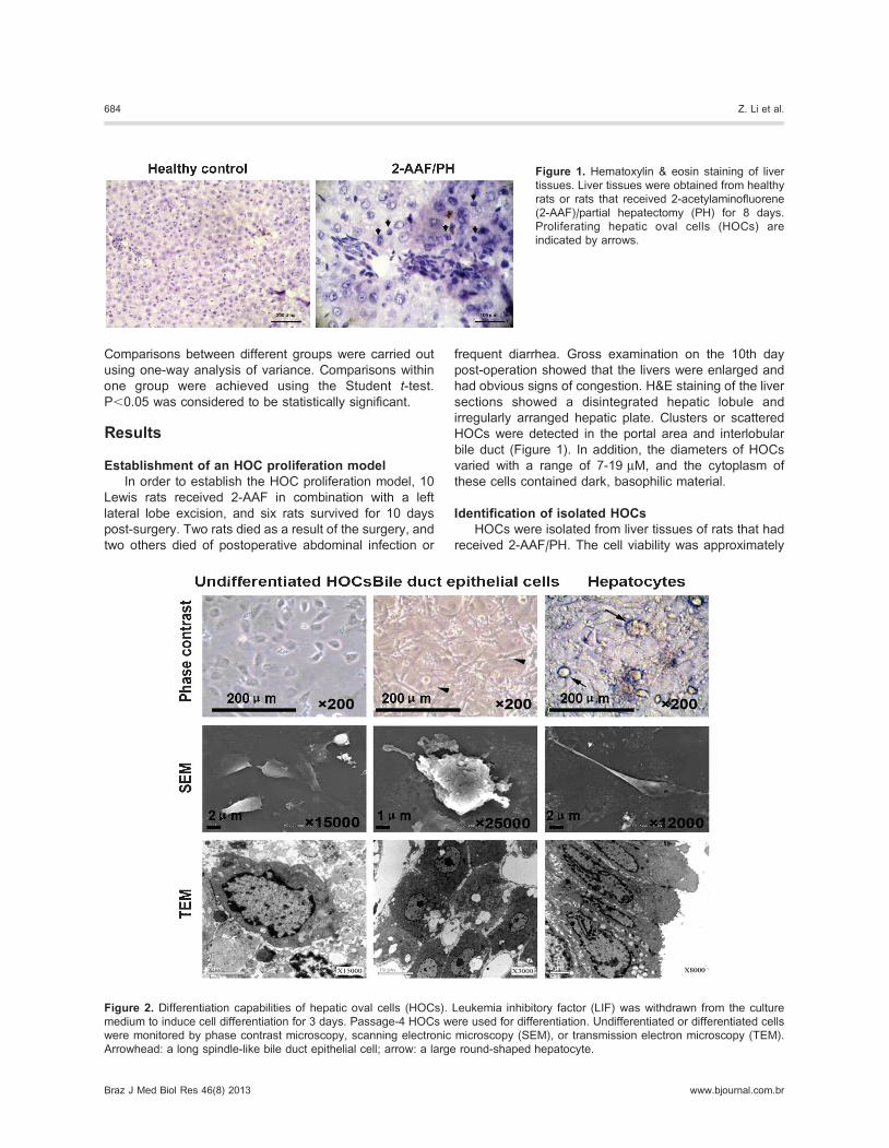
frequent diarrhea. Gross examination on the 10th day
post-operation showed that the livers were enlarged and
had obvious signs of congestion. H&E staining of the liver
sections showed a disintegrated hepatic lobule and
irregularly arranged hepatic plate. Clusters or scattered
HOCs were detected in the portal area and interlobular
bile duct (Figure 1). In addition, the diameters of HOCs
varied with a range of 7-19 mM, and the cytoplasm of
these cells contained dark, basophilic material.
Identification of isolated HOCsHOCs were isolated from liver tissues of rats that had
received 2-AAF/PH. The cell viability was approximately
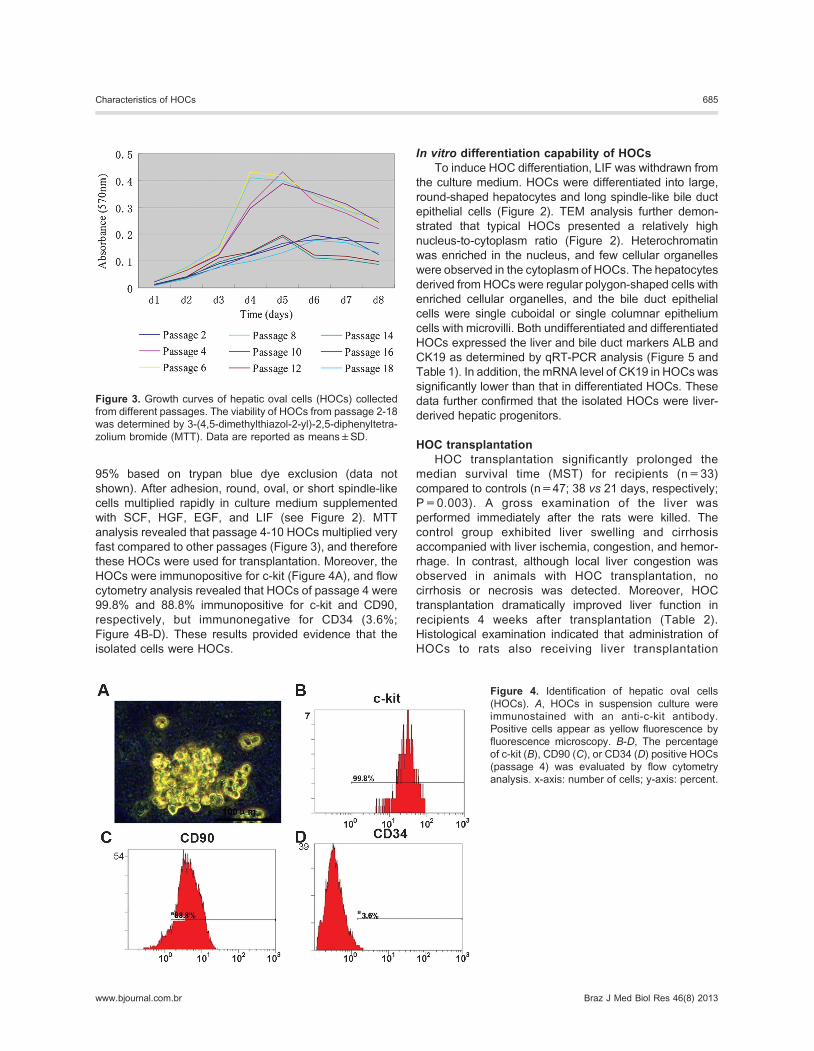
Figure 2. Differentiation capabilities of hepatic oval cells (HOCs). Leukemia inhibitory factor (LIF) was withdrawn from the culture
medium to induce cell differentiation for 3 days. Passage-4 HOCs were used for differentiation. Undifferentiated or differentiated cells
were monitored by phase contrast microscopy, scanning electronic microscopy (SEM), or transmission electron microscopy (TEM).
Arrowhead: a long spindle-like bile duct epithelial cell; arrow: a large round-shaped hepatocyte.
Figure 1. Hematoxylin & eosin staining of liver
tissues. Liver tissues were obtained from healthy
rats or rats that received 2-acetylaminofluorene
(2-AAF)/partial hepatectomy (PH) for 8 days.
Proliferating hepatic oval cells (HOCs) are
indicated by arrows.
684 Z. Li et al.
Braz J Med Biol Res 46(8) 2013 www.bjournal.com.br
95% based on trypan blue dye exclusion (data not
shown). After adhesion, round, oval, or short spindle-like
cells multiplied rapidly in culture medium supplemented
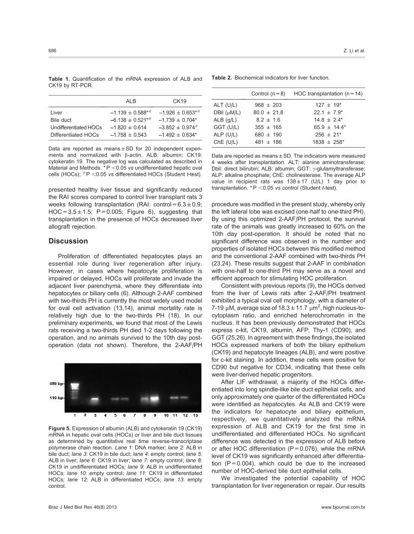
with SCF, HGF, EGF, and LIF (see Figure 2). MTT
analysis revealed that passage 4-10 HOCs multiplied very
fast compared to other passages (Figure 3), and therefore
these HOCs were used for transplantation. Moreover, the
HOCs were immunopositive for c-kit (Figure 4A), and flow
cytometry analysis revealed that HOCs of passage 4 were
99.8% and 88.8% immunopositive for c-kit and CD90,
respectively, but immunonegative for CD34 (3.6%;
Figure 4B-D). These results provided evidence that the
isolated cells were HOCs.
In vitro differentiation capability of HOCsTo induce HOC differentiation, LIF was withdrawn from
the culture medium. HOCs were differentiated into large,
round-shaped hepatocytes and long spindle-like bile duct
epithelial cells (Figure 2). TEM analysis further demon-
strated that typical HOCs presented a relatively high
nucleus-to-cytoplasm ratio (Figure 2). Heterochromatin
was enriched in the nucleus, and few cellular organelles
were observed in the cytoplasm of HOCs. The hepatocytes
derived from HOCs were regular polygon-shaped cells with
enriched cellular organelles, and the bile duct epithelial
cells were single cuboidal or single columnar epithelium
cells with microvilli. Both undifferentiated and differentiated
HOCs expressed the liver and bile duct markers ALB and
CK19 as determined by qRT-PCR analysis (Figure 5 and
Table 1). In addition, the mRNA level of CK19 in HOCswas
significantly lower than that in differentiated HOCs. These
data further confirmed that the isolated HOCs were liver-
derived hepatic progenitors.
HOC transplantationHOC transplantation significantly prolonged the
median survival time (MST) for recipients (n=33)
compared to controls (n=47; 38 vs 21 days, respectively;
P=0.003). A gross examination of the liver was
performed immediately after the rats were killed. The
control group exhibited liver swelling and cirrhosis
accompanied with liver ischemia, congestion, and hemor-
rhage. In contrast, although local liver congestion was
observed in animals with HOC transplantation, no
cirrhosis or necrosis was detected. Moreover, HOC
transplantation dramatically improved liver function in
recipients 4 weeks after transplantation (Table 2).
Histological examination indicated that administration of
HOCs to rats also receiving liver transplantation
Figure 3. Growth curves of hepatic oval cells (HOCs) collected
from different passages. The viability of HOCs from passage 2-18
was determined by 3-(4,5-dimethylthiazol-2-yl)-2,5-diphenyltetra-
zolium bromide (MTT). Data are reported as means±SD.
Figure 4. Identification of hepatic oval cells
(HOCs). A, HOCs in suspension culture were
immunostained with an anti-c-kit antibody.
Positive cells appear as yellow fluorescence by
fluorescence microscopy. B-D, The percentage
of c-kit (B), CD90 (C), or CD34 (D) positive HOCs
(passage 4) was evaluated by flow cytometry
analysis. x-axis: number of cells; y-axis: percent.
Characteristics of HOCs 685
www.bjournal.com.br Braz J Med Biol Res 46(8) 2013
presented healthy liver tissue and significantly reduced
the RAI scores compared to control liver transplant rats 3
weeks following transplantation (RAI: control=6.3±0.9;
HOC=3.5±1.5; P=0.005; Figure 6), suggesting that
transplantation in the presence of HOCs decreased liver
allograft rejection.
Discussion
Proliferation of differentiated hepatocytes plays an
essential role during liver regeneration after injury.
However, in cases where hepatocyte proliferation is
impaired or delayed, HOCs will proliferate and invade the
adjacent liver parenchyma, where they differentiate into
hepatocytes or biliary cells (6). Although 2-AAF combined
with two-thirds PH is currently the most widely used model
for oval cell activation (13,14), animal mortality rate is
relatively high due to the two-thirds PH (18). In our
preliminary experiments, we found that most of the Lewis
rats receiving a two-thirds PH died 1-2 days following the
operation, and no animals survived to the 10th day post-
operation (data not shown). Therefore, the 2-AAF/PH
procedure was modified in the present study, whereby only
the left lateral lobe was excised (one-half to one-third PH).
By using this optimized 2-AAF/PH protocol, the survival
rate of the animals was greatly increased to 60% on the
10th day post-operation. It should be noted that no
significant difference was observed in the number and
properties of isolated HOCs between this modified method
and the conventional 2-AAF combined with two-thirds PH
(23,24). These results suggest that 2-AAF in combination
with one-half to one-third PH may serve as a novel and
efficient approach for stimulating HOC proliferation.
Consistent with previous reports (9), the HOCs derived
from the liver of Lewis rats after 2-AAF/PH treatment
exhibited a typical oval cell morphology, with a diameter of
7-19 mM, average size of 18.3±11.7 mm2, high nucleus-to-
cytoplasm ratio, and enriched heterochromatin in the
nucleus. It has been previously demonstrated that HOCs
express c-kit, CK19, albumin, AFP, Thy-1 (CD90), and
GGT (25,26). In agreement with these findings, the isolated
HOCs expressed markers of both the biliary epithelium
(CK19) and hepatocyte lineages (ALB), and were positive
for c-kit staining. In addition, these cells were positive for
CD90 but negative for CD34, indicating that these cells
were liver-derived hepatic progenitors.
After LIF withdrawal, a majority of the HOCs differ-
entiated into long spindle-like bile duct epithelial cells, and
only approximately one quarter of the differentiated HOCs
were identified as hepatocytes. As ALB and CK19 were
the indicators for hepatocyte and biliary epithelium,
respectively, we quantitatively analyzed the mRNA
expression of ALB and CK19 for the first time in
undifferentiated and differentiated HOCs. No significant
difference was detected in the expression of ALB before
or after HOC differentiation (P=0.076), while the mRNA
level of CK19 was significantly enhanced after differentia-
tion (P=0.004), which could be due to the increased
number of HOC-derived bile duct epithelial cells.
We investigated the potential capability of HOC
transplantation for liver regeneration or repair. Our results
Table 1. Quantification of the mRNA expression of ALB and
CK19 by RT-PCR.
ALB CK19
Liver ––1.139±0.588*# –1.926±0.653*#
Bile duct ––6.138±0.521*# –1.739±0.704*
UndifferentiatedHOCs ––1.820±0.614 ––3.852±0.974#
Differentiated HOCs ––1.758±0.543 ––1.492±0.634*
Data are reported as means±SD for 20 independent experi-
ments and normalized with b-actin. ALB: albumin; CK19:
cytokeratin 19. The negative log was calculated as described in
Material and Methods. * P ,0.05 vs undifferentiated hepatic oval
cells (HOCs); #P ,0.05 vs differentiated HOCs (Student t-test).
Figure 5. Expression of albumin (ALB) and cytokeratin 19 (CK19)
mRNA in hepatic oval cells (HOCs) or liver and bile duct tissues
as determined by quantitative real time reverse-transcriptase
polymerase chain reaction. Lane 1: DNA marker; lane 2: ALB in
bile duct; lane 3: CK19 in bile duct; lane 4: empty control; lane 5:ALB in liver; lane 6: CK19 in liver; lane 7: empty control; lane 8:CK19 in undifferentiated HOCs; lane 9: ALB in undifferentiated
HOCs; lane 10: empty control; lane 11: CK19 in differentiated
HOCs; lane 12: ALB in differentiated HOCs; lane 13: empty
control.
Table 2. Biochemical indicators for liver function.
Control (n=8) HOC transplantation (n=14)
ALT (U/L) 968 ± 203 127 ± 19*
DBil (mM/L) 80.0 ± 21.8 22.1 ± 7.9*
ALB (g/L) 8.2 ± 1.6 14.8 ± 2.4*
GGT (U/L) 355 ± 165 65.9 ± 14.4*
ALP (U/L) 680 ± 190 256 ± 21*
ChE (U/L) 481 ± 186 1838 ± 258*
Data are reported as means±SD. The indicators were measured
4 weeks after transplantation. ALT: alanine aminotransferase;
Dbil: direct bilirubin; ALB: albumin; GGT: c-glutamyltransferase;
ALP: alkaline phosphate; ChE: cholinesterase. The average ALP
value in recipient rats was 138±17 (U/L) 1 day prior to
transplantation. * P ,0.05 vs control (Student t-test).
686 Z. Li et al.
Braz J Med Biol Res 46(8) 2013 www.bjournal.com.br

demonstrated that HOC transplantation greatly prolonged
the MST for recipients compared with the control
(P=0.003). Moreover, 4 weeks after transplantation, the
levels of liver injury biomarkers, including ALT, DBil, GGT,
and ALP, were significantly higher in rats of the control
group than in the recipients of HOC transplantation
(P=0.002; P=0.014; P=0.007; P=0.025). These results
suggested impaired liver function in the controls, which
was improved by HOC transplantation. HOC transplanta-
tion also improved liver synthesis function, because ChE
and ALB levels decreased 4 weeks after surgery
compared to controls. It is possible that HOCs may
proliferate and differentiate in the transplanted liver,
contributing to improved liver synthesis function.
Administration of HOCs to rats also receiving liver
transplantation greatly reduced acute allograft rejection
compared to control liver transplant rats 3 weeks following
transplantation. These results indicate that HOC may
provide a novel option for therapeutic liver regeneration
after orthotopic liver transplantation.
In summary, our study found that HOCs activated in a
modified 2-AAF/PH model proliferate and differentiate into
hepatocytes and bile duct epithelial cells with a prefer-
ential of biliary epithelium differentiation under specific
conditions. The results also raise the possibility that
HOC-based cell therapy can be combined with orthotopic
liver transplantation to stimulate liver regeneration after
surgery.
References
1. Chung H, Kim KH, Kim JG, Lee SY, Yoon YH. Retinal
complications in patients with solid organ or bone marrow
transplantations. Transplantation 2007; 83: 694-699, doi:
10.1097/01.tp.0000259386.59375.8a.
2. PatelH,VoglDT,AquiN,ShakedA,OlthoffK,MarkmannJ,etal.
Posttransplant lymphoproliferative disorder in adult liver trans-
plant recipients: a report of seventeen cases. Leuk Lymphoma
2007; 48: 885-891, doi: 10.1080/10428190701223275.
3. Tamsel S, Demirpolat G, Killi R, Aydin U, Kilic M, Zeytunlu
M, et al. Vascular complications after liver transplantation:
evaluation with Doppler US. Abdom Imaging 2007; 32: 339-
347, doi: 10.1007/s00261-006-9041-z.
4. Tosh D, Strain A. Liver stem cells - prospects for clinical
use. J Hepatol 2005; 42 (Suppl): S75-S84, doi: 10.1016/
j.jhep.2004.12.009.
5. Faris RA, Konkin T, Halpert G. Liver stem cells: a potential
source of hepatocytes for the treatment of human liver
disease. Artif Organs 2001; 25: 513-521, doi: 10.1046/
j.1525-1594.2001.025007513.x.
6. Fausto N, Campbell JS. The role of hepatocytes and oval
cells in liver regeneration and repopulation.Mech Dev 2003;
120: 117-130, doi: 10.1016/S0925-4773(02)00338-6.
7. Paku S, Schnur J, Nagy P, Thorgeirsson SS. Origin and
structural evolution of the early proliferating oval cells in rat
liver. Am J Pathol 2001; 158: 1313-1323, doi: 10.1016/
S0002-9440(10)64082-5.
8. Lemire JM, Shiojiri N, Fausto N. Oval cell proliferation and
the origin of small hepatocytes in liver injury induced by
D-galactosamine. Am J Pathol 1991; 139: 535-552.
9. Wang X, Foster M, Al-Dhalimy M, Lagasse E, Finegold M,
Grompe M. The origin and liver repopulating capacity of
murine oval cells. Proc Natl Acad Sci U S A 2003; 100
(Suppl 1): 11881-11888, doi: 10.1073/pnas.1734199100.
10. Oh SH, Hatch HM, Petersen BE. Hepatic oval ‘stem’ cell in
liver regeneration. Semin Cell Dev Biol 2002; 13: 405-409,
doi: 10.1016/S1084952102001271.
11. Crosby HA, Kelly DA, Strain AJ. Human hepatic stem-like
cells isolated using c-kit or CD34 can differentiate into biliary
epithelium. Gastroenterology 2001; 120: 534-544, doi:
10.1053/gast.2001.21175.
12. Coleman WB, McCullough KD, Esch GL, Faris RA, Hixson
DC, Smith GJ, et al. Evaluation of the differentiation
potential of WB-F344 rat liver epithelial stem-like cells in
vivo. Differentiation to hepatocytes after transplantation into
dipeptidylpeptidase-IV-deficient rat liver. Am J Pathol 1997;
151: 353-359.
13. Paku S, Nagy P, Kopper L, Thorgeirsson SS. 2-acetylami-
nofluorene dose-dependent differentiation of rat oval cells
into hepatocytes: confocal and electron microscopic stu-
dies. Hepatology 2004; 39: 1353-1361, doi: 10.1002/
hep.20178.
14. Evarts RP, Nagy P, Marsden E, Thorgeirsson SS. A
precursor-product relationship exists between oval cells
and hepatocytes in rat liver. Carcinogenesis 1987; 8: 1737-
1740, doi: 10.1093/carcin/8.11.1737.
15. Knight B, Matthews VB, Akhurst B, Croager EJ, Klinken E,
Figure 6. Histological examination of liver allo-
graft rejection. Sections obtained from experi-
mental groups were stained with hematoxylin &
eosin.
Characteristics of HOCs 687
www.bjournal.com.br Braz J Med Biol Res 46(8) 2013

Abraham LJ, et al. Liver inflammation and cytokine
production, but not acute phase protein synthesis, accom-
pany the adult liver progenitor (oval) cell response to chronic
liver injury. Immunol Cell Biol 2005; 83: 364-374, doi:
10.1111/j.1440-1711.2005.01346.x.
16. Chiu CC, Huang GT, Chou SH, Chien CT, Chiou LL, Chang
MH, et al. Characterization of cytokeratin 19-positive
hepatocyte foci in the regenerating rat liver after 2-AAF/
CCl4 injury. Histochem Cell Biol 2007; 128: 217-226, doi:
10.1007/s00418-007-0309-3.
17. Factor VM, Radaeva SA, Thorgeirsson SS. Origin and fate
of oval cells in dipin-induced hepatocarcinogenesis in the
mouse. Am J Pathol 1994; 145: 409-422.
18. Best DH, Coleman WB. Treatment with 2-AAF blocks the
small hepatocyte-like progenitor cell response in retrorsine-
exposed rats. J Hepatol 2007; 46: 1055-1063, doi: 10.1016/
j.jhep.2007.01.040.
19. Yang Y, Fukui K, Koike T, Zheng X. Induction of autophagy
in neurite degeneration of mouse superior cervical ganglion
neurons. Eur J Neurosci 2007; 26: 2979-2988, doi: 10.1111/
j.1460-9568.2007.05914.x.
20. Li N, Cai CJ, Wu YR, Lu MQ. A technique of recipient portal
venoplasty and cuff insertion for portal revascularization in
orthotopic rat liver transplantation. J Surg Res 2012; 176:
317-320, doi: 10.1016/j.jss.2011.09.026.
21. Li J, Hou Y, Liu J, Liu B, Li L. A better way to do small-for-
size liver transplantation in rats. Front Med 2011; 5: 106-
110, doi: 10.1007/s11684-011-0113-2.
22. Solez K. History of the Banff classification of allograft pathology
as it approaches its 20th year. Curr Opin Organ Transplant
2010; 15: 49-51, doi: 10.1097/MOT.0b013e328334fedb.
23. Shiota G, Kunisada T, Oyama K, Udagawa A, Nomi T,
Tanaka K, et al. In vivo transfer of hepatocyte growth factor
gene accelerates proliferation of hepatic oval cells in a 2-
acetylaminofluorene/partial hepatectomy model in rats.
FEBS Lett 2000; 470: 325-330, doi: 10.1016/S0014-
5793(00)01337-5.
24. ZhangW, Chen XP, ZhangWG, Zhang F, Xiang S, Dong HH,
et al. Hepatic non-parenchymal cells and extracellular matrix
participate in oval cell-mediated liver regeneration. World J
Gastroenterol 2009; 15: 552-560, doi: 10.3748/wjg.15.552.
25. Grompe M. The role of bone marrow stem cells in liver
regeneration. Semin Liver Dis 2003; 23: 363-372, doi:
10.1055/s-2004-815560.
26. Petersen BE, Goff JP, Greenberger JS, Michalopoulos GK.
Hepatic oval cells express the hematopoietic stem cell
marker Thy-1 in the rat. Hepatology 1998; 27: 433-445, doi:
10.1002/hep.510270218.
688 Z. Li et al.
Braz J Med Biol Res 46(8) 2013 www.bjournal.com.br