In-vitro inhibition of BVDV, a surrogate model of HCV using novel gold nanoparticles Presented by...
32
In-vitro inhibition of BVDV, a surrogate model of HCV using novel gold nanoparticles Presented by Dr. Mostafa El-Gaffary Lecturer of Clinical Pathology, Faculty of Veterinary medicine, Cairo University, Egypt Cairo University Faculty of Veterinary Medicine
Transcript of In-vitro inhibition of BVDV, a surrogate model of HCV using novel gold nanoparticles Presented by...

In-vitro inhibition of BVDV, a surrogate model of HCV using
novel gold nanoparticles
Presented by
Dr. Mostafa El-Gaffary
Lecturer of Clinical Pathology,
Faculty of Veterinary medicine,
Cairo University, Egypt
Cairo UniversityFaculty of Veterinary Medicine

IntroductionHepatitis C virus (HCV)
• Member of the hepacivirus genus, family Flaviviridae.• is a major cause of human hepatitis throughout the
world. • The World Health Organization estimates that 150 to 170
million people are chronically infected with HCV 3% of world population( around 25-30 million only in Egypt).
• People with chronic infection have the risk of developing liver cirrhosis and hepatocellular carcinoma
• People with hepatitis C virus are almost asymptomatic until cirrhosis of liver occurred

HCV structure

Limitation of experimental researches on HCV
• Unfortunately, study of HCV has been hampered by the inability to propagate the virus efficiently in cell culture
• No experimental animals except chimpanzee (very expensive)

limitations of Current therapies
• Pegylated interferon (IFN)-α in combination with the nucleoside analog ribavirin (1-β-D-ribofuranosyl-1,2,4-triaxole-3-carboxamide) and/or sofosbuvir (brand name sovaldi) according to genotype.
• Limitations:– Very expensive, – Effective only in a subset of patients, – Associated with many side effects such as depression, flu-like
symptoms, fatigue, headache, insomnia and hemolytic anemia, and, due to this, many patients are forced to discontinue therapy.

Why BVDV ?
• HCV shares many molecular and virological similarities with pestiviruses .
• The bovine viral diarrhea virus (BVDV), a member of the Flaviviridae family (genus Pestivirus), shares similarities with HCV (genus Hepacivirus, Flaviviridae family) in terms of their replication cycles, biology and genetic organization, and shows the functionally homologous nature of many of their gene products

BVDV as a model for HCV
• Both utilize the LDL receptor to enter cells, use a functionally similar internal ribosome entry site for translation, have a mechanistically similar NS5B RNA-dependent RNA polymerase, and a seemingly equivalent mechanism of virion maturation, assembly and egress.
• BVDV is easy to culture in vitro, molecular clones are available for genetic studies and the virus undergoes a complete replication cycle]. For these reasons, BVDV is considered to be a valuable surrogate virus model for identifying and characterizing antiviral agents for use against HCV

Nanotechnology
• Nanotechnology is part of science and technology about manipulation of matter at atomic and molecular size.
• Nano is just a prefix of small scale size in which 1 Nano = 10-9 meter. Mean awhile technology is the application of scientific knowledge for practical purposes.
• Nanoparticles made of metals, semiconductors, or oxides are of particular interest for their mechanical, electrical, magnetic, optical, chemical and other properties and exhibiting a number of special properties relative to bulk material.
Nanotechnology
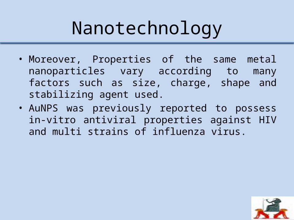
• Moreover, Properties of the same metal nanoparticles vary according to many factors such as size, charge, shape and stabilizing agent used.
• AuNPS was previously reported to possess in-vitro antiviral properties against HIV and multi strains of influenza virus.

Size comparison

Aim of the work
In-vitro Evaluation of antiviral activity of gold nanoparticles on BVDV as a model for HCV.

Material and methods
• Cell line : Madin-Darby bovine kidney (MDBK) cells were maintained in Dulbecco’s modified Eagle medium (DMEM) supplemented with 5% horse serum (DMEM-HS) at 37°C in a humidified, 5% CO2 atmosphere.
• Virus: BVDV-1 (strain NADL) was propagated and titrated according to Reed and Muench (1938).

Gold Nanoparticles• Gold Nanoparticles
Colloidal gold nanoparticles were synthesized by the optimization of classical Citrate Reduction method according to Turkevich et al. (1951).• Surface modification of Gold Nanoparticles
Surface modification of gold nanoparticles necessary to increase its colloidal stability in physiological fluid was performed by adding poly –ethylene glycol (PEG) according to the method described by Alcantar et al. (2000).• Characterization of Gold Nanoparticles
Prepared gold nanoparticles were characterized by using UV-Vis Spectrophotometer, Zetasizer, and high Resolution Transmission Electron Microscopy according to Liu and Lu (2006).

• Cytotoxicity of Gold Nanoparticles by MTT assay
Prior to using the synthesized gold nanoparticles for anti-viral assay, cytotoxicity of nanoparticles was determined using MDBK cell lines by MTT (a tetrazole) assay. MDBK cell lines were trypsinized, suspended in minimal essential medium and counted using trypan blue exclusion dye according to Mosmann (1983).

Determination of AuNPs antiviral activity and interaction of viral particles with AuNPs (Kesarkar et
al., 2012)
• To study the mode of action of Nanoparticles with BVDV, we used two different ways:
• Method I - In the first method, the viral particles were interacted with the AuNPS for 15 minutes and then allowed to interact with the MDBK cells. 25 μl of viral suspension (106 TCD50/ml) was added with 25 μl of the different concentrations of the synthesized AuNPS in PCR tubes and incubated for 15 minutes at 37°C. The concentrations of AuNPS used (2, 4, 6 and 8 ppm) were based on the results of the cytotoxicity assay done for the Nanoparticles showing minimum cytotoxicity.

• Method II – In the second method, the viral particles were first allowed to infect MDBK cells (1 hour)and the AuNPS were added to the suspension afterwards. A volume of 25 μl of viral suspension (106 TCD50/ml) was added to different wells of tissue culture plate containing 5 x 10⁵cells/ml of MDBK cells and incubated for 1 hr in CO2 incubator. After 1 hr of incubation, 2, 4, 6 or 8ppm of Nanoparticles were added and the wells were incubated for 72 hr at 37°C in CO2 incubator
• The following two controls were also kept parallel for incubation.
• 1. Triple control containing only cells and viral suspension.• 2. Triple control containing only cells.
• After incubation, 10μl (5mg/ml) MTT reagent was added in each well and incubated at 37°C for 4 hrs in CO2 incubator. Then 100 μl of 0.1N acidified iso-propanol was added to each well and kept in dark for 30 min at room temperature. The well plates were then kept on a shaker for 1 min and OD was taken at 530 nm wavelength. The average values from triplicate readings were determined and the average value for the blank was subtracted. Absorbances against number of cells/ml were plotted.

Results
Synthesis and characterization of gold nanoparticles
• A) Transmission Electron Microscopy (TEM) imaging
The particles are mostly spherical. The average size (diameter) of particles, measured on the TEM images was 7± 2 nm.
• B) UV-Visible Absorption Spectroscopy
The absorption spectrum for the solution of citrated AuNPs is characterized by the excitation of the Plasmon resonance in the neighborhood of 518 nm.
• C) Zetasizer :The zeta potential of AuNPS was found to be around -12.3 mV.

TEM image showing spherical shape of the prepared AuNPS

Spectrophotometer results of AuNPS characterization showing peak absorption at wave length 518nm
400 500 600 700 800 9000.00
0.05
0.10
0.15
0.20
0.25
0.30
0.35
max
A
bsor
banc
e
wavelength,nm
AuNPS capped citrate=518 nm

Histogram chart providing the size distribution of synthesized AuNPS
2 3 4 5 6 7 8 9 10 11 12 13 14 15 16 17 18 19 20 21 220.0
0.1
0.2
0.3
0.4
0.5
size (nm)
Rela
tive F
requ
enc
y %
AuNPS@citrate

The zeta potential of AuNPS was found to be around -12.3 mV

Results of cytotoxicity assay of prepared AuNPS by MTT (tetrazolium) assay
Group AuNPS concentration(PPM)
Mean(OD)
AuNPS
8 0.17 ± 0.04 b
6 0.20 ± 0.02 b
4 0.28 ± 0.06 a
2 0.28 ± 0.02 a
Cells only (control) (0) 0.28 ± 0.04 a

Results of cytotoxicity assay (MTT) of AuNPS on MDBK cells

Results of antiviral assay (MTT) of AuNPS on MDBK against BVDV by microplate reader
Group AuNPS concentration
(ppm)Mean(OD) method (1)
Mean(OD) method (2)
AuNPS
2 0.17 ± 0.04 bc 0.11 ± 0.08 c
4 0.27± 0.05 a 0.11 ± 0.02 c
6 0.20±0.07ab 0.11 ± 0.05 c
8 0.17 ± 0.01 c 0.10 ± 0.08 c
Cell control 0 0.27±0.07a
+ve virus
control0 0.11 ± 0.02 c

Results of antiviral assay (MTT) method (1) of AuNPS on
MDBK against BVDV by Microplate reader absorbance.

Discussion
• Results showed that the maximum non-toxic concentrations of prepared AuNPS were 2 and 4ppm, which totally agree with Sametband et al. (2011) and Kesarkar et al. (2012).
• Antiviral activity evaluation of prepared AuNPS against BVDV showed greater activity in the test when the viral particles were allowed to interact with the nanoparticles before infecting the cells (method 1) as compared to the test where the cells, previously infected with the virus (method 2), not allowed to interact with the nanoparticles.

Conclusion
• Results confirmed that BVDV NADL strain used in this study was cytopathogenic biotype and the used cell type was suitable as a theater for the following in vitro virus inhibition assays. BVDV can be a surrogate model for HCV on MDBK cells in the antiviral testing procedures.
• AuNPS at 4ppm concentration were found to be effective as virus neutralizing candidate when allowed to interact with cells

References
• Alcantar, N.A.; Aydil, E.S.; Israelachvili, J.N. (2000): Polyethylene glycol-coated
biocompatible surfaces. J. Biomed. Mater. Res., 5, 51(3):343-51.• Bolin, S.R.; Matthews, P.J.; Ridpath, J.F. (1991): Methods for detection and
frequency of contamination of fetal calf serum with bovine viral diarrhea virus and antibodies against Bovine Viral Diarrhea Virus. J. Vet. Diagn. Invest., 3: 199-203.
• Buckwold, V.E.; Beer, B.E.; Donis, R.O. (2003): Bovine viral diarrhea virus as a surrogate model of hepatitis C virus for the evaluation of antiviral agents. Antivir. Res., 60(1): 1–15.
• Chah, S.; Hammond, M.; Zare, R. (2005): Gold nanoparticles as colorimetric sensor for protein conformational changes. Chem. Biol., 12(3): 3 23-8.
• Donis, R.O.; Dubovi, E. J. (1987): Differences in virus-induced polypeptides in cells infected by cytopathic and noncytopathic biotypes of bovine virus diarrhea-mucosal disease virus. Virology J., 158: 168-173.

• Kalaycioglu, T.A.; Russell, P.H.; Howard, C.R. (2007): Selection of mimotopes of Bovine Viral Diarrhoea Virus using a solid-phase peptide library. Vaccine, 25: 7081-7086.
• Kesarkar, R.; Oza, G.; Pandey, S.; Dahake, R.; Mukherjee, S.; Chowdhary, A.; Madhuri Sharon, M. (2012): Gold nanoparticles: effective as both entry inhibitors and virus neutralizing agents against HIV. J. Microbio. And Biotech. es; V (2) , I (2 ) P : 276-283.
• Liu, J., Lu, Y. (2006): Nature Protocols, 1(1), 246• Mosmann, T. (1983): Rapid colorimetric assay for cellular growth and survival:
application to proliferation and cytotoxicity assays. J. immunol. Methods, 65(1-2): 55-63.
• Paeshuyse, J.; Chezal, J.M.; Froeyen, M.; Leyssen, P.; Dutartre, H.; Vrancken, R.; Canard, B.; Letellier, C.; Li, T.; Mittendorfer, H.; Koenen, F.; Kerkhofs, P.; De Clercq, E.; Herdewijn, P.; Puerstinger, G.; Gueiffier, A.; Chavignon, O.; Teulade, J.C.; Neyts, J. (2007): The imidazopyrrolopyridine analogue AG110 is a novel highly selective inhibitor of pestivirus that target the RNA-dependent RNA polymerase at a hot spot for inhibition of viral replication. J. Virol., 81: 11046-11053.

• Paton, D.J.; Carlssson, U.; Lowings, J.P.; Sands, J.J.; Vilcek, S.; Alenius, S. (1995): Identification of herd-specific bovine viral diarrhoea virus isolates from infected cattle and sheep. Vet. Microbiol., 43: 283-294.
• Rai, V.R., Bai, J. A. (2011): Nanoparticles and their Potential Application as Antimicrobials. In: Science Against Microbial Pathogens: Communicating Current Research and Technological Advances, Mendez-Vilas, A. (Ed.). University of Mysore, India, pp: 197-209.
• Reed, L.J.; Muench, H. (1938): A simple method of estimating fifty percent end points. Am. J. Hyg., 27:593-597.
• Rice, C. M. (1996): in Virology, eds. Fields, B. N., Knipe, D. M. & Howley, P. M. (Lippincott, Philadelphia), pp. 931–959.
• Ridpath, J.F.; Bolin, S.R.; Dubovi, E.J. (1994): Segregation of bovine viral diarrhea virus into genotypes. Virology J., 205: 66-74.
• Salim, M.T.A.; Goto, Y.; Hamasaki, T.; Okamoto, M.; Aoyama, H.; Hashimoto, Y.; Musiu, S.; Paeshuyse, J.; Neyts, J.; Froeyen, M.; Herdewijn, P.; Baba, M. (2010): Highly potent and selective inhibition of bovine viral diarrhea virus replication by y-carboline derivatives. Antiviral Res., 88: 263-268.

• Sametband, M.; Shukla, S.; Meningher, T.; Hirsh, S.; Mendelson, E.; Sarid, R.; Gedanken, A.; Mandelboim, M. (2011): Effective multistrain inhibition of influenza virus by anionic gold nanoparticles. Med. Chem. Commun., 2:421-423.
• Tabarrini, O.; Manfroni, G.; Fravolini, A.; Cecchetti, V.; Sabatini, S.; De Clercq, E.; Rozenski, J.; Canard, B.; Dutartre, H.; Paeshuyse, J.; Neyts, J. (2006): Synthesis and anti-BVDV activity of acridones as new potential antiviral agents. J. Med. Apr., 49: 2621-2627.
• Turkevich J.; Stevenson, P. C.; Hillier, J. (1951): A study of the nucleation and growth processes in the synthesis of colloidal gold. Discuss. Faraday Soc., 11, 55-75.
• Vijayakumar, S., Ganesan, S. (2012): Gold nanoparticles as an HIV entry inhibitor. Curr. HIV Res., 10(8): 643-6.
• Wang, L.; Zhao, W.; Tan, W. (2008): Bioconjugated Silica Nanoparticles: Development and Applications. Nano Res., 1 , 2 : 99–99-115
• Yamane, D.; Zahoor, M. A.; Mohamed, Y. M.; Azab, W.; Kato, K.; Tohya, Y.; Akashi, H. (2009): Inhibition of sphingosine kinase by bovine viral diarrhea virus NS3 is crucial for efficient viral replication and cytopathogenesis. J. Biol. Chem., 284, 13648–13659.