![Cytosolic ribosomes on the surface of mitochondria · 4 76. in vitro in a ribosome free system [12, 17-19].Meanwhile, cytosolic ribosomes were 77. detected in the vicinity of mitochondria](https://static.fdocuments.us/doc/165x107/5fa30531bbcc776ccb1369c0/cytosolic-ribosomes-on-the-surface-of-mitochondria-4-76-in-vitro-in-a-ribosome.jpg)
In Vitro Approaches to Assess Mitochondria-Mediated...
33
In Vitro Approaches to Assess Mitochondria-Mediated Drug Toxicity: Advantages and Limitations and a Decade of Learning's Yvonne Will, Ph.D Pfizer Research & Development Groton, CT [email protected]
Transcript of In Vitro Approaches to Assess Mitochondria-Mediated...

In Vitro Approaches to Assess
Mitochondria-Mediated Drug Toxicity:
Advantages and Limitations and a
Decade of Learning's
Yvonne Will, Ph.D
Pfizer Research & Development
Groton, CT

48 Drugs Were Withdrawn for Safety Reasons
Between 1990-2007
18 Hepatotoxic 21 Cardiotoxic 9 Others
Cerivastatin Troglitazone
Tolcapone
Nefazodone

Hepatotoxicity Cardiovascular
Mitochondrial Impairment of Drugs Receiving Black Box Warnings
Antivirals
Abacavir
Didanosine
Emtricitabine
Entecavir
Emtricitabine
Lamivudine
Nevirapine
Telbivudine
Tenofovir
Tipranavir
Stavudine
Zalcitabine
Zidovudine
Anti-Cancer
Flutamide
Dacarbazine
Gemtuzumab
Methotrexate
Pentostatin
Tamoxifen
Antibiotics
Isoniazid
Ketoconazole (oral)
Streptozocin
Trovafloxacin
CNS
Dantrolene
Divalproex Sodium
Felbamate
Naltrexone
Nefazodone
Valproic Acid/
Hypertension
Bosentan
Anthracyclines
Daunorubicin
Doxorubicin
Epirubicin
Idarubicin
NSAIDs
Celecoxib
Diclofenac
Diflunisal
Etodolac
Fenoprofen
Ibuprofen
Indomethacin
Ketoprofen
Mefenamic acid
Meloxicam
Naproxen
Nabumetone
Oxaprozin
Piroxicam
Salsalate
Sulindac
Thioridazine
Tolmetin
Anaesthetic
Bupivacaine
Anti-Cancer
Arsenic Trioxide
Cetuximab
Denileukin diftitox
Mitoxantrone
Tamoxifen
Beta-Blocker]
Atenolol
Antiarhythmic
Amiodarone (oral)
Disopyramide
Dofetilide
Ibutilide
CNS
Amphetamines
Atomoxetin
Droperidol
Methamphetamine
Pergolide
Diabetes
Pioglitazone
Rosiglitazone

Early mitochondrial assessment
allows the identification of
compounds with the desired
efficacy profile, but without
ancillary liabilities.
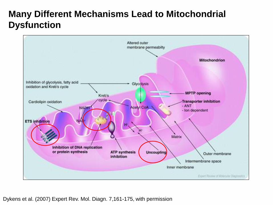
Dykens et al. (2007) Expert Rev. Mol. Diagn. 7,161-175, with permission
Many Different Mechanisms Lead to Mitochondrial
Dysfunction

Objectives/Outline:
• Drug withdrawn from the market exhibit mitochondrial liabilities
• Assays to detect mitochondrial toxicity
• Assay for measuring Oxygen consumption of isolated mitochondria.
• Cell viability assay in (a) Glucose medium, (b) Galactose medium.
• Assay for measuring Oxygen consumption and extracellular acidification of cells.
• Assays for measuring changes in mtDNA and mtDNA-encoded protein levels in cells.
• Summary

Polarographic Mitochondrial
Respiration
Drug Mitos Substrate
ADP
All ADP phosphorylated
Basal Respiration
Maximum Respiration
ADP-Driven O2
Uncoupling
Inhibition
Time (min) 20min

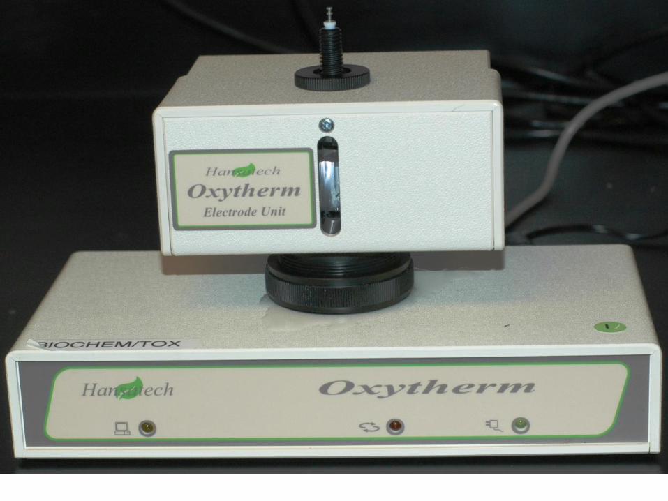
• Phosphorescent
• Water-soluble
• Cell non-invasive
• Non-cytotoxic
• Stable
• Time resolved or prompt
• Compatible with any reader
• Large stoke shift allows for
high signal to noise ratio
• multiplex with “green dyes”
No
rma
lis
ed
In
ten
sit
y
Wavelength (nm)
0
0.2
0.4
0.6
0.8
1
300 400 500 600
0
1
2
3
4
5
600 620 640 660 680 700
Wavelength (nm)
Fo
ld I
nc
rea
se
Deoxygenated
Air-saturated
Oxygen consumption Measurement in
Isolated Mitochondria
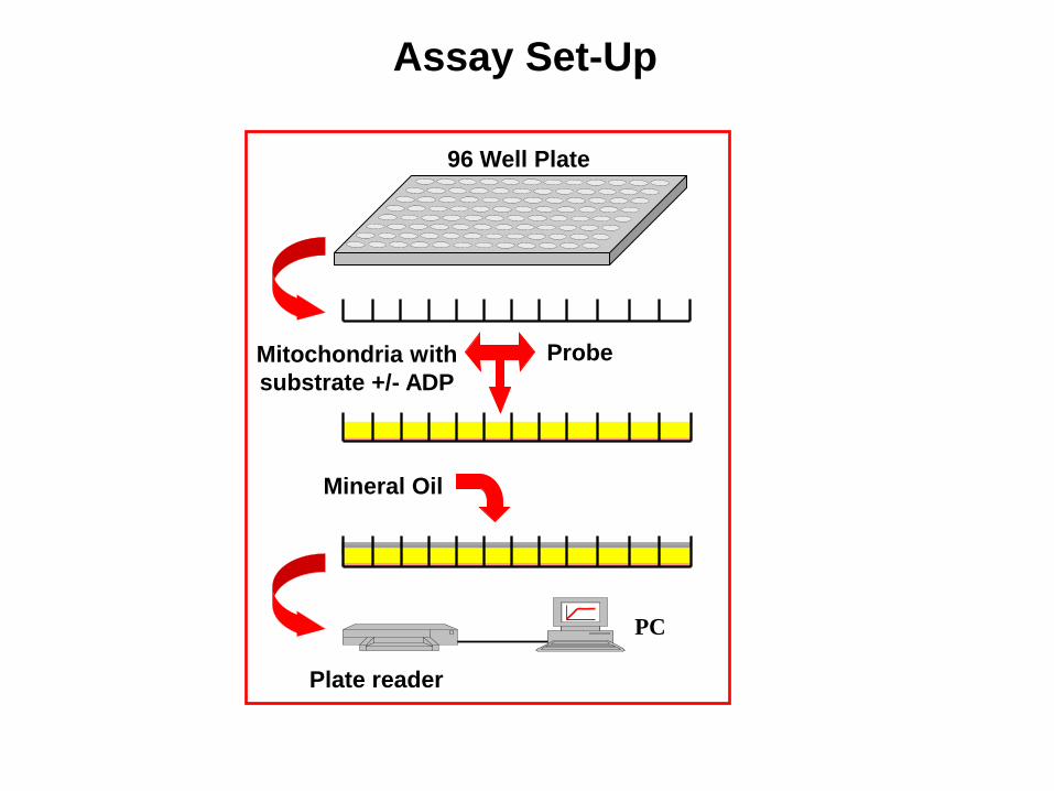
Assay Set-Up
PC
Mitochondria with
substrate +/- ADP
96 Well Plate
Probe
Mineral Oil
Plate reader
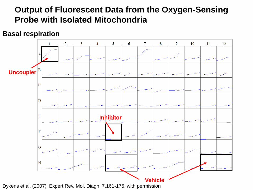
Basal respiration
Output of Fluorescent Data from the Oxygen-Sensing
Probe with Isolated Mitochondria
Vehicle
Uncoupler
Inhibitor
Dykens et al. (2007) Expert Rev. Mol. Diagn. 7,161-175, with permission

State 2
0 2 4 6 8 10 12 14 16 18 20
0
2000
4000
6000
8000
10000
Muraglitazar
Rosiglitazone
Ciglitazone
Darglitazone
Pioglitazone
Troglitazone
Control
Time (min)
RF
U(m
ean
+ S
E)
State 3
0 2 4 6 8 10 12 14 16 18 20
0
3000
6000
9000
12000
15000
Muraglitazar
Rosiglitazone
CiglitazoneDarglitazone
Pioglitazone
Troglitazone
Control
Time (min)
RF
U(m
ean
+ S
E)
Drugs present at 25nmol/mg mitochondrial protein. N=4, except for controls N=48.
Mitochondrial Effects of Thiozolidinediones Vary
Nadanaciva et al. (2007) Toxicol. Appl.
Pharmacol. 223, 277-287, with permission
State 2
0 2 4 6 8 10 12 14 16 18 20
0
2000
4000
6000
8000
10000
Muraglitazar
Rosiglitazone
Ciglitazone
Darglitazone
Pioglitazone
Troglitazone
Control
Time (min)
RF
U(m
ean
+ S
E)
State 3
0 2 4 6 8 10 12 14 16 18 20
0
3000
6000
9000
12000
15000
Muraglitazar
Rosiglitazone
CiglitazoneDarglitazone
Pioglitazone
Troglitazone
Control
Time (min)
RF
U(m
ean
+ S
E)
Drugs present at 25nmol/mg mitochondrial protein. N=4, except for controls N=48.
Withdrawn
Blackbox warning
Basal respiration
ADP-Driven
Withdrawn
Blackbox warning
Dykens JA, Jamieson J, Marroquin L, Nadanaciva S, Billis PA, Will Y. Biguanide-induced mitochondrial dysfunction yields increased lactate production and cytotoxicity of aerobically-poised HepG2 cells and human hepatocytes in vitro. Toxicol Appl Pharmacol. 2008 Dec 1;233(2):203-10. Bioaccumulation
Will Y, Dykens JA, Nadanaciva S, Hirakawa B, Jamieson J, Marroquin LD, Hynes J, Patyna S, Jessen BA. Effect of the multi-targeted tyrosine kinase inhibitors imatinib, dasatinib, sunitinib, and sorafenib on mitochondrial function in isolated rat heart mitochondria and H9c2 cells. Toxicol Sci. 2008 Nov;106(1):153-61.
Liver toxicity of sorafenib
Dykens JA, Jamieson JD, Marroquin LD, Nadanaciva S, Xu JJ, Dunn MC, Smith AR, Will Y. In vitro assessment of mitochondrial dysfunction and cytotoxicity of nefazodone, trazodone, and buspirone. Toxicol Sci. 2008 Jun;103(2):335-45. rank order correct, need additional risk factors
Nadanaciva S, Dykens JA, Bernal A, Capaldi RA, Will Y. Mitochondrial impairment by PPAR agonists and statins identified via immunocaptured OXPHOS complex activities and respiration. Toxicol Appl Pharmacol. 2007 Sep 15;223(3):277-87. rank order correct, Cmax , accumulation
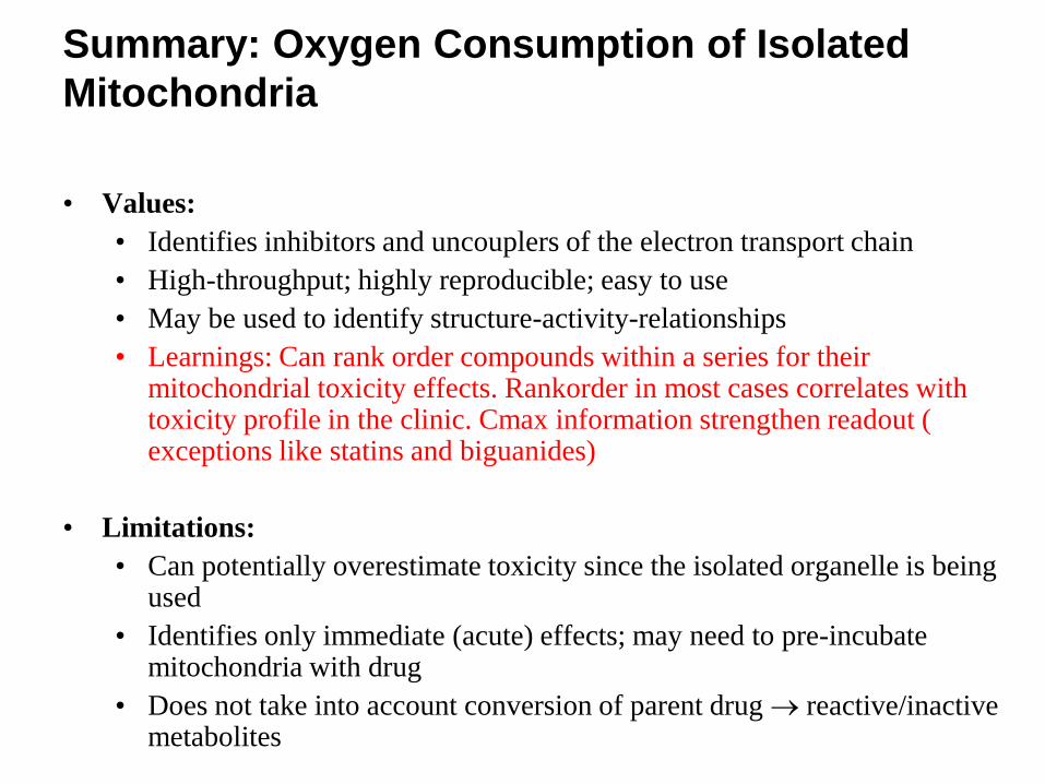
Summary: Oxygen Consumption of Isolated
Mitochondria
• Values:
• Identifies inhibitors and uncouplers of the electron transport chain
• High-throughput; highly reproducible; easy to use
• May be used to identify structure-activity-relationships
• Learnings: Can rank order compounds within a series for their mitochondrial toxicity effects. Rankorder in most cases correlates with toxicity profile in the clinic. Cmax information strengthen readout ( exceptions like statins and biguanides)
• Limitations:
• Can potentially overestimate toxicity since the isolated organelle is being used
• Identifies only immediate (acute) effects; may need to pre-incubate mitochondria with drug
• Does not take into account conversion of parent drug reactive/inactive metabolites
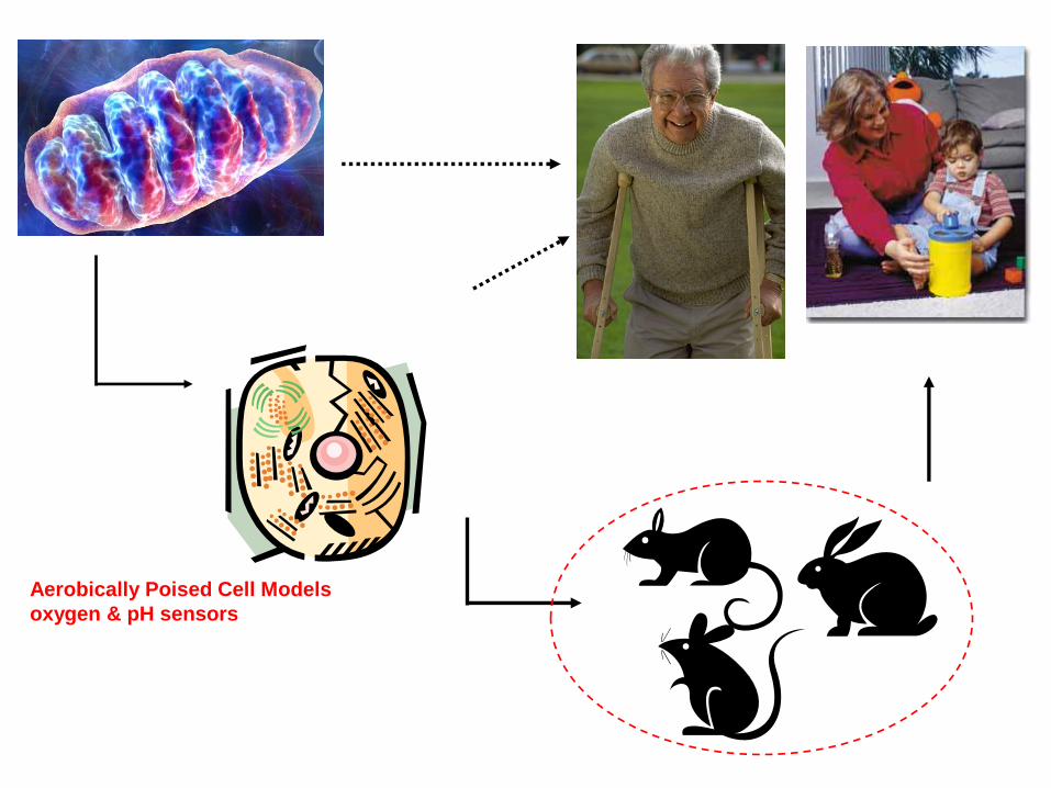
Aerobically Poised Cell Models
oxygen & pH sensors

Objectives/Outline:
• Drug withdrawn from the market exhibit mitochondrial liabilities
• Assays to detect mitochondrial toxicity
• Assay for measuring Oxygen consumption of isolated mitochondria.
• Cell viability assay in (a) Glucose medium, (b) Galactose medium.
• Assay for measuring Oxygen consumption and extracellular acidification of cells.
• Assays for measuring changes in mtDNA and mtDNA-encoded protein levels in cells.
• Summary

Crabtree Effect (1929): inhibition of respiration by elevated glucose.
Warburg Effect (1929): aerobic glycolysis yields lactate despite
competent mitochondria.
Contemporary cell culture often uses 25mM glucose media (5X
physiological!)
Transformed cells are often characterized by low rates of O2
consumption & resistance to mitotoxicants.
Circumventing the Crabtree Effect: The “Glucose-
Galactose” Model
Marroquin et al. (2007) Toxicol. Sci., 97, 539-547
0
20
40
60
80
100
120
0.001 0.01 0.1 1
[Rotenone] M
% A
TP
Co
ntr
ol
*
AT
P
(% C
on
tro
l)
[Antimycin] M
*
AT
P
(% C
on
tro
l)
0
20
40
60
80
100
120
0.01 0.1 1 10 100 1000
[FCCP] M
*
*
AT
P
(% C
on
tro
l)
0
20
40
60
80
100
120
0.0001 0.001 0.01 0.1 1 10
[Oligomycin] M
*
AT
P
(% C
on
tro
l)
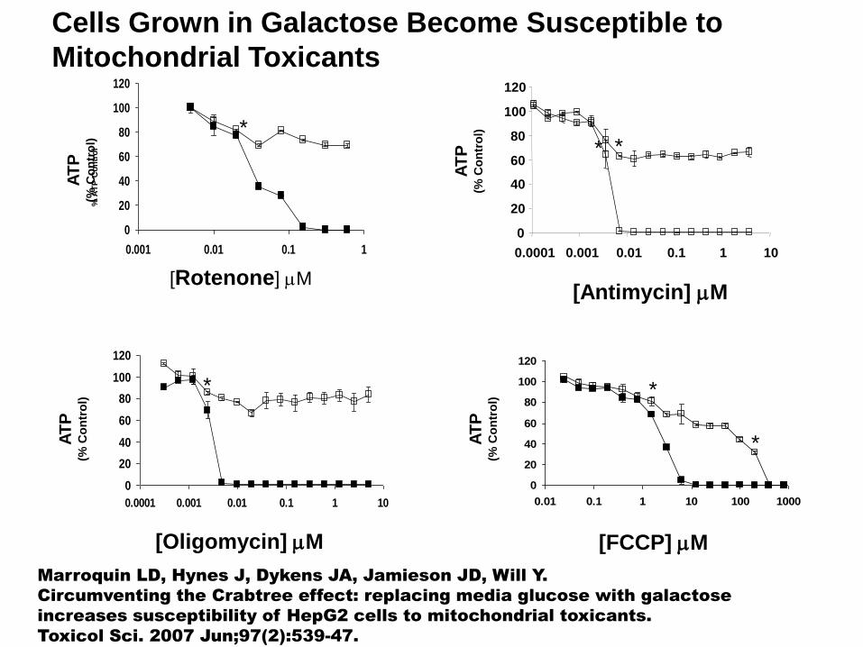
Cells Grown in Galactose Become Susceptible to
Mitochondrial Toxicants
Marroquin LD, Hynes J, Dykens JA, Jamieson JD, Will Y.
Circumventing the Crabtree effect: replacing media glucose with galactose
increases susceptibility of HepG2 cells to mitochondrial toxicants.
Toxicol Sci. 2007 Jun;97(2):539-47.
0
20
40
60
80
100
120
0.0001 0.001 0.01 0.1 1 10
*

Cells Grown in Galactose are More Susceptible to
Mitochondrial Toxicants such as Nefazodone
0
20
40
60
80
100
120
0.01 0.1 1 10 100Concentration (µM)
% A
TP
Co
ntr
ol
8.98
38.4
0
5
10
15
20
25
30
35
40
45
50
Galactose Glucose
Cell Culture Media
IC5
0 (
µM
)
Dykens et al. (2008) Toxicol. Sci., 103, 335-345

Correlating The RST and HepG2 Glu-Gal Assays
RST assay HepG2 Glucose-Galactose assay Mechanism of Toxicity
More toxic in Gal than Glu Toxicity primarily through
mitochondrial effects (<5% of
compounds)
Not toxic in either medium Compound may be converted to inactive
metabolite or does not get into cells
Equally toxic in both media Multiple mechanisms of toxicity
(most of RST positives)
-
More toxic in Gal than Glu Compound may affect apoptosis, impair
fatty acid transport, activate HIF-1a
-
More toxic in Glu than Gal Compound may impair glycolysis
-
Equally toxic in both media Toxicity primarily through
non-mitochondrial “off-targets”
A compound could belong to any of the following categories:
Objectives/Outline:
• Drug withdrawn from the market exhibit mitochondrial liabilities
• Assays to detect mitochondrial toxicity
• Assay for measuring Oxygen consumption of isolated mitochondria.
• Cell viability assay in (a) Glucose medium, (b) Galactose medium.
• Assay for measuring Oxygen consumption and extracellular acidification of cells.
• Assays for measuring changes in mtDNA and mtDNA-encoded protein levels in cells.
• Summary

The O2 Consumption Rate of Cells is a Measure of Mitochondrial
Respiration
Glycolysis
NAD+
NADH ATP
O2
Glucose
Pyruvate
G6P
Acetyl-CoA
Lactic
Acid
H2O
ADP
O2
ATP
ADP
Extracellular Acidification Rate of Cells is a Measure of Glycolysis

Phenformin and Buformin Decrease Oxygen
Consumption in HepG2 cells
DMSO
Metformin
Buformin
Phenformin
125M 125M 125M 125M
Dykens et al. (2008) Toxicol. Appl. Pharmacol. 233, 203-210, with permission
Oxygen Consumption Rate
HepG2 cells
% c
han
ge i
n o
xyg
en
co
nsu
mp
tio
n

Phenformin and Buformin Cause Media Acidification
Phenformin
Buformin
Metformin DMSO
125M 125M 125M 125M
HepG2 cells
Extracellular Acidification Rate %
ch
an
ge i
n p
H
Dykens et al. (2008) Toxicol. Appl. Pharmacol. 233, 203-210, with permission
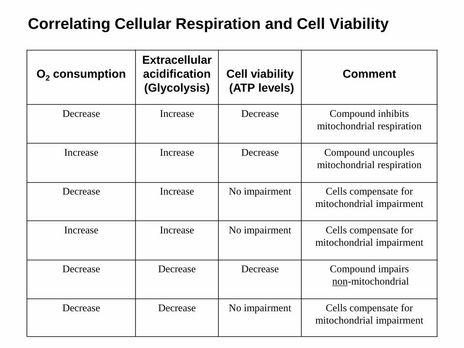
Correlating Cellular Respiration and Cell Viability
O2 consumption
Extracellular
acidification
(Glycolysis)
Cell viability
(ATP levels)
Comment
Decrease Increase Decrease Compound inhibits
mitochondrial respiration
Increase Increase Decrease Compound uncouples
mitochondrial respiration
Decrease Increase No impairment Cells compensate for
mitochondrial impairment
Increase Increase No impairment Cells compensate for
mitochondrial impairment
Decrease Decrease Decrease Compound impairs
non-mitochondrial
Decrease Decrease No impairment Cells compensate for
mitochondrial impairment

Objectives/Outline:
• Drug withdrawn from the market exhibit mitochondrial liabilities
• Assays to detect mitochondrial toxicity
• Assay for measuring Oxygen consumption of isolated mitochondria.
• Cell viability assay in (a) Glucose medium, (b) Galactose medium.
• Assay for measuring Oxygen consumption and extracellular acidification of cells.
• Assays for measuring changes in mtDNA and mtDNA-encoded protein levels in cells.
• Summary

High content screening approach for identifying
antibacterials and anti-retrovirals that cause mitochondrial toxicity
mtDNA-encoded protein
in vehicle-treated cells
mtDNA-encoded protein
in cells grown in 40 M Linezolid
Nuclear DNA-encoded protein
in cells grown in 40 M Linezolid
Nuclear DNA-encoded protein
in vehicle-treated cells

• Many, but not all, drugs causing organ toxicity have a mitochondrial liability.
• Elevated serum liver enzymes = hepatocyte death
• Lactic acidosis is classic hallmark.
• Depending on potency, if a drug has a mitochondrial liability, it will have consequences. Importance of Cmax
– Bioaccumulation alters PK.
– >10,000-fold concentration of some cations in matrix over plasma.
– Additional factors increase risk (BSEP) not discussed today
– Phys-chem property space (promiscuity) drives mitotox
• Severity of such adverse effects is idiosyncratic.
– Function of organ history and genetics (incl. mtDNA).
• Preclinical assessments are done in young, perfectly healthy animals.
– Threshold effects and physiological scope.
Summary:
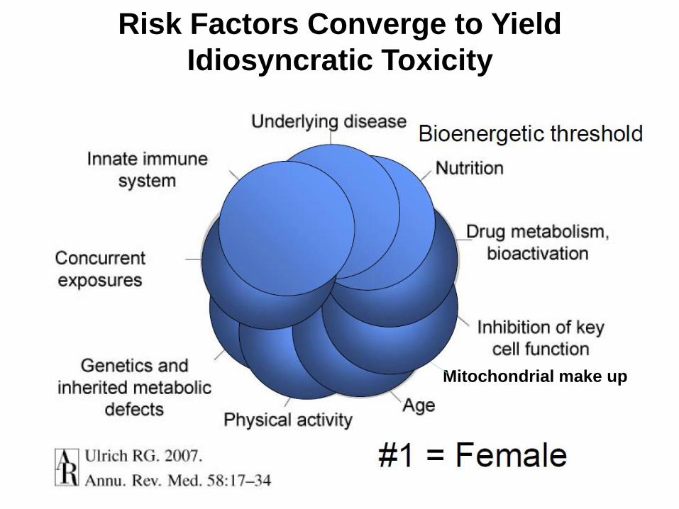
Risk Factors Converge to Yield
Idiosyncratic Toxicity
Mitochondrial make up

Toxicology and Applied Pharmacology, Volume 264, Issue 2, 2012, 167 - 181
.
MEFs from different mouse strains respond
differently to nefazodone when grown in glucose and galactose
Acknowledgements
• Lisa Marroquin, MS
• Dr. James Dykens
• Dr. James Hynes
• Dr Richard Fernandes
• Dr. Roderik Capaldi
• Autumn Bernal, BS
• Dr David Ferrick
• Dr. Sashi Nadanaciva
• Rachel Swiss, Payal Rana
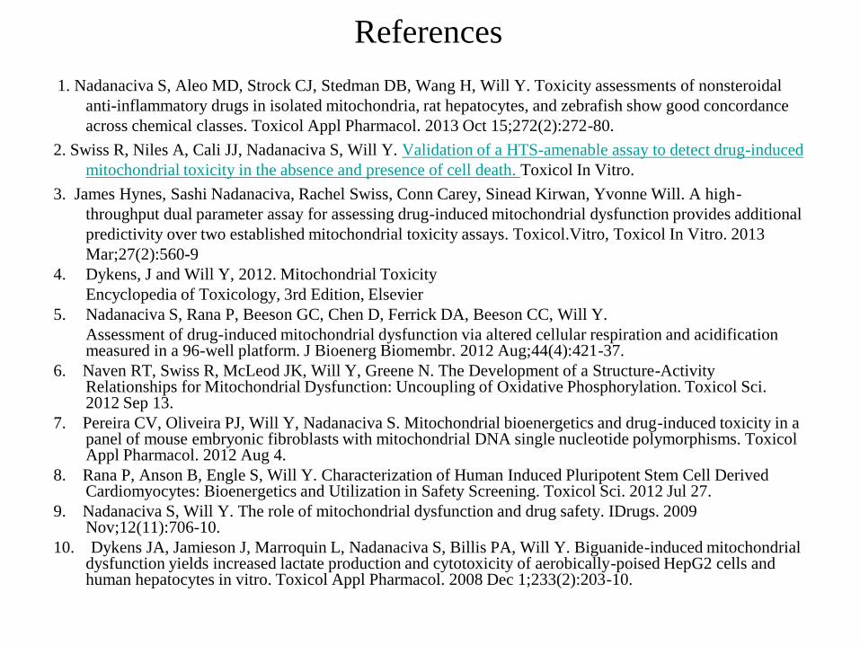
References
1. Nadanaciva S, Aleo MD, Strock CJ, Stedman DB, Wang H, Will Y. Toxicity assessments of nonsteroidal
anti-inflammatory drugs in isolated mitochondria, rat hepatocytes, and zebrafish show good concordance
across chemical classes. Toxicol Appl Pharmacol. 2013 Oct 15;272(2):272-80.
2. Swiss R, Niles A, Cali JJ, Nadanaciva S, Will Y. Validation of a HTS-amenable assay to detect drug-induced
mitochondrial toxicity in the absence and presence of cell death. Toxicol In Vitro.
3. James Hynes, Sashi Nadanaciva, Rachel Swiss, Conn Carey, Sinead Kirwan, Yvonne Will. A high-
throughput dual parameter assay for assessing drug-induced mitochondrial dysfunction provides additional
predictivity over two established mitochondrial toxicity assays. Toxicol.Vitro, Toxicol In Vitro. 2013
Mar;27(2):560-9
4. Dykens, J and Will Y, 2012. Mitochondrial Toxicity
Encyclopedia of Toxicology, 3rd Edition, Elsevier
5. Nadanaciva S, Rana P, Beeson GC, Chen D, Ferrick DA, Beeson CC, Will Y.
Assessment of drug-induced mitochondrial dysfunction via altered cellular respiration and acidification measured in a 96-well platform. J Bioenerg Biomembr. 2012 Aug;44(4):421-37.
6. Naven RT, Swiss R, McLeod JK, Will Y, Greene N. The Development of a Structure-Activity Relationships for Mitochondrial Dysfunction: Uncoupling of Oxidative Phosphorylation. Toxicol Sci. 2012 Sep 13.
7. Pereira CV, Oliveira PJ, Will Y, Nadanaciva S. Mitochondrial bioenergetics and drug-induced toxicity in a panel of mouse embryonic fibroblasts with mitochondrial DNA single nucleotide polymorphisms. Toxicol Appl Pharmacol. 2012 Aug 4.
8. Rana P, Anson B, Engle S, Will Y. Characterization of Human Induced Pluripotent Stem Cell Derived Cardiomyocytes: Bioenergetics and Utilization in Safety Screening. Toxicol Sci. 2012 Jul 27.
9. Nadanaciva S, Will Y. The role of mitochondrial dysfunction and drug safety. IDrugs. 2009 Nov;12(11):706-10.
10. Dykens JA, Jamieson J, Marroquin L, Nadanaciva S, Billis PA, Will Y. Biguanide-induced mitochondrial dysfunction yields increased lactate production and cytotoxicity of aerobically-poised HepG2 cells and human hepatocytes in vitro. Toxicol Appl Pharmacol. 2008 Dec 1;233(2):203-10.
References Continued: 11. Will Y, Dykens JA, Nadanaciva S, Hirakawa B, Jamieson J, Marroquin LD, Hynes J, Patyna
S, Jessen BA. Effect of the multi-targeted tyrosine kinase inhibitors imatinib, dasatinib, sunitinib, and sorafenib on mitochondrial function in isolated rat heart mitochondria and H9c2 cells. Toxicol Sci. 2008 Nov;106(1):153-61.
12. Dykens JA, Jamieson JD, Marroquin LD, Nadanaciva S, Xu JJ, Dunn MC, Smith AR, Will Y. In vitro assessment of mitochondrial dysfunction and cytotoxicity of nefazodone, trazodone, and buspirone. Toxicol Sci. 2008 Jun;103(2):335-45. Dykens JA, Will Y. The significance of mitochondrial toxicity testing in drug development. Drug Discov Today. 2007 Sep;12(17-18):777-85. Review.
13. Nadanaciva S, Dykens JA, Bernal A, Capaldi RA, Will Y. Mitochondrial impairment by PPAR agonists and statins identified via immunocaptured OXPHOS complex activities and respiration. Toxicol Appl Pharmacol. 2007 Sep 15;223(3):277-87.
14. Will Y, Hynes J, Ogurtsov VI, Papkovsky DB. Analysis of mitochondrial function using phosphorescent oxygen-sensitive probes. Nat Protoc. 2006;1(6):2563-72.
15. Marroquin LD, Hynes J, Dykens JA, Jamieson JD, Will Y. Circumventing the Crabtree effect: replacing media glucose with galactose increases susceptibility of HepG2 cells to mitochondrial toxicants. Toxicol Sci. 2007 Jun;97(2):539-47.
16. Nadanaciva S, Bernal A, Aggeler R, Capaldi R, Will Y. Target identification of drug induced mitochondrial toxicity using immunocapture based OXPHOS activity assays. Toxicol In Vitro. 2007 Aug;21(5):902-11
17. Dykens JA, Marroquin LD, Will Y. Strategies to reduce late-stage drug attrition due to mitochondrial toxicity. Expert Rev Mol Diagn. 2007 Mar;7(2):161-75.
18. Hynes J, Marroquin LD, Ogurtsov VI, Christiansen KN, Stevens GJ, Papkovsky DB, Will Y. Investigation of drug-induced mitochondrial toxicity using fluorescence-based oxygen-sensitive probes. Toxicol Sci. 2006 Jul;92(1):186-200.