in the responses of Pinus taeda L and Pinus radiata D. Don to
11
© The Author 2012. Published by Oxford University Press. All rights reserved. For Permissions, please email: [email protected] Tree Physiology 32, 1302–1312 doi:10.1093/treephys/tps091 Research paper Biomarker genes highlight intraspecific and interspecific variations in the responses of Pinus taeda L. and Pinus radiata D. Don to Sirex noctilio F. acid gland secretions John Michael Bordeaux 1 , W. Walter Lorenz 1 and Jeffrey F.D. Dean 1,2,3 1 Warnell School of Forestry and Natural Resources, University of Georgia, Athens, GA 30602, USA; 2 Department of Biochemistry and Molecular Biology, University of Georgia, Athens, GA 30602, USA; 3 Corresponding author ([email protected]) Received March 12, 2012; accepted August 25, 2012; published online October 5, 2012; handling Editor Chung-Jui Tsai Sirex noctilio F., a Eurasian horntail woodwasp recently introduced into North America, oviposits in pines and other conifers and in the process spreads a phytopathogenic fungus that serves as a food source for its larvae. During oviposition the woodwasp also deposits mucus produced in its acid (venom) gland that alters pine defense responses and facilitates infec- tion by the fungus. A 26,496-feature loblolly pine cDNA microarray was used to survey gene expression of pine tissue responding to S. noctilio venom. Six genes were selected for further assessment by quantitative real-time polymerase chain reaction (qRT-PCR), including one that encoded an apparent PR-4 protein and another that encoded a thaumatin-like protein. Expression of both was strongly induced in response to venom, while expression of an apparent actin gene (ACT1) was stable in response to the venom. The pattern of gene response was similar in Pinus taeda L. and Pinus radiata D. Don, but the mag- nitude of response in P. radiata was significantly stronger for each of the induced genes. The magnitude of the biomarker gene response to venom also varied according to genotype within these two species. The qRT-PCR assay was used to dem- onstrate that the primary bioactive component in S. noctilio venom is a polypeptide. Keywords: bioactive peptide, bioassay, conifer, defense response, gene expression, Hymenoptera, loblolly pine, microarray, Monterey pine, venom, woodwasp. Introduction The Eurasian horntail woodwasp, Sirex noctilio Fabricius (Hymenoptera: Siricidae), an invasive insect recently intro- duced into North America, represents a serious economic and ecological threat to pine forests throughout the USA and Canada (Borchert et al. 2007). Females of the species typi- cally attack dead or dying pines in their native range (Europe, Asia and northern Africa), where they cause little if any eco- nomic impact (Coutts and Dolezal 1966, Borchert et al. 2007). The relative importance of S. noctilio to forest health in Europe may be measured by the fact that it is sometimes neglected entirely in discussions on European forest pests (Ciesla 2003). By contrast, S. noctilio is a major pest when introduced as an exotic into commercial pine plantations of the southern hemi- sphere (Talbot 1977). These southern hemisphere pine planta- tions are typically stocked with North American pine species, mostly Monterey (Pinus radiata D. Don) and loblolly pines (Pinus taeda L.), which highlights the danger this woodwasp may pose for North American forests (Ciesla 2003). Unlike other members of the Siricidae family, S. noctilio attacks conifers exclusively (Talbot 1977). Sirex noctilio attack occurs via the act of oviposition in which the female wood- wasps use their specialized ovipositors to drill tunnels into the xylem and deposit their eggs and venom along with arthrospores (oidia) of a fungal symbiont, the white-rot basidiomycete, at University of Pretoria on March 13, 2015 http://treephys.oxfordjournals.org/ Downloaded from
Transcript of in the responses of Pinus taeda L and Pinus radiata D. Don to

© The Author 2012. Published by Oxford University Press. All rights reserved. For Permissions, please email: [email protected]
Tree Physiology 32, 1302–1312doi:10.1093/treephys/tps091
Research paper
Biomarker genes highlight intraspecific and interspecific variations in the responses of Pinus taeda L. and Pinus radiata D. Don to Sirex noctilio F. acid gland secretions
John Michael Bordeaux1, W. Walter Lorenz1 and Jeffrey F.D. Dean1,2,3
1Warnell School of Forestry and Natural Resources, University of Georgia, Athens, GA 30602, USA; 2Department of Biochemistry and Molecular Biology, University of Georgia, Athens, GA 30602, USA; 3Corresponding author ([email protected])
Received March 12, 2012; accepted August 25, 2012; published online October 5, 2012; handling Editor Chung-Jui Tsai
Sirex noctilio F., a Eurasian horntail woodwasp recently introduced into North America, oviposits in pines and other conifers and in the process spreads a phytopathogenic fungus that serves as a food source for its larvae. During oviposition the woodwasp also deposits mucus produced in its acid (venom) gland that alters pine defense responses and facilitates infec-tion by the fungus. A 26,496-feature loblolly pine cDNA microarray was used to survey gene expression of pine tissue responding to S. noctilio venom. Six genes were selected for further assessment by quantitative real-time polymerase chain reaction (qRT-PCR), including one that encoded an apparent PR-4 protein and another that encoded a thaumatin-like protein. Expression of both was strongly induced in response to venom, while expression of an apparent actin gene (ACT1) was stable in response to the venom. The pattern of gene response was similar in Pinus taeda L. and Pinus radiata D. Don, but the mag-nitude of response in P. radiata was significantly stronger for each of the induced genes. The magnitude of the biomarker gene response to venom also varied according to genotype within these two species. The qRT-PCR assay was used to dem-onstrate that the primary bioactive component in S. noctilio venom is a polypeptide.
Keywords: bioactive peptide, bioassay, conifer, defense response, gene expression, Hymenoptera, loblolly pine, microarray, Monterey pine, venom, woodwasp.
Introduction
The Eurasian horntail woodwasp, Sirex noctilio Fabricius (Hymenoptera: Siricidae), an invasive insect recently intro-duced into North America, represents a serious economic and ecological threat to pine forests throughout the USA and Canada (Borchert et al. 2007). Females of the species typi-cally attack dead or dying pines in their native range (Europe, Asia and northern Africa), where they cause little if any eco-nomic impact (Coutts and Dolezal 1966, Borchert et al. 2007). The relative importance of S. noctilio to forest health in Europe may be measured by the fact that it is sometimes neglected entirely in discussions on European forest pests (Ciesla 2003).
By contrast, S. noctilio is a major pest when introduced as an exotic into commercial pine plantations of the southern hemi-sphere (Talbot 1977). These southern hemisphere pine planta-tions are typically stocked with North American pine species, mostly Monterey (Pinus radiata D. Don) and loblolly pines (Pinus taeda L.), which highlights the danger this woodwasp may pose for North American forests (Ciesla 2003).
Unlike other members of the Siricidae family, S. noctilio attacks conifers exclusively (Talbot 1977). Sirex noctilio attack occurs via the act of oviposition in which the female wood-wasps use their specialized ovipositors to drill tunnels into the xylem and deposit their eggs and venom along with arthrospores (oidia) of a fungal symbiont, the white-rot basidiomycete,
at University of Pretoria on M
arch 13, 2015http://treephys.oxfordjournals.org/
Dow
nloaded from
Tree Physiology Online at http://www.treephys.oxfordjournals.org
Amylostereum areolatum (Chaillet ex Fries) Boiden (Coutts and Dolezal 1966, Coutts 1968, 1969a, 1969b). While both the venom and the fungus must be present to kill trees (Coutts 1968, 1969a, 1969b, 1970, Spradbery 1973), the venom alone causes overt changes in host tree phenotype, such as the nee-dle droop (flagging) characteristic of S. noctilio attack, as well as physiological changes that serve to depress host defense responses (Coutts 1969a, 1969b). Since the apparent function of S. noctilio venom is to weaken the tree sufficiently that the fungus can overcome any remaining defense response (Coutts 1969a, 1969b), an understanding of the venom mode of action will be important for unraveling the mechanism of pathology and possibly developing new measures to intercede in this pathosystem.
Bioassays for the effect of S. noctilio venom on pines have typically relied on the visual observation of responses, such as needle flagging and chlorosis (Bordeaux and Dean 2012). A variety of physiological measurements, including needle starch accumulation, dry-weight change, respiration rate, moisture content, amylase and peroxidase activity, leaf pres-sure (total water potential) and radial stem growth have also been used in the past to follow the effects of S. noctilio venom on treated pines (Coutts 1970, Fong and Crowden 1973, Spradbery 1973). Venom action has been tested using intact trees (Coutts 1969a, 1969b, 1970, Fong and Crowden 1973, Spradbery 1973), but such assays are impractical for routine use. Branch and needle explants from various species of pine have been used as indicators of venom activity with varying sensitivity (Coutts 1969a, 1969b, Bowling and Dolezal 1970, Fong and Crowden 1973, Spradbery 1973, Kile et al. 1974, Wong and Crowden 1976), but there exist few reliable quantitative measures for testing S. noctilio venom activity. An issue complicating development of consis-tent, reproducible bioassays has been tree-to-tree variability in response to venom. Although discussed in little detail, reports in the older Sirex literature noted that some P. radiata trees were more sensitive than others to the application of S. noctilio venom (Coutts 1968, 1969a, 1969b, Kile et al. 1974). Noting this variation, researchers used explants from susceptible trees in subsequent studies (Bowling and Dolezal 1970, Fong and Crowden 1973, Spradbery 1973, Wong and Crowden 1976). It remains unclear as to whether these observed variations in venom sensitivity were linked to geno-type or some other factor.
The importance of S. noctilio venom to this pathosystem is reflected in the oversized venom reservoir that fills a large pro-portion of the abdomen in newly emergent female woodwasps. Despite the absence of a reliable quantitative assay for bioac-tivity, early biochemical studies characterized the venom as a protein–polysaccharide complex presenting several enzyme activities, including amylase, esterase, phenoloxidase and pro-tease (Wong and Crowden 1976). Chemical tests were used to
show that non-sulfated acidic mucopolysaccharide is a primary constituent of S. noctilio venom. Activity tests showed the active factor in venom to be heat-stable, retaining activity after boiling for at least 2 h and even after autoclaving (Coutts 1969a, 1969b, 1970). In fact, heating appeared to enhance the activity of diluted venom, which may be related to the slow disaggregation described for the active factor into a low molec-ular mass fraction (2–5 kDa) after dilution in water (Fong and Crowden 1973, Wong and Crowden 1976). This disaggregated factor is most likely the venom component that is transported from the site of woodwasp oviposition on pine stems to photo-synthetic tissues in the upper branches of the tree. Venoms of the more familiar aculeate (stinging) wasps typically are com-plex, containing large and small polypeptides, biogenic amines (histamine, dopamine, adrenaline, noradrenaline and sero-tonin), as well as enzymes such as phospholipases and hyal-uronidases (Gullan and Cranston 2000). However, the biochemical nature of the active factor in S. noctilio venom has remained, until now, unclear.
To better understand S. noctilio venom and its mode of action in Pinus species, work was undertaken to develop bio-marker assays with which to follow venom activity. These bio-marker assays were used to investigate the variability in venom responses between individual trees and in different pine spe-cies. Additional studies tested whether or not polypeptide components in S. noctilio venom are important for venom effects on pines.
Materials and methods
Biological materials
Loblolly pine (P. taeda) or Monterey pine (P. radiata) seeds were soaked in a 1% (v : v) hydrogen peroxide solution at 4 °C for 4 days to hasten germination (Barnett and McLemore 1967). Open-pollinated (OP) P. taeda seeds from a 7–56 genotype mother tree were kindly provided by Dr Michael Cunningham (International Paper Co., Bainbridge, GA, USA), while OP P. radi-ata seeds were provided by Dr Kathleen Jermstad (US Forest Service, Placerville, CA, USA). Seeds were planted in commercial potting soil, and at 6 weeks the seedlings, having developed substantial tap roots, were transferred to Ray Leach Cone-tainers (Stuewe & Sons, Tangent, OR, USA). Seedlings were thereafter kept in growth chambers under a 16 h light/8 h diur-nal cycle and watered every 3–4 days. Fertilizer was applied every 4 weeks as a solution of 14.5 g Miracle-Gro (Scotts, Marysville, OH, USA) and 1.2 g chelated iron per 4 l water. For the P. radiata genotypic variation study, 17 individuals were kept in a greenhouse under ambient conditions and hedged at regu-lar intervals to produce multiple shoot tips. Phenotype assays were performed using individual shoot tips with cut ends sub-merged in 50 µl of 20 mg/ml S. noctilio venom and then trans-ferred to water after venom absorption. To minimize false
Biomarker genes highlight intraspecific and interspecific variations 1303
at University of Pretoria on M
arch 13, 2015http://treephys.oxfordjournals.org/
Dow
nloaded from
Tree Physiology Volume 32, 2012
positives during the incubation period, cut ends were regularly trimmed at the bottom and water was refreshed frequently to prevent tracheal element clogging by microbial growth. The 17 P. radiata individuals were designated as either venom-sensitive or non-responsive based on phenotype (observable wilting and color change in needles). Two venom-sensitive trees and one non-responsive tree were selected for routine use in the poly-merase chain reaction (PCR) biomarker assays.
Sirex noctilio females provided by Kelley Zylstra (USDA APHIS, Syracuse, NY, USA) were frozen immediately upon emergence from pine logs and stored at −80 °C until use. Frozen wasps were dipped in ethanol prior to dissection to remove the venom sac and glands. Pooled venom tissues were ground in water using a fritted-glass homogenizing tube and brought to a concentration of 20 mg/ml whole tissue in deion-ized water. The resultant solution was boiled for 15 min, cooled and dispensed into 1-ml aliquots prior to storage at −20 °C until use. Prior to performing assays, the venom solution was thawed and then centrifuged at 17,000 g for 5–10 min to remove any precipitate. Only the clarified supernatant was used in subsequent assays.
Pine cDNA microarray analysis
To generate a short list of candidate biomarker genes in lob-lolly pine that respond strongly to S. noctilio venom exposure a minimal microarray experiment was performed. The experi-ment used the PtGen2 loblolly pine microarray platform, which contains 26,496 features representing 25,848 unique cDNA amplimers and a unigene set of ~16,000 genes (Lorenz et al. 2011) and employed a reference design (Kerr 2003) to iden-tify genes differentially expressed between venom-treated and control seedlings at two different time points. The reference sample was comprised of total RNA isolated from P. taeda roots, shoots and needles (Lorenz et al. 2011). As the sole purpose of this experiment was to generate a list of potential biomarkers for PCR assays, the experimental design comprised two biological replicates of control and time point samples, as well as two technical replicates. The data from this microarray experiment are not of such statistical significance as to be useful for genome-scale analyses on their own, and their utility is unpredictable beyond the narrow scope of our need to identify useful biomarkers; nonetheless, the data have been placed in the GEO database at NCBI under accession number_GSE40000.
To generate biological samples for the microarrays, 60 5-month-old P. taeda seedlings were cut at the soil line and cut ends were placed in 50 µl each of 20 mg/ml S. noctilio venom or water. After uptake, seedlings were transferred to water until samples were collected. At two time points after treatment (24, 48 h), two sets of 10 venom-treated seedlings were pooled, as was one set of 10 water control seedlings. This yielded a total of six samples for microarray analysis. The seedling pools from
each time point were collected and flash-frozen in liquid nitro-gen. Frozen tissue was ground using a SPEX model 6850 freezer mill (SPEX, Metuchen, NJ, USA), and RNA was extracted using a protocol modified from Chang et al. (1993, Lorenz et al. 2010). Briefly, 3 g of ground tissue was transferred to 20 ml RNA extraction buffer (2% CTAB (w : v), 2% (w : v) polyvinyl pyrrolidinone, 100 mM Tris-HCl pH 8.0, 25 mM EDTA, 2 M NaCl, 0.5 g/l spermidine and 2% (v : v) β-mercaptoethanol), extracted twice with chloroform and cen-trifuged, with retention of the aqueous layer after each centrif-ugation step. RNA was precipitated from the aqueous pool by incubation overnight with 10 M LiCl and centrifuged and the pellet was resuspended in SSTE buffer. Resuspended RNA was further purified by phenol/chloroform, pH 8 (PC8) and neat chloroform extraction, centrifugation and retention of the aque-ous layer. As a final step, RNA was precipitated in ethanol, resuspended in water and treated with RNAse-free DNAse (TurboDNA Free, Ambion, Austin, TX, USA). Complementary DNA was synthesized with incorporation of amino-ally and amino-hexyl modified nucleotides using Invitrogen’s Superscript™ Indirect Labeling Kit (Invitrogen, Carlsbad, CA, USA). Cy-5 and Cy-3 dyes (GE Healthcare Bio-Sciences Corp., Piscataway, NJ, USA) were suspended in 80 µl dimethyl sulfox-ide and coupled to the modified cDNA per manufacturer’s instructions. Dye-coupled cDNAs were purified using pub-lished methods (Alba et al. 2005) with mass and labeling effi-ciencies quantified spectrophotometrically. Microarrays were hybridized with labeled targets as described (Lorenz et al. 2009). Slides were incubated in a HybChamber™ (Genomic Solutions, Ann Arbor, MI, USA) containing 20 µl 100 mM dithiothreitol. HybChambers were wrapped in foil and incu-bated in a 48 °C shaking water bath at 50 rpm for 14–16 h.
A ProScanArray™ confocal scanner (Perkin Elmer, Waltham, MA, USA) was used to scan microarrays at 532 and 635 nm. ImaGene software (ver. 7.5, BioDiscovery Inc., El Segundo, CA, USA) was used to process raw fluorescence data. Data were filtered, log-transformed, normalized (print tip-Lowess) and statistical analyses were performed using BRB-Array Tools Ver. 8.0 (Simon et al. 2007). Spot signals were filtered and flagged as described previously (Wang et al. 2003, Alba et al. 2005), and probes with ≥50% missing data were eliminated from fur-ther analyses. Differentially expressed genes were identified by comparing two treatment groups (venom treatment vs. water) with a test of random variance (Simon et al. 2007) using a false discovery rate of 0.5 and a differential expression cut-off minimum of 1.5-fold.
Biomarker gene analysis
Microarray analysis identified genes whose expression was up-regulated in pine seedlings exposed to S. noctilio venom. From a list of the most highly up-regulated genes (Table 1), three tar-gets were chosen for further testing as potential biomarkers.
1304 Bordeaux et al.
at University of Pretoria on M
arch 13, 2015http://treephys.oxfordjournals.org/
Dow
nloaded from

Tree Physiology Online at http://www.treephys.oxfordjournals.org
Two of the genes were annotated as a putative PR-4 (PR4) pro-tein and a thaumatin-like protein (TLP), while one potential bio-marker was not annotated (unknown, UNK). Two additional genes annotated as actin1 (ACT1) and actin2 (ACT2) were cho-sen for potential use as control (housekeeping) genes. A phenyl-alanine ammonia lyase (PAL) gene previously found to have a relatively steady expression in pine xylem (Nairn et al. 2008) was tested for potential use as a housekeeping control, but was found respond to venom treatment. It was subsequently rele-gated to study as a candidate biomarker. Primers for quantitative real-time PCR (qRT-PCR) analysis of potential biomarker and control genes were designed using Primer 3 v.0.4.0 (Table 2).
Five-month-old loblolly pine seedlings (half-sib family of 7-56) were cut at the soil line and the ends were placed in 50 µl of 20 mg/ml venom solution or water. After absorption of the treatment solution, seedlings were incubated in water. At intervals (4, 24, 48 h), seedlings were flash-frozen in liquid nitrogen. For testing of P. radiata, shoot tips collected from hedged trees were subjected to the same venom and control treatments as were used for loblolly seedlings. At 24 h, one venom-treated and one control shoot tip were flash-frozen, while the remaining three were incubated and observed for phenotypic responses over 3 weeks. Frozen tissues were ground using a mortar and pestle, and RNA was extracted as described above. Complementary DNA was synthesized (as described previously but without modified nucleotides) and further diluted for PCR experiments.
Biomarker gene performance was initially assessed using semi-quantitative PCR as follows. For cDNAs derived from pine tissue samples, a twofold dilution series from 0.25 to 0.0078 ng/µl cDNA was generated as starting template mate-rial for PCR. Reaction mixtures contained 10 µl of cDNA tem-plate in buffer, 3 µl primer (0.5 µM each), 4 µl 10× buffer (100 mM Tris-HCl pH 8.8, 35 mM MgCl2, 250 mM KCl), 2 µl 10 mM dNTP mix and 1 µl Taq polymerase. Reactions (50 µl total volume) were run using a GeneAmp 9700 thermal cycler (Applied Biosystems, Carlsbad, CA, USA). The amplification conditions were 95 °C for 3 min, then 30 cycles of 94 °C for 30 s, 55 °C for 30 s and 72 °C for 90 s, followed by 72 °C for 10 min. Amplification products were visualized in agarose gels
for preliminary, qualitative assessment of differential expres-sion of target genes. Amplimers were visualized by running 5 µl of the PCR product solution on a 1.5% agarose gel in TAE buffer, followed by ethidium bromide staining.
For qRT-PCR, reaction mixtures consisted of 5 µl of 0.5 µM primer pair, 5 µl of 0.5 µM cDNA template and 10 µl SybrGreen 2× Supermix (BioRad, Hercules, CA, USA). Twenty microliter reactions were run using an iCycler thermal cycler (BioRad, Hercules, CA, USA), and reaction conditions were 95 °C for 3 min, then 40 cycles of 95 °C for 30 s, 65 °C for 45 s and 78 °C for 20 s, followed by 95 °C for 1 min, 55 °C for 1 min and 81 cycles of 55 °C for 10 s in 0.5 °C increments to assess product purity via melt temperature. Samples were analyzed in triplicate for each primer set, and runs were considered acceptable when replicates agreed to <0.5% relative standard deviation. Data were generated from two biological replicates and two technical replicates. Means for the technical replicates were used to calcu-late mean and error values for each biological replicate.
Proteolysis
To determine whether the woodwasp venom active factor had a proteinaceous component, venom was subjected to proteolysis.
Biomarker genes highlight intraspecific and interspecific variations 1305
Table 1. Genes identified in P. taeda seedlings showing the greatest fold-expression increases (log2 normalized) at 24 and 48 h post-treatment with S. noctilio venom. Genes noted in boldface were selected for initial development of PCR assays.
Gene ID 24 h 48 h Functional category CLONE_NAME NCBI NR BLASTx NCBI NR annotation
11.9.14 3.0 4.0 Defense NDL1_35_C04_A029 gb|AAC33732.1| PR-4 type protein (PR4) (Vitis vinifera)7.15.6 5.8 3.3 Defense FLD1_38_F11_A029 ref|XP_002275386.1| Class IV chitinase (Vitis vinifera), predicted61.19.9 2.7 2.8 Defense STRR1_17_E07_A033 emb|CAA10492.1| Thaumatin-like protein (TLP) (Pseudotsuga
menziesii)43.20.7 2.9 2.7 Unknown RTCA1_29_F06_A029 Not found Unknown (UNK)23.4.12 4.9 2.7 Unknown RTDS1_10_C05_A015 Not found Unknown43.23.10 4.1 2.6 Defense RTCNT1_8_B10_A029 dbj|BAD93486.1| Pollen allergen CJP38 (Cryptomeria japonica)63.12.9 4.8 2.5 Defense STRR1_57_A10_A033 gb|ABY66958.1| Class IV chitinase (Vitis pseudoreticulata)
Table 2. Gene annotations and primer sequences used in PCR assays.
Gene name Primer sequence (5′ → 3′)
PR4 (PR-4 protein, Vitis vinifera)
5′: TTA TAG GCG GAC GGG TTG TA
3′: GTT TCA GCA CAA ACG CAG AGTLP (Thaumatin-like protein, Pseudotsuga menziesii)
5′: GTT TGC ACC AGA AGC ACA TG
3′: CAC AGG CCT ACA GCT ATG CAUNK (Unknown, Pinus taeda) 5′: CCA TGC ATC CAC CAA CAG TA
3′: GGA TGA GGC CGG GAA TAT APAL1 (Phenylalanine ammonia lyase, Pinus taeda)
5′: TC ACA AGC TGA AGC ACC ATC
3′: T CCC GTC CAA GAC ATA CTC CACT1 (Actin, Pinus taeda) 5′: AA TGG TCA AGG CTG GAT TTG
3′: AG GGC GAC CAA CAA TAC TTGACT2 (Actin, Pinus taeda) 5′: TT GCT GAC CGT ATG AGC AAG
3′: GA GGT GCA ACC ACC TTG ATT at University of Pretoria on M
arch 13, 2015http://treephys.oxfordjournals.org/
Dow
nloaded from
Tree Physiology Volume 32, 2012
Proteinase K (110 µl, 1 mg/ml in 50 mM Tris, pH 8.0, 10 mM CaCl2) was added to 1 ml venom (20 mg/ml in water). A control digestion consisted of prepared Proteinase K that had been deactivated by boiling for 15 min. Both solutions were placed in a 45 °C bath for 24 h, after which they were boiled to deacti-vate proteinase activity (venom activity is heat-stable), followed by centrifugation for 10 min at 17,000 g. Shoot tips from P. radiata trees previously characterized as either highly respon-sive or weakly responsive were treated with venom incubated with heat-inactivated Proteinase K, venom incubated with active Proteinase K or a water control. For each treatment, 50 µl of solution was absorbed by each of five shoot tips after which the shoot tips were transferred to pure water for further incubation. After 24 h, the shoot tips were pooled by treatment, flash-fro-zen in liquid nitrogen, RNA was extracted and cDNA was syn-thesized as described previously. This material was then analyzed in the qRT-PCR assay as described previously.
Results
Phenotypic assays
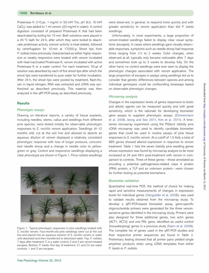
Drawing on literature reports, a variety of tissue explants, including needles, stems, callus and seedlings from different pine species, were tested initially for observable phenotypic responses to S. noctilio venom application. Seedlings (4–12 months old) cut at the soil line and allowed to absorb an aqueous dilution of venom displayed a relatively consistent phenotypic response with loss of turgor pressure, concomi-tant needle droop and a change in needle color to yellow-green or gray. Control and responsive seedlings displaying a clear phenotype are shown in Figure 1. Pinus radiata seedlings
were observed, in general, to respond more quickly and with greater sensitivity to venom application than did P. taeda seedlings.
Unfortunately, in most experiments, a large proportion of venom-treated seedlings failed to display clear visual symp-toms (escapes). In cases where seedlings gave visually observ-able responses, symptoms such as needle droop had response times ranging from 2 h to 2 weeks. Color changes, when observed at all, typically only became noticeable after 7 days and sometimes took up to 3 weeks to develop fully. On the other hand, no control seedlings were ever seen to display the phenotypic changes associated with venom application. The large proportion of escapes in assays using seedlings led us to consider that genetic differences between species and among individual genotypes could be confounding bioassays based on observable phenotypic changes.
Microarray analysis
Changes in the expression levels of genes responsive to biotic and abiotic agents can be measured quickly and with great sensitivity, which is the rationale for developing biomarker gene assays to supplant phenotypic assays (Zimmermann et al. 2008, Jeong and Seo 2011, Kim et al. 2011). A time-series microarray experiment using the PtGen2 loblolly pine cDNA microarray was used to identify candidate biomarker genes that could be used in routine assays of pine tissue responses to S. noctilio venom. At a cutoff of 1.5-fold, a total of 885 genes showed altered expression in response to venom treatment. Table 1 lists the seven loblolly pine seedling genes whose expression was found by microarray analysis to be most increased at 24 and 48 h post-treatment with venom in com-parison to controls. Three of these genes—those annotated as encoding a potential pathogenesis-related class 4 protein (PR4) protein, a TLP and an unknown protein—were chosen for further testing as potential biomarkers.
Biomarker validation
Quantitative real-time PCR, the method of choice for making rapid and sensitive measurements of changes in expression levels for individual genes (Vanguilder et al. 2008), was used to validate results obtained from the microarray study. To develop a qRT-PCR-based biomarker assay, gene-specific oligo nucleotide primers were generated for the three venom-sensitive genes identified in the microarray study. Primers were also designed for three additional genes, two actin genes (ACT1, ACT2) and one PAL gene, identified as useful control (housekeeping) genes in a previous study (Nairn et al. 2008). The complete list of genes used in the qRT-PCR studies and their respective primer sequences are found in Table 2. Preliminary testing showed that all primer pairs yielded single amplimer products when using cDNA templates from either P. taeda or P. radiata.
1306 Bordeaux et al.
Figure 1. Typical phenotypic responses in pine seedlings treated with S. noctilio venom. Four-month-old pine seedlings were cut at the soil line and placed into an aqueous solution of S. noctilio venom or water until absorbed and then transferred to deionized water. Top, P. radiata, 7 days after treatment. C is a water control; 2 and 3 are venom-treated escapes. Bottom, P. taeda, first day of treatment. C1 and C2 are water controls; 1 and 3 are escapes.
at University of Pretoria on M
arch 13, 2015http://treephys.oxfordjournals.org/
Dow
nloaded from
Tree Physiology Online at http://www.treephys.oxfordjournals.org
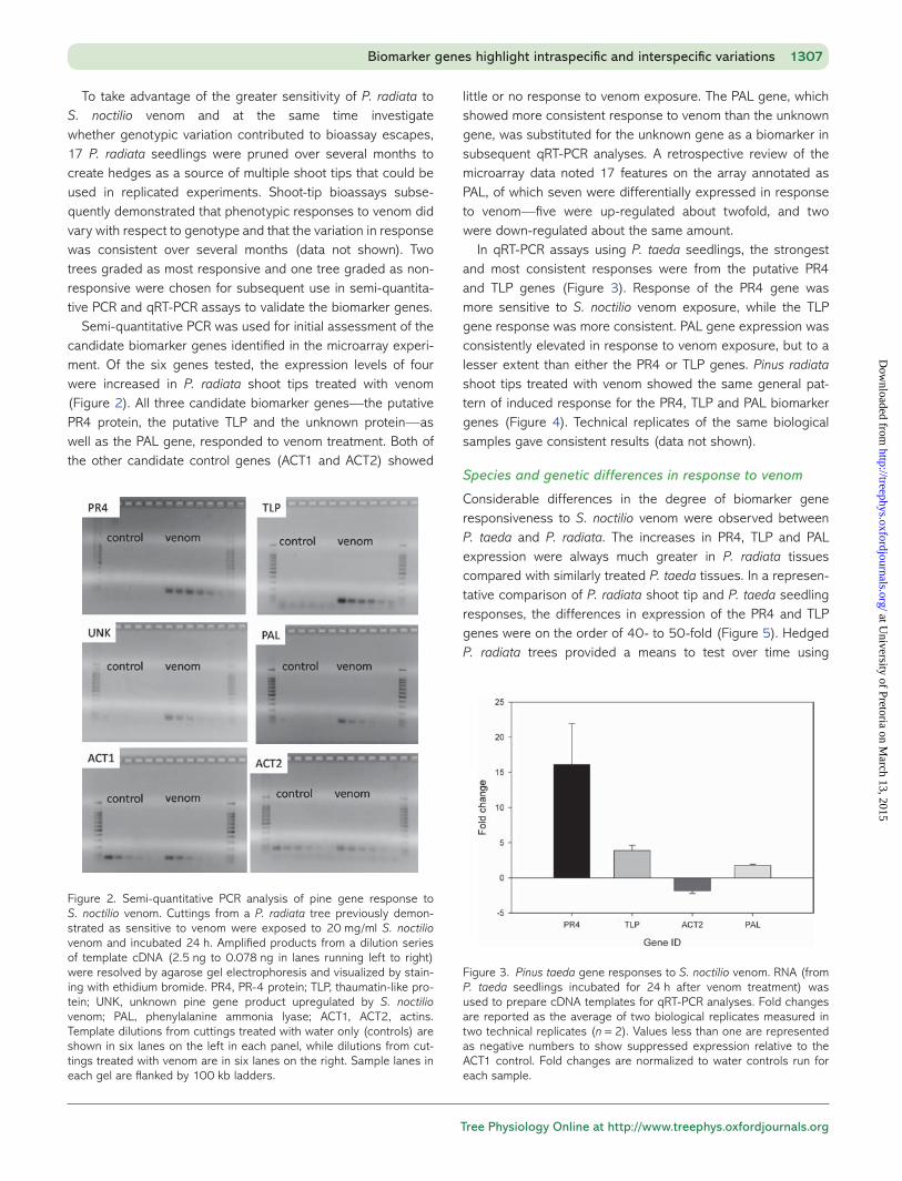
To take advantage of the greater sensitivity of P. radiata to S. noctilio venom and at the same time investigate whether genotypic variation contributed to bioassay escapes, 17 P. radiata seedlings were pruned over several months to create hedges as a source of multiple shoot tips that could be used in replicated experiments. Shoot-tip bioassays subse-quently demonstrated that phenotypic responses to venom did vary with respect to genotype and that the variation in response was consistent over several months (data not shown). Two trees graded as most responsive and one tree graded as non-responsive were chosen for subsequent use in semi-quantita-tive PCR and qRT-PCR assays to validate the biomarker genes.
Semi-quantitative PCR was used for initial assessment of the candidate biomarker genes identified in the microarray experi-ment. Of the six genes tested, the expression levels of four were increased in P. radiata shoot tips treated with venom (Figure 2). All three candidate biomarker genes—the putative PR4 protein, the putative TLP and the unknown protein—as well as the PAL gene, responded to venom treatment. Both of the other candidate control genes (ACT1 and ACT2) showed
little or no response to venom exposure. The PAL gene, which showed more consistent response to venom than the unknown gene, was substituted for the unknown gene as a biomarker in subsequent qRT-PCR analyses. A retrospective review of the microarray data noted 17 features on the array annotated as PAL, of which seven were differentially expressed in response to venom—five were up-regulated about twofold, and two were down-regulated about the same amount.
In qRT-PCR assays using P. taeda seedlings, the strongest and most consistent responses were from the putative PR4 and TLP genes (Figure 3). Response of the PR4 gene was more sensitive to S. noctilio venom exposure, while the TLP gene response was more consistent. PAL gene expression was consistently elevated in response to venom exposure, but to a lesser extent than either the PR4 or TLP genes. Pinus radiata shoot tips treated with venom showed the same general pat-tern of induced response for the PR4, TLP and PAL biomarker genes (Figure 4). Technical replicates of the same biological samples gave consistent results (data not shown).
Species and genetic differences in response to venom
Considerable differences in the degree of biomarker gene responsiveness to S. noctilio venom were observed between P. taeda and P. radiata. The increases in PR4, TLP and PAL expression were always much greater in P. radiata tissues compared with similarly treated P. taeda tissues. In a represen-tative comparison of P. radiata shoot tip and P. taeda seedling responses, the differences in expression of the PR4 and TLP genes were on the order of 40- to 50-fold (Figure 5). Hedged P. radiata trees provided a means to test over time using
Biomarker genes highlight intraspecific and interspecific variations 1307
Figure 2. Semi-quantitative PCR analysis of pine gene response to S. noctilio venom. Cuttings from a P. radiata tree previously demon-strated as sensitive to venom were exposed to 20 mg/ml S. noctilio venom and incubated 24 h. Amplified products from a dilution series of template cDNA (2.5 ng to 0.078 ng in lanes running left to right) were resolved by agarose gel electrophoresis and visualized by stain-ing with ethidium bromide. PR4, PR-4 protein; TLP, thaumatin-like pro-tein; UNK, unknown pine gene product upregulated by S. noctilio venom; PAL, phenylalanine ammonia lyase; ACT1, ACT2, actins. Template dilutions from cuttings treated with water only (controls) are shown in six lanes on the left in each panel, while dilutions from cut-tings treated with venom are in six lanes on the right. Sample lanes in each gel are flanked by 100 kb ladders.
Figure 3. Pinus taeda gene responses to S. noctilio venom. RNA (from P. taeda seedlings incubated for 24 h after venom treatment) was used to prepare cDNA templates for qRT-PCR analyses. Fold changes are reported as the average of two biological replicates measured in two technical replicates (n = 2). Values less than one are represented as negative numbers to show suppressed expression relative to the ACT1 control. Fold changes are normalized to water controls run for each sample.
at University of Pretoria on M
arch 13, 2015http://treephys.oxfordjournals.org/
Dow
nloaded from

Tree Physiology Volume 32, 2012
multiple biological replicates whether individual pine geno-types had differential sensitivity to S. noctilio venom. As shown in Figure 6, there was a nearly 10-fold difference in the response levels of the P. radiata PR4 and TLP genes between trees that had been phenotypically evaluated as either venom-sensitive or non-responsive.
The effect of proteolysis on venom activity
Hymenopteran venoms typically contain a variety of bioactive proteins, peptides and metabolites. To test whether the active
factor in S. noctilio venom was proteinaceous, P. radiata shoot tips were treated with venom that had been incubated with either active or heat-inactivated Proteinase K. As typified by results shown in Figure 7, proteolytic treatment of S. noctilio venom consistently reduced the induction of biomarker gene expression in challenged tissues.
Discussion
Several researchers have in the past used pine tissue explants in bioassays to gauge the response of pines to attack by S. noctilio (Coutts 1969a, 1969b, 1970, Bowling et al. 1970,
1308 Bordeaux et al.
Figure 4. Pinus radiata gene responses to S. noctilio venom. RNA from P. radiata shoot tips incubated for 24 h after venom exposure was used to prepare cDNA templates for qRT-PCR analyses. Fold changes are reported as the average of two biological replicates measured in two technical replicates (n = 2). Values less than 1 are represented as negative numbers to show suppressed expression relative to the ACT1 control. Fold changes are normalized to water controls run for each sample.
Figure 5. Gene expression-level differences between pine species in response to woodwasp venom exposure. Quantitative PCR bioassay measured biomarker gene responses to S. noctilio woodwasp venom. Pinus taeda whole seedlings treated with S. noctilio venom are com-pared with P. radiata shoot tips treated with venom. Both were sam-pled 24 h after exposure. Values less than 1 are represented as negative numbers to show suppressed expression relative to a control. Fold changes are normalized to water controls run for each sample.
Figure 6. Within-species variation in P. radiata biomarker gene response to S. noctilio venom. Shoot tips from hedged seedlings were compared for this test 24 h after venom exposure, using qRT-PCR as the bioassay. Values less than 1 are represented as negative numbers to show suppressed expression relative to a control. Fold changes are normalized to water controls run for each sample.
Figure 7. The effect of proteolysis on induction of biomarker genes by S. noctilio venom. Shoot tips from two P. radiata trees (A and B) previ-ously identified as highly responsive to venom, and one tree (C) identi-fied as weakly responsive to venom were challenged with venom that was incubated with either active Proteinase K or heat-inactivated Proteinase K or with a water blank. Response of the PR4 biomarker gene is displayed as PR4 expression normalized to the ACT1 control gene.
at University of Pretoria on M
arch 13, 2015http://treephys.oxfordjournals.org/
Dow
nloaded from

Tree Physiology Online at http://www.treephys.oxfordjournals.org
Fong and Crowden 1973, Spradbery 1973, Kile et al. 1974, Wong and Crowden 1976). Early in our studies, several pheno-typic assays were tested with variable success. The bioassay developed here, based on gene-expression response, is sig-nificantly faster, more sensitive and more reproducible than previous bioassays based on visual observations or metabolic measures.
Whole-seedling bioassays were used to survey across pine genotypes for phenotypes indicating responsiveness to S. noctilio venom. Initially, P. taeda seedlings grown from OP orchard seed were used, and these were replaced with half-sib 7-56 P. taeda seedlings in an effort to reduce the number of escapes and avoid non-responsive genotypes. Finally, P. radi-ata seedlings were chosen for routine assays because they gave a stronger, more consistent phenotype and were the spe-cies most widely utilized in the early S. noctilio literature. Nonetheless, even using P. radiata, the phenotypic bioassays continued to yield variable results. Frequently, no more than 40% of the seedlings exposed to S. noctilio venom displayed obvious phenotypic responses: loss of needle turgor, growing tip droop and/or chlorosis and browning of needles. In order to develop a more sensitive and consistent bioassay, gene expression in whole P. taeda seedlings exposed to venom was assessed using the PtGen2 microarray.
Explants from hedged trees did not decrease the number of escapes for phenotype assays; thus, up to 60% of the shoot tips from either P. taeda and P. radiata failed to respond, even when taken from the same plant. However, testing of multiple shoots from hedged trees ultimately identified individual trees that were more sensitive or less sensitive to woodwasp venom. These results were used to inform choices of specific trees for the development of quantitative assays.
Gene expression assays
Previous studies have used microarrays to interrogate tree transcription-level responses to insect attack (Ralph et al. 2006a, 2006b, 2007, Philippe et al. 2010). While these stud-ies focused on insect salivary secretions or mechanical wound-ing through herbivory as plant response elicitors, we have focused instead on a hymenopteran venom gland secretion introduced through oviposition as an inducer of pine transcrip-tional changes. The PtGen2 microarray experiments identified numerous pine genes that responded to woodwasp venom. Of the genes that responded most strongly at 24 and 48 h post-exposure, only those that were up-regulated >3-fold compared with control tissues were considered as potential biomarkers. Two of the three biomarker genes selected for testing (PR4 and TLP) have been described in many previous studies as being up-regulated in response to many biotic and abiotic stressors, including drought and various pests (Bertini et al. 2003, Jayaraj et al. 2004, Chung et al. 2005, Li et al. 2010, Liu et al. 2010a, 2010b). While there is no evidence that the
response of either gene is specific for woodwasp venom, they are clearly up-regulated by venom compared with seedlings not exposed to venom and are, thus, of use for following the effects of venom or its components in various tissues and under varying conditions. The responses of these genes were validated using both semi-quantitative and quantitative PCR assays. While visually assessed phenotypic assays of venom-treated seedlings routinely suffered rates of escape as high as 40%, pine seedlings or tissues treated with venom always demonstrated increased transcription of the PR4 and TLP bio-markers relative to controls, even when physical symptoms were absent.
Semi-quantitative PCR analyses showed that PR4, TLP, UNK and PAL were all strongly induced by woodwasp venom, and that ACT1 and ACT2 were suitable as control genes (Figure 2). The results were consistent across both P. taeda and P. radiata. Previous work has shown both PR-4 and TLPs to be induced as part of the water stress response in a wide variety of plant spe-cies (Liu et al. 2010a, 2010b, Wang et al. 2011). Up-regulation of these genes in pine tissues responding to woodwasp venom is consistent with early studies of the Pinus–Sirex pathosystem that hypothesized altered water relations in pine tissues (Coutts 1968, Madden 1977, Madden 1979). Although the PAL gene had been suggested as a potential housekeeping gene on the basis of its steady expression in pine stem tissues, its weak induction by woodwasp venom was not a complete surprise. Induction of phenylpropanoid pathway genes, including PAL, is a common plant response to a variety of biotic and abiotic stresses (Dixon and Paiva 1995).
Interspecific and intraspecific variation of venom response
Variations in response to S. noctilio venom among pine species have been reported previously. Spradbery measured leaf pres-sure changes among 29 Pinus species as a phenotypic response to woodwasp venom (Spradbery 1973). However, this report is the first to quantitatively measure gene expres-sion-level responses to woodwasp venom in different species. In our work, P. radiata and P. taeda clearly showed different trajectories of response to woodwasp venom, and based on 24-h exposure times, P. radiata explants responded more strongly to woodwasp venom than did those from P. taeda. Genotypic variations in response to woodwasp venom were suggested from the earliest studies of this pathosystem (Coutts 1969a, 1969b). Differences in response to S. noctilio venom between genotypes within a single pine species are quantified here for the first time. We have shown that such tree-to-tree dif-ferences are manifest at the level of gene expression in both P. taeda and P. radiata, and we observed that variation across pine species and genotypes, while similar, varied quantitatively.
Demonstration of significant differences in response to woodwasp venom between pine species and individual
Biomarker genes highlight intraspecific and interspecific variations 1309
at University of Pretoria on M
arch 13, 2015http://treephys.oxfordjournals.org/
Dow
nloaded from

Tree Physiology Volume 32, 2012
genotypes allows for practical screening of native pines for venom responsiveness. Such information will be useful for modifying hazard maps for Sirex invasion as the species moves southward in the USA. Application of this screening to individ-ual genotypes may prove a valuable tool in breeding pines resistant to damage by S. noctilio attack. However, it remains to be determined whether a strong response to venom correlates with overall resistance or susceptibility and leads to tree mor-tality. A clear understanding of this relationship will facilitate both hazard map reassessment and establishment of practical breeding programs.
Biochemical nature of active factor(s)
Hymenopteran venoms have been shown to contain high-molecular-weight enzymes and allergenic proteins ( phospholipase, hyaluronidase and proteases), as well as low-molecular-weight entities, such as peptides and kinins (apamin, bradykinin and mastoparan), small amines (histamine, sero-tonin), protease inhibitors (serpins) and neurotoxins (Piek et al. 1986, Piek 1990, De Lima and Brochetto-Braga 2003, Colinet et al. 2009, Monteiro et al. 2009, Hoffman 2010, Asgari and Rivers 2011). However, relatively little is known about the com-position of S. noctilio venom. Boros (1968) characterized S. noctilio bulk venom as an acid–mucopolysaccharide–protein complex, which was similar to reports for closely related endo-parasitoid hymenopteran venoms that commonly contain acidic, often glycosylated proteins (Asgari 2006). Previous characterization efforts left open the question of whether or not the principal active agent in S. noctilio venom was protein-aceous (Wong and Crowden 1976). There was a clear diminu-tion of PR4 gene induction in both highly responsive and weakly responsive trees treated with intact versus proteolyzed venom. A similar pattern was seen for the expression of the TLP biomarker as well (data not shown). That there were quan-titative differences in the magnitude of the gene responses between the different tree genotypes assayed could indicate the presence of more than one active component in venom measured by this assay. Nonetheless, attenuation of the PR4 expression response was consistent among the genotypes tested, clearly indicating that a proteinaceous component in the venom is an important elicitor of the response. This study is the first quantitative examination of the biochemical identity of the active factors in the venom. Work is under way to define the exact biochemical nature of the active component(s) in S. noctilio venom.
The bioassay we have developed will facilitate identification of the specific active factor(s) in S. noctilio venom. With the purified factor(s) in hand we will be able to explore their mech-anisms of action as well as their targets in pine tissues. Findings from those efforts should enable the development of new pro-tective measures, including the breeding of trees with enhanced resistance against S. noctilio attack.
Conclusions
Understanding of the gene-level responses of pines to an intro-duced pathosystem may allow researchers to identify resistant genotypes of valuable species and include them in breeding programs. The present work has identified several pine genes that respond quickly and strongly to woodwasp venom expo-sure. Gene-expression assays based on these biomarkers will facilitate molecular dissection of the venom as well as the host response and provide the information on which to base directed breeding programs. Eventual purification of specific active factor(s) from the venom will give new insight into some of the mechanisms by which insects manipulate host–plant defense responses in order to facilitate infection by damaging plant pathogens.
Acknowledgments
The authors thank Kelley Zylstra (USDA-APHIS, Syracuse, NY) for providing Sirex noctilio wasps, Dr Campbell Nairn for actin and PAL qRT-PCR primer sequences and Stephen Pettis for greenhouse care of P. radiata hedges.
Conflict of interest
None declared.
Funding
The authors wish to acknowledge support from the USDA/CSREES McIntire-Stennis Program (GEOZ-0154-MS) as well as assistantship funding to J.M.B. from the Warnell School of Forestry and Natural Resources.
References
Alba R, Payton P, Fei ZJ, Mcquinn R, Debbie P, Martin GB, Tanksley SD, Giovannoni JJ (2005) Transcriptome and selected metabolite analy-ses reveal multiple points of ethylene control during tomato fruit development. Plant Cell 17:2954–2965.
Asgari S (2006) Venom proteins from polydnavirus-producing endo-parasitoids: their role in host-parasite interactions. Arch Insect Biochem Physiol 61:146–156.
Asgari S, Rivers DB (2011) Venom proteins from endoparasitoid wasps and their role in host-parasite interactions. Annu Rev Entomol 56:313–335.
Barnett JP, McLemore BF (1967) Germination of loblolly pine seed hastened by soakings in aerated cold water. Tree Planter’s Notes 18:24–25.
Bertini L, Leonardi L, Caporale C, Tucci M, Cascone N, Di Berardino I, Buonocore V, Caruso C (2003) Pathogen-responsive wheat PR4 genes are induced by activators of systemic acquired resistance and wounding. Plant Sci 164:1067–1078.
Bordeaux JM, Dean JFD (2012) Susceptibility and response of pines to Sirex noctilio. In: Slippers B, de Groot P, Wingfield MJ (eds) The sirex woodwasp and its fungal symbiont. Springer, Netherlands, pp 31–50.
1310 Bordeaux et al.
at University of Pretoria on M
arch 13, 2015http://treephys.oxfordjournals.org/
Dow
nloaded from

Tree Physiology Online at http://www.treephys.oxfordjournals.org
Borchert D, Fowler G, Jackson L (2007) Organism pest risk analysis: risk to the conterminous United States associated with the wood-wasp, Sirex noctilio Fabricius, and the symbiotic fungus, Amylostereum areolatum (Fries: Fries) Boidin. USDA-APHIS-PPQ-EDP, Raleigh, NC, USA, pp. 1–40.
Boros CB (1968) The relationship between the woodwasp Sirex noc-tilio F. and the wood-rot fungus Amylostereum sp. Thesis, Master of Agricultural Science, University of Adelaide, 65 p.
Bowling JP, Dolezal JE (1970) Initial note on the development of a mucus test to determine resistance of Pinus radiata to attack by Sirex noctilio. Aust For Res 5:57–62.
Chang S, Puryear J, Cairney J (1993) A simple and efficient method for isolating RNA from pine trees. Plant Mol Biol Rep 11:113–116.
Chung S-Y, Lee K-A, Oh K-J, Cho T-J (2005) Molecular characterization of a PR4 gene in Chinese cabbage. Integr Biosci 9:239–244.
Ciesla WM (2003) European woodwasp: a potential threat to North America’s conifer forests. J For 101:18–23.
Colinet D, Dubuffet A, Cazes D, Moreau S, Drezen JM, Poirie M (2009) A serpin from the parasitoid wasp Leptopilina boulardi targets the Drosophila phenoloxidase cascade. Dev Comp Immunol 33:681–689.
Coutts MP (1968) Rapid physiological change in Pinus radiata follow-ing attack by Sirex noctilio and its associated fungus, Amylostereum sp. Aust J Sci 30:275–277.
Coutts MP (1969a) Mechanism of pathogenicity of Sirex noctilio on Pinus radiata. I. Effects of symbiotic fungus Amylostereum sp. (Thelophoraceae). Aust J Biol Sci 22:915–924.
Coutts MP (1969b) Mechanism of pathogenicity of Sirex noctilio on Pinus radiata. 2. Effects of S. noctilio mucus. Aust J Biol Sci 22:1153–1161.
Coutts MP (1970) The physiological effects of the mucus secretion of Sirex noctilio on Pinus radiata. Aust For Res 4:23–26.
Coutts MP, Dolezal JE (1966) Polyphenols and resin in the resistance mechanism of Pinus radiata attacked by the wood wasp, Sirex noctilio, and its associated fungus. Department of National Development, F.a.T.B., Commonwealth of Australia, pp 1–19.
De Lima PR, Brochetto-Braga MR (2003) Hymenoptera venom review focusing on Apis mellifera. J Venom Anim Toxins 9:149–162.
Dixon RA, Paiva NL (1995) Stress-induced phenylpropanoid metabo-lism. Plant Cell 7:1085–1097.
Fong LK, Crowden RK (1973) Physiological effects of mucus from wood wasp, Sirex noctilio F., on foliage of Pinus radiata d. Don. Aust J Biol Sci 26:365–378.
Gullan PJ, Cranston PS (2000) The insects: an outline of entomology. Wiley-Blackwell, Malden, MA, 584 p.
Hoffman DR (2010) Ant venoms. Curr Opin Allergy Clin Immunol 10:342–346.
Jayaraj J, Muthukrishnan S, Liang GH, Velazhahan R (2004) Jasmonic acid and salicylic acid induce accumulation of beta-1,3-glucanase and thaumatin-like proteins in wheat and enhance resistance against Stagonospora nodorum. Biol Plant 48:425–430.
Jeong SW, Seo YR (2011) Transcriptomic analysis of Caenorhabditis elegans exposed to nickel (II) acetate using microarray. BioChip J 5:78–85.
Kerr MK (2003) Design considerations for efficient and effective microarray studies. Biometrics 59:822–828.
Kile GA, Bowling PJ, Dolezal JE, Bird T (1974) The reaction of Pinus radiata twigs to the mucus of Sirex noctilio in relation to resistance to sirex attack. Aust For Res 6:25–34.
Kim H, Watkinson J, Anastassiou D (2011) Biomarker discovery using statistically significant gene sets. J Comput Biol 18:1329–1338.
Li X, Xia B, Jiang Y, Wu Q, Wang C, He L, Peng F, Wang R (2010) A new pathogenesis-related protein, lrpr4, from Lycoris radiata, and its
antifungal activity against Magnaporthe grisea. Mol Biol Rep 37:995–1001.
Liu J-J, Zamani A, Ekramoddoullah AKM (2010a) Expression profiling of a complex thaumatin-like protein family in western white pine. Planta 231:637–651.
Liu JJ, Sturrock R, Ekramoddoullah AKM (2010b) The superfamily of thaumatin-like proteins: its origin, evolution, and expression towards biological function. Plant Cell Rep 29:419–436.
Lorenz W, Yu Y-S, Simões M, Dean JFD (2009) Processing the loblolly pine PtGen2 cDNA microarray. J Vis Exp 25.
Lorenz W, Yu Y-S, Dean JFD (2010) An improved method of RNA isola-tion from loblolly pine (P. taeda L.) and other conifer species. J Vis Exp 36.
Lorenz WW, Alba R, Yu Y-S, Bordeaux JM, Simoes M, Dean JFD (2011) Microarray analysis and scale-free gene networks identify candidate regulators in drought-stressed roots of loblolly pine (P. taeda L.). BMC Genomics 12:264.
Madden JL (1977) Physiological reactions of Pinus radiata to attack by woodwasp, Sirex noctilio F. (Hymenoptera-Siricidae). Bull Entomol Res 67:405–426.
Madden JL, Coutts MP (1979) The role of fungi in the biology and ecology of woodwasps (Hymenoptera: Siricidae). In: Batra LR (ed) Insect-fungus symbiosis: nutrition, mutualism, and commensalism. John Wiley & Sons, New York, pp 165–174.
Monteiro MC, Romao PRT, Soares AM (2009) Pharmacological per-spectives of wasp venom. Protein Pept Lett 16:944–952.
Nairn CJ, Lennon DM, Wood-Jones A, Nairn AV, Dean JFD (2008) Carbohydrate-related genes and cell wall biosynthesis in vascular tissues of loblolly pine (Pinus taeda). Tree Physiol 28:1099–1110.
Philippe RN, Ralph SG, Mansfield SD, Bohlmann J (2010) Transcriptome profiles of hybrid poplar (Populus trichocarpa x deltoides) reveal rapid changes in undamaged, systemic sink leaves after simulated feeding by forest tent caterpillar (Malacosoma disstria). New Phytol 188:787–802.
Piek T (1990) Neurotoxic kinins from wasp and ant venoms. Toxicon 29:139–149.
Piek T, Spanjer W (1986) Chemistry and pharmacology of solitary wasp venoms. In: Piek T (ed) Venoms of the Hymenoptera: bio-chemical, pharmacological, and behavioural aspects. Academic Press, San Diego, pp 161–307.
Ralph S, Park JY, Bohlmann J, Mansfield SD (2006a) Dirigent proteins in conifer defense: gene discovery, phylogeny, and differential wound- and insect-induced expression of a family of DIR and DIR-like genes in spruce (Picea spp.). Plant Mol Biol 60:21–40.
Ralph SG, Yueh H, Friedmann M et al. (2006b) Conifer defence against insects: microarray gene expression profiling of Sitka spruce (Picea sitchensis) induced by mechanical wounding or feeding by spruce budworms (Choristoneura occidentalis) or white pine weevils (Pissodes strobi) reveals large-scale changes of the host transcrip-tome. Plant Cell Environ 29:1545–1570.
Ralph SG, Jancsik S, Bohlmann J (2007) Dirigent proteins in conifer defense II: extended gene discovery, phylogeny, and constitutive and stress-induced gene expression in spruce (Picea spp.). Phytochemistry 68:1975–1991.
Simon R, Lam A, Li M-C, Ngan M, Menenzes S, Zhao Y (2007) Analysis of gene expression data using BRB-array tools. Cancer Inform 3:11–17.
Spradbery JP (1973) A comparative study of the phytotoxic effects of siricid woodwasps on conifers. Ann Appl Biol 75:309–320.
Talbot PHB (1977) Sirex-Amylostereum-Pinus association. Annu Rev Phytopathol. 15:41–54.
Vanguilder HD, Vrana KE, Freeman WM (2008) Twenty-five years of quantitative PCR for gene expression analysis. Biotechniques 44:619–626.
Biomarker genes highlight intraspecific and interspecific variations 1311
at University of Pretoria on M
arch 13, 2015http://treephys.oxfordjournals.org/
Dow
nloaded from

Tree Physiology Volume 32, 2012
Wang NL, Xiao BZ, Xiong LZ (2011) Identification of a cluster of PR4-like genes involved in stress responses in rice. J Plant Physiol 168:2212–2224.
Wang XJ, Hessner MJ, Wu Y, Pati N, Ghosh S (2003) Quantitative qual-ity control in microarray experiments and the application in data filtering, normalization and false positive rate prediction. Bioinformatics 19:1341–1347.
Wong LK, Crowden RK (1976) Preliminary studies on mucus secretion of wood wasp, Sirex noctilio F.1. Physicochemical and biochemical properties. Aust J Biol Sci 29:21–32.
Zimmermann P, Laule O, Schmitz J, Hruz T, Bleuler S, Gruissem W (2008) Genevestigator transcriptome meta-analysis and biomarker search using rice and barley gene expression databases. Mol Plant 1:851–857.
1312 Bordeaux et al.
at University of Pretoria on M
arch 13, 2015http://treephys.oxfordjournals.org/
Dow
nloaded from


