
In the name of GOD
47
-
Upload
marah-meyers -
Category
Documents
-
view
20 -
download
1
description
In the name of GOD. Chest imaging in pneumoconiosis. NOTE. In young persons or in asymptomatic patients a PA projection alone is generally used as a screening procedure. - PowerPoint PPT Presentation
Transcript of In the name of GOD


Chest Chest imagingimaging
in in pneumoconiopneumoconio
sissis

NOTENOTE
In young persons or in asymptomatic In young persons or in asymptomatic patients a PA projection alone is patients a PA projection alone is generally used as a screening generally used as a screening procedure.procedure.
A lateral film should be obtained A lateral film should be obtained whenever chest disease is suspected whenever chest disease is suspected and in screening examination of and in screening examination of patients 40 years of age or olderpatients 40 years of age or older


Male or female? Look for the presence of Male or female? Look for the presence of breast shadows (this will help you to notice a breast shadows (this will help you to notice a mastectomy too). mastectomy too).
Good inspiration? The diaphragms should lie Good inspiration? The diaphragms should lie at the level of the sixth ribs anteriorly. The at the level of the sixth ribs anteriorly. The right hemidiaphragm is usually higher than right hemidiaphragm is usually higher than the left because the liver pushes it upthe left because the liver pushes it up
Good penetration? You should just be able to Good penetration? You should just be able to see the lower thoracic vertebral bodies see the lower thoracic vertebral bodies through the heart through the heart
Is the patient rotated? The spinous processes Is the patient rotated? The spinous processes of the thoracic vertebrae should be midway of the thoracic vertebrae should be midway between the medial ends of the clavicles. between the medial ends of the clavicles.

PA films are better, particularly because the PA films are better, particularly because the heart is not as magnified as on an AP film, heart is not as magnified as on an AP film, making it easier to comment on the heart making it easier to comment on the heart size. size.
First look at the mediastinal contours - run First look at the mediastinal contours - run your eye down the left side of the patient and your eye down the left side of the patient and then up the right. then up the right.
The trachea should be central. The aortic The trachea should be central. The aortic arch is the first structure on the left, followed arch is the first structure on the left, followed by the left pulmonary artery; notice how you by the left pulmonary artery; notice how you can trace the pulmonary artery branches can trace the pulmonary artery branches fanning out through the lungfanning out through the lung
Two thirds of the heart lies on the left side of Two thirds of the heart lies on the left side of the chest, with one third on the right. The the chest, with one third on the right. The heart should take up no more than half of the heart should take up no more than half of the thoracic cavity. The left border of the heart is thoracic cavity. The left border of the heart is made up by the left atrium and left ventricle. made up by the left atrium and left ventricle.

The right border is made up by the right The right border is made up by the right atrium alone (the right ventricle sits atrium alone (the right ventricle sits anteriorly and therefore does not have a anteriorly and therefore does not have a border on the PA chest x ray film - a border on the PA chest x ray film - a question that examiners love to ask. question that examiners love to ask. Above the right heart border lies the Above the right heart border lies the edge of the superior vena cava. edge of the superior vena cava.
The pulmonary arteries and main bronchi The pulmonary arteries and main bronchi arise at the left and right hila. Enlarged arise at the left and right hila. Enlarged lymph nodes can also occur here, as can lymph nodes can also occur here, as can primary tumours. These make the hilum primary tumours. These make the hilum seem bulky seem bulky

Now look at the lungs. Apart from the Now look at the lungs. Apart from the pulmonary vessels (arteries and veins), pulmonary vessels (arteries and veins), they should be black (because they are full they should be black (because they are full of air). of air).
. Force your eye to look at the periphery of . Force your eye to look at the periphery of the lungs - you should not see many lung the lungs - you should not see many lung markings here; if you do then there may be markings here; if you do then there may be disease of the air spaces or interstitium. disease of the air spaces or interstitium.
Make sure you can see the surface of the Make sure you can see the surface of the hemidiaphragms curving downwards, and hemidiaphragms curving downwards, and that the costophrenic and cardiophrenic that the costophrenic and cardiophrenic angles are not blunted - suggesting an angles are not blunted - suggesting an effusion. Check there is no free air under effusion. Check there is no free air under the hemidiaphragm the hemidiaphragm

Finally look at the soft tissues and bones. Are Finally look at the soft tissues and bones. Are both breast shadows present? Is there a rib both breast shadows present? Is there a rib fracture? This would make you look even fracture? This would make you look even harder for a pneumothorax. Are the bones harder for a pneumothorax. Are the bones destroyed or sclerotic? destroyed or sclerotic?
There are only two spaces to look at on the There are only two spaces to look at on the later- al film.later- al film. The heart lies antero-inferiorly. Look at the area The heart lies antero-inferiorly. Look at the area
anterior and superior to the heart. This should be anterior and superior to the heart. This should be black, because it contains aerated lung. Similarly the black, because it contains aerated lung. Similarly the area posterior to the heart should be black right area posterior to the heart should be black right down to the hemidiaphragms. The blackness in these down to the hemidiaphragms. The blackness in these two areas should be equivalent; therefore you can two areas should be equivalent; therefore you can compare one with the other. If the area anterior and compare one with the other. If the area anterior and superior to the heart is opacified, suspect disease in superior to the heart is opacified, suspect disease in the anterior mediastinum or upper lobes. If the area the anterior mediastinum or upper lobes. If the area posterior to the heart is opacified suspect collapse or posterior to the heart is opacified suspect collapse or consolidation in the lower lobes. consolidation in the lower lobes.
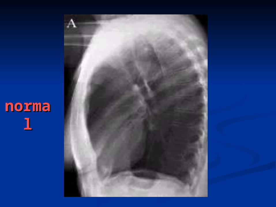
normnormalal

PneumoconiosisPneumoconiosis(Lung Dust)(Lung Dust)
Refers to the pulmonary Refers to the pulmonary manifestations of exposure to a manifestations of exposure to a variety of dusts or aerosolsvariety of dusts or aerosols SilicosisSilicosis Coal workers pneumoconiosisCoal workers pneumoconiosis AsbestosisAsbestosis BerylliosisBerylliosis SiderosisSiderosis

CXRCXRPneumoconPneumocon iosis-iosis- based on based on ILO classification standardsILO classification standards
The accepted means of quantifying dust exposure and retention-The accepted means of quantifying dust exposure and retention-very important in evaluating disability claimsvery important in evaluating disability claims
Profusion of opacities categorized as 1, 2, or 3 with a second sub-Profusion of opacities categorized as 1, 2, or 3 with a second sub-classification to indicate degree of certainty (1, 0/0, 0/1; 1/0, 1/1, classification to indicate degree of certainty (1, 0/0, 0/1; 1/0, 1/1, 1/2, etc.,)1/2, etc.,)
SizeSize Rounded/regular; p <1.5 mm , q 1.5-3 mm, or r >3 to 10 mm Rounded/regular; p <1.5 mm , q 1.5-3 mm, or r >3 to 10 mm
(these are more specific for dust exposure)(these are more specific for dust exposure) Irregular opacities; s, t, or u based on the same sizesIrregular opacities; s, t, or u based on the same sizes Larger opacities are classified as A (1-5 cm), B (>5 cm), or C Larger opacities are classified as A (1-5 cm), B (>5 cm), or C
(equivalent to the entire RUL zone)(equivalent to the entire RUL zone) Progression of disease is usually associated with a change in Progression of disease is usually associated with a change in
profusion, and not size of opacitiesprofusion, and not size of opacities Presence/degree of pleural thickening classified as A (<5 mm), B (5-Presence/degree of pleural thickening classified as A (<5 mm), B (5-
10 mm), C (>10 mm) 10 mm), C (>10 mm)

silicosissilicosis
Silica: active dusts .fibrogenicSilica: active dusts .fibrogenic SilicosisSilicosis has a has a progressive progressive nature nature
despite cessation ofdespite cessation of dust dust exposureexposure X-ray picture is of multiple small rounded X-ray picture is of multiple small rounded
opacities opacities Usually in the upper lobesUsually in the upper lobes May occasionally calcify (20%) May occasionally calcify (20%) Lymph Lymph node enlargement is commonnode enlargement is common Large opacities are conglomerations of small Large opacities are conglomerations of small
opacities opacities

Progressive Massive Fibrosis (PMF) Progressive Massive Fibrosis (PMF) Cavitate from tuberculosis or ischemic necrosis Cavitate from tuberculosis or ischemic necrosis Massive fibrosis and conglomerate mass formation Massive fibrosis and conglomerate mass formation
in upper lobes with scarring and retraction of hila in upper lobes with scarring and retraction of hila upwards upwards
Eggshell calcificationEggshell calcification of hilar nodes of hilar nodes in 5%in 5% Caplan’s syndrome Caplan’s syndrome consists of large consists of large
necrobiotic nodules superimposed on necrobiotic nodules superimposed on silicosissilicosis
Silicosis Silicosis predisposes to TBpredisposes to TB The radiologic findings of tuberculosis developed in The radiologic findings of tuberculosis developed in
the patients with silicosis include pleural effusion, the patients with silicosis include pleural effusion, newly-developed consolidation, bronchovascular newly-developed consolidation, bronchovascular infiltrations, cavitary change in pre-existing PMF, infiltrations, cavitary change in pre-existing PMF, etc. etc.

C.W.PC.W.P Coal dusts: A combination of active and inert Coal dusts: A combination of active and inert
materialmaterial Coal dust is deposited in the alveolar Coal dust is deposited in the alveolar
macrophages which migrate to, and leave, macrophages which migrate to, and leave, coal dust deposits around the respiratory coal dust deposits around the respiratory bronchiole bronchiole Here a very small fibrous reaction occurs Here a very small fibrous reaction occurs
Complicated CWP occurs as large masses Complicated CWP occurs as large masses in either the upper lobes or the superior in either the upper lobes or the superior segments of the lower lobes segments of the lower lobes Unlike silicosis, the large upper lobe lesions of CWP Unlike silicosis, the large upper lobe lesions of CWP
are single (rather than conglomerate) black masses are single (rather than conglomerate) black masses with a with a liquid core, not a fibrous tissue coreliquid core, not a fibrous tissue core

The masses may undergo The masses may undergo cavitation cavitation either from TB or ischemiaeither from TB or ischemia
The rounded opacities of CWP, found The rounded opacities of CWP, found predominantly in the upper lobes predominantly in the upper lobes
Massive fibrosis are round or oval and Massive fibrosis are round or oval and tend to migrate toward the hila tend to migrate toward the hila creating peripheral areas of creating peripheral areas of emphysema and bulla.emphysema and bulla.

AsbestosisAsbestosis Asbestos particles invoke a hemorrhagic Asbestos particles invoke a hemorrhagic
response in the lung response in the lung Fibers are then coated with a ferritin-like Fibers are then coated with a ferritin-like
material resulting in material resulting in ferruginous bodiesferruginous bodies Does its damage in respiratory bronchioles and Does its damage in respiratory bronchioles and
alveoli alveoli Affects lowerAffects lower lobes firstlobes first Opacities are small and irregularly shaped Opacities are small and irregularly shaped Cardiac silhouette Cardiac silhouette may become may become shaggyshaggy Almost all patients have some pleural Almost all patients have some pleural
involvement-pleural plaque, diffuse pleural involvement-pleural plaque, diffuse pleural thickening, calcification or effusion thickening, calcification or effusion

Pleural involvement without Pleural involvement without parenchymal disease is commonparenchymal disease is common
Parietal pleural plaques in the mid lung Parietal pleural plaques in the mid lung are the most common asbestos-related are the most common asbestos-related disorder and are usually bilateral disorder and are usually bilateral
Pleural Pleural calcification occurs in about calcification occurs in about 50%50% with asbestos-related disease, with asbestos-related disease, especially diaphragmatic pleura especially diaphragmatic pleura
Diffuse pleural thickening involves Diffuse pleural thickening involves diaphragmatic pleura, blunting of diaphragmatic pleura, blunting of costophrenic sulci and lateral chest costophrenic sulci and lateral chest wall thickening wall thickening

Effusion alone may occurEffusion alone may occur early in the early in the disease (first 20 years) in about 3% of disease (first 20 years) in about 3% of cases cases
Asbestos-related lung cancer is either Asbestos-related lung cancer is either squamous cell or adenocarcinomasquamous cell or adenocarcinoma
Bronchogenic ca is almost always Bronchogenic ca is almost always associated with cigarette smoking associated with cigarette smoking
In contrast to silicosis, hilar In contrast to silicosis, hilar lymph nodeslymph nodes are are rarely affectedrarely affected
HRCT:thickend interlobular septal HRCT:thickend interlobular septal lines,curvilinear subpleural lines,curvilinear subpleural lines,parenchymal bands and lines,parenchymal bands and honycombing.honycombing.

Asbestos and Cigarette Smoking Asbestos and Cigarette Smoking Interaction on Chest X-ray ILO CategoryInteraction on Chest X-ray ILO Category
Asbestos causes pulmonary Asbestos causes pulmonary fibrosis, while smoking usually fibrosis, while smoking usually causes emphysema causes emphysema (destruction of alveolar surface (destruction of alveolar surface area).area).
In those with asbestosis who In those with asbestosis who have also been heavy smokers, have also been heavy smokers, there is (on average) an there is (on average) an increase in the profusion of increase in the profusion of small linear opacities on chest small linear opacities on chest x-ray.x-ray.
A smoker may have one half A smoker may have one half category higher profusion than category higher profusion than a non-smoker with equivalent a non-smoker with equivalent asbestos exposureasbestos exposure
Weiss, Weiss, Am Rev Respir DisAm Rev Respir Dis 1984; 130:293- 1984; 130:293-301.301.Barnhart, Barnhart, Am Rev Respir DisAm Rev Respir Dis 1990; 1990; 141:1102141:1102


Egg shellEgg shell

PMF(silicosis)PMF(silicosis)

normnormalal

This picture shows This picture shows complicated coal workers complicated coal workers pneumoconiosis.pneumoconiosis. There are There are diffuse, massive light areas diffuse, massive light areas that run together in the that run together in the upper and middle parts of upper and middle parts of both lungs. These are both lungs. These are superimposed on a superimposed on a background of small and background of small and poorly distinguishable light poorly distinguishable light areas that are diffuse and areas that are diffuse and located in both lungs. located in both lungs. Diseases which may explain Diseases which may explain these X-ray findings include, these X-ray findings include, but are not limited to: but are not limited to: complicated coal workers complicated coal workers pneumoconiosis (CWP), pneumoconiosis (CWP), silico-tuberculosis, and silico-tuberculosis, and metastatic lung cancer metastatic lung cancer


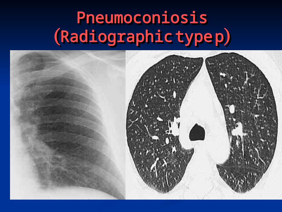
Pneumoconiosis Pneumoconiosis (Radiographic type p) (Radiographic type p)


normnormalal
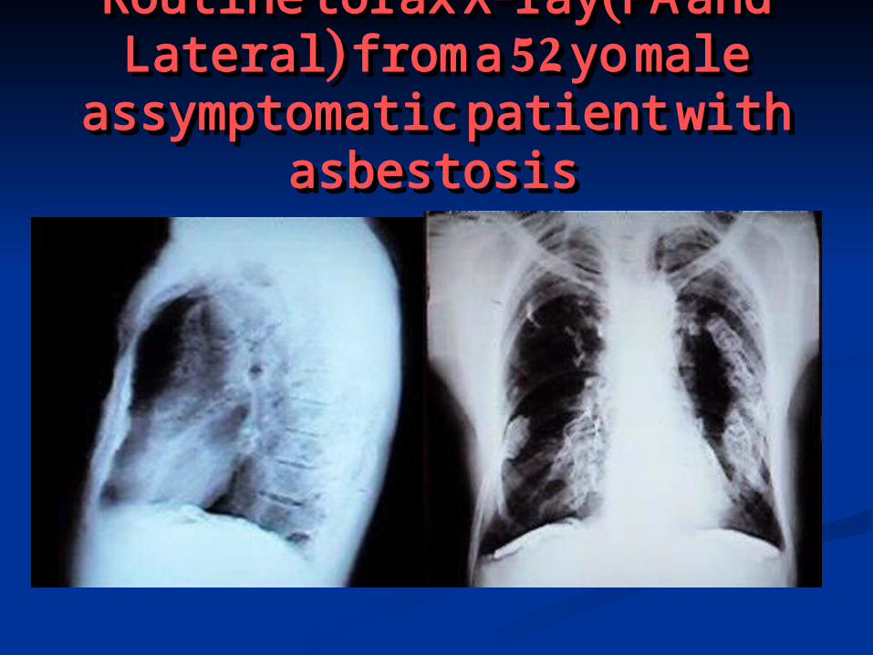
Routine torax X-ray(PA and Routine torax X-ray(PA and Lateral) from a 52 yo male Lateral) from a 52 yo male
assymptomatic patient with assymptomatic patient with asbestosisasbestosis

HRCT scan (left) shows thickened intra- and HRCT scan (left) shows thickened intra- and interlobular lines (A). HRCT (right) shows interlobular lines (A). HRCT (right) shows
subpleural curvilinear density. (B)subpleural curvilinear density. (B)
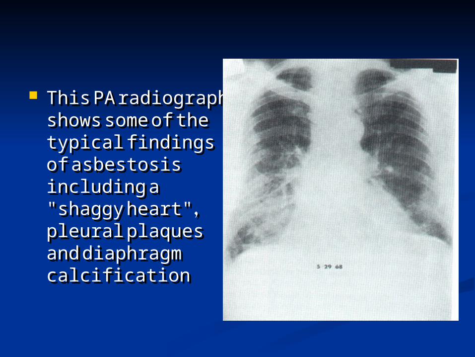
This PA radiograph This PA radiograph shows some of the shows some of the typical findings of typical findings of asbestosis asbestosis including a including a "shaggy heart", "shaggy heart", pleural plaques pleural plaques and diaphragm and diaphragm calcification calcification


This picture shows This picture shows complicated coal workers complicated coal workers pneumoconiosis.pneumoconiosis. There are There are diffuse, small, light areas diffuse, small, light areas (3 to 5 mm) in all areas on (3 to 5 mm) in all areas on both sides of the lungs. both sides of the lungs. There are large light areas There are large light areas which run together with which run together with poorly defined borders in poorly defined borders in the upper areas on both the upper areas on both sides of the lungs. sides of the lungs. Diseases which may Diseases which may explain these X-ray explain these X-ray findings include findings include complicated coal workers complicated coal workers pneumoconiosis (CWP), pneumoconiosis (CWP), silico-tuberculosis, silico-tuberculosis, disseminated tuberculosis, disseminated tuberculosis, metastatic lung cancer, metastatic lung cancer, and other diffuse and other diffuse infiltrative pulmonary infiltrative pulmonary diseases. diseases.

CaplanCaplan
Description: X-ray Description: X-ray showing lung showing lung nodules in a nodules in a patient with RA patient with RA (note differential (note differential diagnosis: diagnosis: Wegener's Wegener's granulomatosis, granulomatosis, metastatic cancer metastatic cancer (eg kidney)TB) (eg kidney)TB)

Small roundedSmall rounded opacityopacity

Small rounded opacitySmall rounded opacity

caplancaplan

55 year old man who was pensioned 55 year old man who was pensioned early from his job as a coal worker. early from his job as a coal worker.
Features in the image Features in the image There are well defined nodules in There are well defined nodules in
both lungs with a mid-zone both lungs with a mid-zone predominance. The nodules appear predominance. The nodules appear more confluent in the left upper more confluent in the left upper zone, where larger ill-defined zone, where larger ill-defined masses are present, distorting and masses are present, distorting and elevating the left hilum. Both hila elevating the left hilum. Both hila appear enlarged and lobulated. appear enlarged and lobulated. Although the overlying nodules may Although the overlying nodules may make the hila look large, the make the hila look large, the present appearance suggests hilar present appearance suggests hilar lymphadenopathy. In addition to the lymphadenopathy. In addition to the slightly prominent basal segment slightly prominent basal segment lower lobe bronchial markings, there lower lobe bronchial markings, there is the appearance of additional is the appearance of additional perihilar strands. A horizontal line, perihilar strands. A horizontal line, crossing the basal vessels, is crossing the basal vessels, is probably a linear fibrotic scar.probably a linear fibrotic scar.
DX: Pneumoconiosis,silicosis DX: Pneumoconiosis,silicosis developing massive fibrosis not developing massive fibrosis not pure anthracosis,which produces pure anthracosis,which produces less fibrotic reactionless fibrotic reaction

This chest X-ray This chest X-ray shows coal shows coal workers workers pneumoconiosis -. pneumoconiosis -. There are diffuse, There are diffuse, small (2 to 4 mm) small (2 to 4 mm) light areas on light areas on both sides of the both sides of the lungs. Diseases lungs. Diseases which may which may explain these X-explain these X-ray findings ray findings include simple include simple coal workers coal workers pneumoconiosis pneumoconiosis (CWP) simple (CWP) simple silicosis, silicosis, disseminated disseminated tuberculosis, tuberculosis, metastatic lung metastatic lung cancer, and other cancer, and other diffuse infiltrative diffuse infiltrative pulmonary pulmonary diseases. diseases.
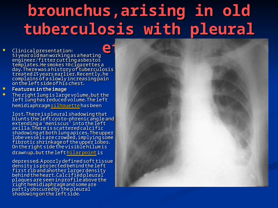
Carcinoma Carcinoma brounchus,arising in old brounchus,arising in old tuberculosis with pleural tuberculosis with pleural
effusioneffusion Clinical presentation:Clinical presentation:53 year old man working as a 53 year old man working as a heating engineer/fitter cutting heating engineer/fitter cutting asbestos templates. He smokes 10 asbestos templates. He smokes 10 cigarettes a day. There was a history cigarettes a day. There was a history of tuberculosis treated 25 years of tuberculosis treated 25 years earlier. Recently, he complains of a earlier. Recently, he complains of a slowly increasing pain on the left slowly increasing pain on the left side of his chest. side of his chest.
Features in the image Features in the image The right lung is large volume, but The right lung is large volume, but
the left lung has reduced volume. the left lung has reduced volume. The left hemidiaphragm The left hemidiaphragm silhouettesilhouette has been lost. There is pleural has been lost. There is pleural shadowing that blunts the left costo-shadowing that blunts the left costo-phrenic angle and extending a phrenic angle and extending a 'meniscus' into the left axilla. There 'meniscus' into the left axilla. There is scattered calcific shadowing at is scattered calcific shadowing at both lung apices. The upper lobe both lung apices. The upper lobe vessels are crowded, implying some vessels are crowded, implying some fibrotic shrinkage of the upper lobes. fibrotic shrinkage of the upper lobes. On the right side the visible hilum is On the right side the visible hilum is drawn up, but the left drawn up, but the left hilar pointhilar point is is depressed. A poorly defined soft depressed. A poorly defined soft tissue density is projected behind tissue density is projected behind the left first rib and another larger the left first rib and another larger density behind the heart. Calcified density behind the heart. Calcified pleural plaques are seen in profile pleural plaques are seen in profile above the right hemidiaphragm and above the right hemidiaphragm and some are partly obscured by the some are partly obscured by the pleural shadowing on the left side. pleural shadowing on the left side.

Elderly male, former pottery maker Elderly male, former pottery maker There are multiple fairly dense There are multiple fairly dense
nodules, mostly in both mid and lower nodules, mostly in both mid and lower zones. The right hilum appears bulky zones. The right hilum appears bulky with some lobulation lateral to the with some lobulation lateral to the main bronchi. This resembles hilar main bronchi. This resembles hilar lymphadenopathy, despite the lymphadenopathy, despite the enlarging effect that overlying enlarging effect that overlying nodules have on the hilar appearance. nodules have on the hilar appearance. There are additional radiating strands, There are additional radiating strands, extending from the right hilum, extending from the right hilum, perhaps too thick for 'Kerley B' lines, perhaps too thick for 'Kerley B' lines, but not corresponding to dilated but not corresponding to dilated bronchi alone. The vessel count in the bronchi alone. The vessel count in the left upper lobe is reduced and there is left upper lobe is reduced and there is amorphous calcific shadowing at the amorphous calcific shadowing at the left apex. The left hilum is distorted, left apex. The left hilum is distorted, undersized and is associated with a undersized and is associated with a horizontal strand of fibrosis in the left horizontal strand of fibrosis in the left mid-zone. mid-zone.
Dx:silicosis,with perihilar interstitial Dx:silicosis,with perihilar interstitial shadowing. Old tuberculosis of the shadowing. Old tuberculosis of the left upper lobe.possible early left upper lobe.possible early bronchiectasis.bronchiectasis.
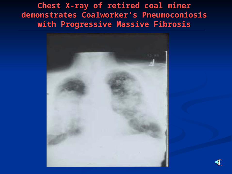
Chest X-ray of retired coal miner Chest X-ray of retired coal miner demonstrates Coalworker’s Pneumoconiosis demonstrates Coalworker’s Pneumoconiosis
with Progressive Massive Fibrosiswith Progressive Massive Fibrosis

Normal Chest ( X-Ray ILO Category 0/0)Normal Chest ( X-Ray ILO Category 0/0)

Interaction of Asbestos and Cigarette Interaction of Asbestos and Cigarette Smoking to Increase X-ray Markings Smoking to Increase X-ray Markings
(Asbestosis, ILO Category 2/2)(Asbestosis, ILO Category 2/2)
Smoking increases the profusion of small opacities onSmoking increases the profusion of small opacities onx-rays in asbestosis Weiss W. x-rays in asbestosis Weiss W. Am Rev Respir DisAm Rev Respir Dis 1984; 130:293-301 1984; 130:293-301


