Improving success in the development of novel PET ligands ...
21
Improving success in the development of novel PET ligands for CNS indications Elizabeth Beck for the Pfizer Neuroscience PET unit
Transcript of Improving success in the development of novel PET ligands ...

Improving success in the development of novel PET ligands for CNS indications
Elizabeth Beck for the Pfizer Neuroscience PET unit
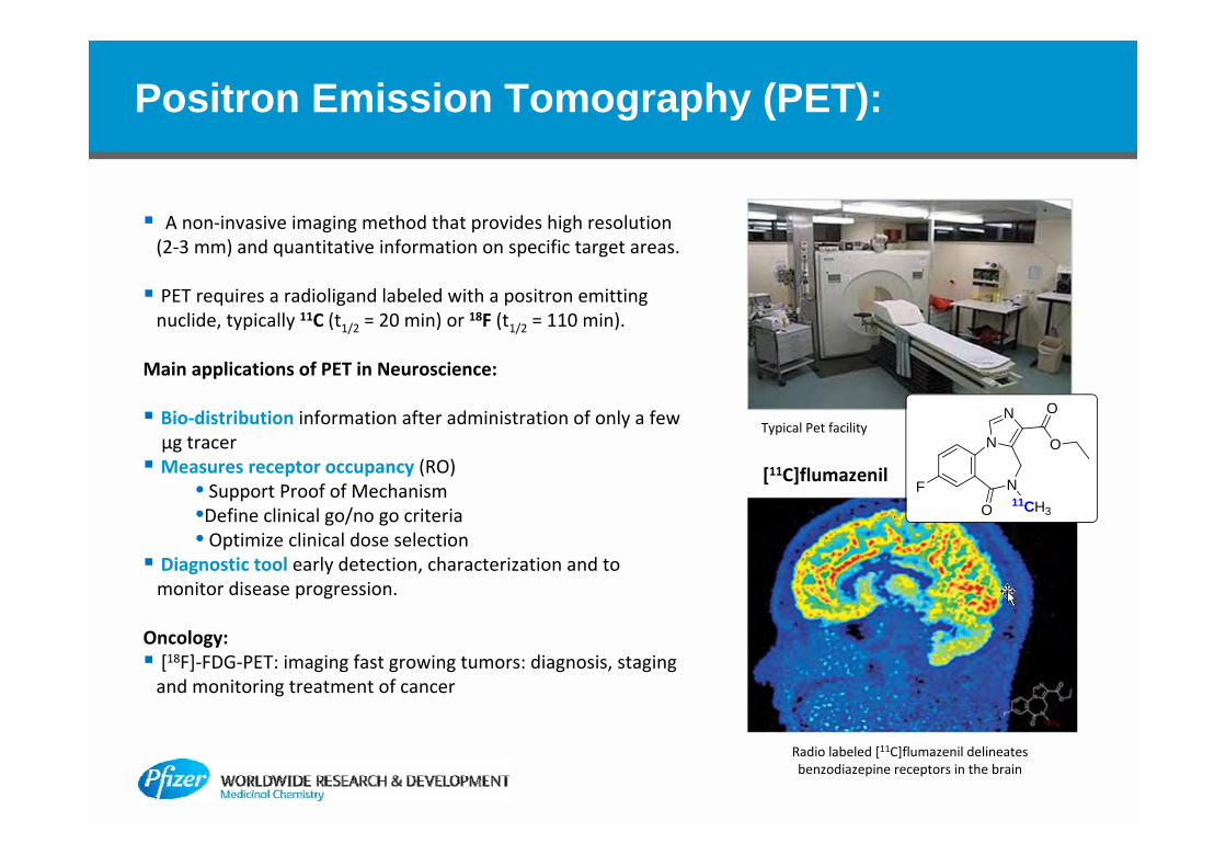
Positron Emission Tomography (PET):
A non‐invasive imaging method that provides high resolution (2‐3 mm) and quantitative information on specific target areas.
PET requires a radioligand labeled with a positron emitting nuclide, typically 11C (t1/2 = 20 min) or 18F (t1/2 = 110 min).
Main applications of PET in Neuroscience:
Bio‐distribution information after administration of only a few μg tracer Measures receptor occupancy (RO)
• Support Proof of Mechanism•Define clinical go/no go criteria• Optimize clinical dose selection
Diagnostic tool early detection, characterization and tomonitor disease progression.
Oncology: [18F]‐FDG‐PET: imaging fast growing tumors: diagnosis, staging and monitoring treatment of cancer
Radio labeled [11C]flumazenil delineates benzodiazepine receptors in the brain
Typical Pet facility
N
N
FO 11CH3
N O
O
[11C]flumazenil

PET Ligand Design Criteria
Pharmacology:Target density / Mode of action
Affinity:: Bmax (nM)/Kd > 10 (Bmax: max Conc. of target binding site)
Selectivity: >30‐100x selective
Synthesis:Synthesis limited to [11C] or [18F];
Rapid synthesis ‐ due to short T1/2Proximity to cyclotron – limited reagents
Pharmacokinetics:
Brain permeability
No brain‐penetrating radioactive metabolites
Low non‐specific binding
Safety:Lower safety hurdle: single dose IV micro dosing
‐ Structure alerts/Gentox may be tolerated
‐ Establish safety window at multiple of projected clinical dose (g scale)
In‐silico tools and high through‐put assays to predict:‐ good brain permeability ‐ minimal non‐specific binding
Improve do‐ability of labeling→ enable greater flexibility in the design of ligands
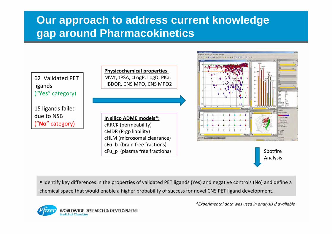
Our approach to address current knowledge gap around Pharmacokinetics
62 Validated PET ligands(“Yes” category)
15 ligands failed due to NSB(“No” category)
Physicochemical properties: MWt, tPSA, cLogP, LogD, PKa, HBDOR, CNS MPO, CNS MPO2
In silico ADME models*: cRRCK (permeability)cMDR (P‐gp liability)cHLM (microsomal clearance)cFu_b (brain free fractions)cFu_p (plasma free fractions) Spotfire
Analysis
Identify key differences in the properties of validated PET ligands (Yes) and negative controls (No) and define a
chemical space that would enable a higher probability of success for novel CNS PET ligand development.
*Experimental data was used in analysis if available
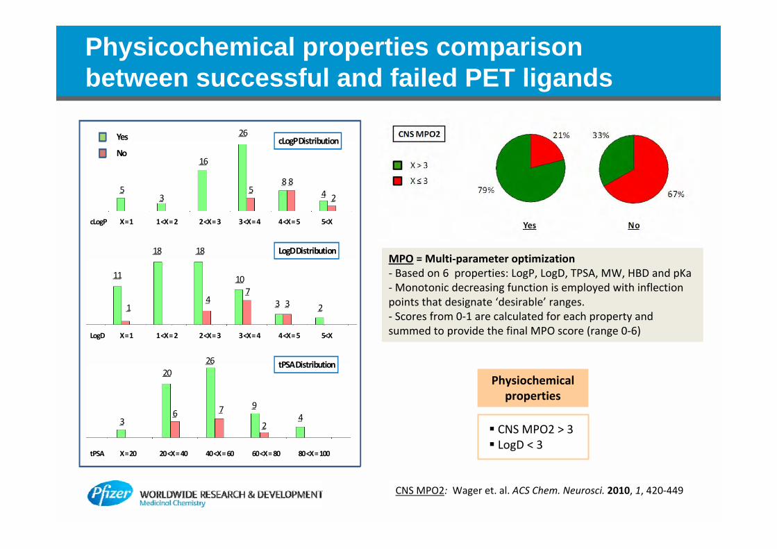
Physicochemical properties comparison between successful and failed PET ligands
26
58 8
4 2
16
35
X = 1 1 <X = 2 2 <X = 3 3 <X = 4 4 <X = 5 5<X
cLogP DistributionYes
No
X = 1 1 <X = 2 2 <X = 3 3 <X = 4 4 <X = 5 5<X
cLogP
LogD
11
1
18 18
4
107
3 3 2
LogD Distribution
X = 20 20 <X = 40 40 <X = 60 60 <X = 80 80 <X = 100tPSA
3
20
6
26
7 9
24
tPSA Distribution
CNS MPO2: Wager et. al. ACS Chem. Neurosci. 2010, 1, 420‐449
Physiochemical properties
CNS MPO2 > 3 LogD < 3
MPO = Multi‐parameter optimization‐ Based on 6 properties: LogP, LogD, TPSA, MW, HBD and pKa‐Monotonic decreasing function is employed with inflection points that designate ‘desirable’ ranges.‐ Scores from 0‐1 are calculated for each property and summed to provide the final MPO score (range 0‐6)
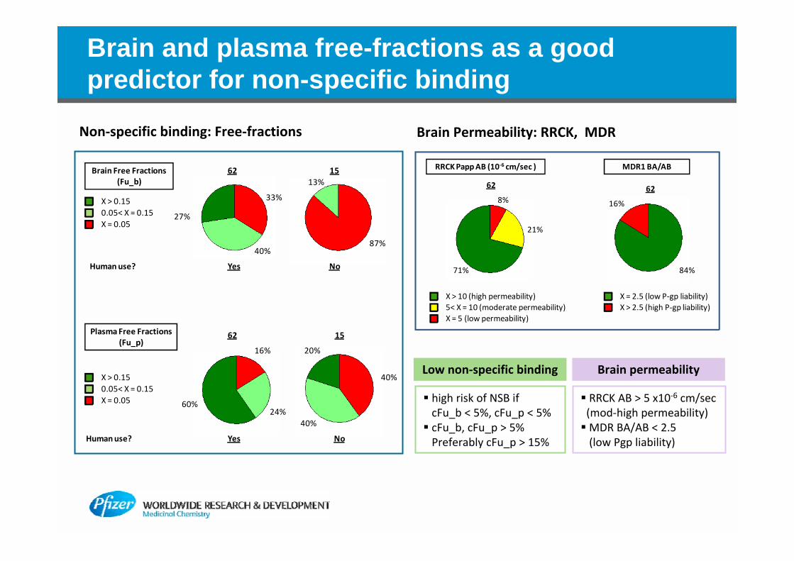
Brain and plasma free-fractions as a good predictor for non-specific binding
71%
8%
RRCK Papp AB (10‐6 cm/sec )
62
84%
16%
MDR1 BA/AB
62
21%
X > 10 (high permeability)5< X = 10 (moderate permeability)X = 5 (low permeability)
X = 2.5 (low P‐gp liability)X > 2.5 (high P‐gp liability)
62 15
Yes No
33%
40%
27%
87%
Human use?
Brain Free Fractions (Fu_b)
X > 0.150.05< X = 0.15X = 0.05
13%
16%
60%
40%
20%
Plasma Free Fractions (Fu_p)
X > 0.150.05< X = 0.15X = 0.05
Yes NoHuman use?
62 15
24%40%
Brain Permeability: RRCK, MDRNon‐specific binding: Free‐fractions
Low non‐specific binding
high risk of NSB if cFu_b < 5%, cFu_p < 5% cFu_b, cFu_p > 5%Preferably cFu_p > 15%
Brain permeability
RRCK AB > 5 x10‐6 cm/sec(mod‐high permeability)MDR BA/AB < 2.5 (low Pgp liability)
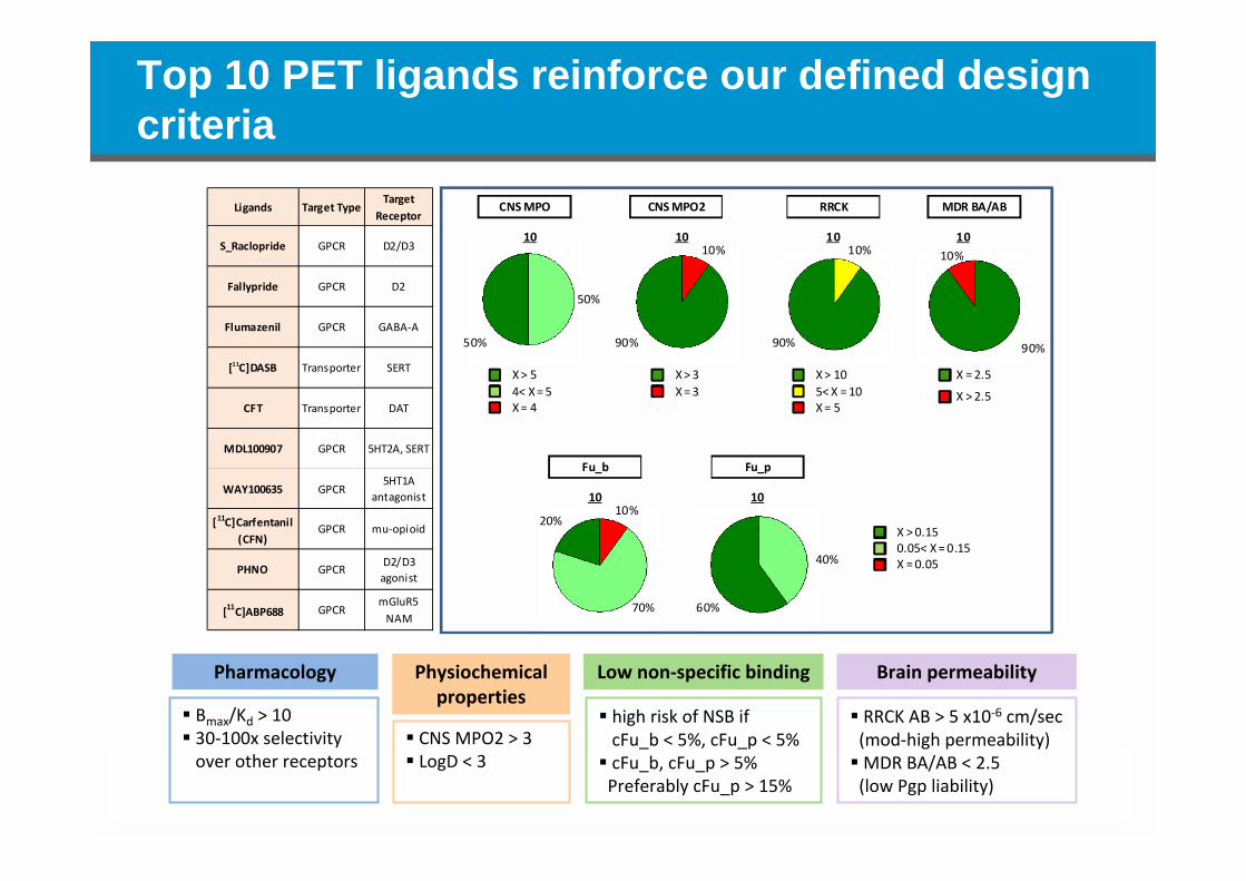
Top 10 PET ligands reinforce our defined design criteria
CNS MPO
10
50%
50%
CNS MPO2
1010%
90%
RRCK
1010%
90%
MDR BA/AB
1010%
90%
X > 54< X = 5X = 4
X > 3X = 3
X > 105< X = 10X = 5
X > 2.5
X = 2.5
Fu_b
1010%
20%
Fu_p
10
40%
60%70%
X > 0.150.05< X = 0.15X = 0.05
Ligands Target TypeTarget
Receptor
S_Raclopride GPCR D2/D3
Fallypride GPCR D2
Flumazenil GPCR GABA‐A
[11C]DASB Transporter SERT
CFT Transporter DAT
MDL100907 GPCR 5HT2A, SERT
WAY100635 GPCR5HT1A
antagonist
[11C]Carfentanil (CFN)
GPCR mu‐opioid
PHNO GPCRD2/D3 agonist
[11C]ABP688 GPCRmGluR5 NAM
Pharmacology
Bmax/Kd > 10 30‐100x selectivityover other receptors
Physiochemical properties
CNS MPO2 > 3 LogD < 3
Low non‐specific binding
high risk of NSB if cFu_b < 5%, cFu_p < 5% cFu_b, cFu_p > 5%Preferably cFu_p > 15%
Brain permeability
RRCK AB > 5 x10‐6 cm/sec(mod‐high permeability)MDR BA/AB < 2.5 (low Pgp liability)
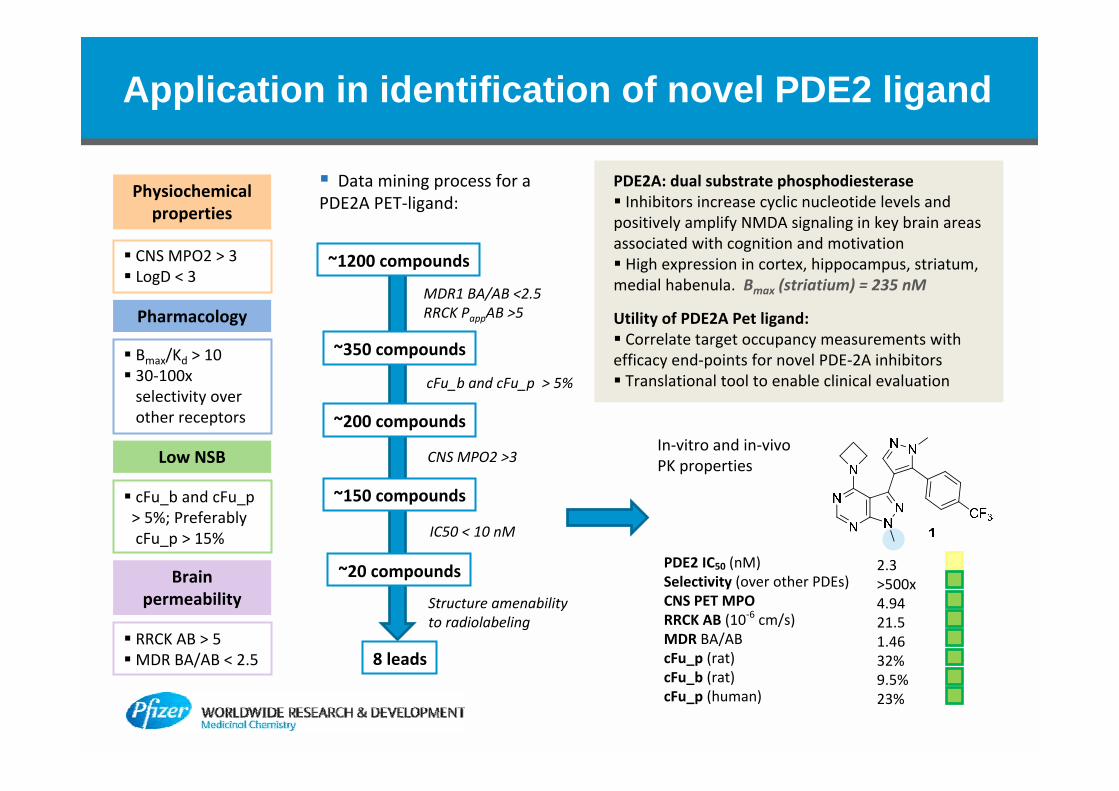
Application in identification of novel PDE2 ligand
Data mining process for a PDE2A PET‐ligand:
~1200 compounds
~350 compounds
~200 compounds
~150 compounds
~20 compounds
8 leads
MDR1 BA/AB <2.5RRCK PappAB >5
cFu_b and cFu_p > 5%
CNS MPO2 >3
IC50 < 10 nM
Structure amenability to radiolabeling
PDE2 IC50 (nM)Selectivity (over other PDEs)CNS PET MPORRCK AB (10‐6 cm/s)MDR BA/ABcFu_p (rat)cFu_b (rat)cFu_p (human)
2.3>500x4.9421.51.4632%9.5%23%
In‐vitro and in‐vivo PK properties
PDE2A: dual substrate phosphodiesterase Inhibitors increase cyclic nucleotide levels and positively amplify NMDA signaling in key brain areas associated with cognition and motivation High expression in cortex, hippocampus, striatum, medial habenula. Bmax (striatium) = 235 nM
Utility of PDE2A Pet ligand: Correlate target occupancy measurements with efficacy end‐points for novel PDE‐2A inhibitors Translational tool to enable clinical evaluation
Pharmacology
Bmax/Kd > 10 30‐100x selectivity over other receptors
Physiochemical properties
CNS MPO2 > 3 LogD < 3
Low NSB
cFu_b and cFu_p> 5%; Preferably cFu_p > 15%
Brain permeability
RRCK AB > 5MDR BA/AB < 2.5
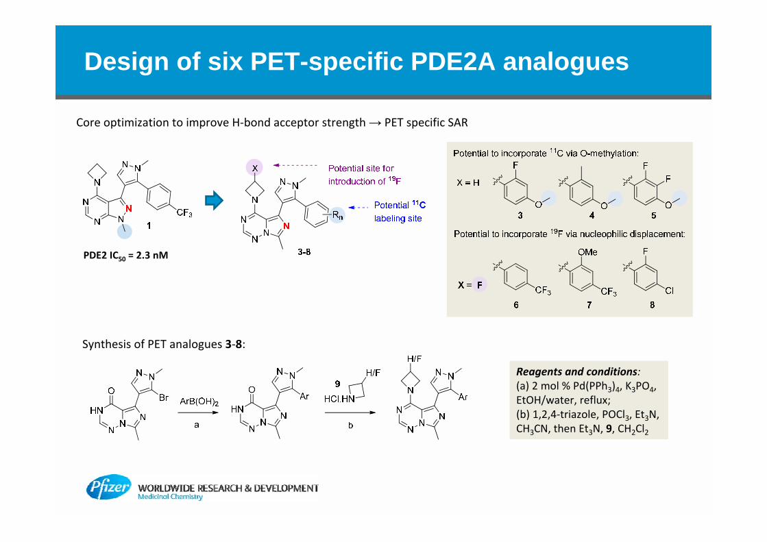
Design of six PET-specific PDE2A analogues
PDE2 IC50 = 2.3 nM
Core optimization to improve H‐bond acceptor strength → PET specific SAR
Reagents and conditions: (a) 2 mol % Pd(PPh3)4, K3PO4, EtOH/water, reflux; (b) 1,2,4‐triazole, POCl3, Et3N, CH3CN, then Et3N, 9, CH2Cl2
Synthesis of PET analogues 3‐8:
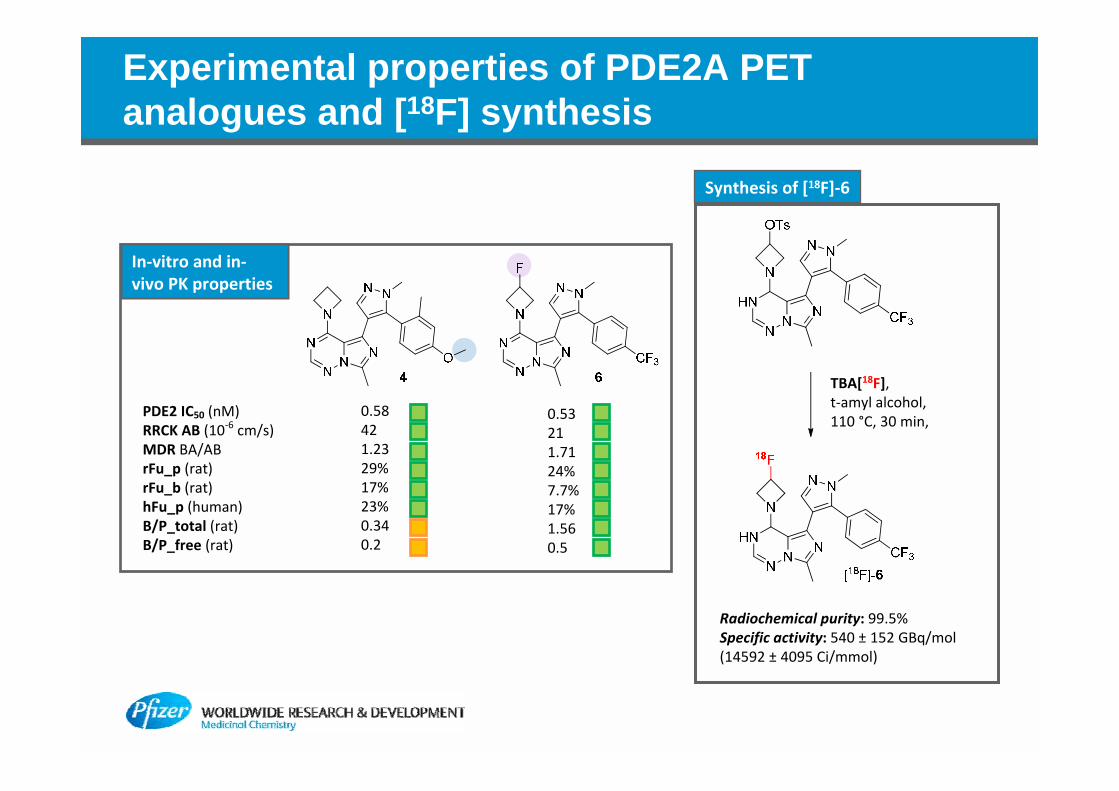
Experimental properties of PDE2A PET analogues and [18F] synthesis
PDE2 IC50 (nM)RRCK AB (10‐6 cm/s)MDR BA/ABrFu_p (rat)rFu_b (rat)hFu_p (human)B/P_total (rat)B/P_free (rat)
0.58421.2329%17%23%0.340.2
0.53211.7124%7.7%17%1.560.5
In‐vitro and in‐vivo PK properties
Synthesis of [18F]‐6
TBA[18F], t‐amyl alcohol, 110 °C, 30 min,
Radiochemical purity: 99.5% Specific activity: 540 ± 152 GBq/mol (14592 ± 4095 Ci/mmol)
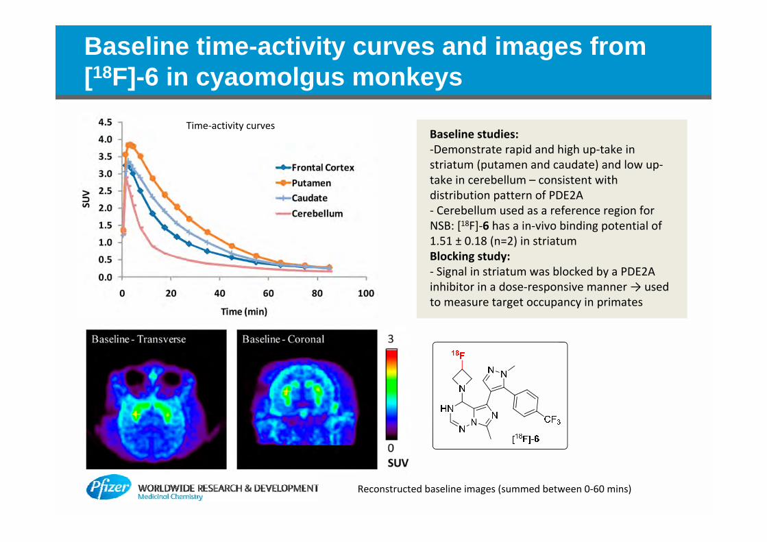
Baseline time-activity curves and images from [18F]-6 in cyaomolgus monkeys
Baseline studies:‐Demonstrate rapid and high up‐take in striatum (putamen and caudate) and low up‐take in cerebellum – consistent with distribution pattern of PDE2A‐ Cerebellum used as a reference region for NSB: [18F]‐6 has a in‐vivo binding potential of 1.51 ± 0.18 (n=2) in striatumBlocking study: ‐ Signal in striatum was blocked by a PDE2A inhibitor in a dose‐responsive manner → used to measure target occupancy in primates
Time‐activity curves
Reconstructed baseline images (summed between 0‐60 mins)
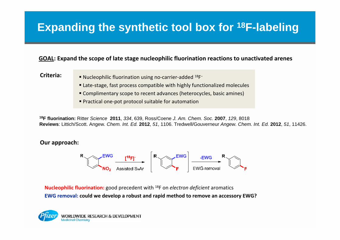
Expanding the synthetic tool box for 18F-labeling
Nucleophilic fluorination: good precedent with 18F on electron deficient aromatics
EWG removal: could we develop a robust and rapid method to remove an accessory EWG?
Nucleophilic fluorination using no‐carrier‐added 18F−
Late‐stage, fast process compatible with highly functionalized molecules
Complimentary scope to recent advances (heterocycles, basic amines)
Practical one‐pot protocol suitable for automation
GOAL: Expand the scope of late stage nucleophilic fluorination reactions to unactivated arenes
Our approach:
Criteria:
18F fluorination: Ritter Science 2011, 334, 639, Ross/Coene J. Am. Chem. Soc. 2007, 129, 8018Reviews: Littich/Scott. Angew. Chem. Int. Ed. 2012, 51, 1106. Tredwell/Gouverneur Angew. Chem. Int. Ed. 2012, 51, 11426.
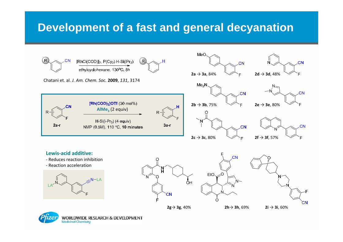
Development of a fast and general decyanation
N
O
EtO O
NN
FCN
F
NN CN
N
F
CN
Chatani et. al. J. Am. Chem. Soc. 2009, 131, 3174
N
F
N LALA
Lewis‐acid additive:‐ Reduces reaction inhibition‐ Reaction acceleration
2a→ 3a, 84% 2d→ 3d, 48%
2e→ 3e, 80%
2f→ 3f, 57%2c→ 3c, 80%
2b→ 3b, 75%
2i→ 3i, 60%2h→ 3h, 69%2g→ 3g, 40%
AlMe3 (2 equiv)

Application of [18F] fluorination/decyanation sequence on model substrate
RCY = 5‐12%, Chemical purity 90%High specific activity (>20,000 Ci/mmol)
Yield Radiochem purity Radio‐HPLC Yield[18F] Separation cartridge
PS‐HCO3QMA
57%22%
99%95%
99.8%95.0%
QMA
Manual
Automated Reactor
Radiosynthesis
Provides a novel and practical [18F]‐labelling strategy that is complimentary to the recent advances in late‐stage electrophilic fluorination and enables greater flexibility in the design of future PET ligands

Summary
PET is a powerful translational tool for preclinical and clinical evaluations of candidate compounds.‐ enables measurement of target occupancy via blocking studies with inhibitors.
Our Approaches to improving success in PET ligand development:
A set of preferred design and selection parameters to enable prospective design and prioritization:
Zhang et al. J. Med. Chem., 2013, 56, 4568–4579
Expanding the synthetic tool box for 18F‐labeling through development of a new strategies for late stage fluorination of aromatics o within complex molecules.
Physicochemical Properties:
•CNS MPO2 > 3•LogD = 3
Pharmacology Brain PermeabilityLow non-specific binding
•Bmax/Kd > 10•>30-100x Selectivity over other receptors.
• high risk of NSB if cFu_b = 0.05 and cFu_p = 0.05;•cFu_b and cFu_p > 0.05; preferably cFu_p > 0.15.
• RRCK AB > 5 x 10⁻⁶ cm/sec (moderate to high permeability)• MDR BA/AB = 2.5 (low Pgpliability)

Acknowledgments
Medicinal ChemistryLei Zhang
Anabella Villalobos
Tom Chappie
Chris Helal
Xinjun Hou
John Humphrey
Jiemin Lu
Patrick Verhoest
Travis Wager
Medicinal ChemistryLei Zhang
Anabella Villalobos
Tom Chappie
Chris Helal
Xinjun Hou
John Humphrey
Jiemin Lu
Patrick Verhoest
Travis Wager
Bioimaging CenterMarc Skaddan
Rick Coelho
Kurt Fan
Kenneth Zasadny
Laigao Chen
Aijun Zhu
Thomas Bocan
Bioimaging CenterMarc Skaddan
Rick Coelho
Kurt Fan
Kenneth Zasadny
Laigao Chen
Aijun Zhu
Thomas Bocan
NSRUSarah Grimwood
Elena Drummond
Deborah Smith
Joe Hedde
Greg Preston
Christopher Schmidt
Rebecca O’Connor
NSRUSarah Grimwood
Elena Drummond
Deborah Smith
Joe Hedde
Greg Preston
Christopher Schmidt
Rebecca O’Connor
DADASteve HeckBruce Lefker
DADASteve HeckBruce Lefker
PharmSciKlaas Schildknegt Andrew ProudKathy Haas
PharmSciKlaas Schildknegt Andrew ProudKathy Haas
Precision MedicineTimothy McCarthyRikki Waterhouse
Precision MedicineTimothy McCarthyRikki Waterhouse
Collaborators and Sponsors
Yale Alliance:Richard CarsonYu‐Shin DingHenry Huang
Karolinska Institute, SwedenChrister Halldin
King’s College, UKAntony Gee
M. J. Fox Foundation
Collaborators and Sponsors
Yale Alliance:Richard CarsonYu‐Shin DingHenry Huang
Karolinska Institute, SwedenChrister Halldin
King’s College, UKAntony Gee
M. J. Fox Foundation
DSRDBetty PettersenJoseph Brady
DSRDBetty PettersenJoseph Brady
BACK-UPS

Identification of a novel PDE2A PET ligand
Develop a selective PDE2A PET ligand to serve as a translational tool‐ Pre‐clinical pharmacology studies (eg. correlating target occupancy measurements with efficacy end‐points for novel PDE‐2A inhibitors)‐ Clinical evaluation (Translation of RO‐PD correlations between species)
• High expression in cortex, hippocampus, striatum, medial habenula• Critical regions for cognition, motivation• All behaviors impacted in schizophrenia
Stephenson et al. JHC 2009; 57(10):933‐49
PDE2A localizationPDE2A Inhibitors: Positive Amplifiers of NMDA and DA Signaling
PDE2A Inhibition
Ca2+s
NMDA Receptor
cAMP
glutamate
NMDA and DADependentSYNAPTICPLASTICITY
Dopamine D1
cGMP
dopamineAMPAReceptor
+
+
+
Develop a selective PDE2A PET ligand to serve as a translational tool
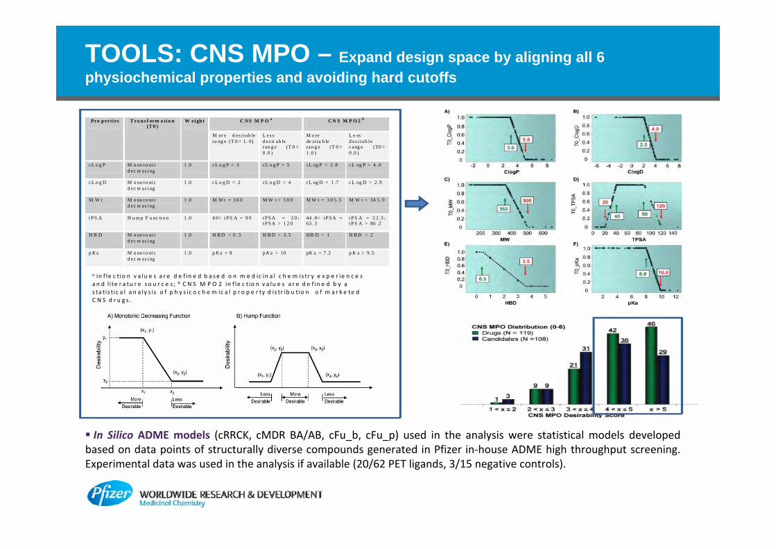
TOOLS: CNS MPO – Expand design space by aligning all 6 physiochemical properties and avoiding hard cutoffs
Pro p erti es T ra n s f orm a ti o n (T 0 )
W ei gh t C NS M P O a CN S M P O 2 b
M or e d es i rab le ra ng e (T 0 = 1 . 0)
L es s d es ir ab le ran g e (T 0 = 0 .0 )
M o re de si ra b le ran g e (T 0 = 1. 0 )
L e ss d es i rab l e r ang e (T0 = 0 .0 )
cL o g P M o no t o ni c d ec re as i ng
1 .0 cL o g P = 3 cL o g P > 5 cL og P = 2 .8 cL og P > 4 . 0
cL o g D M o no t o ni c d ec re as i ng
1 .0 cL o g D = 2 cL o g D > 4 cL og D = 1 .7 cL og D > 2 .9
M W t M o no t o ni c d ec re as i ng
1 .0 M W t = 3 6 0 M W t > 5 0 0 M W t = 3 0 5. 3 M W t > 34 5. 9
t P S A H u m p F u nc ti o n 1 .0 4 0< t P S A = 9 0 t P S A = 2 0 ; t P S A > 1 2 0
44 . 8< t P S A = 63 . 3
t P S A = 3 2 .3 ; t P S A > 86 .2
H B D M o no t o ni c d ec re as i ng
1 .0 H B D = 0 . 5 H B D > 3 .5 HB D = 1 H B D > 2
p K a M o no t o ni c d ec re as i ng
1 .0 p K a = 8 p K a > 10 pK a = 7 .2 p K a > 9 .5
a in f le c t io n v a lu e s a r e d e f in e d b a se d o n m e d ic in a l c h e m is tr y e x p e r ie n c e s a n d l i te r a tu r e s o u r c e s ; b C N S M P O 2 in f le c t io n v a lu e s a r e d e f in e d b y a s ta tis t ic a l a n a ly s is o f p h y s ic o c h e m ic a l p r o p e r ty d is tr ib u tio n o f m a r k e te d C N S d r u g s .
In Silico ADME models (cRRCK, cMDR BA/AB, cFu_b, cFu_p) used in the analysis were statistical models developed based on data points of structurally diverse compounds generated in Pfizer in‐house ADME high throughput screening. Experimental data was used in the analysis if available (20/62 PET ligands, 3/15 negative controls).
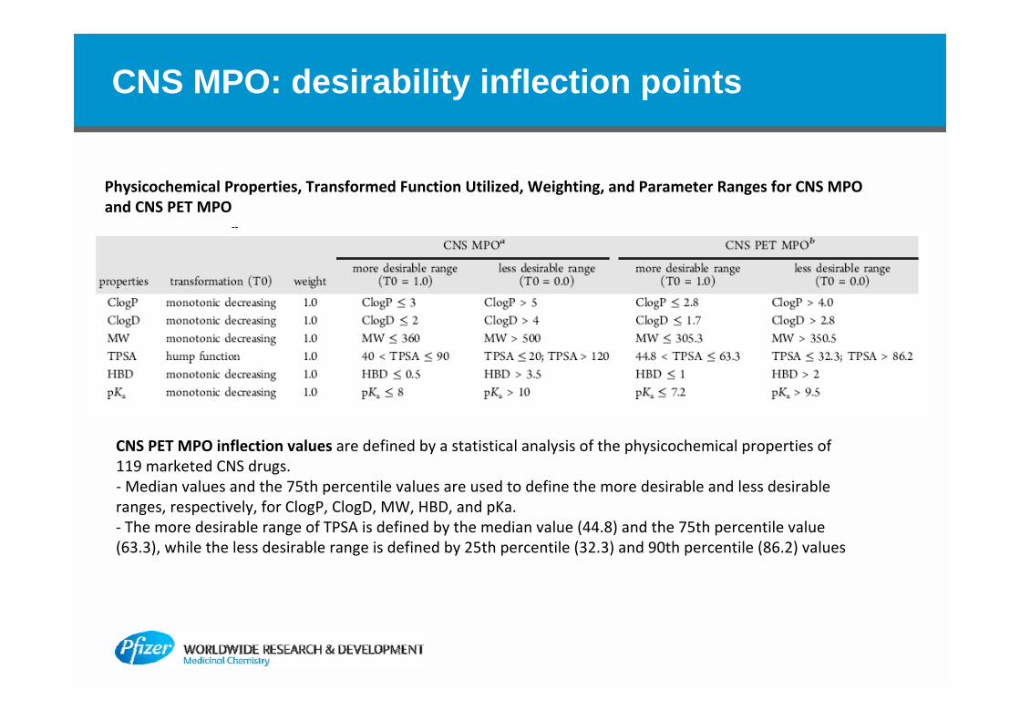
CNS MPO: desirability inflection points
Physicochemical Properties, Transformed Function Utilized, Weighting, and Parameter Ranges for CNS MPO and CNS PET MPO
CNS PET MPO inflection values are defined by a statistical analysis of the physicochemical properties of 119 marketed CNS drugs. ‐Median values and the 75th percentile values are used to define the more desirable and less desirable ranges, respectively, for ClogP, ClogD, MW, HBD, and pKa. ‐ The more desirable range of TPSA is defined by the median value (44.8) and the 75th percentile value (63.3), while the less desirable range is defined by 25th percentile (32.3) and 90th percentile (86.2) values
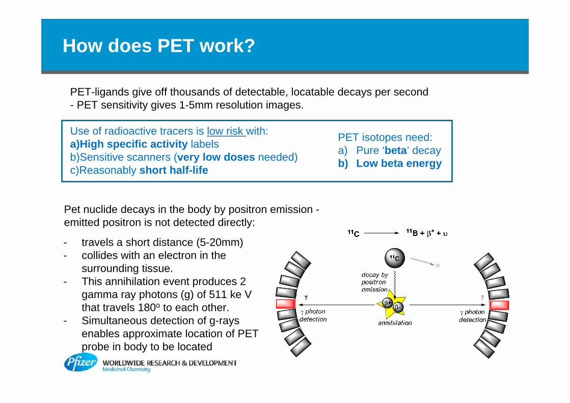
How does PET work?
PET-ligands give off thousands of detectable, locatable decays per second- PET sensitivity gives 1-5mm resolution images.
Use of radioactive tracers is low risk with:a)High specific activity labelsb)Sensitive scanners (very low doses needed)c)Reasonably short half-life
- travels a short distance (5-20mm)- collides with an electron in the
surrounding tissue. - This annihilation event produces 2
gamma ray photons (g) of 511 ke V that travels 180o to each other.
- Simultaneous detection of g-rays enables approximate location of PET probe in body to be located
PET isotopes need:a) Pure ‘beta’ decayb) Low beta energy
Pet nuclide decays in the body by positron emission -emitted positron is not detected directly: