Improved anchoring nails: design and analysis of ...
8
RESEARCH ARTICLE Open Access Improved anchoring nails: design and analysis of resistance ability Tensile test and finite element analysis (FEA) of improved anchoring nails used in temporomandibular joint (TMJ) disc anchor Z. H. Zhou 1† , X. Z. Chen 1† , X. W. Chen 1† , Y. X. Wang 1 , S. Y. Zhang 1,3* , S. F. Sun 2* and J. Z. Zhen 1* Abstract Background: Anchorage is one of the most important treatments for severe temporomandibular joint disorder (TMD). Anchoring nails have shown great success in clinical trials; however, they can break under pressure and are difficult to remove. In this study, we aimed to evaluate an improved anchoring nail and its mechanical stability. Methods: The experiment consisted of two parts: a tensile test and finite element analysis (FEA). First, traditional and improved anchoring nails were implanted into the condylar cortical bone and their tensile strength was measured using a tension meter. Second, a three-dimensional finite element model of the condyles with implants was established and FEA was performed with forces from three different directions. Results: The FEA results showed that the total force of the traditional and improved anchoring nails is 48.2 N and 200 N, respectively. The mean (±s.d.) maximum tensile strength of the traditional anchoring nail with a 3–0 suture was 27.53 ± 5.47 N. For the improved anchoring nail with a 3–0 suture it was 25.89 ± 2.64 N and with a 2–0 suture it was above 50 N. The tensile strengths of the traditional and improved anchoring nails with a 3–0 suture was significantly different (P = 0.033–< 0.05). Furthermore, the difference between the traditional anchoring nail with a 3–0 suture and the improved anchoring nail with a 2–0 suture was also significantly different (P = 0.000–< 0.01). Conclusion: The improved anchoring nail, especially when combined with a 2–0 suture, showed better resistance ability compared with the traditional anchoring nail. Keywords: Temporalmandibular joint disc anchorage, Mandibular condyle, Anchoring nail, Suture, Tensile test, Finite element analysis, FEA Background Temporomandibular joint disorder (TMD) is a common condition, with an approximate prevalence ranging from 13 to 87% [1]. Considering the limitations of non-surgical treatments, including medications [2], splints [3], physical therapy [4], and trigger point injections [5], surgical inter- vention is needed in severe cases of TMD [6, 7]. Previous clinical reports reveal that the results of sur- gical temporomandibular joint (TMJ) disc repositioning procedures have been variable due to long-term instabil- ity [8]. In 2001, an open joint procedure using Mitek an- choring nails (Mitek mini anchor, Mitek Products Inc., Westwood, Mass) showed great success in both clinical trials and radiography [9]. Despite their success, MiTek anchoring nails still have the following problems: (1) once fixed in the cortical bone, compared with other * Correspondence: [email protected]; [email protected]; [email protected] † Z. H. Zhou, X. Z. Chen and X. W. Chen contributed equally to this work. 1 Department of Oral and Maxillofacial Surgery, School of Medicine, Ninth People’s Hospital, Shanghai Jiao Tong University, 639 Zhi Zao Ju Road, Shanghai 200011, People’s Republic of China 2 Department of Stomatology, Tongren Hospital Affiliated to Shanghai Jiao Tong University School of Medicine, Shanghai 200336, People’s Republic of China Full list of author information is available at the end of the article © The Author(s). 2018 Open Access This article is distributed under the terms of the Creative Commons Attribution 4.0 International License (http://creativecommons.org/licenses/by/4.0/), which permits unrestricted use, distribution, and reproduction in any medium, provided you give appropriate credit to the original author(s) and the source, provide a link to the Creative Commons license, and indicate if changes were made. The Creative Commons Public Domain Dedication waiver (http://creativecommons.org/publicdomain/zero/1.0/) applies to the data made available in this article, unless otherwise stated. Zhou et al. BMC Oral Health (2018) 18:150 https://doi.org/10.1186/s12903-018-0606-3
Transcript of Improved anchoring nails: design and analysis of ...

RESEARCH ARTICLE Open Access
Improved anchoring nails: design andanalysis of resistance abilityTensile test and finite element analysis (FEA) of improvedanchoring nails used in temporomandibular joint (TMJ) discanchorZ. H. Zhou1†, X. Z. Chen1†, X. W. Chen1†, Y. X. Wang1, S. Y. Zhang1,3*, S. F. Sun2* and J. Z. Zhen1*
Abstract
Background: Anchorage is one of the most important treatments for severe temporomandibular joint disorder(TMD). Anchoring nails have shown great success in clinical trials; however, they can break under pressure and aredifficult to remove. In this study, we aimed to evaluate an improved anchoring nail and its mechanical stability.
Methods: The experiment consisted of two parts: a tensile test and finite element analysis (FEA). First, traditionaland improved anchoring nails were implanted into the condylar cortical bone and their tensile strength was measuredusing a tension meter. Second, a three-dimensional finite element model of the condyles with implants was establishedand FEA was performed with forces from three different directions.
Results: The FEA results showed that the total force of the traditional and improved anchoring nails is 48.2 N and 200 N,respectively. The mean (±s.d.) maximum tensile strength of the traditional anchoring nail with a 3–0 suture was 27.53 ±5.47 N. For the improved anchoring nail with a 3–0 suture it was 25.89 ± 2.64 N and with a 2–0 suture it was above 50 N.The tensile strengths of the traditional and improved anchoring nails with a 3–0 suture was significantly different(P = 0.033–< 0.05). Furthermore, the difference between the traditional anchoring nail with a 3–0 suture and theimproved anchoring nail with a 2–0 suture was also significantly different (P = 0.000–< 0.01).
Conclusion: The improved anchoring nail, especially when combined with a 2–0 suture, showed better resistanceability compared with the traditional anchoring nail.
Keywords: Temporalmandibular joint disc anchorage, Mandibular condyle, Anchoring nail, Suture, Tensile test, Finiteelement analysis, FEA
BackgroundTemporomandibular joint disorder (TMD) is a commoncondition, with an approximate prevalence ranging from13 to 87% [1]. Considering the limitations of non-surgicaltreatments, including medications [2], splints [3], physical
therapy [4], and trigger point injections [5], surgical inter-vention is needed in severe cases of TMD [6, 7].Previous clinical reports reveal that the results of sur-
gical temporomandibular joint (TMJ) disc repositioningprocedures have been variable due to long-term instabil-ity [8]. In 2001, an open joint procedure using Mitek an-choring nails (Mitek mini anchor, Mitek Products Inc.,Westwood, Mass) showed great success in both clinicaltrials and radiography [9]. Despite their success, MiTekanchoring nails still have the following problems: (1)once fixed in the cortical bone, compared with other
* Correspondence: [email protected]; [email protected];[email protected]†Z. H. Zhou, X. Z. Chen and X. W. Chen contributed equally to this work.1Department of Oral and Maxillofacial Surgery, School of Medicine, NinthPeople’s Hospital, Shanghai Jiao Tong University, 639 Zhi Zao Ju Road,Shanghai 200011, People’s Republic of China2Department of Stomatology, Tongren Hospital Affiliated to Shanghai Jiao TongUniversity School of Medicine, Shanghai 200336, People’s Republic of ChinaFull list of author information is available at the end of the article
© The Author(s). 2018 Open Access This article is distributed under the terms of the Creative Commons Attribution 4.0International License (http://creativecommons.org/licenses/by/4.0/), which permits unrestricted use, distribution, andreproduction in any medium, provided you give appropriate credit to the original author(s) and the source, provide a link tothe Creative Commons license, and indicate if changes were made. The Creative Commons Public Domain Dedication waiver(http://creativecommons.org/publicdomain/zero/1.0/) applies to the data made available in this article, unless otherwise stated.
Zhou et al. BMC Oral Health (2018) 18:150 https://doi.org/10.1186/s12903-018-0606-3

types of Mitek anchor, Mitek mini anchoring nails tendto break easily under pressure; (2) once the wings aretwisted, extraction of the anchoring nail becomes diffi-cult. Spallaccia et al. [10] (2013) described an anchoragesurgery using bioabsorbable microanchor nails. Postop-erative MRI showed a low reposition rate (65.7% in 35patients). To improve the success rate, reposition stabil-ity and implant safety, He et al. [11] (2015) appliedChinese-made anchoring nails in modified disc anchor-age surgery. As the shape of the anchoring nail does notfit perfectly with the anatomical structure of a condyle,7.47% of patients experienced postoperative friction inthe parotideomasseteric region [12]. Our group has pre-viously modified the traditional Chinese-made anchoringnail to reduce discomfort and improve stability. The an-choring nail is designed to be fully threaded, totally im-planted in the cortical bone, and fixed with a 2–0suture. However, the properties and safety of the im-proved anchoring nail have not yet been studied.Therefore, in this study, we aimed to assess the mech-
anical performance of the improved anchoring nail com-pared with the traditional anchoring nail. In previousstudies, tensile tests have been used to estimate the re-sistance ability of Mitek anchors [9]. With advances incomputer science, finite element analysis (FEA) has be-come a useful tool that addresses the limitations associ-ated with the TMJ structure and has tremendousadvantages in many aspects [13, 14]. FEA is capable ofmodeling and analyzing shape, structure, and resistanceability and has become the most popular numerical the-oretical method for TMJ biomechanics analysis. In ourstudy, we used tensile tests and FEA to estimate the re-sistance ability of the improved anchoring nail. The hy-pothesis of this study was that the improved anchoringnail would show greater tensile strength compared withthe traditional anchoring nail.
MethodsTensile testSubjectsFrom April 2015 to June 2016, 10 patients (4 males and6 females, aged 20–72 years old) undergoing TMJ re-placement at the Department of Oral Surgery, the NinthPeople’s Hospital Affiliated to Shanghai Jiaotong Univer-sity School of Medicine were selected. Condyle speci-mens were collected, wrapped with wool yarn immersedin normal saline, and preserved at − 20 °C. This studywas approved by the Ethics Committee of Shanghai Jiao-Tong University School of Medicine.
Anchoring nailsBoth traditional and improved anchoring nails (Cixi CityCibei Dental Instrument Co., Ltd., Cixi, Zhejiang, China)were made of titanium alloy. The total length of the
Fig. 1 The two types of anchoring nails. a Traditional anchoring nail.b Improved anchoring nail
Fig. 2 Condyle specimen. Traditional and modified anchoring nailswere implanted in the condyle
Zhou et al. BMC Oral Health (2018) 18:150 Page 2 of 8

traditional anchoring nail was 7 mm, with 1.5 mm nutthicknesses, 5.5 mm thread length, 2.8 mm nut diame-ters, and 2.0 mm thread diameters. The transition be-tween the head and thread was smooth and a groovewas designed for the knotting of the suture, which canonly be fixed with a 3–0 suture (Fig. 1a). The improvedanchoring nail had a length of 6 mm and a diameter of3.0 mm. There was a small hole in the upper-middlepart of the anchoring nail, with two grooves connectedto the head. The upper part of the grooves was smoothfor the placing and knotting of the suture (Fig. 1b).
SuturesIn this study, 3–0 and 2–0 nylon sutures (Ethibond*Excel, Green Braided Polyester Suture, Ethicon, Inc.)were used. The sutures were 90 cm in length with onesuture needle at each end. Each suture was divided intotwo in the middle.
Implantation procedureTraditional and improved anchoring nails were im-planted 10–15 mm below the inferior margin of the pos-terior inclined plane of the condylar process. The two
anchoring nails were placed symmetrically and the dis-tance between them was more than 3 mm (Fig. 2).
Tensile testA tension meter ((Cixi City Cibei Dental InstrumentCo., Ltd., Cixi, Zhejiang, China) was used for tensiletests. In the lower part, specimens were immobilizedwith steel wires to a clamping board. In the upper part,the suture was directly immobilized to the clampingboard.
Statistical analysisSPSS 17.0 software (Chicago, IL, USA) was used for stat-istical analysis. The maximum bearable tension forces ofthe sutures were analyzed by descriptive statistics andreported as x ± s. d. The difference in tension readingsbetween the two sutures used for the traditional anchor-ing nail was compared using a t-test. (The traditional an-choring nail does not match a 2–0 suture.) Thedifference in tension readings between the two suturesused for the improved anchoring nail was comparedusing a Kruskal-Wallis test. P < 0.05 was consideredsignificant.
Fig. 3 Finite element computer-aided design model
Fig. 4 Three types of force
Zhou et al. BMC Oral Health (2018) 18:150 Page 3 of 8

Finite element analysisFEA toolWe used three-dimensional modeling software (Hyper-mesh, Altair Engineering Inc.) and analysis programs(LS-DYNA, LSTC Inc.) to regulate the network struc-ture and make it more homogeneous, create the solidmodel, and conduct the stress analysis using the finiteelement procedure.
Finite element modelA three-dimensional computer-aided design model ofthe anchoring nails was established and used as themesh model for the FEA. The finite element model con-sisted of a first-order tetrahedral mesh, a total of139,000 units, and 29,000 nodes. (Fig. 3).
Data processingThe FEA results are a stress result accumulated grad-ually by deformation. Consequently, FEA transforms anengineering stress–strain curve to a true stress–straincurve. The formulae for the transformation are asfollows:
True stress = (1 + engineering strain) × engineeringstress.True strain = ln(1 + engineering strain).
ProcessThe directions of force, including vertical, level, and ver-tical rotation forces, were selected according to previousstudies on TMJ disk movement [15–17]. The main stresspoint of the anchoring nail was analyzed byFEM.(Fig. 4).
ResultsNeither fracture of the cortical bone nor fracture orloosening of the anchoring nails occurred during the im-plantation, and the anchoring nails were all successfullyimplanted into the cortical bone of the condylar process.Only sutures were damaged during the tensile tests, andneither the anchoring nails nor the cortical bone weredamaged. As the 2–0 suture used for the improved an-choring nail was still not fractured at the maximum ten-sion reading on the tension meter of 50 N, the tensilestrength was recorded as above 50 N (Table 1).According to the FEA of the traditional anchoring nail
in condyle, the total vertical force is 481.467 N (Fig. 5),the total level force is 261.587 N (Fig. 6), and the totalvertical rotation force is 48.2 N. Therefore, the totalforce of the traditional nail is 48.2 N. For the improvedanchoring nail, the total vertical force is 795.88 N (Fig. 7),the total vertical force is 516 N (Fig. 8) and the total ver-tical rotation force is 200 N. Therefore, the total force ofthe improved nail is 200 N, which is twice that of the
Table 1 Mean maximum bearable tension forces of sutures under different conditions (N, x ± s)
Suture Number of measurements Mean maximum bearable tension force (F/N) Range (F/N)
3–0 suture 20 28.74 ± 3.52 21.15~ 34.17
Conventional anchor nail (with 3–0 suture) 20 27.53 ± 5.47 17.84~ 37.80
Improved anchor nail (with 3–0 suture) 20 25.89 ± 2.64 21.32~ 33.83
Improved anchor nail (with 2–0 suture) 20 50 ≥50
2–0 suture 20 50 ≥50
Fig. 5 The total vertical force of the traditional anchoring nail is 481.467 N
Zhou et al. BMC Oral Health (2018) 18:150 Page 4 of 8

traditional anchoring nail. In the FEA of the differentanchoring nail, regardless of the direction the force, themain force points are in the condylar cortical bone ra-ther than the cancellous bone (Fig. 9).
DiscussionCommon applications of anchoring nails include repairof the medial canthal ligament, muscle reattachment,TMJ disc repositioning, and other craniofacial surgery[17–19]. Mehra and Wolford [9](2001) reported the firstcase of using MiTek anchoring nails to reposition theTMJ disc, which achieved good clinical outcomes. How-ever, the specimens they used were from non-living pa-tients. Therefore, the bones had a lower bone density,higher brittleness, and higher risk of fractures, comparedwith bones taken from living patients, which was themain reason for the frequent bone damage observed. Inthe tensile test, we performed a control study byimplanting the different types of nails in the same freshspecimen to exclude the effect of individual variationsand to mimic the real clinical situation. The force ap-plied in the tensile tests was parallel to the long axis of
the anchoring nail and the minimum pull-out force wasabove 50 N. The improved anchoring nail was superiorto the traditional anchoring nail.Both of the anchoring nails used by Mehra and Wol-
ford were implanted into the cortical bone after using aspecial puncher. If rejection occurs or the anchoringnails are deformed, damaged or misplaced, it is very dif-ficult and traumatic to remove them. Furthermore, a fewpatients using traditional anchoring nails complained ofdiscomfort in the anterior wall of the external auditorycanal, which may be related to the protrusion of the an-choring nail nut. We modified the design of the anchor-ing nail based on MiTek anchoring nails and theanchoring nails used by He et al. [11](2015). The im-proved anchoring nail is much easier to implant andextract.Tensile tests were conducted for the traditional and
improved anchoring nails. It was found that the max-imum tensile strength of the sutures used in the differ-ent anchoring nails varied. For the 3–0 suture, the t-testindicated a significant difference in tensile strength be-tween the improved and traditional anchoring nails (P =
Fig. 6 The total level force of the traditional anchoring nail is 261.6 N
Fig. 7 The total vertical force of the improved anchoring nail is 795.88 N
Zhou et al. BMC Oral Health (2018) 18:150 Page 5 of 8

0.033–< 0.05). The tensile strength of the conventionalanchoring nail with a 3–0 suture was higher than that ofthe improved anchoring nail with a 3–0 suture. TheKruskal-Wallis test showed that the difference was sig-nificant between the 3–0 and 2–0 sutures (P = 0.000–< 0.1). Therefore, the improved anchoring nail with a 2–0 suture was superior to the traditional and improvedanchoring nails with a 3–0 suture (Table 2). After im-plantation, the tensile strength of the 3–0 suture did notvary considerably between the different anchoring nails,and the maximum tensile strength of the sutures in thedifferent anchoring nails was generally smaller than themodulus of elasticity of the sutures. The method of tyingsutures to the anchoring nails had little impact on thetensile strength of the sutures.Compared with larger-suture anchoring nails used in
plastic surgery [19] the two anchoring nails in our studyhad a lower retention force. Other studies have generallybeen conducted in swine thighbones or in other placesin the human body where the bone density is higher.The cortical bone is thicker and the contact area of theanchoring nails was greater. Our measurements
indicated that the retention force of the improved an-choring nails was above 50 N, which is higher than thelowest value reported in the above studies. Moreover,the length of the improved anchoring nail embedded inthe cortical bone and the thread diameter was largercompared with the traditional anchoring nail.In the analysis of the finite element model of the two
types of anchoring nails, the force and form are giventhe same analysis. Moreover, we avoided the influence ofcondylar origin. When the vertical force is applied, thelongitudinal pulling out force is often relatively largercompared with the considerable frictional force, due tothe limited axial rotation of the anchoring nail. As forthe horizontal force, an inversely proportional relation-ship existed between the lateral force size and force armL. As the anchoring nail is an asymmetric structure, thelateral pull-out force would be slightly different follow-ing the change of lateral forces. When the verticalpull-out force is applied, it is assumed that the coeffi-cient of friction between the anchoring nail and the boneis close to infinity. Furthermore, as both the anchoringnail and the bone have small coefficients of friction, the
Fig. 8 The total vertical force of the improved anchoring nail is 516 N
Fig. 9 The stress distribution between the cortical bone and the anchoring nail
Zhou et al. BMC Oral Health (2018) 18:150 Page 6 of 8
anchoring nail is mainly planted in the weaker intrinsicbone, which can barely resist anchoring nail rotation.Hence, a small longitudinal tension can make the an-choring nail come out. In the experiment, we used twotypes of anchoring nail under identical conditions to ef-fectively simulate the actual situation of anchoring nailsunder stress.Despite the advantages of this study, there are still
some disadvantages. The sample size is small and weaim to enlarge the sample size to obtain more precise re-sults. Moreover, we only performed this study in vitro.In this regard, we aim to verify the effectiveness in vivoand, eventually, in a clinical study.
ConclusionBoth traditional and improved anchoring nails can besuccessfully implanted into the condyle without fractureof the anchoring nail or destruction of the cortical bone.The improved anchoring nail can resist a strongerpulling-out force compared with the traditional anchor-ing nail. It can be fixed with 2–0 suture, which substan-tially improves its resistance ability. It is also moreconvenient compared with a Mitek anchoring nail interms of the implant and extraction processes [9].Byconducting tensile tests and FEA of the two anchoringnails in the mandibular condyle, we conclude that theimproved anchoring nail has better resistance abilitycompared with the traditional anchoring nail. The im-proved anchoring nail has the potential for clinical appli-cation; however, further research in animals and clinicalexperience is required.
AbbreviationsFEA: Finite element analysis; FEM: Finite element analysis; TMD: Temporomandibularjoint disorder; TMJ: Temporomandibular joint
AcknowledgmentsWe would like to thank the native English speaking scientists of ElixigenCompany (Huntington Beach, California) for editing our manuscript.
FundingThis work was supported by a grant form the Clinical Research Program of9th People’s Hospital, Shanghai Jiao Tong University School of Medicine.
Availability of data and materialsThe datasets used and/or analysed during the current study available fromthe corresponding author on reasonable request.
Authors’ contributionsSYZ was responsible for the analysis and interpretation of the data; XZC andXWC recorded the data; SFS and YXW built the 3-D model; JZZ collectedsamples; ZHZ was responsible for the conception, design of the study and
critical revision of the manuscript. All authors read the final version of themanuscript and approved the publication of this article.
Competing interestThe authors declare that they have no competing interests.
Ethics approval and consent to participateThe study design was approved by the Ethical Committee of Shanghai NinthPeople’s Hospital. The registration number is 2017422 T318. All participantsgave informed consent and consent was written.
Consent for publicationNot applicable.
Publisher’s NoteSpringer Nature remains neutral with regard to jurisdictional claims inpublished maps and institutional affiliations.
Author details1Department of Oral and Maxillofacial Surgery, School of Medicine, NinthPeople’s Hospital, Shanghai Jiao Tong University, 639 Zhi Zao Ju Road,Shanghai 200011, People’s Republic of China. 2Department of Stomatology,Tongren Hospital Affiliated to Shanghai Jiao Tong University School ofMedicine, Shanghai 200336, People’s Republic of China. 3Department of OralSurgery, Shanghai Key Laboratory of Stomatology & Shanghai Research Instituteof Stomatology, College of Stomatology, Ninth People’s Hospital, Shanghai JiaoTong University School of Medicine, No. 639, Zhi-Zao-Ju Road, 200011Shanghai, People’s Republic of China.
Received: 23 March 2018 Accepted: 8 August 2018
References1. Schiffman EL, Friction JR, Haley DP, Shapiro BL. The prevalence and
treatment needs of subjects with temporomandibular disorders. Am DentAssoc. 1990;120:295–303.
2. Dionne RA. Pharmacologic treatments for temporomandibular disorders.Oral Surg Oral Med Oral Pathol Oral Radiol Endod. 1997;83:134–42.
3. Okeson JP, Moody PM, Kemper JT, et al. Evaluation of occlusal splinttherapy. J cranio-mandibular Pract. 1983;1:47.
4. Dominique Royle M. TMJ disorders – management of the craniomandibularcomplex. Physiotherapy 1988, 74:590–590.
5. Mercuri LG, Giobbie-Hurder A. Long-term outcomes after total alloplastictemporomandibular joint reconstruction following exposure to failedmaterials. J Oral Maxillofac Surg. 2004;62:1088–96.
6. Wilkes CH. Internal derangements of the temporomandibular joint.Pathological variations. Arch Otolaryngol Head Neck Surg. 1989;115(115):469–77.
7. Zhang S, Liu X, Yang X, et al. Temporomandibular joint disc repositioningusing bone anchors: an immediate post surgical evaluation by magneticresonance imaging. BMC Musculoskelet Disord. 2010;11:262.
8. Kerstens HC, Tuinzing DB, Wa VDK. Eminectomy and discoplasty forcorrection of the displaced temporomandibular joint disc. J Oral MaxillofacSurg. 1989;47:150–2.
9. Mehra P, Wolford LM. Use of the MITEK anchor in temporomandibular jointdisc-repositioning surgery. Proceedings. 2001;14:22.
10. Spallaccia F, Rivaroli A, Basile E, et al. Disk repositioning surgery of thetemporomandibular joint with bioabsorbable anchor. J Craniofac Surg. 2013;24:1792–5.
11. He D, Yang C, Zhang S, et al. Modified Temporomandibular Joint DiscRepositioning With Miniscrew Anchor: Part I—Surgical Technique. J OralMaxillofac Surg. 2015;73(1):47.e1–9.
12. Li H, Sun S, Fan B, Shen P, Zheng J, Zhang S. Prevention of adhesions in thetemporomandibular joint by the use of chitosan membrane in goats ☆. BrJ Oral Maxillofac Surg. 2017;55(1):26–30.
13. Roarth CM, Grosland NM. Adaptive meshing technique applied to anorthopaedic finite element contact problem. Iowa Orthop J. 2004;24:21–9.
14. Thresher KW, Saito GE. The stress analysis of humal teeth. J Biomech.1973;6:443.
15. Tanaka E, Van ET. Biomechanical behavior of the temporomandibular jointdisc. Crit Rev Oral Biol Med. 2003;14:138–50.
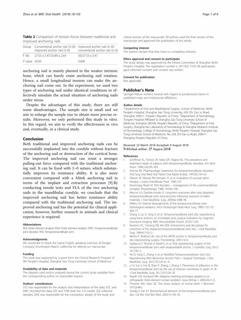
Table 2 Comparison of tension forces between traditional andimproved anchoring nails
Group Conventional anchor nail (3–0)/improved anchor nail (3–0)
Improved anchor nail (2–0)/conventional anchor nail (3–0)
F (N) 27.53 ± 5.47/25.89 ± 2.64 50/27.53 ± 5.47
P value 0.033 0.000
Zhou et al. BMC Oral Health (2018) 18:150 Page 7 of 8
16. Koolstra JH, van Eijden TM. Combined finite-element and rigid-body analysisof human jaw joint dynamics. J Biomech. 2005;38:2431–9.
17. Liu Z, Fan Y, Qian Y. Comparative evaluation on three-dimensional finiteelement models of the temporomandibular joint. Clin Biomech. 2008;23:S53.
18. Barnes SJ, Coleman SG, Gilpin D. Repair of avulsed insertion of biceps. Anew technique in four cases. J Bone Joint Surg Br. 1993;75:938.
19. Farrall LA. Arthroscopic rotator cuff repairs using suture anchors. AORN J.1995;62:737–50.
Zhou et al. BMC Oral Health (2018) 18:150 Page 8 of 8