
Importance of venous return, venous resistance, and mean
9
DOI 10.1378/chest.92.5.906 1987;92;906-912 Chest M A Bressack and T A Raffin and management of shock. and mean circulatory pressure in the physiology Importance of venous return, venous resistance, http://chestjournal.chestpubs.org/content/92/5/906.citation services can be found online on the World Wide Web at: The online version of this article, along with updated information and ) ISSN:0012-3692 http://chestjournal.chestpubs.org/site/misc/reprints.xhtml ( without the prior written permission of the copyright holder. distributed rights reserved. No part of this article or PDF may be reproduced or College of Chest Physicians, 3300 Dundee Road, Northbrook, IL 60062. All has been published monthly since 1935. Copyright 1987 by the American CHEST is the official journal of the American College of Chest Physicians. It © 1987 American College of Chest Physicians by guest on October 11, 2009 chestjournal.chestpubs.org Downloaded from
Transcript of Importance of venous return, venous resistance, and mean
DOI 10.1378/chest.92.5.906 1987;92;906-912Chest
M A Bressack and T A Raffin and management of shock.and mean circulatory pressure in the physiology Importance of venous return, venous resistance,
http://chestjournal.chestpubs.org/content/92/5/906.citation
services can be found online on the World Wide Web at: The online version of this article, along with updated information and
) ISSN:0012-3692http://chestjournal.chestpubs.org/site/misc/reprints.xhtml(without the prior written permission of the copyright holder.
distributedrights reserved. No part of this article or PDF may be reproduced or College of Chest Physicians, 3300 Dundee Road, Northbrook, IL 60062. Allhas been published monthly since 1935. Copyright 1987 by the American CHEST is the official journal of the American College of Chest Physicians. It
© 1987 American College of Chest Physicians by guest on October 11, 2009chestjournal.chestpubs.orgDownloaded from

acc�counciloncriticalcareImportance of Venous Return, Venous Resistance, andMean Circulatory Pressure in the Physiology andManagement of Shock*Michael A. Bressack, M.D. ;t and Thomas A. Raffin, M.D. , F. C. C.? 1
CARDIACOUTPUT
FIGURE 1. Cardiac function curves demonstrating the effects of
preload (atrial pressure), inotropy, and afterload on cardiac’
VENOUSRETURN
ATRIAL PRESSURE
906 Physiology and Management of Shock (Bressak, Raffin)
PHYSIoLoGIC BACKGROUND
A reasonable, unifying definition of shock at the
cellular level is inadequate oxygen consumption
by the cell for its metabolic needs. The negative
consequence is the accumulation of an oxygen debt
with eventual cellular dysfunction and death.
Inadequate oxygen consumption (shock) can result
from the following: (1) inadequate oxygen content of
the blood; (2) inadequate circulation of the blood
(cardiac output); (3) failure to deliver the circulated
blood to the cell (microperfusion); and (4) failure of the
cell to utilize the delivered oxygen (cellular dysfunc-
tion).
In our discussion, we shall focus on inadequate
circulation (cardiac output) as the limiting factor in
oxygen consumption (shock). Traditionally, cardiac
function has been viewed as the sole determinant of
cardiac output. This function has been divided into
preload, inotropy, afterload, and heart rate; cardiac
output can be maximized by increasing preload and
inotropy, decreasing afterload, and optimizing heart
rate. 1.2
Guyton et aP emphasized that the regulation of
cardiac output is determined by the interaction be-
tween the vasculature and the heart. In steady state,
the heart cannot eject more blood than it receives from
the vasculature and the vasculature cannot return
more blood than it receives from the heart. Cardiac
output must equal venous return.
The physiology ofthese separate systems can best be
understood graphically. A family of Starling curves
shows the regulation ofcardiac output by the heart (Fig
1); maximal cardiac output requires adequate preload
(indirectly measured as atrial pressure) and inotropy
*From the Departments of Pediatrics and Medicine, Stanford
University, Stanford, CA.tClinical Associate Professor of Pediatrics.
tAssociate Professor of Medicine.Reprint requests: Dr Raffin, Division of Respiratory Medicine,C-356, Stanford University Medical Center. Stanford 94305
and minimal afterload (principally measured as
vascular resistance).
0
ATRIAL PRESSUREFIGURE 2. Venous return curve demonstrating the pressure-flow
relationship of the venous system. The pressure-axis intercept
equals mean circulatory pressure (Pmc). The inverse of the slope
equals resistance to venous return (Rv). The plateau in the curve is
due to the great veins functioning as Starling resistors.’ Although our
graphs refer to atnal pressure in general, it should be noted that right
atrial pressure has a much greater effect than left atrial pressure on
venous return.
© 1987 American College of Chest Physicians by guest on October 11, 2009chestjournal.chestpubs.orgDownloaded from
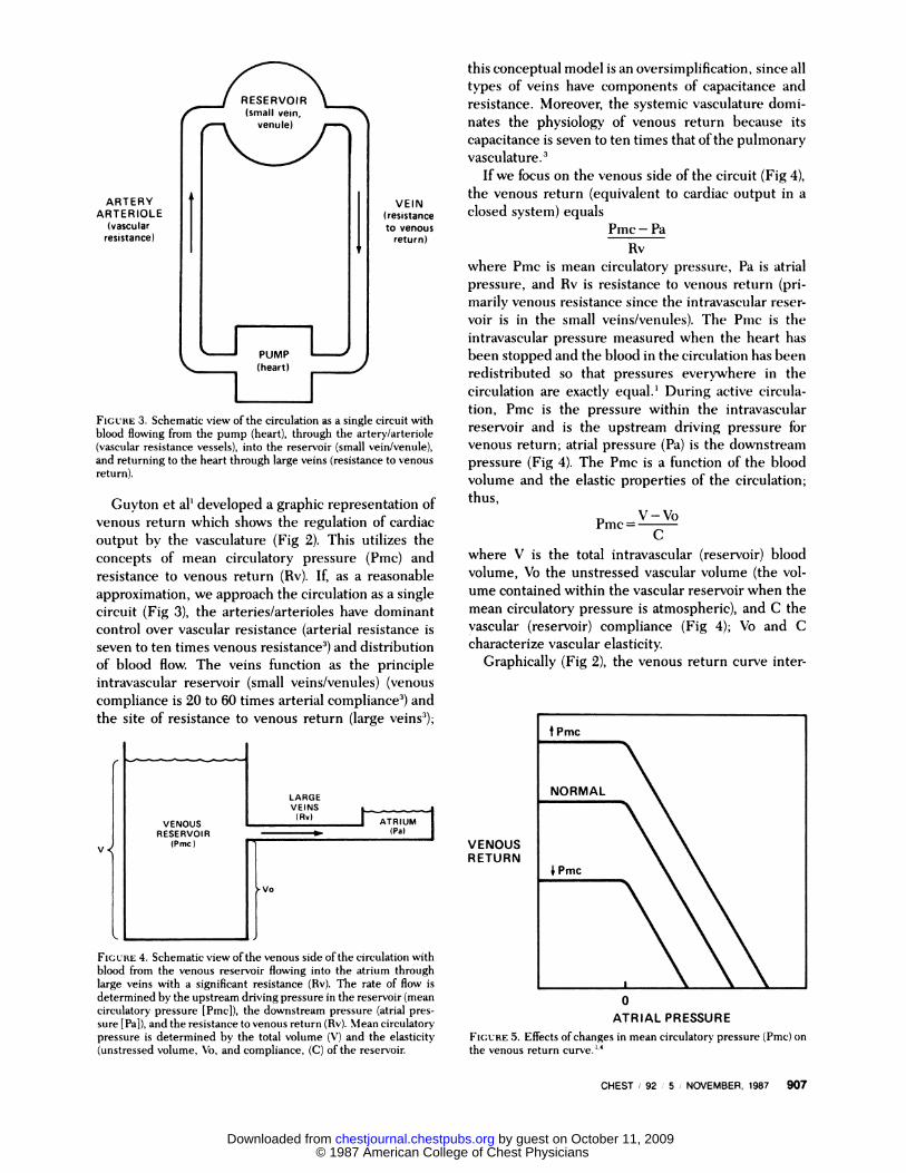
ARTERY
ARTERIOLE(vascular
resistance)
VEIN( resistanceto venous
return)
VENOUSRESERVOIR
(Pmc)
LARGEVEINS(Rv)
Vo
VENOUSRETURN
V
FIGURE 4. Schematic view ofthe venous side ofthe circulation with
blood from the venous reservoir flowing into the atrium through
large veins with a significant resistance (Rv). The rate of flow is
determined by the upstream driving pressure in the reservoir (mean
circulatory pressure [Pmc]), the downstream pressure (atrial pres-
sure [Pa]), and the resistance to venous return (Rv). Mean circulatory
pressure is determined by the total volume (V) and the elasticity
(unstressed volume, Vo, and compliance, (C) ofthe reservoir.
0
ATRIAL PRESSURE
FIGURE 5. Effects ofchanges in mean circulatory pressure (Pmc) on
the venous return curve.”4
CHEST I 92 : 5 � NOVEMBER, 1987 907
FIGURE 3. Schematic view of the circulation as a single circuit with
blood flowing from the pump (heart), through the artery/arteriole
(vascular resistance vessels), into the reservoir (small veinlvenule),
and returning to the heart through large veins (resistance to venous
return).
Guyton et al’ developed a graphic representation of
venous return which shows the regulation of cardiac
output by the vasculature (Fig 2). This utilizes the
concepts of mean circulatory pressure (Pmc) and
resistance to venous return (Rv). If, as a reasonable
approximation, we approach the circulation as a single
circuit (Fig 3), the arteries/arterioles have dominant
control over vascular resistance (arterial resistance is
seven to ten times venous resistance3) and distribution
of blood flow. The veins function as the principle
intravascular reservoir (small veins/venules) (venous
compliance is 20 to 60 times arterial compliance3) and
the site of resistance to venous return (large veins3);
this conceptual model is an oversimplification, since all
types of veins have components of capacitance and
resistance. Moreover, the systemic vasculature domi-
nates the physiology of venous return because its
capacitance is seven to ten times that ofthe pulmonary
3
If we focus on the venous side of the circuit (Fig 4),
the venous return (equivalent to cardiac output in aclosed system) equals
Pmc - Pa
Rv
where Pmc is mean circulatory pressure, Pa is atrial
pressure, and Rv is resistance to venous return (pri-
manly venous resistance since the intravascular reser-
voir is in the small veins/venules). The Pmc is the
intravascular pressure measured when the heart has
been stopped and the blood in the circulation has been
redistributed so that pressures everywhere in the
circulation are exactly equal.’ During active circula-
tion, Pmc is the pressure within the intravascular
reservoir and is the upstream driving pressure for
venous return; atrial pressure (Pa) is the downstream
pressure (Fig 4). The Pmc is a function of the blood
volume and the elastic properties of the circulation;
thus,
V - VoPmc=
C
where V is the total intravascular (reservoir) blood
volume, Vo the unstressed vascular volume (the vol-
ume contained within the vascular reservoir when the
mean circulatory pressure is atmospheric), and C the
vascular (reservoir) compliance (Fig 4); Vo and C
characterize vascular elasticity.
Graphically (Fig 2), the venous return curve inter-
© 1987 American College of Chest Physicians by guest on October 11, 2009chestjournal.chestpubs.orgDownloaded from

0
ATRIAL PRESSURE
908 Physiology and Management of Shock (Bressak, Raffin)
VENOUSRETURN
FIGURE 6. Effects ofchanges in resistance to venous return (Rv) on
the venous return �j�1�4
cepts the pressure axis at a Pa which is equal to mean
circulatory pressure (Pmc) and the inverse ofthe slope
of the venous return curve is resistance to venous
return (Rv). Although our graphs refer to atrial pres-
sure (Pa), right atnal pressure has a much greater effect
on venous return than left atrial pressure because the
systemic compliance is so much larger than pulmonary
compliance.3 The plateau in the venous return curve is
due to the fact that the great veins entering the chest
function as Starling resistors, collapsing as the right
atrial pressure (Pa) becomes subatmospheric. A family
of venous return curves can be constructed showing
the effects of isolated changes in mean circulatory
pressure or resistance to venous return. Venous return
can be impaired by a decrease in mean circulatory
pressure (Pmc) (Fig 5) or an increase in resistance to
venous return (Rv) (Fig 6). The Pmc decreases due to
the loss ofintravascular blood volume (V) or a decrease
in vascular elasticity (increased Vo, C). Decreased
vascular elasticity is caused by dilatation/relaxation (an
active process due to circulating vasoactive mediators
or autonomic changes, or a passive relaxation due to
increased flow) of the reservoir vessels (small veins/
venules).
The veins primarily determining the resistance to
venous return include central large veins such as the
vena cavae, which are passively affected, and periph-
eral large- and medium-sized veins which can be
passively affected or responsive to circulatory vasoac-
tive mediators and autonomic stimulation. Increased
resistance to venous return can occur due to active
vasoconstriction of the peripheral large- and medium-
sized veins, passive narrowing of the veins (extrinsic
pressure due to increased intraabdominal/pleural
pressure or passive elastic recoil due to low flow), or to
hyperviscosity (polycythemia).
The resistance to venous return (Rv) can be greatly
affected by the distribution ofblood flow (flow distribu-
tion is determined by artery/arteriolar function). The
systemic vasculature can be divided into vascular beds
with short time constants (striated muscle, kidney) and
long time constants (hepatosplanchnic); flow through
vascular beds with short time constants will decrease
resistance and therefore, improve venous return while
flow through vessels with long time constants will
increase resistance and therefore interfere with venous
4
Guyton et al’ realized that since venous return must
equal cardiac output in a closed system, the same
graphic parameters could be used for both the cardiac
function and venous return curves. The superimposed
curves demonstrate the interaction of the two systems
in determining cardiac output (Fig 7). The intersection
point of the curve (point A) will determine the cardiac
output and atrial pressure based on the momentary
pumping ability ofthe heart (inotropy, afterload, heart
rate) and the characteristics ofthe circulation (V, C, Vo,
Rv). Both the heart and the circulation may be abnor-
mal. However, ifcardiac function is the limiting factor
in blood flow (decreased inotropy or increased after-
load), there will be a low cardiac output associated with
an increased atrial pressure (Fig 8, point B). On the
other hand, if venous return is the principle limiting
factor (decreased mean circulatory pressure or in-
creased resistance to venous return), there will be a low
cardiac output associated with a decreased atrial pres-
sure (Fig 9, point B, C).
CLINICAL RELEVANCE
Well and Shubin3 classified clinical shock as hypo-
volemic, cardiogenic, distributive, and obstructive. In
cardiogenic and obstructive (pulmonary embolism,
cardiac tamponade) shock, the primary abnormality is
cardiac function; cardiac output is decreased in associa-
CARDIACOUTPUT
ORVENOUSRETURN
0
ATRIAL PRESSURE
FIGURE 7. Superimposition of the cardiac function and venous
return curves. The point of intersection (point A) determines the
cardiac output and atrial pressure.’
© 1987 American College of Chest Physicians by guest on October 11, 2009chestjournal.chestpubs.orgDownloaded from

0
0
ATRIAL PRESSURE
0
ATRIAL PRESSURE
CHEST I 92 I 5 I NOVEMBER, 1987 909
CAR D I ACOUTPUT
ORVENOUSRETURN
ATRIAL PRESSURE
FIGURE 8. Effects of decreased cardiac function with no change in
venous return. Cardiac output decreases and atrial pressure in-
creases (point A to B).’4
tion with an increased atrial pressure (Fig 8, Point A to
B) (the exact shape of the cardiac function curve will
depend on the specific disorder). Although there can
be primary or secondary abnormalities in the venous
return curve, cardiac function curve abnormalities
limit the cardiac output.
In hypovolemic shock, the principle abnormality is
the loss of intravascular volume (V); although secon-
dary changes in vascular and cardiac function may
develop, venous return is primarily decreased due to a
low mean circulatory pressure (Pmc)6 (Fig 9, point A to
C). Cardiac output is depressed in association with a
decreased atrial pressure.
The mechanisms involved in distributive shock are
much more complex and less understood; moreover,
this form of shock is heterogeneous, including septic,
CARDIACOUTPUT
ORVENOUSRETURN
FIGURE 9. Effects of decreased mean circulatory pressure (Pmc)
(p oint A to C) or increased resistance to venous return (Rv) (point A
to B) with no change in cardiac function. Cardiac output and atnal
pressure �
anaphylactic, and neurogenic shock. Focusing on only
septic shock, we clinically find patients with a high,
normal, or low vascular resistance and cardiac output;
many of these findings are probably related to the age
of the patient, the specific bacteria involved, and the
associated vasoactive mediators.7 Although arterial
tone determines total vascular resistance, perfusion
pressure, and distribution of blood flow,3 venous tone
with its effects on Pmc and Rv determines venous
return, and therefore, cardiac output early in septic
shock (before cardiac dysfunction). The study of the
physiology of septic shock has dealt primarily with the
arteries and not the veins. If the sepsis involves active
venoconstriction, venous resistance (peripheral large
veins) and elasticity (small veins/venules) will both be
increased. The effect on venous return (cardiac output)
will depend on which determinant (Pmc or Rv) domi-
nates; increased Rv would decrease venous return
while increased Pmc would increase venous return.
Studies in dogs (E coli and endotoxin7) and piglets
(group B streptococci8) have shown a decrease in
venous return (cardiac output) because of the domi-
nant effect ofan increased resistance to venous return
(Fig 10, point A to B). The Rv is further increased by
passive elastic recoil of the veins (caused by a low
cardiac output) and by the blockage of small veins/
venules (microvascular agglutination andlor clotting).
If the venoconstriction is associated with significant
arterioconstriction (elevated afterload), the cardiac
output could be further harmed by a worsening of the
cardiac function curve . S evere arterioconstriction
could also elevate Rv by increasing the capacitance of
proximal arteries.
If active venodilatation is part of the sepsis,9 resist-
ance to venous return and elasticity will be decreased.
CARDIACOUTPUT
ORVENOUSRETURN
FIGURE 10. Sepsis with venoconstriction causing an increased
resistance to venous return (Rv) and mean circulatory pressure
(Pmc); cardiac function is unchanged. Cardiac output (venous
return) and atrial pressure are decreased because of the dominant
effect of Rv (point A to B). With fluid therapy cardiac output and
atrial pressure are normalized by further increasing Pmc (point A).
© 1987 American College of Chest Physicians by guest on October 11, 2009chestjournal.chestpubs.orgDownloaded from
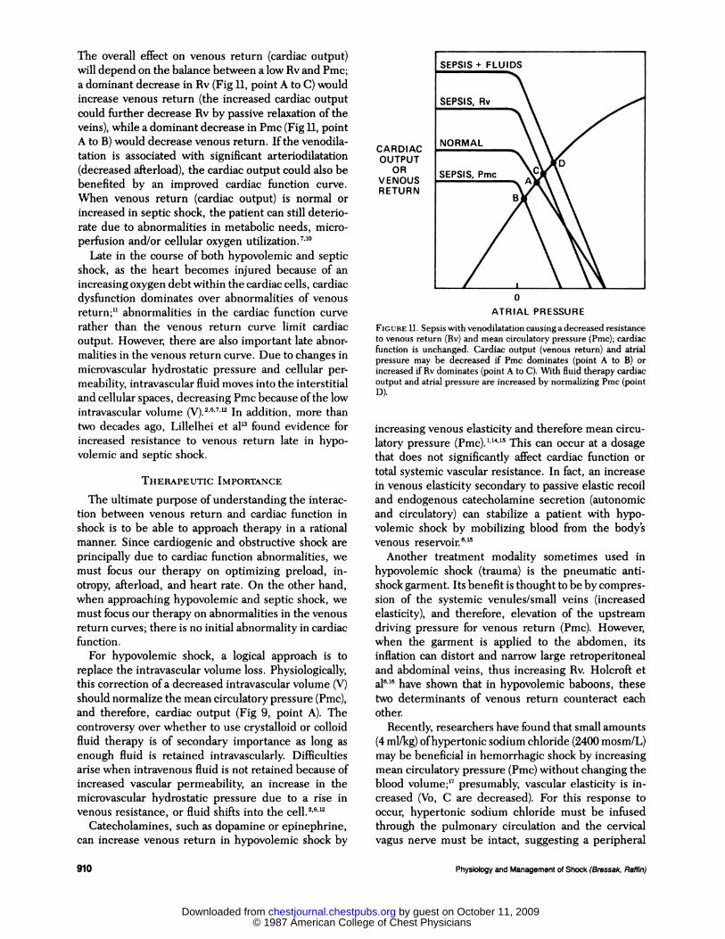
CARDIACOUTPUT
ORVENOUSRETURN
FIGURE II. Sepsis with venodilatation causing a decreased resistance
to venous return (Rv) and mean circulatory pressure (Pmc); cardiac
function is unchanged. Cardiac output (venous return) and atrial
pressure may be decreased if Pmc dominates (point A to B) or
increased ifRv dominates (point A to C). With fluid therapy cardiac
output and atrial pressure are increased by normalizing Pmc (point
D).
0
ATRIAL PRESSURE
910 Physiology and Management of Shock (Bressak, Raffin)
The overall effect on venous return (cardiac output)
will depend on the balance between a low Rv and Pmc;
a dominant decrease in Rv (Fig II, point A to C) would
increase venous return (the increased cardiac output
could further decrease Rv by passive relaxation of the
veins), while a dominant decrease in Pmc (Fig II, point
A to B) would decrease venous return. Ifthe venodila-
tation is associated with significant arteriodilatation
(decreased afterload), the cardiac output could also be
benefited by an improved cardiac function curve.
When venous return (cardiac output) is normal or
increased in septic shock, the patient can still deterio-
rate due to abnormalities in metabolic needs, micro-
perfusion and/or cellular oxygen utilization.7”0
Late in the course of both hypovolemic and septic
shock, as the heart becomes injured because of an
increasing oxygen debt within the cardiac cells, cardiac
dysfunction dominates over abnormalities of venous
return;11 abnormalities in the cardiac function curve
rather than the venous return curve limit cardiac
output. However, there are also important late abnor-
malities in the venous return curve. Due to changes in
microvascular hydrostatic pressure and cellular per-
meability, intravascular fluid moves into the interstitial
and cellular spaces, decreasing Pmc because ofthe low
intravascular volume (V).2’6’7”� In addition, more than
two decades ago, Lillelhei et al’3 found evidence for
increased resistance to venous return late in hypo-
volemic and septic shock.
THERAPEUTIC IMPORTANCE
The ultimate purpose of understanding the interac-
tion between venous return and cardiac function in
shock is to be able to approach therapy in a rational
manner. Since cardiogenic and obstructive shock are
principally due to cardiac function abnormalities, we
must focus our therapy on optimizing preload, in-
otropy, afterload, and heart rate. On the other hand,
when approaching hypovolemic and septic shock, we
must focus our therapy on abnormalities in the venous
return curves; there is no initial abnormality in cardiac
function.
For hypovolemic shock, a logical approach is to
replace the intravascular volume loss. Physiologically,
this correction of a decreased intravascular volume (V)
should normalize the mean circulatory pressure (Pmc),
and therefore, cardiac output (Fig 9, point A). The
controversy over whether to use crystalloid or colloid
fluid therapy is of secondary importance as long as
enough fluid is retained intravascularly. Difficulties
arise when intravenous fluid is not retained because of
increased vascular permeability, an increase in the
microvascular hydrostatic pressure due to a rise in
venous resistance, or fluid shifts into the cell.2’6-’�
Catecholamines, such as dopamine or epinephrine,
can increase venous return in hypovolemic shock by
increasing venous elasticity and therefore mean circu-
latory pressure � This can occur at a dosage
that does not significantly affect cardiac function or
total systemic vascular resistance. In fact, an increase
in venous elasticity secondary to passive elastic recoil
and endogenous catecholamine secretion (autonomic
and circulatory) can stabilize a patient with hypo-
volemic shock by mobilizing blood from the body’s
venous reservoir.6”5
Another treatment modality sometimes used in
hypovolemic shock (trauma) is the pneumatic anti-
shock garment. Its benefit is thought to be by compres-
sion of the systemic venules/small veins (increased
elasticity), and therefore, elevation of the upstream
driving pressure for venous return (Pmc). However,
when the garment is applied to the abdomen, its
inflation can distort and narrow large retroperitoneal
and abdominal veins, thus increasing Rv. Hoicroft et
al6”6 have shown that in hypovolemic baboons, these
two determinants of venous return counteract each
other.
Recently, researchers have found that small amounts
(4 mllkg) ofhypertonic sodium chloride (2400 mosm/L)
may be beneficial in hemorrhagic shock by increasing
mean circulatory pressure (Pmc) without changing the
blood volume;’7 presumably, vascular elasticity is in-
creased (Vo, C are decreased). For this response to
occur, hypertonic sodium chloride must be infused
through the pulmonary circulation and the cervical
vagus nerve must be intact, suggesting a peripheral
© 1987 American College of Chest Physicians by guest on October 11, 2009chestjournal.chestpubs.orgDownloaded from

CARDIACOUTPUT
ORVENOUSRETURN
0
ATRIAL PRESSURE
CHEST I 92 I 5 I NOVEMBER, 1987 911
reflex of pulmonary origin.
In addition to specific antibiotic therapy, treatment
for septic shock should depend on the specific abnor-
mal determinants of venous return. When there is
venoconstriction with an increase in resistance to
venous return, the resulting low venous return (cardiac
output) (Fig 10, point B) can be treated by elevation of
Pmc with enough intravenous fluid therapy to over-
come the increased Rv (increase the upstream driving
pressure for venous return) (Fig 10, point A) or by
normalization of the increased resistance to venous
return. The increased resistance might be improved
pharmacologically by venodilatation with agents such
as nitroglycerin, calcium channel blockers, or beta-2
catecholamines. The ultimate effects of vasoactive
agents on venous return are confusing and unresolved
because ofpossible actions on venous elasticity (Vo, C),
resistance to venous return (Rv), and the distribution of
blood flow through vascular beds with long or short
time constants.’3”8”9 As already stated, small veins/
venules tend to be the site of the intravascular reser-
voir, while large veins are the primary site of resistance
to venous return (Fig 3). Ideally, a therapeutic agent
would improve venous return by decreasing venous
resistance without decreasing venous elasticity and
therefore Pmc. Unfortunately, researchers have so far
not been able to find vasoactive agents that will clearly
separate venous resistance and capacitance func-
lions;’3”8”9 this may partially be due to the fact that all
types of vasoactive-responsive veins have components
of capacitance and resistance. Vasoactive agents may
also affect venous return by altering arteriolar tone;
increased perfusion of vascular beds with short time
constants will improve venous return while perfusion
ofvascular beds with long time constants will decrease
venous return. Future research in these physiologic
areas will be quite important for selecting vasoactive
therapeutic agents. Finally, if the venoconstriction is
associated with significant arterioconstriction (in-
creased total vascular resistance), treatment of the
elevated afterload may be important ifcardiac function
is abnormal.
When septic shock involves venodilatation, and
therefore, increased capacitance, maintaining an ade-
quate mean circulatory pressure (Pmc) with in-
travenous fluid therapy is crucial to maintaining a
normal or elevated cardiac output (Fig II, point D); the
decreased Rv will help venous return. As with hypo-
volemic shock, catecholamines such as dopamine may
benefit this form of septic shock by increasing venous
elasticity and therefore Pmc.9 However, as already
mentioned, vasoactive agents can have unpredictable
effects on venous return because of differential effects
on venous elasticity, resistance to venous return, and
distribution of blood �
Vasoactive mediators are probably a major cause of
FIGURE 12. Effects of positive end-expiratory pressure (PEEP) on
venous return and cardiac function curves. The venous return curve
shows an increase in mean circulatory pressure (Pmc) and resistanceto venous return (Rv). The cardiac function curve is shifted to the
right and depressed. PEEP results in a decrease in cardiac output
and an increase in atrial pressure (point A to B). ‘#{176}
the distributive changes (venoconstriction or veno-
dilatation) in septic � Discovering and reversing
these mediators may reverse the abnormalities in
venous return in a more specific manner. Therapy for
venous occlusion (caused by microvascular agglutina-
tion and/or clotting) may be necessary to prevent
abnormalities of Rv and Pmc.
Late in the course of all clinical types of shock, as
cardiac decompensation becomes the dominant prob-
lem, therapy must focus on heart inotropy, afterload,
and heart rate (cardiac function curve). However, as
vascular and cellular permeability increase, a loss of
intravascular fluid (V) into the interstitial and cellular
space must be replaced with enough intravenous fluid
to maintain an adequate mean circulatory pressure
(Pmc) for venous return.”6’7’�
Finally, we must mention the important effects
positive pressure ventilation has on venous return and
cardiac function because of its frequent usage in
critically ill patients in shock; this becomes relevant as
the pleural and abdominal pressures increase at high
levels ofpositive end-expiratory pressure. The venous
return curve shows an increase in Pmc and Rv (Fig
12);20 this is due to compression of intrathoracic and
intraabdominal venous structures. The cardiac func-
tion curve is adversely affected by the increased
afterload on the right ventricle and a decrease in
cardiac compliance.�#{176} Even without any adverse car-
diac effects, the cardiac function curve will be shifted
to the right on the basis of the positive pleural
pressure. Thus, the impairment in cardiac output
during positive end-expiratory pressure (Fig 12, point
A to B) can be explained by the relationship between
the venous return and cardiac function curves.
© 1987 American College of Chest Physicians by guest on October 11, 2009chestjournal.chestpubs.orgDownloaded from

912 Physiology and Management of Shock (Bressak, Raffin)
CONCLUSION
We have discussed the physiology of venous return
and its importance in the understanding and treatment
ofshock. Cardiac output is regulated by the interaction
between the vasculature and the heart. When ap-
proaching a patient in shock, we must determine
whether cardiac output is being limited by the
vasculature (venous return) or cardiac function; ther-
apy will be quite different for each. Moreover, the
physiology of shock will probably change over time.
Therapy must be chosen based on an understanding of
the changing relationship between venous return and
cardiac function. In order to understand and improve
our therapeutic options, there is a great need for
research into the differential effects of vasoactive
agents on venous resistance, capacitance, and flow
distribution. Adequate treatment for cardiac output
does not assure survival. Abnormalities in microperfu-
sion and/or cellular oxygen utilization can still lead to
cellular dysfunction and death.
ACKNOWLEDGMENTS: We thank Dr. J. Green for reviewing themanuscript and Kathy Stephens for outstanding assistance in prepar-ing the manuscript.
REFERENCES
1 Guyton AC, Jones CE, Coleman TC. Circulatory physiology:
cardiac output and its regulation, 2nd ed. Philadelphia: W. B.
Saunders, 1973
2 Perkin RM, Levin DL. Shock in the pediatric patient: Part II.
Therapy. J Pediatr 1982; 101:319-32
3 Green JF. Circulatory mechanics. In: Fundamental car-
diovascular and pulmonary physiology. Philadelphia: Lea &
Febiger, 1982:101-10
4 Green JF. Determinants ofsystemic blood flow. In: Guyton AC,
Young DB, eds. International review of physiology: car-
diovascular physiology III, vol 18. Baltimore: University Park
Press, 1979:34-65
5 Weil MH, Shubin H. Proposed reclassification of shock states
with special reference to distributive defects. Adv Exp Med Biol
1972; 23:13-22
6 Holcroft JW. Impairment ofvenous return in hemorrhagic shock.
Surg Clin N Am 1982; 62:17-28
7 Hinshaw LB. Release of vasoactive agents and the vascular
effects of endotoxin. In: Kadis S, Weinbaum G, Ajl SJ, eds.
Microbiol toxins. New York: Academic Press, 1971:209-55
8 Bressack MA, Morton NS, Hortop J. Group B streptococcal
sepsis in the piglet: effects of fluid therapy on venous return,
organ edema, and organ blood flow. Circ Res (in press)
9 Hinshaw LB. Shambour LL, Greenfield U, Coalson JJ. Mecha-
nism ofdecreased venous return. Arch Surg 1970; 100:600-06
10 Pollack MM, Fields AL, Ruttimann UE. Sequential cardiopul-
monary variables of infants and children in septic shock. Crit
Care Med 1984; 12:554-59
11 Hinshaw LB. Role ofthe heart in the pathogenesis of endotoxic
shock. J Surg Res 1974; 17:134-45
12 Shoemaker WC, Hauser CH. Critique of crystalloid versus
colloid therapy in shock and shock lung. Crit Care Med 1979; 7:
117-24
13 Lillehei RC, Longerbeam JK, BlochJH, Manax WG. The nature
of irreversible shock: experimental and clinical observations.
Ann Surg 1964; 160:682-708
14 Marmno RJ, Romagnoli A, Keats AS. Selective venoconstriction
by dopamine in comparison with isoproterenol and phenyle-
phrine. Anesthesiology 1975; 43:570-72
15 Rothe CE Venous system: physiology ofthe capacitance vessels.
In: JT Shepherd, FM Abboud, eds. Handbook of physiology:
peripheral circulation and organ blood flow. Ed 2. Bethesda:
American Physiology Society, 1983; 397-452
16 Holcroft JW, Link DP, Lantz BMT, Green JE Venous return and
the pneumatic antishock garment in hypovolemic baboons. JTrauma 1984; 24:928-35
17 Lopes OU, Velasco IT, Guertzenstein PG, Silva MR. Pontieri V.
Hypertonic sodium chloride restores mean circulatory filling
pressure in severely hypovolemic dogs. Hypertension 8 (suppl
1); 1986; I 195-99
18 Ogilvie R. Comparative effects of vasodilator drugs on flow
distribution and venous return. Can J Physiol Pharmacol 1985;
63:1345-55
19 Ito H, Hirakawa S. Effects of vasodilators on the systemic
capacitance vessels, a study with the measurement of the mean
circulatory pressure in dogs. Jap Circ J 1984; 48:388-404
20 Sylvester JT, Goldberg HS, Permutt S. The role of the
vasculature in the regulation ofcardiac output. Clin Chest Med
1983; 4:111-28
© 1987 American College of Chest Physicians by guest on October 11, 2009chestjournal.chestpubs.orgDownloaded from

DOI 10.1378/chest.92.5.906 1987;92; 906-912Chest
M A Bressack and T A Raffinpressure in the physiology and management of shock.
Importance of venous return, venous resistance, and mean circulatory
October 11, 2009This information is current as of
& ServicesUpdated Information
http://chestjournal.chestpubs.org/content/92/5/906.citationfigures, can be found at:Updated Information and services, including high-resolution
Citations
related-urlshttp://chestjournal.chestpubs.org/content/92/5/906.citation#This article has been cited by 1 HighWire-hosted articles:
Open Access Freely available online through CHEST open access option
Permissions & Licensing
http://www.chestjournal.org/site/misc/reprints.xhtmltables) or in its entirety can be found online at: Information about reproducing this article in parts (figures,
Reprints http://www.chestjournal.org/site/misc/reprints.xhtml
Information about ordering reprints can be found online:
Email alerting serviceup in the box at the top right corner of the online article.Receive free email alerts when new articles cite this article. Sign
formatImages in PowerPoint
article figure for directions teaching purposes in PowerPoint slide format. See any online Figures that appear in CHEST articles can be downloaded for
© 1987 American College of Chest Physicians by guest on October 11, 2009chestjournal.chestpubs.orgDownloaded from