Implications for function and therapy of a 2.9 å structure of binary-complexed antithrombin
6
COMMUNICATION Implications for Function and Therapy of a 2.9 A ˚ Structure of Binary-complexed Antithrombin Richard Skinner 1 , Wun-Shaing W. Chang 1,2 , Lei Jin 1 , Xue Pei 1 James A. Huntington 1 , Jan-Pieter Abrahams 3 , Robin W. Carrell 1 and David A. Lomas 1,2 * 1 Department of Haematology and 2 Respiratory Medicine Unit, Department of Medicine University of Cambridge MRC Centre, Hills Road Cambridge CB2 2QH 3 Department of Biophysical Structural Chemistry University of Leiden Netherlands The crystal structure of a binary complex of human antithrombin with a peptide of the same sequence as its reactive loop (P 14 -P 3 ) has been deter- mined at 2.9 A ˚ . The peptide binds as the middle strand s4A in the A b-sheet, homologously to that of the reactive loop in the latent and cleaved forms of antithrombin. Peptide binding results in the complete expulsion of the hinge region of the loop from the A b-sheet although the conformation differs from that of heparin-activated antithrombin. The 36-fold increase in the rate of reaction of the binary complex with factor Xa indicates that full loop expulsion alone is not sufficient for complete heparin activation of antithrombin but that this is also dependent on the overall conformation of the molecule. Previous studies have demon- strated that reactive loop peptides can block or reverse the polymeris- ation of serpins associated with cirrhosis and thrombosis. The antithrombin binary complex structure defines the precise localisation of the blocking peptide in a serpin and provides the basis for rational drug design for mimetics that will prevent polymerisation in vivo and so ame- liorate the associated disease. # 1998 Academic Press Keywords: serpin; polymerisation; a 1 -antitrypsin; peptide *Corresponding author The function of serpins as proteinase inhibitors is dependent on a profound conformational change that follows cleavage at their P 1 reactive centre. The structure of cleaved a 1 -antitrypsin showed that the otherwise exposed 14-residue peptide loop (P 14 -P 1 ), aminoterminal to the site of cleavage, had become incorporated as a new central strand (s4A) in the otherwise five-stranded A b-sheet of the molecule (Loebermann et al., 1984). It was also shown that exogenous peptides with the sequence of the reac- tive loop could be annealed to intact serpins to give a change in biophysical properties that mimicked the changes associated with reactive centre cleavage in a 1 -antitrypsin (Schulze et al., 1990) and similarly in antithrombin (Bjo ¨rk et al., 1992). Subsequently, many functional studies of the serpins have been based on the logical deduction from these results, that the reactive loop (P 14 -P 3 ) peptides in the binary complexes of the serpins are annealed in the s4A position (Chang et al., 1996; Fitton et al., 1997; Lomas et al., 1993; Mast et al., 1992; Schulze et al., 1992). However, the certainty of this assumption was questioned by a later structural finding that dimers of antithrombin formed in vitro were linked through the P 8 -P 3 portion of their reactive loop by strand replacement to the C-sheet of a second mol- ecule (Carrell et al., 1994; Schreuder et al., 1994). The question this raised is of importance as the for- mation of P 14 -P 3 binary complexes has been shown in vitro to completely prevent the more extensive polymerisation that is a contributory cause to a number of disease consequences associated with genetic variants of serpins including emphysema, cirrhosis, angio-oedema and thrombosis (Aulak et al., 1993; Bruce et al., 1994; Eldering et al., 1995; Lomas et al., 1992). The ability of annealing peptides to prevent and to reverse this loop-sheet polymeris- ation opens possibilities for the rational design of therapies, but before this can be undertaken there Present addresses: R. Skinner, Unilever Research, Port Sunlight Laboratory, Wirral, UK; W.-S.W. Chang, Institute of Biomedical Sciences, Academia Sinica, Taipei, Taiwan. E-mail address of the corresponding author: [email protected] Article No. mb982083 J. Mol. Biol. (1998) 283, 9–14 0022 – 2836/98/410009–06 $30.00/0 # 1998 Academic Press
-
Upload
richard-skinner -
Category
Documents
-
view
212 -
download
0
Transcript of Implications for function and therapy of a 2.9 å structure of binary-complexed antithrombin

Article No. mb982083 J. Mol. Biol. (1998) 283, 9±14
COMMUNICATION
Implications for Function and Therapy of a 2.9 AÊ
Structure of Binary-complexed Antithrombin
Richard Skinner1, Wun-Shaing W. Chang1,2, Lei Jin1, Xue Pei1
James A. Huntington1, Jan-Pieter Abrahams3, Robin W. Carrell1
and David A. Lomas1,2*
1Department of Haematologyand 2Respiratory MedicineUnit, Department of MedicineUniversity of CambridgeMRC Centre, Hills RoadCambridge CB2 2QH3Department of BiophysicalStructural ChemistryUniversity of LeidenNetherlands
Present addresses: R. Skinner, UnSunlight Laboratory, Wirral, UK; WInstitute of Biomedical Sciences, AcTaipei, Taiwan.
E-mail address of the [email protected]
0022±2836/98/410009±06 $30.00/0
The crystal structure of a binary complex of human antithrombin with apeptide of the same sequence as its reactive loop (P14-P3) has been deter-mined at 2.9 AÊ . The peptide binds as the middle strand s4A in the Ab-sheet, homologously to that of the reactive loop in the latent andcleaved forms of antithrombin. Peptide binding results in the completeexpulsion of the hinge region of the loop from the A b-sheet althoughthe conformation differs from that of heparin-activated antithrombin. The36-fold increase in the rate of reaction of the binary complex with factorXa indicates that full loop expulsion alone is not suf®cient for completeheparin activation of antithrombin but that this is also dependent on theoverall conformation of the molecule. Previous studies have demon-strated that reactive loop peptides can block or reverse the polymeris-ation of serpins associated with cirrhosis and thrombosis. Theantithrombin binary complex structure de®nes the precise localisation ofthe blocking peptide in a serpin and provides the basis for rational drugdesign for mimetics that will prevent polymerisation in vivo and so ame-liorate the associated disease.
# 1998 Academic Press
Keywords: serpin; polymerisation; a1-antitrypsin; peptide
*Corresponding authorThe function of serpins as proteinase inhibitors isdependent on a profound conformational changethat follows cleavage at their P1 reactive centre. Thestructure of cleaved a1-antitrypsin showed that theotherwise exposed 14-residue peptide loop (P14-P1),aminoterminal to the site of cleavage, had becomeincorporated as a new central strand (s4A) in theotherwise ®ve-stranded A b-sheet of the molecule(Loebermann et al., 1984). It was also shown thatexogenous peptides with the sequence of the reac-tive loop could be annealed to intact serpins to givea change in biophysical properties that mimickedthe changes associated with reactive centre cleavagein a1-antitrypsin (Schulze et al., 1990) and similarlyin antithrombin (BjoÈ rk et al., 1992). Subsequently,many functional studies of the serpins have been
ilever Research, Port.-S.W. Chang,ademia Sinica,
ing author:
based on the logical deduction from these results,that the reactive loop (P14-P3) peptides in the binarycomplexes of the serpins are annealed in the s4Aposition (Chang et al., 1996; Fitton et al., 1997;Lomas et al., 1993; Mast et al., 1992; Schulze et al.,1992). However, the certainty of this assumptionwas questioned by a later structural ®nding thatdimers of antithrombin formed in vitro were linkedthrough the P8-P3 portion of their reactive loop bystrand replacement to the C-sheet of a second mol-ecule (Carrell et al., 1994; Schreuder et al., 1994). Thequestion this raised is of importance as the for-mation of P14-P3 binary complexes has been shownin vitro to completely prevent the more extensivepolymerisation that is a contributory cause to anumber of disease consequences associated withgenetic variants of serpins including emphysema,cirrhosis, angio-oedema and thrombosis (Aulaket al., 1993; Bruce et al., 1994; Eldering et al., 1995;Lomas et al., 1992). The ability of annealing peptidesto prevent and to reverse this loop-sheet polymeris-ation opens possibilities for the rational design oftherapies, but before this can be undertaken there
# 1998 Academic Press
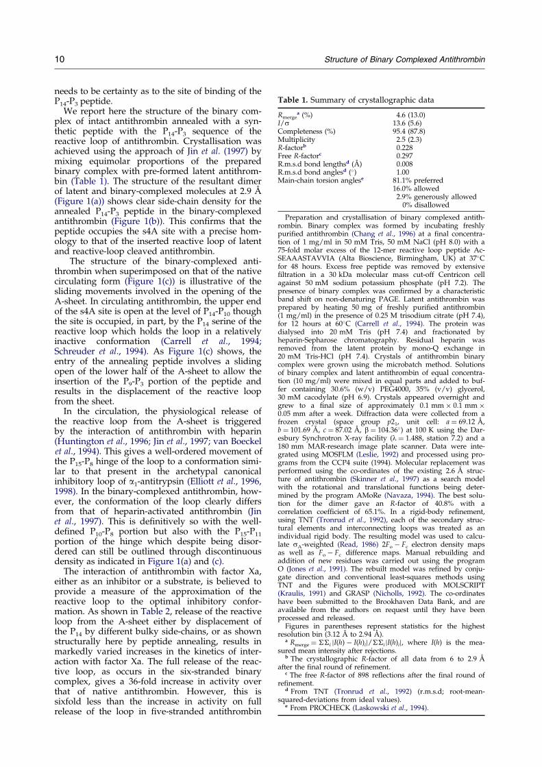
Table 1. Summary of crystallographic data
Rmergea (%) 4.6 (13.0)
I/s 13.6 (5.6)Completeness (%) 95.4 (87.8)Multiplicity 2.5 (2.3)R-factorb 0.228Free R-factorc 0.297R.m.s.d bond lengthsd (AÊ ) 0.008R.m.s.d bond anglesd (�) 1.00Main-chain torsion anglese 81.1% preferred
16.0% allowed2.9% generously allowed
0% disallowed
Preparation and crystallisation of binary complexed antith-rombin. Binary complex was formed by incubating freshlypuri®ed antithrombin (Chang et al., 1996) at a ®nal concentra-tion of 1 mg/ml in 50 mM Tris, 50 mM NaCl (pH 8.0) with a75-fold molar excess of the 12-mer reactive loop peptide Ac-SEAAASTAVVIA (Alta Bioscience, Birmingham, UK) at 37�Cfor 48 hours. Excess free peptide was removed by extensive®ltration in a 30 kDa molecular mass cut-off Centricon cellagainst 50 mM sodium potassium phosphate (pH 7.2). Thepresence of binary complex was con®rmed by a characteristicband shift on non-denaturing PAGE. Latent antithrombin wasprepared by heating 50 mg of freshly puri®ed antithrombin(1 mg/ml) in the presence of 0.25 M trisodium citrate (pH 7.4),for 12 hours at 60�C (Carrell et al., 1994). The protein wasdialysed into 20 mM Tris (pH 7.4) and fractionated byheparin-Sepharose chromatography. Residual heparin wasremoved from the latent protein by mono-Q exchange in20 mM Tris-HCl (pH 7.4). Crystals of antithrombin binarycomplex were grown using the microbatch method. Solutionsof binary complex and latent antithrombin of equal concentra-tion (10 mg/ml) were mixed in equal parts and added to buf-fer containing 30.6% (w/v) PEG4000, 35% (v/v) glycerol,30 mM cacodylate (pH 6.9). Crystals appeared overnight andgrew to a ®nal size of approximately 0.1 mm � 0.1 mm �0.05 mm after a week. Diffraction data were collected from afrozen crystal (space group p21, unit cell: a � 69.12 AÊ ,b � 101.69 AÊ , c � 87.02 AÊ , b � 104.36�) at 100 K using the Dar-esbury Synchrotron X-ray facility (l � 1.488, station 7.2) and a180 mm MAR-research image plate scanner. Data were inte-grated using MOSFLM (Leslie, 1992) and processed using pro-grams from the CCP4 suite (1994). Molecular replacement wasperformed using the co-ordinates of the existing 2.6 AÊ struc-ture of antithrombin (Skinner et al., 1997) as a search modelwith the rotational and translational functions being deter-mined by the program AMoRe (Navaza, 1994). The best solu-tion for the dimer gave an R-factor of 40.8% with acorrelation coef®cient of 65.1%. In a rigid-body re®nement,using TNT (Tronrud et al., 1992), each of the secondary struc-tural elements and interconnecting loops was treated as anindividual rigid body. The resulting model was used to calcu-late sA-weighted (Read, 1986) 2Fo ÿ Fc electron density mapsas well as Fo ÿ Fc difference maps. Manual rebuilding andaddition of new residues was carried out using the programO (Jones et al., 1991). The rebuilt model was re®ned by conju-gate direction and conventional least-squares methods usingTNT and the Figures were produced with MOLSCRIPT(Kraulis, 1991) and GRASP (Nicholls, 1992). The co-ordinateshave been submitted to the Brookhaven Data Bank, and areavailable from the authors on request until they have beenprocessed and released.
Figures in parentheses represent statistics for the highestresolution bin (3.12 AÊ to 2.94 AÊ ).
a Rmerge � ��i jI(h) ÿ I(h)ij/��i jI(h)ij, where I(h) is the mea-sured mean intensity after rejections.
b The crystallographic R-factor of all data from 6 to 2.9 AÊ
after the ®nal round of re®nement.c The free R-factor of 898 re¯ections after the ®nal round of
re®nement.d From TNT (Tronrud et al., 1992) (r.m.s.d; root-mean-
squared-deviations from ideal values).e From PROCHECK (Laskowski et al., 1994).
10 Structure of Binary Complexed Antithrombin
needs to be certainty as to the site of binding of theP14-P3 peptide.
We report here the structure of the binary com-plex of intact antithrombin annealed with a syn-thetic peptide with the P14-P3 sequence of thereactive loop of antithrombin. Crystallisation wasachieved using the approach of Jin et al. (1997) bymixing equimolar proportions of the preparedbinary complex with pre-formed latent antithrom-bin (Table 1). The structure of the resultant dimerof latent and binary-complexed molecules at 2.9 AÊ
(Figure 1(a)) shows clear side-chain density for theannealed P14-P3 peptide in the binary-complexedantithrombin (Figure 1(b)). This con®rms that thepeptide occupies the s4A site with a precise hom-ology to that of the inserted reactive loop of latentand reactive-loop cleaved antithrombin.
The structure of the binary-complexed anti-thrombin when superimposed on that of the nativecirculating form (Figure 1(c)) is illustrative of thesliding movements involved in the opening of theA-sheet. In circulating antithrombin, the upper endof the s4A site is open at the level of P14-P10 thoughthe site is occupied, in part, by the P14 serine of thereactive loop which holds the loop in a relativelyinactive conformation (Carrell et al., 1994;Schreuder et al., 1994). As Figure 1(c) shows, theentry of the annealing peptide involves a slidingopen of the lower half of the A-sheet to allow theinsertion of the P9-P3 portion of the peptide andresults in the displacement of the reactive loopfrom the sheet.
In the circulation, the physiological release ofthe reactive loop from the A-sheet is triggeredby the interaction of antithrombin with heparin(Huntington et al., 1996; Jin et al., 1997; van Boeckelet al., 1994). This gives a well-ordered movement ofthe P15-P8 hinge of the loop to a conformation simi-lar to that present in the archetypal canonicalinhibitory loop of a1-antitrypsin (Elliott et al., 1996,1998). In the binary-complexed antithrombin, how-ever, the conformation of the loop clearly differsfrom that of heparin-activated antithrombin (Jinet al., 1997). This is de®nitively so with the well-de®ned P10-P8 portion but also with the P15-P11
portion of the hinge which despite being disor-dered can still be outlined through discontinuousdensity as indicated in Figure 1(a) and (c).
The interaction of antithrombin with factor Xa,either as an inhibitor or a substrate, is believed toprovide a measure of the approximation of thereactive loop to the optimal inhibitory confor-mation. As shown in Table 2, release of the reactiveloop from the A-sheet either by displacement ofthe P14 by different bulky side-chains, or as shownstructurally here by peptide annealing, results inmarkedly varied increases in the kinetics of inter-action with factor Xa. The full release of the reac-tive loop, as occurs in the six-stranded binarycomplex, gives a 36-fold increase in activity overthat of native antithrombin. However, this issixfold less than the increase in activity on fullrelease of the loop in ®ve-stranded antithrombin

Figure 1(a±b) (legend on page 12)
Structure of Binary Complexed Antithrombin 11

Figure 1. (a) Dimer of latent(yellow) and binary-complexed(red) antithrombin showing thelinkage between the reactive loopof the binary complex molecule(blue) and the C b-sheet of latentantithrombin. The binary complexpeptide is shown in black displa-cing the reactive loop in blue. Thereactive loop in the latent moleculeis poorly de®ned over the portion396 to 404 and the uncoloured lineindicates the position of the loop inour other, higher resolution, struc-ture of the antithrombin dimer(Jin et al., 1997). The P15-P12 portionof the reactive loop of the binarycomplexed molecule is disorderedand is shown as an uncoloured linefrom discontinuous density. Therewas clear density for the connec-
tion of the reactive centre loop to the C-sheet at the back of the molecule showing that it was the binary complexpeptide and not the reactive loop that was inserted into the A b-sheet of the red molecule. (b) A sA-weighted differ-ence map calculated after omitting the peptide P14-P3 is displayed at a contour level of 2.5s within volume 3.5 AÊ
around the omitted atoms. Superimposed on the omitmaps are the atoms of the peptide and a ribbon representationof antithrombin. P14 serine is uppermost. (c) Comparison of the Ca backbone of binary-complexed (in blue) withnative antithrombin (in red) illustrates the conformational transition following peptide annealing.
12 Structure of Binary Complexed Antithrombin
produced by the substitution of a bulky cysteine-¯uorescein at P14 (Huntington & Gettins, 1998) andsevenfold less than that produced by pentasacchar-ide binding (Jin et al., 1997; Olson et al., 1992). Thisresult indicates that the conformational activationof antithrombin is dependent not only on therelease of the loop from the A b-sheet but also onthe overall conformation of the antithrombin mol-ecule.
It had been deduced, on the basis of elution pro-®les from heparin-af®nity chromatography, thatthe six-stranded (A-sheet) forms of antithrombin,latent, reactive-centre cleaved and binary-com-plexed, all shared the same low af®nity for the
Table 2. Interaction of different antithrombin conformers wi
AntithrombinRate of reaction factor Xa
(Mÿ1 sÿ1)
Circulating (native) 2.3 � 103 a
Plus pentasaccharide 6.1 � 105 a
P14 tryptophan 1.5 � 104 b
P14 Cys-fluorescein 4.7 � 105 c
Binary complex 8.2 � 104
Plus pentasaccharide 7.5 � 104
Latent NACleaved (P1-P1
0) NA
a Olson et al. (1992); b Huntington et al. (1996); c Huntington & Gof the binary complex of antithrombin by bovine factor Xa (Boehriquanti®ed by densitometry carried out after staining with Coomassmodel 300A). Initial velocities were taken from time points where le3.4 to 6.8 nM and the binary complex concentration ranged from 5 ttration of the binary complex and the slope of the curve was takenwas included in the reaction. Pentasaccharide binding to the varioutryptophan ¯uorescence with excitation at 295 nm and emission fro20 nm/minute on a Perkin Elmer LS 50B ¯uorometer. Titrations wfor each titration point. The fractional ¯uorescence enhancement atand ®tted as described previously (Olson et al., 1993). All six-strandno signi®cant shift in lmax. The reported dissociation constants are
binding of heparin (BjoÈ rk et al., 1992). However,when we compared the structures of the three six-stranded forms of antithrombin it was clear thatthey differed signi®cantly in conformational detailat the heparin binding site, as indicated in thecharge-contour depictions in Figure 2. Despitethese differences, the Kd values obtained for penta-saccharide binding to the latent, cleaved andbinary-complexed forms are indeed similar, as arethe rates of factor Xa cleavage of the binary com-plex with and without pentasaccharide (Table 2).These data support deductions from other evi-dence (Pike et al., 1997) that the conformationalchange responsible for the shift from low to high
th factor Xa and pentasaccharide
Fold enhancement Pentasaccharide Kd (M)
1 3.6 � 10ÿ8 a
265 NA6.5 ND
204 ND36 1.8 � 10ÿ5
33 NANA 2.0 � 10ÿ5
NA 3.4 � 10ÿ5
ettins (1998); NA, not applicable; ND, not determined. Cleavagenger Mannheim) was followed by non-reducing SDS-PAGE andie brilliant blue (Molecular Dynamics Computing Densitometer,ss than 30% cleavage had occurred. Factor Xa concentration waso 20 mM. Initial velocities were plotted against the initial concen-as kcat/Km. Pentasaccharide was saturating in the cases where its six-stranded forms of antithrombin was assessed by change inm 320 to 360 nm using slit widths of 4 nm and a scan speed ofere carried out at 25�C and the average of two scans was taken336.5 nm was plotted against total pentasaccharide concentrationed forms gave a ¯uorescence enhancement of around 10% withthe average of two titrations.

Figure 2. Heparin binding sites. Electrostatic surface potential maps (red, negative; blue, positive) of the latent andbinary-complexed forms of the dimer. The crystallographic structure of the complex of antithrombin with the heparinpentasaccharide (Jin et al., 1997) (right) de®nes the binding site, but despite the difference in positive charge densityat this site in latent and binary-complexed forms, both have equivalent af®nities for the pentasaccharide (Table 2).
Structure of Binary Complexed Antithrombin 13
heparin af®nity of antithrombin is not just due tochanges at the binding site itself but also involvesa shift in the molecule to the ®ve-stranded form(van Boeckel et al., 1994).
The ability to control such conformational tran-sitions in variant serpins is clearly a prime thera-peutic goal in the endeavour to prevent pathology,particularly with respect to the liver cirrhosis thatresults from the polymerisation of the common Zvariant of a1-antitrypsin (Lomas et al., 1992). TheP14-P3 peptide has been shown to block andreverse the loop-sheet polymerisation of a1-anti-trypsin in vitro but shorter peptides, or preferablymimetics based on them, will be required for tar-geting to hepatocytes in vivo. The determination ofthe structure of the antithrombin binary complexnow provides a de®ned basis for the design ofsuch therapeutic strategies.
Protein data bank accession number
The coordinates have been deposited in theBrookhaven Protein Data Bank with accessionnumber 1br8 for the coordinates and r1br8sf forthe structure factor amplitudes.
Acknowledgements
We thank Linda Butler, Department of Haematology,University of Cambridge for the preparation of the latentantithrombin, and Tim Dafforn for helpful discussions.This work was supported by the Wellcome Trust, theBritish Heart Foundation and the Medical Research
Council (UK). R.S. and W.-S.W.C. contributed equally tothis work.
References
Aulak, K. S., Eldering, E., Hack, C. E., Lubbers, Y. P. T.,Harrison, R. A., Mast, A., Cicardi, M. & Davis,A. E., III (1993). A hinge region mutation in C1-inhibitor (Ala436! Thr) results in nonsubstrate-likebehavior and in polymerization of the molecule.J. Biol. Chem. 268, 18088±18094.
BjoÈrk, I., YlinenjaÈrvi, K., Olson, S. T. & Bock, P. E.(1992). Conversion of antithrombin from an inhibi-tor of thrombin to a substrate with reduced heparinaf®nity and enhanced conformational stability bybinding of a tetradecapeptide corresponding to theP1±P14 region of the putative reactive bond loop ofthe inhibitor. J. Biol. Chem. 267, 1976±1982.
Bruce, D., Perry, D. J., Borg, J.-Y., Carrell, R. W. &Wardell, M. R. (1994). Thromboembolic disease dueto thermolabile conformational changes of antith-rombin Rouen VI (187 Asn! Asp). J. Clin. Invest.94, 2265±2274.
Carrell, R. W., Stein, P. E., Fermi, G. & Wardell, M. R.(1994). Biological implications of a 3 AÊ structure ofdimeric antithrombin. Structure, 2, 257±270.
Chang, W.-S. W., Wardell, M. R., Lomas, D. A. &Carrell, R. W. (1996). Probing serpin reactive loopconformations by proteolytic cleavage. Biochem. J.314, 647±653.
Collaborative Computational Project Number 4 (1994).Programs for protein crystallography. Acta Crystal-log. sect. D, 50, 760±763.
Eldering, E., Verpy, E., Roem, D., Meo, T. & Tosi, M.(1995). COOH-terminal substitutions in the serpinC1 inhibitor that cause loop overinsertion and sub-sequent multimerization. J. Biol. Chem. 270, 2579±2587.

14 Structure of Binary Complexed Antithrombin
Elliott, P. R., Lomas, D. A., Carrell, R. W. & Abrahams,J.-P. (1996). Inhibitory conformation of the reactiveloop of a1-antitrypsin. Nature Struct. Biol. 3, 676±681.
Elliott, P. R., Abrahams, J.-P. & Lomas, D. A. (1998).Wildtype a1±antitrypsin is in the canonical inhibi-tory conformation. J. Mol. Biol. 275, 419±425.
Fitton, H. L., Pike, R. N., Carrell, R. W. & Chang, W.-S.W. (1997). Mechanisms of antithrombin polymeris-ation and heparin activation probed by insertion ofsynthetic reactive loop peptides. Biol. Chem. 378,1059±1063.
Huntington, J. A. & Gettins, P. G. W. (1998). Confor-mational conversion of antithrombin to a fully acti-vated substrate of factor Xa without need forheparin. Biochemistry, 37, 3272±3277.
Huntington, J. A., Olson, S. T., Fan, B. & Gettins, P. G. W.(1996). Mechanism of heparin activation of antith-rombin. Evidence for reactive center loop preinser-tion with expulsion upon heparin binding.Biochemistry, 35, 8495±8503.
Jin, L., Abrahams, J.-P., Skinner, R., Petitou, M., Pike, R.& Carrell, R. W. (1997). The anticoagulant activationof antithrombin by heparin. Proc. Natl Acad. Sci.USA, 94, 14683±14688.
Jones, T. A., Zou, J.-Y., Cowan, S. W. & Kjeldgaard, M.(1991). Improved methods for building proteinmodels in electron density maps and the location oferrors in these models. Acta Crystallog. sect. A, 47,110±119.
Kraulis, P. J. (1991). MOLSCRIPT: a program to produceboth detailed and schematic plots of protein struc-tures. J. Appl. Crystallog. 24, 946±950.
Laskowski, R. A., MacArthur, M. W., Moss, D. S. &Thornton, J. M. (1994). PROCHECK: a program tocheck the stereo chemical quality of protein struc-tures. J. Appl. Crystallog. 26, 283±291.
Leslie, A. W. G. (1992). In Joint CCP4 and ESF-EACMBNewsletter on Protein Crystallography, vol. 26, Dares-bury Laboratory, Warrington, UK.
Loebermann, H., Tokuoka, R., Deisenhofer, J. & Huber,R. (1984). Human a1±proteinase inhibitor. Crystalstructure analysis of two crystal modi®cations, mol-ecular model and preliminary analysis of the impli-cations for function. J. Mol. Biol. 177, 531±556.
Lomas, D. A., Evans, D. L., Finch, J. T. & Carrell, R. W.(1992). The mechanism of Z a1±antitrypsin accumu-lation in the liver. Nature, 357, 605±607.
Lomas, D. A., Evans, D. L., Stone, S. R., Chang, W.-S.W. & Carrell, R. W. (1993). Effect of the Z mutationon the physical and inhibitory properties of a1-anti-trypsin. Biochemistry, 32, 500±508.
Mast, A. E., Enghild, J. J. & Salvesen, G. (1992). Confor-mation of the reactive site loop of a1-proteinase
inhibitor probed by limited proteolysis. Biochemistry,31, 2720±2728.
Navaza, J. (1994). AMoRe: an automated package formolecular replacement. Acta Crystallog. sect. A, 50,157±163.
Nicholls, A. (1992). GRASP: graphical representationand analysis of surface properties, Columbia Uni-versity, New York.
Olson, S. T., BjoÈrk, I., Sheffer, R., Craig, P. A., Shore,J. D. & Choay, J. (1992). Role of antithrombin-bind-ing pentasaccharide in heparin acceleration ofantithrombin-proteinase reactions. J. Biol. Chem. 267,12528±12538.
Olson, S. T., BjoÈrk, I. & Shore, J. D. (1993). Kineticcharacterization of heparin-catalyzed and uncata-lyzed inhibition of blood coagulation proteinases byantithrombin. Methods Enzymol. 524, 525±559.
Pike, R. N., Potempa, J., Skinner, R., Fitton, H. L.,McGraw, W. T., Travis, J., Owen, M., Jin, L. &Carrell, R. W. (1997). Heparin-dependent modi®-cation of the reactive center arginine of antithrom-bin and consequent increase in heparin bindingaf®nity. J. Biol. Chem. 272, 19652±19655.
Read, R. J. (1986). Improved Fourier coef®cients formaps using phases from partial structures witherrors. Acta Crystallog. sect. A, 42, 140±149.
Schreuder, H. A., de Boer, B., Dijkema, R., Mulders, J.,Theunissen, H. J. M., Grootenhuis, P. D. J. & Hol,W. G. J. (1994). The intact and cleaved humanantithrombin III complex as a model for serpin-pro-teinase interactions. Nature Struct. Biol. 1, 48±54.
Schulze, A. J., Baumann, U., Knof, S., Jaeger, E., Huber,R. & Laurell, C.-B. (1990). Structural transition ofa1-antitrypsin by a peptide sequentially similar tob-strand s4A. Eur. J. Biochem. 194, 51±56.
Schulze, A. J., Frohnert, P. W., Engh, R. A. & Huber, R.(1992). Evidence for the extent of insertion of theactive site loop of intact a1 proteinase inhibitor inb-sheet A. Biochemistry, 31, 7560±7565.
Skinner, R., Abrahams, J.-P., Whisstock, J. C., Lesk,A. M., Carrell, R. W. & Wardell, M. R. (1997). The2.6 AÊ structure of antithrombin indicates a confor-mational change at the heparin binding site. J. Mol.Biol. 266, 601±609.
Tronrud, D. E., Ten, Eyck L. F. & Matthews, B. W.(1992). An ef®cient general-purpose least-squaresre®nement program for macromolecular structures.Acta Crystallog. sect. A, 43, 489±501.
van Boeckel, C. A. A., Grootenhuis, P. D. J. & Visser, A.(1994). A mechanism for heparin-induced poten-tiation of antithrombin III. Nature Struct. Biol. 1,423±425.
Edited by R. Huber
(Received 23 April 1998; received in revised form 17 July 1998; accepted 17 July 1998)