Implant Retained Auricular Prostheses - InTech -...
21
3 Implant Retained Auricular Prostheses Metin Sencimen 1 and Aydin Gulses 2 1 Gulhane Military Medical Academy, Department of Oral and Maxillofacial Surgery 2 2 nd Army Corps, Commando Troop No 5. Dental Service, Gokceada Canakkale Turkey 1. Introduction Reconstruction of a facial defect is a complex modality either surgically or prosthetically, depending on the site, size, etiology, severity, age, and the patient’s expectation. The loss of an auricle, in the presence of an auditory canal, affects hearing, because the auricle gathers sound and directs it into the canal. The auricle acts as a resonator to slightly amplify lower frequency sounds and helps to localize sounds, especially in conjunction with the other ear. (Wright et al., 2008 Karakoca et al., 2010, Toljanic et al., 2005) Recently developed surgical reconstruction techniques, including microsurgical tissue transfer and autogenous or alloplastic grafts, have been used for the reconstruction of auricular defects. More than 40 different cartilaginous, osseous, and alloplastic frame materials for auricular reconstruction have been described since 1891. Reconstructive techniques for auricular defects include second intention healing simple linear closures, skin grafts if the perichondrium and soft tissue are intact, local rotation flaps, two-lobed advancement flaps from the post-auricular sulcus, and post-auricular interpolation flaps for larger defects of the ear ear rim(Vergilis-Kalner et al, 2010, Goldberg et al, 1996). Away from the helical rim, donor skin from the posterior surface of the ear is easily obtainable and the defect can be closed with a vertically oriented side-to-side closure. Other reconstruction options for an auricular defect, adjacent to and on the helical rim, include the helical rim advancement flap, helical advancement flap, wedge excision, or a post-auricular interpolation flap from the scalp (Justiniano & Eisen, 2009, Vergilis-Kalner et al, 2010). Most of the local options involve extensive undermining, often into the hair-bearing portions of the scalp (Cordeiro et al, 2007, Vergilis-Kalner et al, 2010) [3]. Closing the ear defects still represents a reconstructive challenge because of the lack of available freely mobile skin anteriorly, superiorly, and inferiorly to the defect. (Vergilis-Kalner et al, 2010) According to Vergilis-Kalner et al., the choice of the bilobed flap circumvents this challenge by using skin from the posterior surface of the ear and, as necessary, from the post-auricular groove. In addition, bilobe flap is a one-stage repair in which donor tissue is transferred from the area of excess, such as from the post-auricular sulcus, lower pole of the posterior ear, or superior neck adjacent to the posterior ear, rotated anteriorly, folded forward, and fitted into the defect over the exposed cartilage. (Vergilis-Kalner et al, 2010) Vergilis Kalner et al suggested that, the bilobed flap is a useful technique for transferring local tissue while simultaneously minimizing donor-site deformity and described two cases in which a bilobed flap was used to rotate skin from the post-auricular surface to reconstruct full www.intechopen.com
Transcript of Implant Retained Auricular Prostheses - InTech -...
3
Implant Retained Auricular Prostheses
Metin Sencimen1 and Aydin Gulses2 1Gulhane Military Medical Academy, Department of Oral and Maxillofacial Surgery
22ndArmy Corps, Commando Troop No 5. Dental Service, Gokceada Canakkale Turkey
1. Introduction
Reconstruction of a facial defect is a complex modality either surgically or prosthetically, depending on the site, size, etiology, severity, age, and the patient’s expectation. The loss of an auricle, in the presence of an auditory canal, affects hearing, because the auricle gathers sound and directs it into the canal. The auricle acts as a resonator to slightly amplify lower frequency sounds and helps to localize sounds, especially in conjunction with the other ear. (Wright et al., 2008 Karakoca et al., 2010, Toljanic et al., 2005)
Recently developed surgical reconstruction techniques, including microsurgical tissue transfer and autogenous or alloplastic grafts, have been used for the reconstruction of auricular defects. More than 40 different cartilaginous, osseous, and alloplastic frame materials for auricular reconstruction have been described since 1891. Reconstructive techniques for auricular defects include second intention healing simple linear closures, skin grafts if the perichondrium and soft tissue are intact, local rotation flaps, two-lobed advancement flaps from the post-auricular sulcus, and post-auricular interpolation flaps for larger defects of the ear ear rim(Vergilis-Kalner et al, 2010, Goldberg et al, 1996). Away from the helical rim, donor skin from the posterior surface of the ear is easily obtainable and the defect can be closed with a vertically oriented side-to-side closure. Other reconstruction options for an auricular defect, adjacent to and on the helical rim, include the helical rim advancement flap, helical advancement flap, wedge excision, or a post-auricular interpolation flap from the scalp (Justiniano & Eisen, 2009, Vergilis-Kalner et al, 2010). Most of the local options involve extensive undermining, often into the hair-bearing portions of the scalp (Cordeiro et al, 2007, Vergilis-Kalner et al, 2010) [3]. Closing the ear defects still represents a reconstructive challenge because of the lack of available freely mobile skin anteriorly, superiorly, and inferiorly to the defect. (Vergilis-Kalner et al, 2010) According to Vergilis-Kalner et al., the choice of the bilobed flap circumvents this challenge by using skin from the posterior surface of the ear and, as necessary, from the post-auricular groove. In addition, bilobe flap is a one-stage repair in which donor tissue is transferred from the area of excess, such as from the post-auricular sulcus, lower pole of the posterior ear, or superior neck adjacent to the posterior ear, rotated anteriorly, folded forward, and fitted into the defect over the exposed cartilage. (Vergilis-Kalner et al, 2010) Vergilis Kalner et al suggested that, the bilobed flap is a useful technique for transferring local tissue while simultaneously minimizing donor-site deformity and described two cases in which a bilobed flap was used to rotate skin from the post-auricular surface to reconstruct full
www.intechopen.com
Current Concepts in Plastic Surgery
50
thickness skin defects involving the helical rim and posterior ear, with excellent cosmetic resultsCombined with coverage of the framework by a temporoparietal fascia flap and autologous skin grafts, this surgical approach of auricular reconstruction is reported not only to yield reliable results but also to be associated with a low complication rate. However, an auricular prosthesis is the efficient alternative, when aesthetic and functional demands cannot be surgically fulfilled. Complete rehabilitation of patients with auricular defect is achieved using a multidisciplinary team approach, involving surgical and prosthetic personnel. Treatment requires cooperation between those treating the disease and those responsible for the emotional wellbeing of the patient. Retention and stability of prostheses improve the patient’s confidence and sense of security.
However, especially in pediatric patients, the impact of surgical invasion and donor-site morbidity can be severe, and the collectable volume of autologous cartilage is limited. Therefore, Yanaga et al (Yanaga et al ,2009) proposed regenerative surgery for microtia using cultured ear chondrocytes. Through the development of a multilayer chondrocyte culture system and two-stage implantation technique, the authors successfully generated human ears. In culture, the chondrocytes are expanded to a sufficiently large volume, produce rich chondroid matrix, and form immature cartilaginous tissues. First, the cultured chondrocytes are injection-implanted into the lower abdomen of the patient, where the cells grow into a large, newly generated cartilage with neoperichondrium in 6 months. Following this, the cartilage is harvested surgically, sculptured into an ear framework, and implanted subcutaneously into the position of the new ear. The cultured chondrocytes formed a mature cartilage block with sufficient elasticity for use as an auricular cartilage. The formed block had the same histologic origin as elastic cartilage. The ear framework was implanted into the auricular defect area, and an auricle with a smooth curvature and shape was subsequently configured. In the 2 to 5 years of postoperative follow up, the neocartilage maintained good shape, without absorption. The authors have suggested that, the benefits of the technique are minimal surgical invasion, lower donor-site morbidity, lessened chance of immunologic rejection, and implantation stability. (Yanaga et al, 2009)
The use of medical-grade skin adhesives, solvents, eyeglasses, the use of hard and soft tissue undercuts, and other modalities became traditional means of retaining facial prostheses. However these techniques were often wrought with difficulties associated with retention, stability, adverse tissue reactions, discoloration and prosthesis deterioration, inconvenience of use or application, poor hygiene, discomfort, and lack of acceptance. The use of osseointegrated implants in craniofacial reconstruction has minimized some of these disadvantages and has provided patients with predictable cosmetics, improved retention, and stability of the episthesis. (Wright et al., 2008 Karakoca et al., 2010, Toljanic et al., 2005, Karayazgan Saracoglu et al., 2010, Tolman& Taylor, 1996)
Nowadays, methods of retention varied within each prosthesis type. Retention methods for auricular prostheses are bars, adhesives, magnets, and mechanical devices. Since the early 1970s, the use of osseointegrated implants to retain facial prostheses has become an integral part of treatment planning for facial reconstruction. Implant retention is currently considered the standard of care in many situations because of the advantages it offers over conventional retention methods such as the use of adhesives. (Arcuri & Rubinstein, 1998, Karakoca et al., 2010, Toljanic et al., 2005, Karayazgan Saracoglu et al., 2010, Tolman& Taylor, 1996, Gumieiro et al, 2009, Niparko et al., 1993)
www.intechopen.com
Implant Retained Auricular Prostheses
51
This chapter reviews the history, planning, surgical technique and complications of osseointegrated implants in auricular reconstruction and briefly discusses the surgical and non surgical treatment alternatives of auricular defects. In adition, a simple surgical technique was described herein.
2. Implant retained auricular prosthesis
2.1 Historical perspective
Since the introduction of endosseous implants for use with bone conduction hearing aids in 1970s, the use of osseointegrated implants to retain facial prostheses has acquired an important role in the prosthetic rehabilitation of patients with craniofacial defects and became an integral part of treatment planning for facial reconstruction. (Granström, 2007, Brånemark & Albrektsson, 1982, Scolozzi & Jaques, 2004)
Implant retention is currently considered as the gold standard in prosthetic reconstruction of these structures. The success of bone-anchored auricular prostheses could base upon the patients’ acceptance, contribution to quality of life and use of the prostheses as replacement prosthesis for either a developmental defect or acquired defect. (Karayazgan Saracoglu et al., 2010, Karakoca et al., 2010)
The use of cranial implants has also provided an alternative approach towards rehabilitating patients with severe auricular defects since 1977 (Niparko et al., 1993) and has become a viable option that can offers several advantages over traditional reconstructive techniques. (Miles et al, 2006) It has been suggested that, auricular implants enhance retention and stability of prostheses, improving the patient’s confidence and sense of security.(Karakoca et al., 2010) In addition, attachment systems aid in the proper positioning of prostheses, facilitating insertion by the individuals with auricular defects. The etiology of the loss of an auricle can be either acquired or congenital. Among acquired cases, gun shot injuries, traffic accidents etc, burns, ablative cancer surgeries are the reasons. (Karakoca et al., 2010, Toljanic et al., 2005, Karayazgan Saracoglu et al., 2010, Tolman& Taylor, 1996) (Table 1)
Defect etiology
Gunshot injuries Traffic accidents
Dog bite
Burns
Ablative cancer surgeries
Congenital
Table 1. The etiology of the auricular defects
Another advantage of the implant retained auricular prostheses is that the skin and mucosa are less subject to mechanical and chemical irritation from mechanical retention or adhesives. (Karakoca et al., 2010) Cosmetically, a fine feathered margin in implant-retained prostheses allows the creation and maintenance of more esthetic results and patient satisfaction. The elimination of the marginal degradation due to daily application and removal of adhesives improves extension in functional life of the prostheses. (Arcuri &
www.intechopen.com
Current Concepts in Plastic Surgery
52
Rubinstein, 1998, Karakoca et al., 2010, Toljanic et al., 2005, Karayazgan Saracoglu et al., 2010, Tolman& Taylor, 1996)
The use of osseointegrated implants in extraoral prosthetic rehabilitation has resulted in several studies which are primarily focused on implant osseointegration success and soft tissue complication rates. (Karakoca et al., 2010, Tolman& Taylor, 1996, Karayazgan Saracoglu et al., 2010)
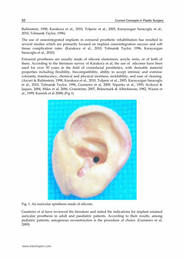
Extraoral prostheses are usually made of silicone elastomers, acrylic resin, or of both of these. According to the literature survey of Karakoca et al, the use of silicones have been used for over 50 years in the field of craniofacial prosthetics, with desirable material properties including flexibility, biocompatibility, ability to accept intrinsic and extrinsic colorants, translucency, chemical and physical inertness, moldability, and ease of cleaning. (Arcuri & Rubinstein, 1998, Karakoca et al., 2010, Toljanic et al., 2005, Karayazgan Saracoglu et al., 2010, Tolman& Taylor, 1996, Gumieiro et al, 2009, Niparko et al., 1993, Scolozzi & Jaques, 2004, Miles et al, 2006, Granström, 2007, Brånemark & Albrektsson, 1982, Wazen et al., 1999, Kamish et al 2008) (Fig 1)
Fig. 1. An auricular episthesis made of silicone.
Gumieiro et al have reviewed the literature and stated the indications for implant retained auricular prosthesis in adult and paediatric patients. According to their results, among pediatric patients, autogenous reconstruction is the procedure of choice. (Gumieiro et al, 2009)
www.intechopen.com
Implant Retained Auricular Prostheses
53
Adult patients Paediatric patients
The presence of an acquired total or subtotal auricular defect, most often traumatic or ablative in origin
When plastic surgery is impossible or when the final cosmetic result is unsatisfactory
Failed autogenous reconstruction
Severe soft-tissue/skeletal hypoplasia
A low or unfavorable hairline
Lack of adequate tissue for reconstruction
Severe congenital or acquired microtia
Absence of the lower half of the ear
Failed attempts at reconstruction
Major cancer excision
Poor operative risks
Selection of the technique by the patient
Table 2. Indications for implant retained auricular prosthesis in adult and paediatric patients. (Adopted from Gumieiro et al., 2009)
2.2 Retentive system
Although the concept of osseointegration is the same whether implants are placed
intraorally or extraorally, craniofacial implants should have modified design features to
match the anatomical and biomechanical differences in the facial area. (Kamish et al 2008)
Compared with the maxilla and the mandible, in spite of having limited thickness, facial
bones are dense. The load and frequency of loading forces on craniofacial implants are
limited when compared to implants placed intraorally. In the literature, some researchers
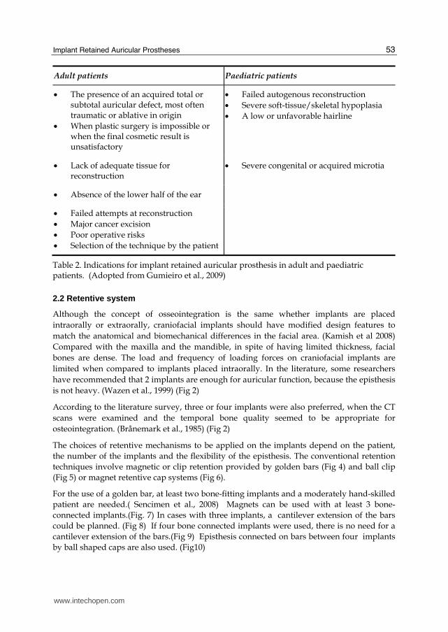
have recommended that 2 implants are enough for auricular function, because the episthesis
is not heavy. (Wazen et al., 1999) (Fig 2)
According to the literature survey, three or four implants were also preferred, when the CT
scans were examined and the temporal bone quality seemed to be appropriate for
osteointegration. (Brånemark et al., 1985) (Fig 2)
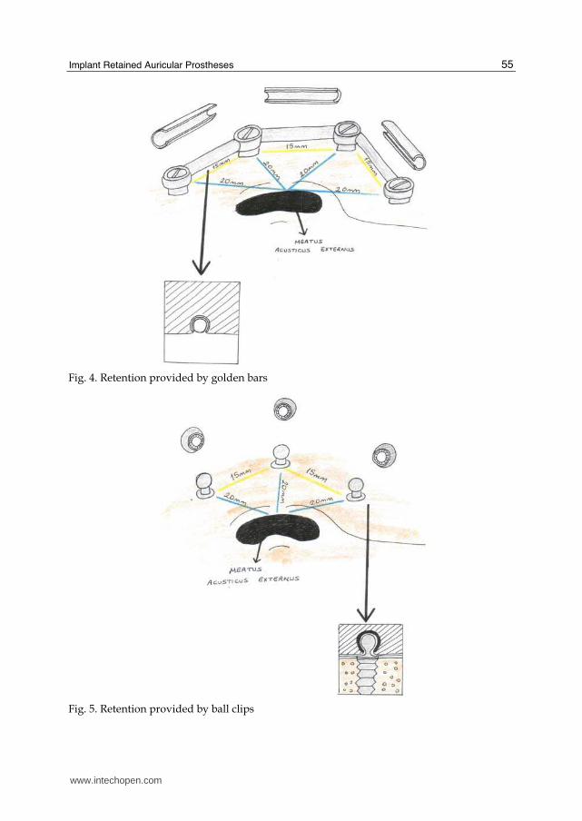
The choices of retentive mechanisms to be applied on the implants depend on the patient,
the number of the implants and the flexibility of the episthesis. The conventional retention
techniques involve magnetic or clip retention provided by golden bars (Fig 4) and ball clip
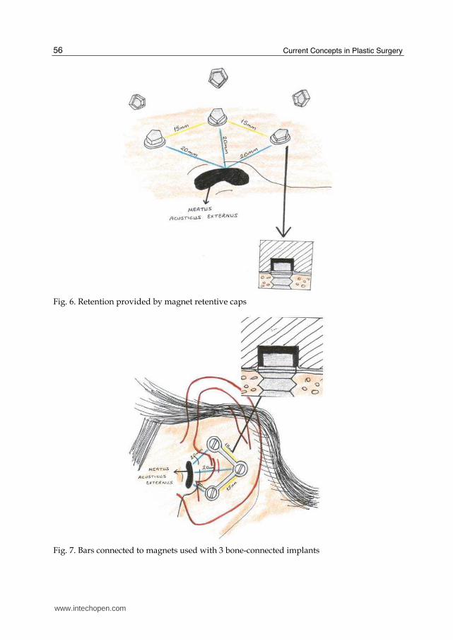
(Fig 5) or magnet retentive cap systems (Fig 6).
For the use of a golden bar, at least two bone-fitting implants and a moderately hand-skilled
patient are needed.( Sencimen et al., 2008) Magnets can be used with at least 3 bone-
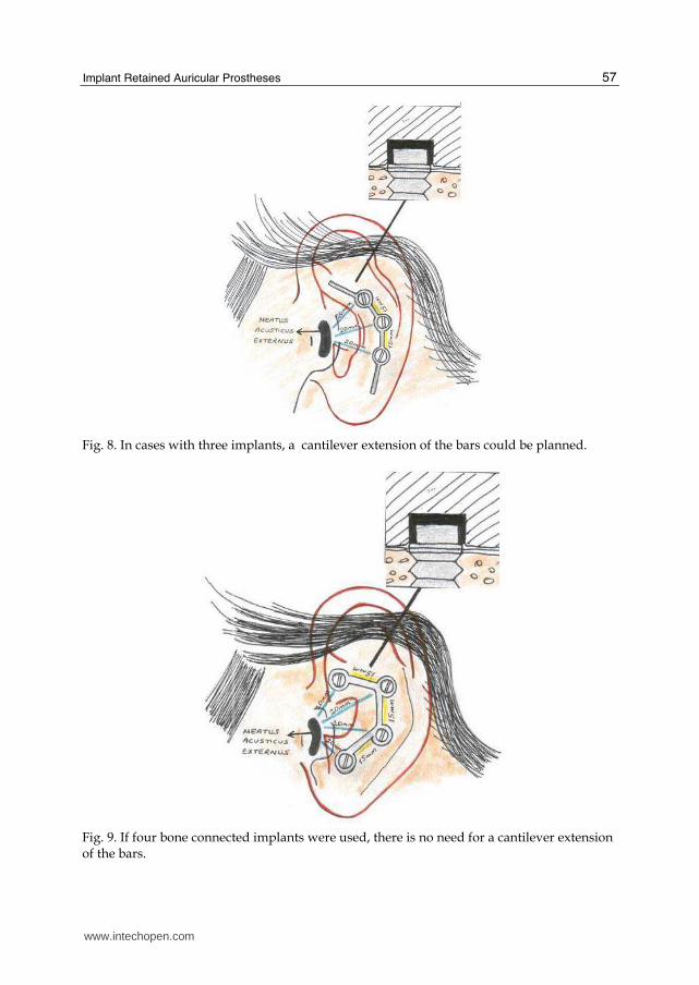
connected implants.(Fig. 7) In cases with three implants, a cantilever extension of the bars
could be planned. (Fig 8) If four bone connected implants were used, there is no need for a
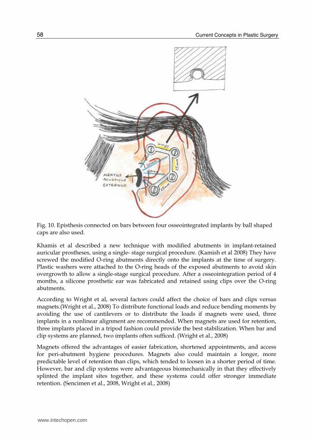
cantilever extension of the bars.(Fig 9) Episthesis connected on bars between four implants
by ball shaped caps are also used. (Fig10)
www.intechopen.com
Current Concepts in Plastic Surgery
54
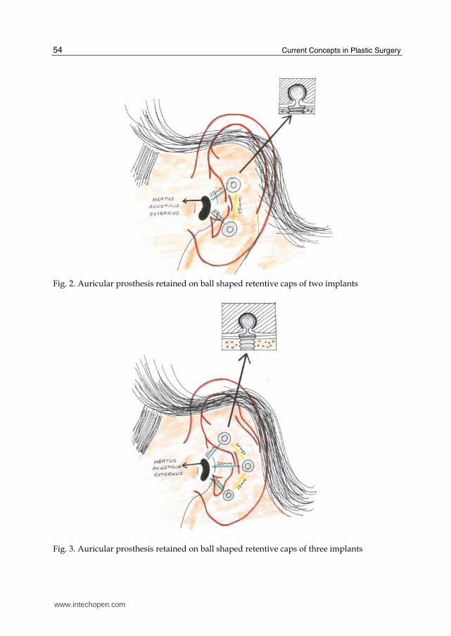
Fig. 2. Auricular prosthesis retained on ball shaped retentive caps of two implants
Fig. 3. Auricular prosthesis retained on ball shaped retentive caps of three implants
www.intechopen.com
Implant Retained Auricular Prostheses
55
Fig. 4. Retention provided by golden bars
Fig. 5. Retention provided by ball clips
www.intechopen.com
Current Concepts in Plastic Surgery
56
Fig. 6. Retention provided by magnet retentive caps
Fig. 7. Bars connected to magnets used with 3 bone-connected implants
www.intechopen.com
Implant Retained Auricular Prostheses
57
Fig. 8. In cases with three implants, a cantilever extension of the bars could be planned.
Fig. 9. If four bone connected implants were used, there is no need for a cantilever extension of the bars.
www.intechopen.com
Current Concepts in Plastic Surgery
58
Fig. 10. Episthesis connected on bars between four osseointegrated implants by ball shaped caps are also used.
Khamis et al described a new technique with modified abutments in implant-retained auricular prostheses, using a single- stage surgical procedure. (Kamish et al 2008) They have screwed the modified O-ring abutments directly onto the implants at the time of surgery. Plastic washers were attached to the O-ring heads of the exposed abutments to avoid skin overgrowth to allow a single-stage surgical procedure. After a osseointegration period of 4 months, a silicone prosthetic ear was fabricated and retained using clips over the O-ring abutments.
According to Wright et al, several factors could affect the choice of bars and clips versus magnets.(Wright et al., 2008) To distribute functional loads and reduce bending moments by avoiding the use of cantilevers or to distribute the loads if magnets were used, three implants in a nonlinear alignment are recommended. When magnets are used for retention, three implants placed in a tripod fashion could provide the best stabilization. When bar and clip systems are planned, two implants often sufficed. (Wright et al., 2008)
Magnets offered the advantages of easier fabrication, shortened appointments, and access for peri-abutment hygiene procedures. Magnets also could maintain a longer, more predictable level of retention than clips, which tended to loosen in a shorter period of time. However, bar and clip systems were advantageous biomechanically in that they effectively splinted the implant sites together, and these systems could offer stronger immediate retention. (Sencimen et al., 2008, Wright et al., 2008)
www.intechopen.com
Implant Retained Auricular Prostheses
59
2.3 Planning
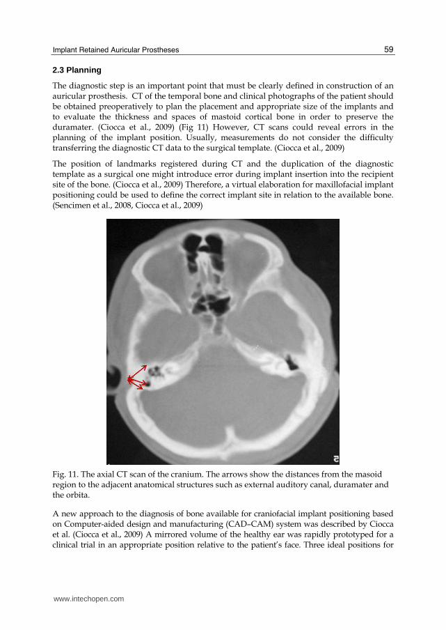
The diagnostic step is an important point that must be clearly defined in construction of an auricular prosthesis. CT of the temporal bone and clinical photographs of the patient should be obtained preoperatively to plan the placement and appropriate size of the implants and to evaluate the thickness and spaces of mastoid cortical bone in order to preserve the duramater. (Ciocca et al., 2009) (Fig 11) However, CT scans could reveal errors in the planning of the implant position. Usually, measurements do not consider the difficulty transferring the diagnostic CT data to the surgical template. (Ciocca et al., 2009)
The position of landmarks registered during CT and the duplication of the diagnostic template as a surgical one might introduce error during implant insertion into the recipient site of the bone. (Ciocca et al., 2009) Therefore, a virtual elaboration for maxillofacial implant positioning could be used to define the correct implant site in relation to the available bone. (Sencimen et al., 2008, Ciocca et al., 2009)
Fig. 11. The axial CT scan of the cranium. The arrows show the distances from the masoid region to the adjacent anatomical structures such as external auditory canal, duramater and the orbita.
A new approach to the diagnosis of bone available for craniofacial implant positioning based on Computer-aided design and manufacturing (CAD–CAM) system was described by Ciocca et al. (Ciocca et al., 2009) A mirrored volume of the healthy ear was rapidly prototyped for a clinical trial in an appropriate position relative to the patient’s face. Three ideal positions for
www.intechopen.com
Current Concepts in Plastic Surgery
60
the implant were chosen in the inner of the volume of the mirrored ear. The same positions were transferred to a diagnostic template that was rapidly prototyped with a positioning arm extending to the zygomatic arch, and two craniofacial implants were correctly positioned in the temporal bone. Ciocca et al have stated that this protocol allows the correct diagnosis of the available bone and perfect transfer in the surgical environment. In addition, the use of CAD–CAM technology allowed visualization in a virtual environment that was previously elab orated on film, and allowed to prototype the final volume of the prosthesis and its consequent surgical template with a 3D printer. This feature assured perfect transfer of the projected and CT-registered implant positions to the surgical template. (Ciocca et al., 2009)
In cases with aetiology of cancer, the surgeon should be aware of the risk of osseointegration failures and such patients who have undergone irradiation should be treated with caution, because differences in volume and density could result in irradiation having a more destructive effect on the vascularity of this site, thereby compromising the potential for osseointegration. (Gumieiro et al, 2009)Basically, the adverse biological changes that occur when osseous tissues are exposed to ionizing radiation results from alterations in the cellular components of bone, involving significant reductions in the numbers of viable osteoblasts and osteocytes, as well as the development of areas of fatty degeneration within the bone marrow spaces. In addition, regional ischemia could also be seen as results of the blood vessels undergo progressive endarteritis, hyalinization and fibrosis. As a conclusion, radiotherapy is not a contraindication for the use of osseointegrated implants in the maxillofacial region, but the loss of implants is higher in irradiated sites than in non-irradiated sites. (Gumieiro et al, 2009)
2.4 Surgical technique
It has been suggested that the mastoid region as a recipient site could offer the best results in implant retained auricular epistheses. Wright et al have stated that, the mastoid region in nonirradiated patients has provided a high degree of predictable individual implant survival. (Wright et al., 2008)
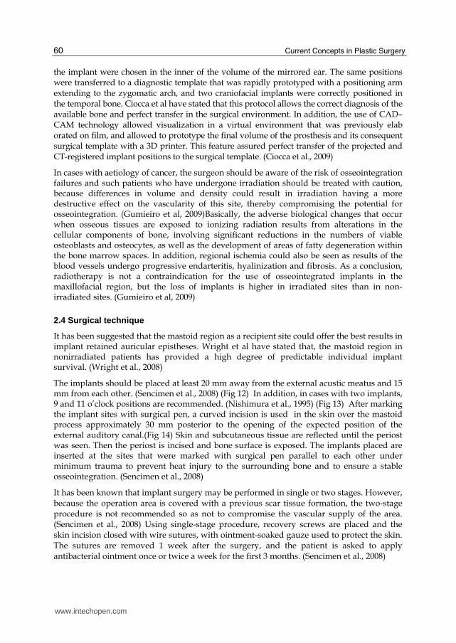
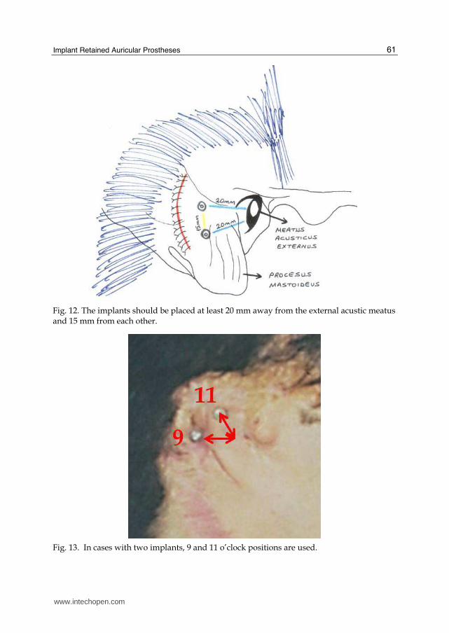
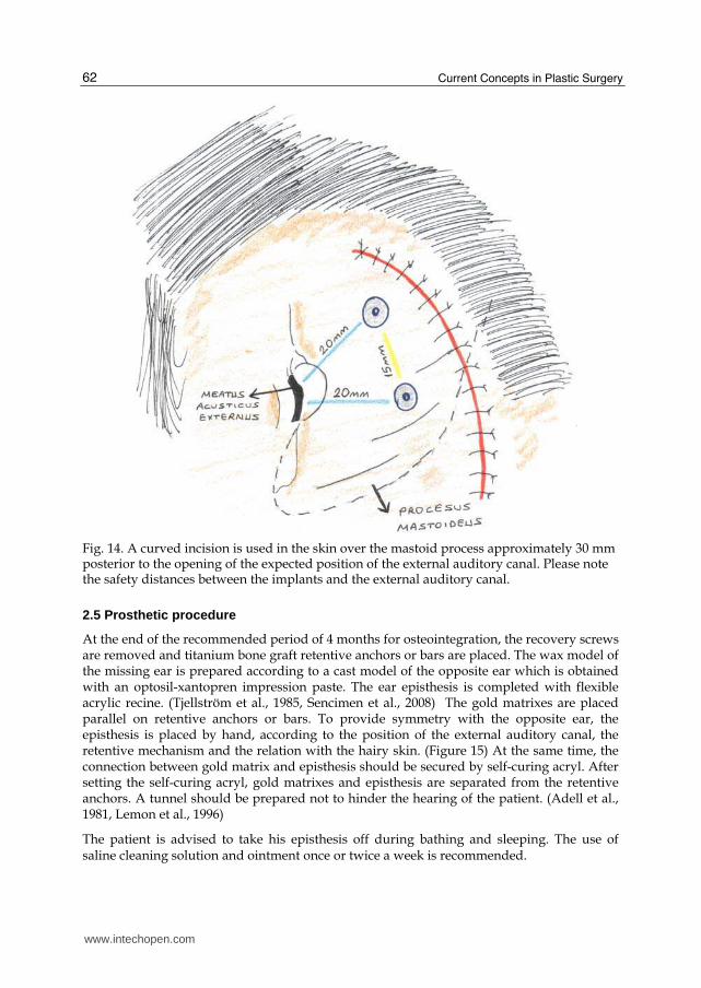
The implants should be placed at least 20 mm away from the external acustic meatus and 15 mm from each other. (Sencimen et al., 2008) (Fig 12) In addition, in cases with two implants, 9 and 11 o’clock positions are recommended. (Nishimura et al., 1995) (Fig 13) After marking the implant sites with surgical pen, a curved incision is used in the skin over the mastoid process approximately 30 mm posterior to the opening of the expected position of the external auditory canal.(Fig 14) Skin and subcutaneous tissue are reflected until the periost was seen. Then the periost is incised and bone surface is exposed. The implants placed are inserted at the sites that were marked with surgical pen parallel to each other under minimum trauma to prevent heat injury to the surrounding bone and to ensure a stable osseointegration. (Sencimen et al., 2008)
It has been known that implant surgery may be performed in single or two stages. However, because the operation area is covered with a previous scar tissue formation, the two-stage procedure is not recommended so as not to compromise the vascular supply of the area. (Sencimen et al., 2008) Using single-stage procedure, recovery screws are placed and the skin incision closed with wire sutures, with ointment-soaked gauze used to protect the skin. The sutures are removed 1 week after the surgery, and the patient is asked to apply antibacterial ointment once or twice a week for the first 3 months. (Sencimen et al., 2008)
www.intechopen.com
Implant Retained Auricular Prostheses
61
Fig. 12. The implants should be placed at least 20 mm away from the external acustic meatus and 15 mm from each other.
Fig. 13. In cases with two implants, 9 and 11 o’clock positions are used.
www.intechopen.com
Current Concepts in Plastic Surgery
62
Fig. 14. A curved incision is used in the skin over the mastoid process approximately 30 mm posterior to the opening of the expected position of the external auditory canal. Please note the safety distances between the implants and the external auditory canal.
2.5 Prosthetic procedure
At the end of the recommended period of 4 months for osteointegration, the recovery screws are removed and titanium bone graft retentive anchors or bars are placed. The wax model of the missing ear is prepared according to a cast model of the opposite ear which is obtained with an optosil-xantopren impression paste. The ear episthesis is completed with flexible acrylic recine. (Tjellström et al., 1985, Sencimen et al., 2008) The gold matrixes are placed parallel on retentive anchors or bars. To provide symmetry with the opposite ear, the episthesis is placed by hand, according to the position of the external auditory canal, the retentive mechanism and the relation with the hairy skin. (Figure 15) At the same time, the connection between gold matrix and episthesis should be secured by self-curing acryl. After setting the self-curing acryl, gold matrixes and episthesis are separated from the retentive anchors. A tunnel should be prepared not to hinder the hearing of the patient. (Adell et al., 1981, Lemon et al., 1996)
The patient is advised to take his episthesis off during bathing and sleeping. The use of saline cleaning solution and ointment once or twice a week is recommended.
www.intechopen.com
Implant Retained Auricular Prostheses
63
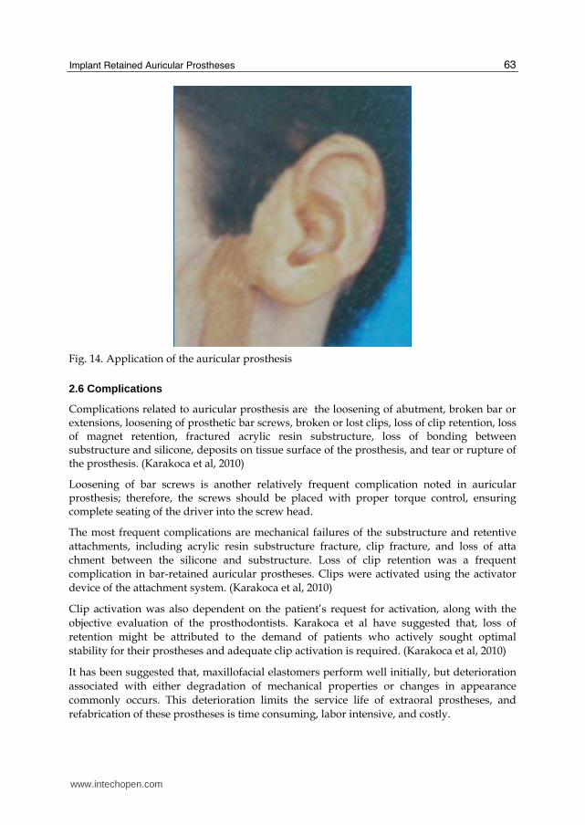
Fig. 14. Application of the auricular prosthesis
2.6 Complications
Complications related to auricular prosthesis are the loosening of abutment, broken bar or extensions, loosening of prosthetic bar screws, broken or lost clips, loss of clip retention, loss of magnet retention, fractured acrylic resin substructure, loss of bonding between substructure and silicone, deposits on tissue surface of the prosthesis, and tear or rupture of the prosthesis. (Karakoca et al, 2010)
Loosening of bar screws is another relatively frequent complication noted in auricular prosthesis; therefore, the screws should be placed with proper torque control, ensuring complete seating of the driver into the screw head.
The most frequent complications are mechanical failures of the substructure and retentive
attachments, including acrylic resin substructure fracture, clip fracture, and loss of atta
chment between the silicone and substructure. Loss of clip retention was a frequent
complication in bar-retained auricular prostheses. Clips were activated using the activator
device of the attachment system. (Karakoca et al, 2010)
Clip activation was also dependent on the patient’s request for activation, along with the
objective evaluation of the prosthodontists. Karakoca et al have suggested that, loss of
retention might be attributed to the demand of patients who actively sought optimal
stability for their prostheses and adequate clip activation is required. (Karakoca et al, 2010)
It has been suggested that, maxillofacial elastomers perform well initially, but deterioration
associated with either degradation of mechanical properties or changes in appearance
commonly occurs. This deterioration limits the service life of extraoral prostheses, and
refabrication of these prostheses is time consuming, labor intensive, and costly.
www.intechopen.com
Current Concepts in Plastic Surgery
64
According to the literature survey of Gumieiro et al, auricular osseointegrated implants have presented survival rates varying according to the length of follow-up, ranging from 92% after 8 years to 100% with shorter follow-up. (Gumieiro et al, 2009) However, there have been limited clinical studies on the life span of extraoral prostheses. (Aydin et al., 2008, Jebreil, 1980) Two studies reported on the life span of adhesive retained prostheses.(Jani & Schaaf, 1978, Jebreil, 1980) Jani and Schaaf indicated that 36% of prostheses were refabricated within 6 months, 33.6% within 7 to 12 months, 17.6% within 13 to 18 months, 8% within 19 to 24 months, and 4.8% were refabricated after 24 months.( Jani & Schaaf, 1978) Jebreil reported that adhesive-retained orbital prostheses were refabricated after 6-9 months. (Jebreil, 1980)
Wright et al have reported on the survival rate of 16 patients treated with extraoral implants in the auricular region and encountered no surgical complication, implant failures, or prosthetic failures; however, the follow-up visits were scheduled at 1 week, 6 months, and 1year. Hooper et al reported a 14-month mean life span for implant-retained extraoral prostheses. A study performed by Aydin et al. demonstrated a 17-month mean survival time for implant-retained auricular prostheses. (Aydin et al, 2008) In a recent study performed by Visser et al, it was indicated that a new prosthesis had to be made every 1.5 to 2 years. (Visser et al, 2008, Karakoca et al, 2010)
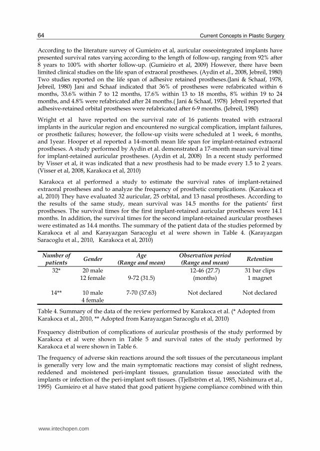
Karakoca et al performed a study to estimate the survival rates of implant-retained extraoral prostheses and to analyze the frequency of prosthetic complications. (Karakoca et al, 2010) They have evaluated 32 auricular, 25 orbital, and 13 nasal prostheses. According to the results of the same study, mean survival was 14.5 months for the patients’ first prostheses. The survival times for the first implant-retained auricular prostheses were 14.1 months. In addition, the survival times for the second implant-retained auricular prostheses were estimated as 14.4 months. The summary of the patient data of the studies peformed by Karakoca et al and Karayazgan Saracoglu et al were shown in Table 4. (Karayazgan Saracoglu et al., 2010, Karakoca et al, 2010)
Number of patients
Gender Age
(Range and mean) Observation period (Range and mean)
Retention
32*
14**
20 male 12 female
10 male 4 female
9-72 (31.5)
7-70 (37.63)
12-46 (27.7) (months)
Not declared
31 bar clips 1 magnet
Not declared
Table 4. Summary of the data of the review performed by Karakoca et al. (* Adopted from Karakoca et al., 2010, ** Adopted from Karayazgan Saracoglu et al, 2010)
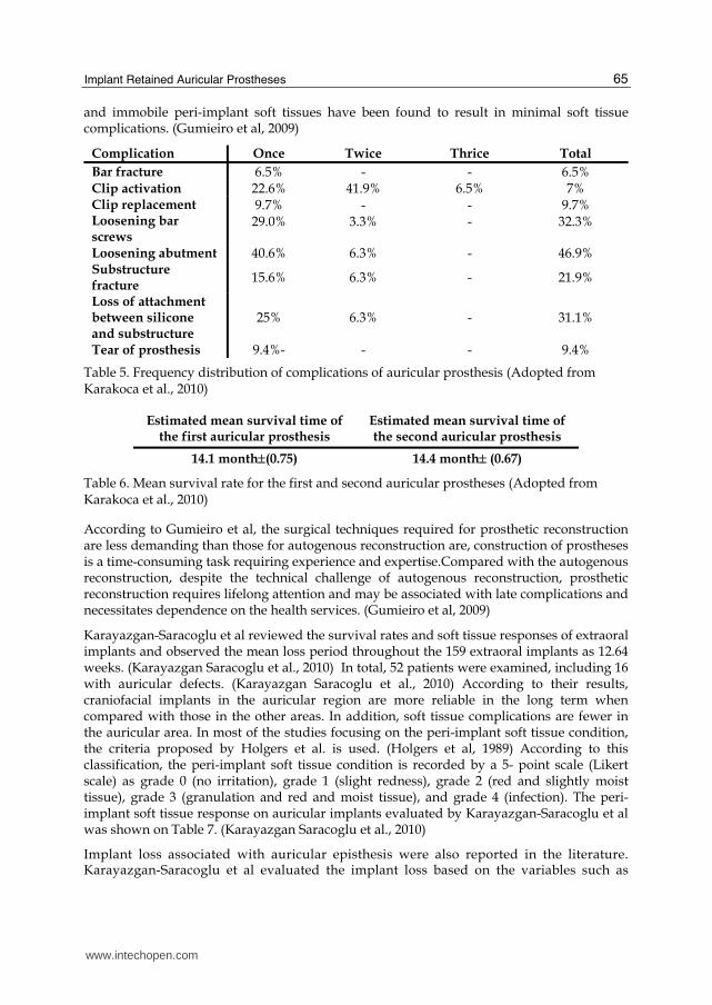
Frequency distribution of complications of auricular prosthesis of the study performed by Karakoca et al were shown in Table 5 and survival rates of the study performed by Karakoca et al were shown in Table 6.
The frequency of adverse skin reactions around the soft tissues of the percutaneous implant is generally very low and the main symptomatic reactions may consist of slight redness, reddened and moistened peri-implant tissues, granulation tissue associated with the implants or infection of the peri-implant soft tissues. (Tjellström et al, 1985, Nishimura et al., 1995) Gumieiro et al have stated that good patient hygiene compliance combined with thin
www.intechopen.com
Implant Retained Auricular Prostheses
65
and immobile peri-implant soft tissues have been found to result in minimal soft tissue complications. (Gumieiro et al, 2009)
Complication Once Twice Thrice Total
Bar fracture 6.5% - - 6.5% Clip activation 22.6% 41.9% 6.5% 7% Clip replacement Loosening bar screws
9.7% 29.0%
- 3.3%
- -
9.7% 32.3%
Loosening abutment 40.6% 6.3% - 46.9% Substructure fracture
15.6% 6.3% - 21.9%
Loss of attachment between silicone and substructure
25% 6.3% - 31.1%
Tear of prosthesis 9.4%- - - 9.4%
Table 5. Frequency distribution of complications of auricular prosthesis (Adopted from Karakoca et al., 2010)
Estimated mean survival time of the first auricular prosthesis
Estimated mean survival time of the second auricular prosthesis
14.1 month(0.75) 14.4 month (0.67)
Table 6. Mean survival rate for the first and second auricular prostheses (Adopted from Karakoca et al., 2010)
According to Gumieiro et al, the surgical techniques required for prosthetic reconstruction are less demanding than those for autogenous reconstruction are, construction of prostheses is a time-consuming task requiring experience and expertise.Compared with the autogenous reconstruction, despite the technical challenge of autogenous reconstruction, prosthetic reconstruction requires lifelong attention and may be associated with late complications and necessitates dependence on the health services. (Gumieiro et al, 2009)
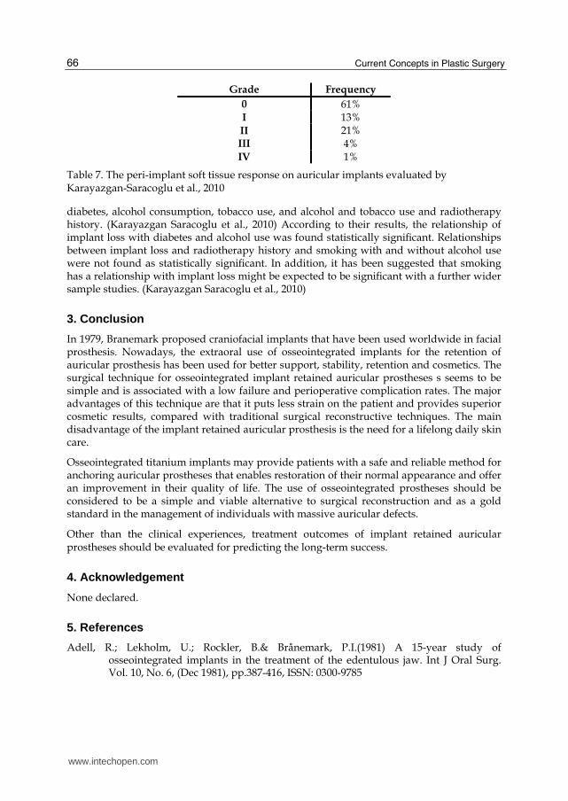
Karayazgan-Saracoglu et al reviewed the survival rates and soft tissue responses of extraoral implants and observed the mean loss period throughout the 159 extraoral implants as 12.64 weeks. (Karayazgan Saracoglu et al., 2010) In total, 52 patients were examined, including 16 with auricular defects. (Karayazgan Saracoglu et al., 2010) According to their results, craniofacial implants in the auricular region are more reliable in the long term when compared with those in the other areas. In addition, soft tissue complications are fewer in the auricular area. In most of the studies focusing on the peri-implant soft tissue condition, the criteria proposed by Holgers et al. is used. (Holgers et al, 1989) According to this classification, the peri-implant soft tissue condition is recorded by a 5- point scale (Likert scale) as grade 0 (no irritation), grade 1 (slight redness), grade 2 (red and slightly moist tissue), grade 3 (granulation and red and moist tissue), and grade 4 (infection). The peri-implant soft tissue response on auricular implants evaluated by Karayazgan-Saracoglu et al was shown on Table 7. (Karayazgan Saracoglu et al., 2010)
Implant loss associated with auricular episthesis were also reported in the literature. Karayazgan-Saracoglu et al evaluated the implant loss based on the variables such as
www.intechopen.com
Current Concepts in Plastic Surgery
66
Grade Frequency
0 61% I 13% II III
21% 4%
IV 1%
Table 7. The peri-implant soft tissue response on auricular implants evaluated by Karayazgan-Saracoglu et al., 2010
diabetes, alcohol consumption, tobacco use, and alcohol and tobacco use and radiotherapy history. (Karayazgan Saracoglu et al., 2010) According to their results, the relationship of implant loss with diabetes and alcohol use was found statistically significant. Relationships between implant loss and radiotherapy history and smoking with and without alcohol use were not found as statistically significant. In addition, it has been suggested that smoking has a relationship with implant loss might be expected to be significant with a further wider sample studies. (Karayazgan Saracoglu et al., 2010)
3. Conclusion
In 1979, Branemark proposed craniofacial implants that have been used worldwide in facial prosthesis. Nowadays, the extraoral use of osseointegrated implants for the retention of auricular prosthesis has been used for better support, stability, retention and cosmetics. The surgical technique for osseointegrated implant retained auricular prostheses s seems to be simple and is associated with a low failure and perioperative complication rates. The major advantages of this technique are that it puts less strain on the patient and provides superior cosmetic results, compared with traditional surgical reconstructive techniques. The main disadvantage of the implant retained auricular prosthesis is the need for a lifelong daily skin care.
Osseointegrated titanium implants may provide patients with a safe and reliable method for anchoring auricular prostheses that enables restoration of their normal appearance and offer an improvement in their quality of life. The use of osseointegrated prostheses should be considered to be a simple and viable alternative to surgical reconstruction and as a gold standard in the management of individuals with massive auricular defects.
Other than the clinical experiences, treatment outcomes of implant retained auricular prostheses should be evaluated for predicting the long-term success.
4. Acknowledgement
None declared.
5. References
Adell, R.; Lekholm, U.; Rockler, B.& Brånemark, P.I.(1981) A 15-year study of osseointegrated implants in the treatment of the edentulous jaw. Int J Oral Surg. Vol. 10, No. 6, (Dec 1981), pp.387-416, ISSN: 0300-9785
www.intechopen.com
Implant Retained Auricular Prostheses
67
Arcuri, M.R.& Rubenstein, J.T.(1998) Facial implants. Dent Clin North Am. Vol. 42, No.1, (Jan 1998), pp. 161-175, ISSN: 1558-0512
Aydin, C.; Karakoca, S.; Yilmaz, H.& Yilmaz, C.(2008) Implant-retained auricular prostheses: an assessment of implant success and prosthetic complications. Int J Prosthodont. Vol. 21, No. 3, (May-Jun 2008), pp.241-244, ISSN: 1942-4426
Brånemark, P.I., & Albrektsson, T.(1982)Titanium implants permanently penetrating human skin. Scand J Plast Reconstr Surg, Vol. 16, No. 1, (June 2008), pp. 17-21, ISSN: 0036-5556
Brånemark,, P.I.; Zarb, G.A.,;& Albrentsson, T. (1985) Tissue-iıntegrated pros theses osseointegration in clinical dentistry. Chicago, London, Berlin, Sao Paulo, Tokyo, Quintessence Publishing, 1985, ISBN: 10: 0867151293
Ciocca, L.; Mingucci, R.; Bacci, G.& Scotti, R.(2009) CAD-CAM construction of an auricular template for craniofacial implant positioning: a novel approach to diagnosis. Eur J Radiol. Vol 71, No. 2, (Aug 2009), pp. :253-256, ISSN: 1872-7727
Cordeiro, C.N.; McCarthy, C.M.; Mastorakos, D.P.& Cordeiro, P.G.(2007) Repair of postauricular defects using cervical donor skin: a novel use of the bilobed flap. Ann Plast Surg, Vol. 59, No. 4, (Oct 2007), pp. 451-452, ISSN: 1536-3708
Goldberg, L.H.; Mauldin, D.V.& Humphreys, T.R. (1996) The postauricular cutaneous advancement flap for repairing ear rim defects. Dermatol Surg Vol 22, No. 1, (Jan 1996), pp. 28-31. ISSN:1087-2108
Granström, G.(2007) Craniofacial osseointegration. Oral Dis. Vol. 13, No. 3.,(May 2007), pp. 261-269, ISSN: 1601-0825
Gumieiro, E.H.; Dib, L.L.; Jahn, R.S.; Santos Junior, J.F.; Nannmark, U.; Granström, G.& Abrahão, M.(2009) Bone-anchored titanium implants for auricular rehabilitation: case report and review of literature. Sao Paulo Med J. Vol. 127, No. 3, (2009), pp. 160-165, ISSN: 1806-9460
Holgers, K.M.; Bjursten, L.M.; Thomsen, P.; Ericson, L.E.& Tjellström, A.(1989) Experience with percutaneous titanium implants in the head and neck: a clinical and histological study. J Invest Surg. Vol. 2, No. 1, (1989), pp.7-16, ISSN: 1521-0553a
Hooper, S.M.; Westcott, T.; Evans, P.L.; Bocca, A.P.& Jagger, D.C.(2005)Implant-supported facial prostheses provided by a maxillofacial unit in a U.K. regional hospital: longevity and patient opinions. J Prosthodont. Vol. 14, No. 1,(Mar 2005), pp.32-38, ISSN: 1532-849X
Jani, R.M.& Schaaf, N.G.(1978) An evaluation of facial prostheses. J Prosthet Dent. Vol. 39, No. 5, (May 1978), pp.546-550, ISSN: 1097-6841
Jebreil, K.(1980) Accetability of orbital prostheses. J Prosthet Dent. Vol. 43, No. 1, (Jan 1980), pp.82-85, ISSN: 1097-6841
Justiniano, H. & Eisen, D,B.(2009) Pearls for perfecting the mastoid interpolation flap. Dermatology Online Journal, Vol.15, No. 6, (June 2009),pp. 2. ISSN:1087-2108
Karakoca, S.; Aydin, C.; Yilmaz, H.& Bal, B.T. (2010) Retrospective study of treatment outcomes with implant-retained extraoral prostheses: survival rates and prosthetic complications. J Prosthet Dent. Vol. 103, No. 2, (Feb 2010), pp. 118-126, ISSN: 1097-6841
Karayazgan-Saracoglu, B.; Zulfikar, H.; Atay, A.& Gunay, Y.(2010) Treatment outcome of extraoral implants in the craniofacial region. J Craniofac Surg. Vol. 21, No. 3, (May 2010), pp.751-758.
www.intechopen.com
Current Concepts in Plastic Surgery
68
Khamis, M.M.; Medra, A.& Gauld, J.(2008) Clinical evaluation of a newly designed single-stage craniofacial implant: a pilot study. J Prosthet Dent. Vol. 100, No. 5, (Nov 2008), pp. 375-383, ISSN: 1097-6841
Lemon, J.C.; Chambers, M.S.; Wesley, P.J.& Martin, J.W.(1996) Technique for fabricating a mirror-image prosthetic ear. J Prosthet Dent.Vol. 75, No. 3, (Mar 1996), pp.292-293, ISSN: 1097-6841
Miles, B.A.; Sinn, D.P.& Gion, G.G.(2006) Experience with cranial implant-based prosthetic reconstruction. J Craniofac Surg. Vol. 17, No. 5, (Sep 2006), pp. 889-897, ISSN: 1536-3732
Niparko, J.K.; Langman, A.W.; Cutler, D.S.& Carroll, W.R.(1993), Tissue-integrated prostheses in the rehabilitation of auricular defects: results with percutaneous mastoid implants. Am J Otol.Vol. 14, No. 4, (Jul 1993), pp. 343-348, ISSN: 0192-9763
Nishimura, R.D.; Roumanas, E.; Sugai, T.& Moy, P.K.(1995) Auricular prostheses and osseointegrated implants: UCLA experience. J Prosthet Dent. Vol. 73, No. 6, (Jun 1995), pp. 553-558, ISSN: 1097-6841
Scolozzi,P.& Jaques, B. (2004)Treatment of midfacial defects using prostheses supported by ITI dental implants. Plast Reconstr Surg. Vol. 114, No.6, (Nov 2004), pp. 1395-404, ISSN: 1529-4242
Sencimen, M.; Bal, H.E.; Demiroğullari, M.; Kocaoglu, M.& Dogan, N.(2008) Auricular episthesis retained by an attachment system (2 case reports). Oral Surg Oral Med Oral Pathol Oral Radiol Endod. Vol. 105, No. 2, (Feb 2008),pp. e28-34, ISSN: 1528-395X
Tjellström, A.; Yontchev, E.; Lindström, J.& Brånemark, P.I.(1985) Five years' experience with bone-anchored auricular prostheses. Otolaryngol Head Neck Surg. Vol. 93, No. 3, (Jun 1985), pp.366-372, ISSN: 1097-6817
Toljanic, J.A.; Eckert, S.E.; Roumanas, E.; Beumer, J.; Huryn, J.M.; Zlotolow, I.M.; Reisberg, D.J.; Habakuk, S.W.; Wright, R.F.; Rubenstein, J.E.; Schneid, T.R.; Mullasseril, P.; Garcia, L.T.; Bedard, J.F.& Choi, Y.G. (2005) Osseointegrated craniofacial implants in the rehabilitation of orbital defects: an update of a retrospective experience in the United States. J Prosthet Dent. Vol. 94, No. 2, (Aug 2005), pp. 177-182, ISSN: 1097-6841
Tolman, D.E.& Taylor, P.F. Bone-anchored craniofacial prosthesis study: irradiated patients. Int J Oral Maxillofac Implants. Vol. 11, No. 5, (Sep-Oct 1996), pp. 612-619, ISSN: 1942-4434
Tolman, D.E.& Taylor, P.F. Bone-anchored craniofacial prosthesis study. Int J Oral Maxillofac Implants.Vol. 11, No. 2, (Mar-Apr 1996), pp. 159-168. ISSN: 1942-4434
Vergilis-Kalner, I.J.& Goldberg, L.H. (2010) Bilobed flap for reconstruction of defects of the helical rim and posterior ear.Dermatol Online J.Vol.10, No. 15, (Oct 2010), pp. 9, ISSN:1087-2108
Visser, A.; Raghoebar, G.M.; van Oort, R.P.& Vissink, A.(2008) Fate of implant-retained craniofacial prostheses: life span and aftercare. Int J Oral Maxillofac Implants. Vol. 23, No. 1,(Jan-Feb 2008),pp.89-98, ISSN: 1942-4434
Wazen, J.J.; Wright, R.; Hatfield, R.B.& Asher, E.S.(1999) Auricular rehabilitation with bone-anchored titanium implants. Laryngoscope.Vol. 109, No. 4, (Apr 1999), pp. 523-527,ISSN: 1531-4995
Wright, R.F.; Zemnick, C.; Wazen, J.J. & Asher, E. (2008) Osseointegrated implants and auricular defects: a case series study. J Prosthodont., Vol. 17, No. 6, (August 2008), pp. 468-475. ISSN: 1532-849X
Yanaga, H.; Imai, K.; Fujimoto, T.& Yanaga, K. (2009) Generating ears from cultured autologous auricular chondrocytes by using two-stage implantation in treatment of microtia. Plast Reconstr Surg. Vol.124, No. 3, (Sep 2009), pp.817-825, ISSN:0032-105
www.intechopen.com
Current Concepts in Plastic SurgeryEdited by Dr. Frank Agullo
ISBN 978-953-51-0398-1Hard cover, 264 pagesPublisher InTechPublished online 23, March, 2012Published in print edition March, 2012
InTech EuropeUniversity Campus STeP Ri Slavka Krautzeka 83/A 51000 Rijeka, Croatia Phone: +385 (51) 770 447 Fax: +385 (51) 686 166www.intechopen.com
InTech ChinaUnit 405, Office Block, Hotel Equatorial Shanghai No.65, Yan An Road (West), Shanghai, 200040, China
Phone: +86-21-62489820 Fax: +86-21-62489821
Plastic surgery continues to be a rapidly growing field in medicine. There have been multiple recentadvancements in the field. Specifically, there has been a continuously growing interest in fat grafting, bodycontouring, minimally invasive surgery, and plastic surgery education. At the same time, there have beencontinued advances and modifications in surgical techniques, which translate into better and improved resultsfor our patients while increasing safety and efficacy. The title of the book is Current Concepts in PlasticSurgery and, as such, it highlights some of the "hot topics" in recent years. We have invited renownedspecialists from around the world to share their valued expertise and experience. Most of the chapters willexpose the reader to multiple techniques for achieving desired results, with emphasis on the author's preferredmethodology.
How to referenceIn order to correctly reference this scholarly work, feel free to copy and paste the following:
Metin Sencimen and Aydin Gulses (2012). Implant Retained Auricular Prostheses, Current Concepts in PlasticSurgery, Dr. Frank Agullo (Ed.), ISBN: 978-953-51-0398-1, InTech, Available from:http://www.intechopen.com/books/current-concepts-in-plastic-surgery/implant-retained-auricular-prostheses


![fibrilacion auricular[1]](https://static.fdocuments.us/doc/165x107/577d2baa1a28ab4e1eab09b6/fibrilacion-auricular1.jpg)