Impairment in Delayed Nonmatching to Sample … in Delayed Nonmatching to Sample Following Lesions...
9
Impairment in Delayed Nonmatching to Sample Following Lesions of Dorsal Prefrontal Cortex Tara L. Moore Boston University School of Medicine Stephen P. Schettler University of California, Los Angeles Ronald J. Killiany Boston University School of Medicine Douglas L. Rosene and Mark B. Moss Boston University School of Medicine and Emory University The prefrontal cortex has been identified as essential for executive function, as well as for aspects of rule learning and recognition memory. As part of our studies to assess prefrontal cortical function in the monkey, we evaluated the effects of damage to the dorsal prefrontal cortex (DPFC) on the Category Set Shifting Task (CSST), a test of abstraction and set-shifting, and on the Delayed Nonmatching to Sample (DNMS) task, a benchmark test of rule learning and recognition memory. The DPFC lesions in this study included dorsolateral and dorsomedial aspects of the PFC. In a previous report, we published evidence of an impairment on the CSST as a consequence of DPFC lesions (Moore, Schettler, Killiany, Rosene, & Moss, 2009). Here we report that monkeys with lesions of the DPFC were also markedly impaired relative to controls on both the acquisition (rule learning) and performance (recognition memory) conditions of trial-unique DNMS. The presence and extent of the deficits that we observed were of some surprise and support the possibility that the dorsal prefrontal cortex plays a more direct role in learning and recognition memory than had been previously thought. Keywords: dorsal prefrontal cortex, delayed nonmatching to sample, recognition memory, rule learning, rhesus monkey The work of Jacobsen (1936) using nonhuman primates was among the first to implicate the prefrontal cortex (PFC) in cogni- tive function in an animal model. During the past 70 years, this work was extended to show that the prefrontal cortices subserve several aspects of cognitive function, including aspects of execu- tive functions such as abstraction, cognitive flexibility, category shifting (Pribram, Mishkin, Rosvold, & Kaplan, 1952; Mishkin & Weiskrantz, 1958; Butter, Mishkin, & Mirsky, 1968; Butters & Panyda, 1969; Pohl, 1973; Mishkin & Manning, 1978; Oscar- Berman, 1978; Dias, Robbins, & Roberts, 1996; Monchi, Petrides, Petre, Worsley, & Dagher, 2001; Buckley et al., 2009: Mansouri, Tanaka, & Buckley, 2009; Moore, Schettler, Killiany, Rosene, & Moss, 2009; Tsujimoto et al., 2011) as well as conditional asso- ciative learning and working memory (Passingham, 1985; Petrides, 1985a, 1985b; Bachevalier & Mishkin, 1986; Gaffan & Harrison, 1989; Kojima, Kojima, & Goldman-Rakic, 1982; Petrides, 1991; Levy & Goldman-Rakic, 1999, 2000). Though the PFC has been implicated strongly with executive function and working memory, evidence for a role in rule learning and various aspects of recognition memory have also been well established. (Goldman-Rakic, 1991; Rowe & Passingham, 2001; Sakai, Rowe, & Passingham, 2002; Passingham & Sakai, 2004; Passingham, Rowe, & Sakai, 2005; Warden & Miller, 2010). As part of our studies of prefrontal cortical function (see Moore et al., 2009), we assessed monkeys with lesions of the dorsal prefrontal cortex (DPFC) (including dorsolateral and dorsomedial aspects of the PFC, extending laterally from the ventral lip of sulcus principalis and to the dorsal lip of the cingulate sulcus) on the acquisition and delay conditions of trial-unique Delayed Non- matching to Sample (DNMS). The acquisition phase of the DNMS task serves as a test of rule learning since it requires the monkey to learn to choose a novel object when presented together with a familiar one that had been originally presented 10 seconds earlier. The delay phase of the DNMS task serves as a test of recognition memory since the delay between the presentation of the sample stimulus and its representation with the novel one is lengthened from 10 sec, to increasingly longer intervals. In the present study, the administration of the DNMS task was virtually identical to that This article was published Online First October 22, 2012. Tara L. Moore and Ronald J. Killiany, Department of Anatomy and Neurobiology and Department of Neurology, Boston University School of Medicine; Stephen P. Schettler, Department of Pathology and Laboratory Medicine, UCLA; Douglas L. Rosene, Department of Anatomy and Neu- robiology, Boston University School of Medicine and Yerkes National Primate Research Center, Emory University; Mark B. Moss, Department of Anatomy and Neurobiology and Department of Neurology, Boston Uni- versity School of Medicine; and Yerkes National Primate Research Center, Emory University. The research was supported by NIH Grant PO1-AG00001. We thank Jerry Edwards, Beverly Duryea-Steiger, and Dana Whitelaw for their valuable assistance with this project. Correspondence concerning this article should be addressed to Tara L. Moore, Department of Anatomy and Neurobiology, Boston University School of Medicine, 700 Albany Street, W701, Boston, MA 02118. E-mail: [email protected] Behavioral Neuroscience © 2012 American Psychological Association 2012, Vol. 126, No. 6, 772–780 0735-7044/12/$12.00 DOI: 10.1037/a0030493 772
Transcript of Impairment in Delayed Nonmatching to Sample … in Delayed Nonmatching to Sample Following Lesions...

Impairment in Delayed Nonmatching to Sample Following Lesions ofDorsal Prefrontal Cortex
Tara L. MooreBoston University School of Medicine
Stephen P. SchettlerUniversity of California, Los Angeles
Ronald J. KillianyBoston University School of Medicine
Douglas L. Rosene and Mark B. MossBoston University School of Medicine and Emory University
The prefrontal cortex has been identified as essential for executive function, as well as for aspects of rulelearning and recognition memory. As part of our studies to assess prefrontal cortical function in themonkey, we evaluated the effects of damage to the dorsal prefrontal cortex (DPFC) on the Category SetShifting Task (CSST), a test of abstraction and set-shifting, and on the Delayed Nonmatching to Sample(DNMS) task, a benchmark test of rule learning and recognition memory. The DPFC lesions in this studyincluded dorsolateral and dorsomedial aspects of the PFC. In a previous report, we published evidenceof an impairment on the CSST as a consequence of DPFC lesions (Moore, Schettler, Killiany, Rosene,& Moss, 2009). Here we report that monkeys with lesions of the DPFC were also markedly impairedrelative to controls on both the acquisition (rule learning) and performance (recognition memory)conditions of trial-unique DNMS. The presence and extent of the deficits that we observed were of somesurprise and support the possibility that the dorsal prefrontal cortex plays a more direct role in learningand recognition memory than had been previously thought.
Keywords: dorsal prefrontal cortex, delayed nonmatching to sample, recognition memory, rule learning,rhesus monkey
The work of Jacobsen (1936) using nonhuman primates wasamong the first to implicate the prefrontal cortex (PFC) in cogni-tive function in an animal model. During the past 70 years, thiswork was extended to show that the prefrontal cortices subserveseveral aspects of cognitive function, including aspects of execu-tive functions such as abstraction, cognitive flexibility, categoryshifting (Pribram, Mishkin, Rosvold, & Kaplan, 1952; Mishkin &Weiskrantz, 1958; Butter, Mishkin, & Mirsky, 1968; Butters &Panyda, 1969; Pohl, 1973; Mishkin & Manning, 1978; Oscar-Berman, 1978; Dias, Robbins, & Roberts, 1996; Monchi, Petrides,
Petre, Worsley, & Dagher, 2001; Buckley et al., 2009: Mansouri,Tanaka, & Buckley, 2009; Moore, Schettler, Killiany, Rosene, &Moss, 2009; Tsujimoto et al., 2011) as well as conditional asso-ciative learning and working memory (Passingham, 1985;Petrides, 1985a, 1985b; Bachevalier & Mishkin, 1986; Gaffan &Harrison, 1989; Kojima, Kojima, & Goldman-Rakic, 1982;Petrides, 1991; Levy & Goldman-Rakic, 1999, 2000). Though thePFC has been implicated strongly with executive function andworking memory, evidence for a role in rule learning and variousaspects of recognition memory have also been well established.(Goldman-Rakic, 1991; Rowe & Passingham, 2001; Sakai, Rowe,& Passingham, 2002; Passingham & Sakai, 2004; Passingham,Rowe, & Sakai, 2005; Warden & Miller, 2010).
As part of our studies of prefrontal cortical function (see Mooreet al., 2009), we assessed monkeys with lesions of the dorsalprefrontal cortex (DPFC) (including dorsolateral and dorsomedialaspects of the PFC, extending laterally from the ventral lip ofsulcus principalis and to the dorsal lip of the cingulate sulcus) onthe acquisition and delay conditions of trial-unique Delayed Non-matching to Sample (DNMS). The acquisition phase of the DNMStask serves as a test of rule learning since it requires the monkeyto learn to choose a novel object when presented together with afamiliar one that had been originally presented 10 seconds earlier.The delay phase of the DNMS task serves as a test of recognitionmemory since the delay between the presentation of the samplestimulus and its representation with the novel one is lengthenedfrom 10 sec, to increasingly longer intervals. In the present study,the administration of the DNMS task was virtually identical to that
This article was published Online First October 22, 2012.Tara L. Moore and Ronald J. Killiany, Department of Anatomy and
Neurobiology and Department of Neurology, Boston University School ofMedicine; Stephen P. Schettler, Department of Pathology and LaboratoryMedicine, UCLA; Douglas L. Rosene, Department of Anatomy and Neu-robiology, Boston University School of Medicine and Yerkes NationalPrimate Research Center, Emory University; Mark B. Moss, Department ofAnatomy and Neurobiology and Department of Neurology, Boston Uni-versity School of Medicine; and Yerkes National Primate Research Center,Emory University.
The research was supported by NIH Grant PO1-AG00001. We thankJerry Edwards, Beverly Duryea-Steiger, and Dana Whitelaw for theirvaluable assistance with this project.
Correspondence concerning this article should be addressed to Tara L.Moore, Department of Anatomy and Neurobiology, Boston UniversitySchool of Medicine, 700 Albany Street, W701, Boston, MA 02118. E-mail:[email protected]
Behavioral Neuroscience © 2012 American Psychological Association2012, Vol. 126, No. 6, 772–780 0735-7044/12/$12.00 DOI: 10.1037/a0030493
772

used in previous studies assessing damage to various regions of thePFC (Bachevalier & Mishkin, 1986; Kowalska, Bachevalier, &Mishkin, 1991; Levy & Goldman-Rakic, 1999). It is important tonote, however, that in all of these prior studies, monkeys weretrained on the acquisition phase of the DNMS task to a stringentlearning criterion prior to surgery, and then tested for retention ofthe task and performance on the delay phase after surgery. Incontrast, in our study monkeys were administered the DNMS taskfor the first time after surgery, and thus acquired the task in theabsence of a prefrontal cortex, a paradigm unlike the previousstudies where the monkeys first learned the task with an intactprefrontal cortex and were then tested for retention of the taskfollowing damage to the prefrontal cortex, producing a differentset of test conditions.
Method
The subjects were eight, behaviorally naive, young adult (5–10years of age), male rhesus monkeys (Macaca mulatta), weighingbetween 6.0 kg and 14.5 kg at the beginning of this study. All ofthe monkeys were obtained from a national primate researchfacility or breeding facility and had known birth dates and com-plete health records. Before entering the study, monkeys receivedmedical examinations that included serum chemistry, hematology,urine analysis, and fecal analysis. In addition, all monkeys under-went MRI to ensure there was no occult neurological damage andto provide a baseline for lesion reconstruction. Results of themedical exams and MRIs revealed that all monkeys were healthyat the time of the study. While on study, monkeys were individ-ually housed in colony rooms where they were in constant auditoryand visual range of other monkeys in the Laboratory AnimalScience Center (LASC) of Boston University School of Medicine.This facility is fully AAALAC approved and animal maintenanceand research were conducted in accordance with the guidelines ofthe National Institutes of Health and the Institute of LaboratoryAnimal Resources Guide for the Care and Use of LaboratoryAnimals (2011). All procedures were approved by the Institu-tional Animal Care and Use Committee of the Boston Univer-sity Medical Campus. Diet consisted of Purina Monkey Chow(Purina Mills, St. Louis, MO) supplemented by fruit with feedingtaking place once per day, immediately following behavioral test-ing. All monkeys were fed 12–15 biscuits per day based on theirweight. During testing, raisins or small pieces of apple were usedas rewards; as there were 20 trials per day, monkeys receivedapproximately 20–40 rewards each day. Water was availablecontinuously. The monkeys were housed under a 12-hr light�darkcycle with cycle changes occurring in a graded fashion over thecourse of an hour. Following a quarantine period and acclimationto the colony room, four monkeys were randomly assigned to thesurgical group for bilateral removal of the prefrontal cortex(PFC�1, 2, 3, 4), and the remaining four monkeys served asunoperated controls.
Surgical Procedures
Monkeys were sedated with ketamine hydrochloride (10 mg/kg)and cuff blood pressures and electrocardiograms were taken. Anintravenous line was established via the saphenous vein and slowinfusion of lactated Ringers solution was begun. A surgical level of
anesthesia was induced with intravenous sodium pentobarbital(approximately 25 mg/kg) in titrated doses to effect. The monkeyswere intubated and heart rate, respiration rate, and muscle tonuswere continuously monitored throughout surgery to ensure that adeep surgical level of anesthesia was maintained. Body tempera-ture was monitored and maintained with a heating pad.
After opening the skin and retracting fascia and muscle, a boneflap was opened bilaterally over the prefrontal cortex extendingapproximately 5 cm caudally from the frontal sinus and about 5 cmin width at its caudal margin. The dura was then opened and thecortical lesion accomplished in one stage by using a small glasspipette and subpial aspiration to separate the pia and its bloodvessels from the underlying superficial layer of the cortex. Thisresults in degeneration of the cortical gray matter without directdamage to underlying white matter tracks. As shown in Figures 1and 2, the intended lesion was targeted to include all of area 46 inthe banks and depths of the sulcus principalis as well as the moredorsally located area 9 beginning on the dorsal bank of sulcusprincipalis and continuing rostrally toward the frontal pole, firstbeneath the superior limb of the arcuate sulcus and to its tip andthen extending medially to the cingulate sulcus. The lesion alsotargeted the adjacent parts of area 8 on the rostral lip of the arcuatesulcus. Care was taken to avoid extending the lesion into the bankof cingulate sulcus. Upon completion of the lesion within theseboundaries, the dura was closed, the bone flap sutured back inplace and the incision closed in layers.
Figure 1. Lateral and medial view of the rhesus monkey brain. Theintended area of the lesion is outlined.
773DELAYED NONMATCHING AFTER PREFRONTAL LESIONS
At the conclusion of surgery, the monkeys were administered600,000 units of Bicillin-LA intramuscularly to guard againstinfection, extubated and maintained in an incubator until theyemerged from anesthesia. They were administered analgesic totreat postoperative pain (Banamine IM, 1.0 mg/kg). Analgesia wascontinued for 48 to 96 hours or longer if needed as determined bythe veterinary staff. One week after surgery, the skin sutures wereremoved.
MRI
Prior to behavioral testing, which began 3 to 4 weeks postop-eratively, all monkeys underwent a MRI scan in order to charac-terize the locus and extent of the lesion. For all MRI procedures,monkeys were anesthetized with a mixture of ketamine and xyla-zine and their head was stabilized using an MRI compatiblestereotactic machine. A coronal T1 weighted high-resolution ana-tomical scan (3-D SPGR with slice thickness of 1.5 mm) as wellas an axial T2 weighted scan were acquired on a GE 1.5 TeslaSigna scanner.
Behavioral Training
Preoperative familiarization. Preoperatively all monkeyswere initially familiarized with behavioral testing in a Wisconsin
General Testing Apparatus (WGTA). All monkeys were trainedonly to displace a single gray plaque placed pseudorandomly overone of the three food wells to obtain a reward. Raisins or smallpieces of apples were used as rewards during testing. Monkeyswere trained until they responded for 20 consecutive trials on twosuccessive days.
Postoperative testing. Though changes in overt behaviors canoccur following lesions to the PFC, throughout the postoperativeperiod we did not see any overt changes in the disposition orday-to-day behavior/demeanor of any of the monkeys in this study.The monkeys continually demonstrated efficient attention and didnot display evidence of abnormal disinhibition, perseveration, orhyperactivity.
Delayed nonmatching to sample (DNMS). Three to 4 weeksfollowing surgery, all monkeys began the acquisition phase of theDelayed Nonmatching to Sample task in the WGTA. The DNMStask assesses the subject’s ability to identify a novel from afamiliar stimulus and was administered in two parts. First, in thebasic task or acquisition phase, there is a short delay (10 seconds)between the sample and the nonmatch portion of each trial, themonkey over time and trials acquires the rule that was necessaryfor the successful completion of this task—that is, the “novel”stimulus is always rewarded. Once criterion was reached on thebasic task, (90% correct over 100 trials) the monkey was tested in
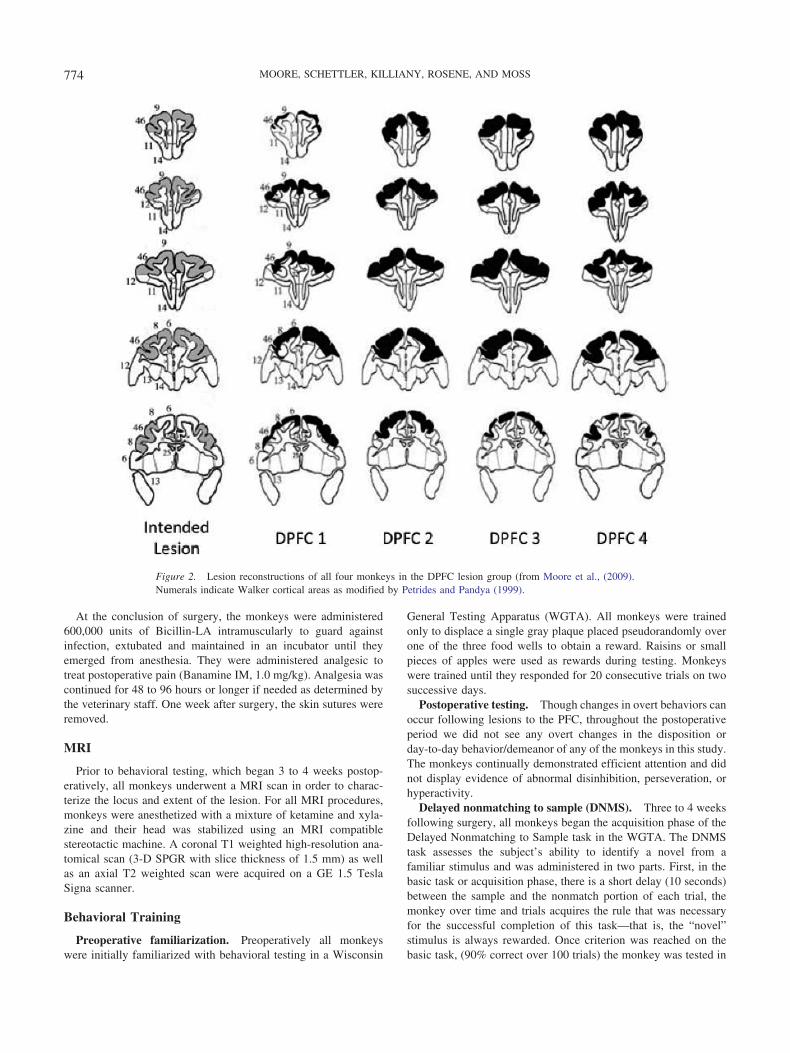
Figure 2. Lesion reconstructions of all four monkeys in the DPFC lesion group (from Moore et al., (2009).Numerals indicate Walker cortical areas as modified by Petrides and Pandya (1999).
774 MOORE, SCHETTLER, KILLIANY, ROSENE, AND MOSS

stages on two delay conditions with the intratrial interval set at twominute and then ten minute delays.
DNMS basic task. The trial begins with a sample objectpresented over the central baited food well. The monkey waspermitted to displace the object and obtain the reward. The doorwas then lowered, and the original, now familiar, sample objectwas placed over an unbaited lateral well and a new, novel, unfa-miliar object is placed over the other lateral well that was baited.Ten seconds after the original sample trial, this choice trial wasbegun and the monkey must choose the unfamiliar, novel object inorder to obtain the reward. Twenty seconds later, the next trial wasinitiated with a new, novel sample object presented over the baitedcentral well followed 10 seconds later by another recognition trialusing that second sample object and another new novel object. Theposition of the two objects on successive recognition trials wasvaried from left to right lateral wells in a predetermined pseudo-random order. A noncorrection procedure was used, and 20 trialsper day were given until the monkey reaches a learning criterion of90 correct responses in 100 consecutive trials or a maximum of1,500 trials. Objects were drawn from a pool of 600 “junk” objects,and paired so that in each daily session of 20 trials, 40 of theobjects were used. Once all of the initial pairings were used, (30days of 20 trials per day), the 600 objects were randomly recom-bined to produce new pairs so that the pairings presented continuedto be new and unique on each trial.
DNMS delays. Following administration of the basic task, the10-sec delay between the presentation of the sample object and therecognition trial was increased, in stages, first to two minute andthen to ten minute. Ten trials a day for 10 days at each delayinterval were given with the monkey remaining in the testingapparatus during the delay interval. A total of 100 trials were givenover 10 days at each of the two delays.
Perfusion and Lesion Reconstruction
Following completion of testing on the DNMS task, monkeyswere tested on other tasks that are reported elsewhere (Moore etal., 2009). After all testing was completed, monkeys were sedatedwith Ketamine (10 mg/kg) and were then given an overdose ofsodium pentobarbital and were killed by exsanguination duringtranscardial perfusion of the brain with 4% paraformaldehyde.Following perfusion, both hemispheres of the brain were blockedin situ in the coronal stereotactic plane for serial sectioning andtransferred to cryoprotectant solution to eliminate freezing artifact(Rosene, Roy, & Davis, 1986). The cryoprotected blocks werethen flash frozen and stored at �80 °C until they were cut on amicrotome into eight interrupted series of 30-�m-thick frozensections and one 60-�m-thick series. The 60-�m series and one30-�m series were immediately mounted on microscope slides,stained with thionin, and used to reconstruct the lesions.
For lesion reconstructions, each monkey’s preoperative T1weighted MRI scans were used with a standard rhesus monkeybrain template to create an individualized coronal section atlas ofthe frontal lobe from the arcuate sulcus to the rostral extent of thefrontal pole. Walker’s cytoarchotechtonic areas (as revised byBarbas & Pandya, 1989) were marked on each section of thisindividualized atlas. Area measurements (mm2) were determinedfor each of the cytoarchotechtonic areas on each section of theindividualized map using NIH Image J software.
To reconstruct the lesions, thionin-stained sections throughoutthe rostral/caudal extent of the lesion were superimposed onto theatlas sections and the extent of the lesion was marked. The bordersof lesions were then checked at higher power under the lightmicroscope and adjusted accordingly. Each section was thenscanned into the computer and NIH Image J software was used toobtain area measurements of the lesion. The percent of corticaltissue damaged for each cytoarchitectonic area was then calculatedby dividing each area of damage by the baseline area establishedon the preoperative MRI slices. These relative lesion sizes areshown in Figure 2 and the percent of tissue damaged is presentedin Table 1.
Data Analysis
Acquisition scores for the DNMS basic task, both trials anderrors to criterion, were analyzed separately with one-way analysesof variance. Performance scores for 2- and 10-min delays werebased on the percentage of correct responses for each delay con-dition. These scores were analyzed with a two-way, repeatedmeasures analysis of variance with group as a between-subjectsvariable and delay as a within-subjects variable.
Results
Lesion Reconstructions
The results of the lesion reconstructions are tabulated in Table 1and illustrated in drawings in Figure 2 and representative thioninsections from 1 monkey in Figure 3. These results show that, asintended, all monkeys had nearly complete damage to areas 46 and9 where damage ranged from a low of about 71% up to a high ofalmost 95% of each area. In addition there was significant damageto area 8, largely on the surface and rostral bank of the arcuatesuclus, and this ranged from about 44% up to almost 66%. Therewas also some encroachment of the lesion into areas 6, 10, 12where the damage ranged from as little as 4% up to 32%. Due tothe small group size in this study and the relative homogeneousperformance on the DNMS task, it was not possible to determineif the variability in extraneous damage in areas 6, 10, 12 wasrelated to individual impairment on the DNMS task.
DNMS Basic Task
Individual data for both trials and errors to criterion for theDNMS basic task are shown in Table 2. Monkeys in the control
Table 1Extent of Damage in Four Monkeys With Lesions of PrefrontalCortex by Cytoarchitectonic Areas
Monkey Area 46 Area 8 Area 9 Area 10 Area 12 Area 6
DPFC 1 70.8 65.73 79.27 4.03 18.30 31.63DPFC 2 89.92 53.64 85.54 20.13 17.26 32.29DPFC 3 87.47 45.70 84.30 24.35 3.18 31.55DPFC 4 94.33 43.92 90.34 30.29 2.99 22.18
Note. (From Moore, Schettler, Killiany, Moss, & Rosene, 2009.) Valuesrepresent percent of damaged tissue bilaterally based on comparison ofthionin-stained sections with preoperative MRI scans.
775DELAYED NONMATCHING AFTER PREFRONTAL LESIONS

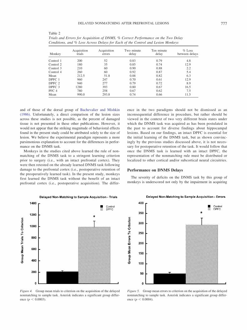
group learned the basic task within an average of 213 trials,whereas monkeys in the lesion group required an average of 990trials. Data for both trials and errors to criterion are shown inFigures 4 and 5. Separate one-way analyses of variance demon-
strated that there was a significant difference between the controland lesion groups on the number of trials [F(1, 6) � 53.70, p �.0003] and also in the number of errors [F(1, 6) � 50.16, p �.0004] to criterion on the postoperative acquisition of the DNMStask.
DNMS Delays
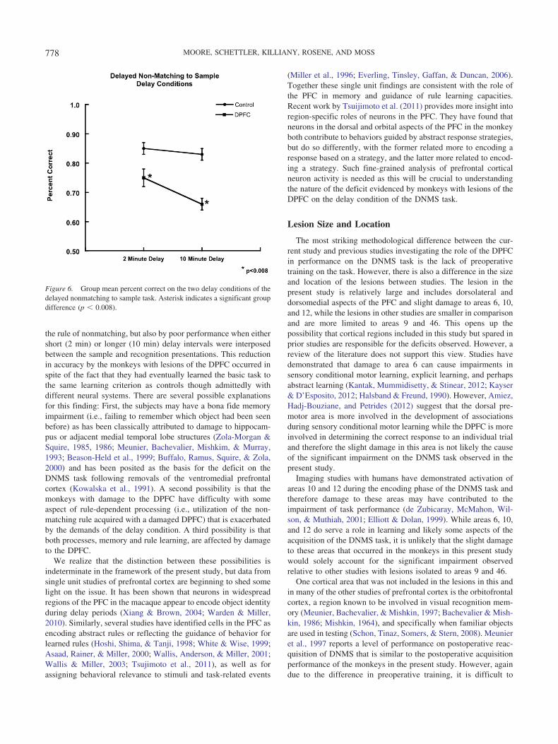
As shown in Table 2, monkeys in the control group performedat an 88% level of accuracy on the 2-min delay condition of theDNMS task, whereas those in the DPFC group performed at a 74%level of accuracy. On the 10-min delay, performance of the controlgroup dropped slightly to 82% accuracy, while that of the DPFCgroup declined to levels of only 66%. Separate two way repeatedmeasures analysis of variance revealed a significant overall effectof group [F(1, 16) � 15.43, p � .008], and delay [F(1, 16) � 29.4,p � .002] for the total percent correct for delay trials (see Figure6). There was no significant group-by-delay interaction [F(1, 6) �1.35, p � .289] for the total percent correct for delay trials.
Discussion
Two principal findings emerged from this study: 1) Relative tothe control group, monkeys with lesions of the DPFC were sig-nificantly impaired in the initial acquisition of the DNMS task (i.e.,learning to choose a novel from a familiar stimulus); and 2)Monkeys with lesions of the DPFC evidenced a degradation inperformance relative to controls when delays were increased be-tween the sample and recognition trials. These findings are dis-cussed in separate sections below.
Acquisition of Delayed Nonmatching to Sample
Monkeys with DPFC lesions required an average of 990 trialsto acquire the DNMS task, a level over four times greater thanthat of monkeys in the control group. In fact, this level ofimpairment is even greater than that observed in monkeys withselective hippocampal lesions who required an average of 650trials to acquire the DNMS task postoperatively (Beason-Held,Rosene, Killiany, & Moss, 1999). The severity of this effectwas striking, and somewhat surprising given previous findingsthat suggested that the locus for impairment of acquisition ofthe DNMS task is the more ventral, but not dorsal, aspects ofprefrontal cortex (Bachevalier & Mishkin, 1986; Kowalska etal., 1991). Bachevalier and Mishkin (1986) found that monkeyswith damage to the ventromedial prefrontal cortex, but not thedorsal prefrontal cortex, were markedly impaired in postoper-ative relearning of the basic DNMS task relative to controls.Kowalska et al. (1991), using the same experimental paradigmas that of Bachevalier and Mishkin (1986), found that monkeyswith lesions of the inferior convexity alone or in combinationwith the dorsal prefrontal cortex, evidenced a moderate degreeof impairment on the postoperative retention of the DNMS task.Finally, Levy and Goldman-Rakic (1999) showed that monkeyswith lesions restricted to either the dorsal prefrontal cortex(areas 46 and 8a) or the dorsomedial prefrontal cortex (areas 9and 8b), were unimpaired on the postoperative retention of theDNMS task.
The lesions in the present study were larger than those in themonkeys in the dorsal group of Levy and Goldman-Rakic (1999)
Figure 3. Representative thionin sections from one monkey in the study.Level of sections approximately match those shown in Figure 2.
776 MOORE, SCHETTLER, KILLIANY, ROSENE, AND MOSS
and of those of the dorsal group of Bachevalier and Mishkin(1986). Unfortunately, a direct comparison of the lesion sizesacross these studies is not possible, as the percent of damagedtissue is not presented in these other publications. However, itwould not appear that the striking magnitude of behavioral effectsfound in the present study could be attributed solely to the size oflesion. We believe the experimental paradigm represents a moreparsimonious explanation to account for the differences in perfor-mance on the DNMS task.
Monkeys in the studies cited above learned the rule of non-matching of the DNMS task to a stringent learning criterionprior to surgery (i.e., with an intact prefrontal cortex). Theywere then retested on the already learned DNMS task followingdamage to the prefrontal cortex (i.e., postoperative retention ofthe preoperatively learned task). In the present study, monkeysfirst learned the DNMS task without the benefit of an intactprefrontal cortex (i.e., postoperative acquisition). The differ-
ence in the two paradigms should not be dismissed as aninconsequential difference in procedure, but rather should beviewed in the context of two very different brain states underwhich the DNMS task was acquired as has been postulated inthe past to account for diverse findings about hippocampallesions. Based on our findings, an intact DPFC is essential forthe initial learning of the DNMS task, but as shown convinc-ingly by the previous studies discussed above, it is not neces-sary for postoperative retention of the task. It would follow thatonce the DNMS task is learned with an intact DPFC, therepresentation of the nonmatching rule must be distributed orlocalized to other cortical and/or subcortical neural circuitries.
Performance on DNMS Delays
The severity of deficits on the DNMS task by this group ofmonkeys is underscored not only by the impairment in acquiring
Figure 5. Group mean errors to criterion on the acquisition of the delayednonmatching to sample task. Asterisk indicates a significant group differ-ence (p � 0.0004).
Table 2Trials and Errors for Acquisition of DNMS, % Correct Performance on the Two DelayConditions, and % Loss Across Delays for Each of the Control and Lesion Monkeys
MonkeyAcquisition
trialsAcquisition
errorsTwo minute
delayTen minute
delay% Loss
between delays
Control 1 200 52 0.83 0.79 4.8Control 2 180 35 0.85 0.74 12.9Control 3 210 60 0.90 0.88 2.2Control 4 260 60 0.92 0.87 5.4Mean 212.5 51.8 0.88 0.82 6.3DPFC 1 960 247 0.70 0.61 12.9DPFC 2 940 277 0.79 0.72 8.9DPFC 3 1280 393 0.80 0.67 16.5PFC 4 780 258 0.67 0.62 7.5Mean 990.0 293.8 0.74 0.66 11.5
Figure 4. Group mean trials to criterion on the acquisition of the delayednonmatching to sample task. Asterisk indicates a significant group differ-ence (p � 0.0003).
777DELAYED NONMATCHING AFTER PREFRONTAL LESIONS
the rule of nonmatching, but also by poor performance when eithershort (2 min) or longer (10 min) delay intervals were interposedbetween the sample and recognition presentations. This reductionin accuracy by the monkeys with lesions of the DPFC occurred inspite of the fact that they had eventually learned the basic task tothe same learning criterion as controls though admittedly withdifferent neural systems. There are several possible explanationsfor this finding: First, the subjects may have a bona fide memoryimpairment (i.e., failing to remember which object had been seenbefore) as has been classically attributed to damage to hippocam-pus or adjacent medial temporal lobe structures (Zola-Morgan &Squire, 1985, 1986; Meunier, Bachevalier, Mishkim, & Murray,1993; Beason-Held et al., 1999; Buffalo, Ramus, Squire, & Zola,2000) and has been posited as the basis for the deficit on theDNMS task following removals of the ventromedial prefrontalcortex (Kowalska et al., 1991). A second possibility is that themonkeys with damage to the DPFC have difficulty with someaspect of rule-dependent processing (i.e., utilization of the non-matching rule acquired with a damaged DPFC) that is exacerbatedby the demands of the delay condition. A third possibility is thatboth processes, memory and rule learning, are affected by damageto the DPFC.
We realize that the distinction between these possibilities isindeterminate in the framework of the present study, but data fromsingle unit studies of prefrontal cortex are beginning to shed somelight on the issue. It has been shown that neurons in widespreadregions of the PFC in the macaque appear to encode object identityduring delay periods (Xiang & Brown, 2004; Warden & Miller,2010). Similarly, several studies have identified cells in the PFC asencoding abstract rules or reflecting the guidance of behavior forlearned rules (Hoshi, Shima, & Tanji, 1998; White & Wise, 1999;Asaad, Rainer, & Miller, 2000; Wallis, Anderson, & Miller, 2001;Wallis & Miller, 2003; Tsujimoto et al., 2011), as well as forassigning behavioral relevance to stimuli and task-related events
(Miller et al., 1996; Everling, Tinsley, Gaffan, & Duncan, 2006).Together these single unit findings are consistent with the role ofthe PFC in memory and guidance of rule learning capacities.Recent work by Tsuijimoto et al. (2011) provides more insight intoregion-specific roles of neurons in the PFC. They have found thatneurons in the dorsal and orbital aspects of the PFC in the monkeyboth contribute to behaviors guided by abstract response strategies,but do so differently, with the former related more to encoding aresponse based on a strategy, and the latter more related to encod-ing a strategy. Such fine-grained analysis of prefrontal corticalneuron activity is needed as this will be crucial to understandingthe nature of the deficit evidenced by monkeys with lesions of theDPFC on the delay condition of the DNMS task.
Lesion Size and Location
The most striking methodological difference between the cur-rent study and previous studies investigating the role of the DPFCin performance on the DNMS task is the lack of preoperativetraining on the task. However, there is also a difference in the sizeand location of the lesions between studies. The lesion in thepresent study is relatively large and includes dorsolateral anddorsomedial aspects of the PFC and slight damage to areas 6, 10,and 12, while the lesions in other studies are smaller in comparisonand are more limited to areas 9 and 46. This opens up thepossibility that cortical regions included in this study but spared inprior studies are responsible for the deficits observed. However, areview of the literature does not support this view. Studies havedemonstrated that damage to area 6 can cause impairments insensory conditional motor learning, explicit learning, and perhapsabstract learning (Kantak, Mummidisetty, & Stinear, 2012; Kayser& D’Esposito, 2012; Halsband & Freund, 1990). However, Amiez,Hadj-Bouziane, and Petrides (2012) suggest that the dorsal pre-motor area is more involved in the development of associationsduring sensory conditional motor learning while the DPFC is moreinvolved in determining the correct response to an individual trialand therefore the slight damage in this area is not likely the causeof the significant impairment on the DNMS task observed in thepresent study.
Imaging studies with humans have demonstrated activation ofareas 10 and 12 during the encoding phase of the DNMS task andtherefore damage to these areas may have contributed to theimpairment of task performance (de Zubicaray, McMahon, Wil-son, & Muthiah, 2001; Elliott & Dolan, 1999). While areas 6, 10,and 12 do serve a role in learning and likely some aspects of theacquisition of the DNMS task, it is unlikely that the slight damageto these areas that occurred in the monkeys in this present studywould solely account for the significant impairment observedrelative to other studies with lesions isolated to areas 9 and 46.
One cortical area that was not included in the lesions in this andin many of the other studies of prefrontal cortex is the orbitofrontalcortex, a region known to be involved in visual recognition mem-ory (Meunier, Bachevalier, & Mishkin, 1997; Bachevalier & Mish-kin, 1986; Mishkin, 1964), and specifically when familiar objectsare used in testing (Schon, Tinaz, Somers, & Stern, 2008). Meunieret al., 1997 reports a level of performance on postoperative reac-quisition of DNMS that is similar to the postoperative acquisitionperformance of the monkeys in the present study. However, againdue to the difference in preoperative training, it is difficult to
Figure 6. Group mean percent correct on the two delay conditions of thedelayed nonmatching to sample task. Asterisk indicates a significant groupdifference (p � 0.008).
778 MOORE, SCHETTLER, KILLIANY, ROSENE, AND MOSS

directly compare the effects of the lesions in different regions ofthe PFC on the DNMS task.
Conclusions
The main finding of this study was that monkeys with extensivedamage to the DPFC were impaired in the acquisition of theDNMS task, a deficit that has been typically reported followingdamage to the hippocampal formation when a postoperative ac-quisition paradigm is used (Zola-Morgan & Squire, 1986; Beason-Held et al., 1999). Together with recent findings in humans thatboth the prefrontal cortex and hippocampal formation are activatedin various visual recognition memory paradigms (e.g., Kirwan,Wixted, & Squire, 2008; Trivedi et al., 2008), it is tempting toposit that both structures may participate in mediating recognitionmemory, at least that required in performing the DNMS task. Evenmore intriguing is the possibility that the two regions not onlyparticipate in subserving these functions (see Corkin, 2001), butthat they do so in an interdependent fashion. Support for the notionof a functional relationship between the medial temporal lobe andprefrontal cortex has, in fact, begun to accumulate. Using postop-erative retention paradigms, Gaffan, Easton, and Parker (2002)demonstrated impairments in object associative learning followingcrossed lesions of frontal and inferotemporal cortices in the mon-key, and Bussey, Wise, and Murray (2002) has observed deficits inconditional visuomotor associations following crossed unilaterallesions of the orbital and ventral prefrontal cortices together withthe inferotemporal cortex in the monkey. The nature of the inter-action between the prefrontal cortices, and the medial temporallobe in memory and rule learning, await further delineation.
References
Amiez, C., Hadj-Bouziane, F., & Petrides, M. (2012). Response selectionversus feedback analysis in conditional visuo-motor learning. NeuroIm-age, 59, 3723–3735. doi:10.1016/j.neuroimage.2011.10.058
Asaad, W. F., Rainer, G., & Miller, E. K. (2000). Task-specific neuralactivity in the primate prefrontal cortex. Journal of Neurophysiology, 84,451–459.
Bachevalier, J., & Mishkin, M. (1986). Visual recognition impairmentfollows ventromedial but not dorsolateral prefrontal lesions in monkeys.Behavioural Brain Research, 20, 249 –261. doi:10.1016/0166-4328(86)90225-1
Barbas, H., & Pandya, D. N. (1989). Architecture and intrinsic connectionsof the prefrontal cortex in the rhesus monkey. The Journal of Compar-ative Neurology, 286, 353–375. PMID: 2768563.
Beason-Held, L. L., Rosene, D. L., Killiany, R. J., & Moss, M. B. (1999).Hippocampal formation lesions produce memory impairment in therhesus monkey. Hippocampus, 9, 562–574. doi:10.1002/(SICI)1098-1063(1999)9:5�562::AID-HIPO10�3.0.CO;2-X
Buckley, M. J., Mansouri, F. A., Hoda, H., Mahboubi, M., Browning,P. G., Kwok, S. C., . . . Tanaka, K. (2009). Dissociable components ofrule-guided behavior depend on distinct medial and prefrontal regions.Science, 325, 52–58. doi:10.1126/science.1172377
Buffalo, E. A., Ramus, S. J., Squire, L. R., & Zola, S. M. (2000).Perception and recognition memory in monkeys following lesion of areaTE and perirhnial cortex. Learning & Memory, 7, 375–382. doi:10.1101/lm.32100
Bussey, T. J., Wise, S. P., & Murray, E. A. (2002). Interaction of ventraland orbital prefrontal cortex with inferotemporal cortex in conditionalvisuomotor learning. Behavioral Neuroscience, 116, 703–715. doi:10.1037/0735-7044.116.4.703
Butter, C. M., Mishkin, M., & Mirsky, A. F. (1968). Emotional responsestoward humans in monkeys with selective frontal lesions. Physiology &Behavior, 3, 213–215. doi:10.1016/0031-9384(68)90087-5
Butters, N., & Pandya, D. (1969). Retention of delayed alteration: Effect ofselective lesions of sulcus principalis. Science, 165, 1271–1273. doi:10.1126/science.165.3899.1271
Corkin, S. (2001). Beware of frontal lobe deficits in hippocampal clothing.Trends in Cognitive Sciences, 5, 321–323. doi:10.1016/S1364-6613(00)01709-5
de Zubicaray, G. I., McMahon, K., Wilson, S. J., & Muthiah S. (2001).Brain activity during the encoding, retention, and retrieval of stimulusrepresentations. Learning & Memory, 8, 243–51. doi:10.1101/lm.40301
Dias, R., Robbins, T. W., & Roberts, A. C. (1996). Primate analogue of theWisconsin Card Sorting Test: Effects of excitotoxic lesions of theprefrontal cortex in the marmoset. Behavioral Neuroscience, 110, 872–886. doi:10.1037/0735-7044.110.5.872
Elliott, R., & Dolan R. J. (1999). Differential neural responses duringperformance of matching and nonmatching to sample tasks at two delayintervals. The Journal of Neuroscience, 19, 5066–5073.
Everling, S., Tinsley, C. J., Gaffan, D., & Duncan, J. (2006). Selectiverepresentation of task-relevant objects and locations in the monkeyprefrontal cortex. European Journal of Neuroscience, 23, 2197–2214.doi:10.1111/j.1460-9568.2006.04736.x
Gaffan, D., Easton, A., & Parker, A. (2002). Interaction of inferior tem-poral cortex with frontal cortex and basal forebrain: Double dissociationin strategy implementation and associative learning. The Journal ofNeuroscience, 22, 7288–7296.
Gaffan, D., & Harrison, S. (1989). A comparison of the effects of fornixtransection and sulcus principalis ablation upon spatial learning bymonkeys. Behavioural Brain Research, 31, 207–220. doi:10.1016/0166-4328(89)90003-X
Goldman-Rakic, P. S. (1991). Cellular and circuit basis of working mem-ory in prefrontal cortex of nonhuman primates. Progress in BrainResearch, 85, 325–336. doi:10.1016/S0079-6123(08)62688-6
Halsband, U., & Freund, H. J. (1990). Premotor cortex and conditionalmotor learning in man. Brain, 113, 207–222. doi:10.1093/brain/113.1.207
Hoshi, E., Shima, K., & Tanji, J. (1998). Task-dependent selectivity ofmovement-related neuronal activity in the primate prefrontal cortex.Journal of Neurophysiology, 80, 3392–3397.
Institute for Laboratory Animal Research, National Research Council.(2011). Guide for the care and use of laboratory animals. Washington,DC: National Academies Press.
Jacobsen, C. F. (1936). Studies of cerebral function in primates. I. Thefunctions of the frontal association areas in monkeys. ComparativePsychology Monographs, 13, 1–60.
Kantak, S. S., Mummidisetty, C. K., & Stinear, J. W. (2012). Primarymotor and premotor cortex in implicit sequence learning�evidence forcompetition between implicit and explicit human motor memory sys-tems. European Journal of Neuroscience, 36, 2710–2715. doi:10.1111/j.1460-9568.2012.08175.x
Kayser, A. S., & D’Esposito, M. (2012). Abstract rule learning: Thedifferential effects of lesions in frontal cortex. Cerebral Cortex. Ad-vance online publication. doi:10.1093/cercor/bhs013
Kirwan, C. B., Wixted, J. T., & Squire, L. R. (2008). Activity in the medialtemporal lobe predicts memory strength whereas activity in the prefron-tal cortex predicts recollection. The Journal of Neuroscience, 28, 10541–10548. doi:10.1523/JNEUROSCI.3456-08.2008
Kojima, S., Kojima, M., & Goldman-Rakic, P. S. (1982). Operant behav-ioral analysis of memory loss in monkeys with prefrontal lesions. BrainResearch, 248, 51–59. doi:10.1016/0006-8993(82)91146-5
Kowalska, D. M., Bachevalier, J., & Mishkin, M. (1991). The role of theinferior prefrontal convexity in performance of delayed nonmatching-
779DELAYED NONMATCHING AFTER PREFRONTAL LESIONS

to-sample. Neuropsychologia, 29, 583– 600. doi:10.1016/0028-3932(91)90012-W
Levy, R., & Goldman-Rakic, P. (1999). Association of storage and pro-cessing functions in the dorsolateral prefrontal cortex of the nonhumanprimate. The Journal of Neuroscience, 19, 5149–5158.
Levy, R., & Goldman-Rakic, P. S. (2000). Segregation of working memoryfunctions within the dorsolateral prefrontal cortex. Experimental BrainResearch, 133, 23–32. doi:10.1007/s002210000397
Mansouri, F. A., Tanaka, K., & Buckley, M. J. (2009). Conflict-inducedbehavioural adjustment: A clue to the executive functions of the pre-frontal cortex. Natore Reviews: Neuroscience, 10, 141–152.
Meunier, M., Bachevalier, J., Mishkin, M., & Murray, E. A. (1993). Effectson visual recognition of combined and separate ablations of the ento-rhinal and perirhinal cortex in rhesus monkeys. Journal of Neuroscience,13, 5418–5432.
Meunier, M., Bachevalier, J., & Mishkin, M. (1997). Effects of orbitalfrontal and anterior cingulate lesions on object and spatial memory inrhesus monkeys. Neuropsychologia, 35, 999–1015. doi:10.1016/S0028-3932(97)00027-4
Miller, E. K., Erickson, C. A., & Desimone, R. (1996). Neural mechanismsof visual working memory in prefrontal cortex of the macaque. TheJournal of Neuroscience, 16, 5154–5167.
Mishkin, M. (1964). Perseveration of central sets after frontal lesions inman. In J. M. Warren and K. Akert (Eds.), The frontal granular cortexand behavior (pp. 219–294). New York: McGraw-Hill.
Mishkin, M., & Manning, F. J. (1978). Non-spatial memory after selectiveprefrontal lesions in monkeys. Brain Research, 143, 313–323. doi:10.1016/0006-8993(78)90571-1
Mishkin, M., & Weiskrantz, L. (1958). Effects of delayed reward onvisual-discrimination performance in monkeys with frontal lesions.Journal of Comparative and Physiological Psychology, 51, 276–281.doi:10.1037/h0047280
Monchi, O., Petrides, M., Petre, V., Worsley, K., & Dagher, A. (2001).Wisconsin Card Sorting revisited: Distinct neural circuits participatin indifferent stages of the task identified by ebven-related magnetic reso-nance imaging. The Journal of Neuroscience, 21, 7733–7741,
Moore, T. L., Schettler, S. P., Killiany, R. J., Rosene, D. L., & Moss, M. B.(2009). Effects on executive function following damage to the prefrontalcortex in the rhesus monkey. Behavioral Neuroscience, 123, 231–41.
Oscar-Berman, M. (1978). The effects of dorsolateral-frontal andventrolateral-orbitofrontal lesions on nonspatial test performance. Neu-ropsychologia, 16, 259–267.
Passingham, R. E. (1985). Memory of monkeys (Macaca mulatta) withlesions in prefrontal cortex. Behavioral Neuroscience, 99, 3–21.
Passingham, R. E., Rowe, J. B., & Sakai, K. (2005). Prefrontal cortex andattention to action. In G. Humphreys & M. J. Riddoch (Eds.), Attentionin action (pp. 263–286). New York, NY: Psychology Press.
Passingham, R. E., & Sakai, K. (2004). The prefrontal cortex and workingmemory: Physiology and brain imaging. Current Opinion in Neurobi-ology, 14, 163–168.
Petrides, M. (1985a). Deficits in non-spatial conditional associative learn-ing after periarcuate lesions in the monkey. Behavioural Brain Research,16, 95–101.
Petrides, M. (1985b). Deficits on conditional association learning tasksafter frontal-and temporal-lobe lesions in man Neuropsychologia, 23,601–614.
Petrides, M. (1991). Functional specialization within the dorsolateral fron-tal cortex for serial order memory. Proceedings, Biological Sciences:The Royal Society, 246, 299–306.
Petrides, M., & Pandya, D. N. (1999). Dorsolateral prefrontal cortex:comparative cytoarchitectonic analysis in the human and the macaquebrain and corticocortical connection patterns. European Journal of Neu-roscience, 11, 1011–1036. PMID: 10103094.
Pohl, W. (1973). Dissociation of spatial discrimination deficits followingfrontal and parietal lesions in monkeys. Journal of Comparative andPhysiological Psychology, 82, 227–239.
Pribram, K. H., Mishkin, M., Rosvold, H. E., & Kaplan, S. J. (1952).Effects on delayed-response performance of lesions of dorsolateral andventromedial frontal cortex of baboon. Journal of Comparative andPhysiological Psychology, 45, 565–575.
Rosene, D. L., Roy, N. J., & Davis, B. J. (1986). A cryoprotection methodthat facilitates cutting frozen sections of whole monkey brains forhistological and histochemical processing without freezing artifact.Journal of Histochemistry and Cytochemistry. 34, 1301–1315. PMID:3745909.
Rowe, J., & Passingham, R. E. (2001). Working memory for location andtime: Activity in prefrontal area 46 relates to selection rather than tomaintenance in memory. NeuroImage, 14, 77–86.
Sakai, K., Rowe, J. B., & Passingham, R. E. (2002). Active maintenancecreates distractor-resistant memory: Critical role of sustained activity inprefrontal area 46. Nature Neuroscience, 5, 479–484.
Schon, K., Tinaz, S., Somers, D. C., & Stern, C. E. (2008). Delayed matchto object or place: An event-related fMRI study of short-term stimulusmaintenance and the role of stimulus pre-exposure. Neuroimage, 39,857–872.
Trivedi, M. A., Murphy, C. M., Goetz, C., Shah, R. C., Gabrieli, J. D.,Whitfield-Gabrieli, S., . . . Stebbins, G. T. (2008). fMRI activationchanges during successful episodic memory encoding and recognition inamnestic mild cognitive impairment relative to cognitively healthy olderadults. Dementia and Geriatric Cognitive Disorders, 26, 123–127.
Tsujimoto, S., Genovesio, A., & Wise, S. P. (2011). Comparison ofstrategy signals in the dorsolateral and orbital prefrontal cortex. TheJournal of Neuroscience, 31, 4583–4592.
Wallis, J. D., Anderson, K. C., & Miller, E. K. (2001). Single neurons inprefrontal cortex encode abstract rules. Nature, 411, 953–956.
Wallis, J. D., & Miller, E. K. (2003). From rule to response: Neuronalprocesses in the premotor and prefrontal cortex. Journal of Neurophys-iology, 90, 1790–1806.
Warden, M. R., & Miller, E. K. (2010). Task-dependent changes inshort-term memory in the prefrontal cortex. The Journal of Neurosci-ence, 30, 15802–15810.
White, I. M., & Wise, S. P. (1999). Rule-dependent neuronal activity in theprefrontal cortex. Experimental Brain Research, 126, 315–335.
Xiang, J. Z., & Brown, M. W. (2004). Neuronal responses related tolong-term recognition memory processes in prefrontal cortex. Neuron,42, 817–829.
Zola-Morgan, S., & Squire, L. R. (1985). Medial temporal lesions inmonkeys impair memory on a variety of tasks sensitive to humanamnesia, Behavioral Neuroscience, 99, 22–23.
Zola-Morgan, S., & Squire, L. R. (1986). Memory impairment in monkeysfollowing lesions limited to the hippocampus. Behavioral Neuroscience,100, 155–160.
Received June 2, 2012Revision received September 17, 2012
Accepted September 18, 2012 �
780 MOORE, SCHETTLER, KILLIANY, ROSENE, AND MOSS