Impact of Beta-Adrenergic Agonist Supplementation and Heat ...
154
University of Nebraska - Lincoln DigitalCommons@University of Nebraska - Lincoln eses and Dissertations in Animal Science Animal Science Department 7-2019 Impact of Beta-Adrenergic Agonist Supplementation and Heat Stress on the Microbiome and Gastrointestinal Transcriptome of Sheep Erin M. Duffy University of Nebraska-Lincoln, erinduff[email protected] Follow this and additional works at: hps://digitalcommons.unl.edu/animalscidiss Part of the Agriculture Commons , Animal Sciences Commons , Comparative and Laboratory Animal Medicine Commons , and the Veterinary Microbiology and Immunobiology Commons is Article is brought to you for free and open access by the Animal Science Department at DigitalCommons@University of Nebraska - Lincoln. It has been accepted for inclusion in eses and Dissertations in Animal Science by an authorized administrator of DigitalCommons@University of Nebraska - Lincoln. Duffy, Erin M., "Impact of Beta-Adrenergic Agonist Supplementation and Heat Stress on the Microbiome and Gastrointestinal Transcriptome of Sheep" (2019). eses and Dissertations in Animal Science. 186. hps://digitalcommons.unl.edu/animalscidiss/186
Transcript of Impact of Beta-Adrenergic Agonist Supplementation and Heat ...

University of Nebraska - LincolnDigitalCommons@University of Nebraska - Lincoln
Theses and Dissertations in Animal Science Animal Science Department
7-2019
Impact of Beta-Adrenergic AgonistSupplementation and Heat Stress on theMicrobiome and Gastrointestinal Transcriptome ofSheepErin M. DuffyUniversity of Nebraska-Lincoln, [email protected]
Follow this and additional works at: https://digitalcommons.unl.edu/animalscidiss
Part of the Agriculture Commons, Animal Sciences Commons, Comparative and LaboratoryAnimal Medicine Commons, and the Veterinary Microbiology and Immunobiology Commons
This Article is brought to you for free and open access by the Animal Science Department at DigitalCommons@University of Nebraska - Lincoln. It hasbeen accepted for inclusion in Theses and Dissertations in Animal Science by an authorized administrator of DigitalCommons@University of Nebraska- Lincoln.
Duffy, Erin M., "Impact of Beta-Adrenergic Agonist Supplementation and Heat Stress on the Microbiome and GastrointestinalTranscriptome of Sheep" (2019). Theses and Dissertations in Animal Science. 186.https://digitalcommons.unl.edu/animalscidiss/186

IMPACT OF BETA-ADRENERGIC AGONIST SUPPLEMENTATION AND HEAT
STRESS ON THE MICROBIOME AND GASTROINTESTINAL TRANSCRIPTOME
OF SHEEP
By
Erin Margaret Duffy
A THESIS
Presented to the Faculty of
The Graduate College at the University of Nebraska
In Partial Fulfillment of Requirements
For the Degree of Master of Science
Major: Animal Science
Under the Supervision of Professor Jessica Lynn Petersen
Lincoln, Nebraska
August, 2019

IMPACT OF BETA-ADRENERGIC AGONIST SUPPLEMENTATION AND HEAT
STRESS ON THE MICROBIOME AND GASTROINTESTINAL TRANSCRIPTOME
OF SHEEP
Erin Margaret Duffy, M.S.
University of Nebraska, 2019
Advisor: Jessica L. Petersen
Improving animal growth and efficiency are critical points of research as the world’s
population and demand for agriculture products increase. Therefore, adaptions or changes
in the gut are of interest to maximize growth efficiency and wellbeing of livestock. The
gastrointestinal tract of the rumen plays many critical roles with the assistance of the
associated microbial community. One way to improve animal performance is
supplementation of β-adrenergic agonists (β-AA) which are commonly fed to cattle
during the last 20-40 days of the finishing period, improving muscle growth by
decreasing adipose deposition and increasing muscle accretion. Two β-AA, Ractopamine
HCl (β1-AA) and Zilpaterol HCl (β2-AA) are currently approved for use in beef cattle in
the United States. Alternatively to the beneficial effects of β-AA, heat stress in livestock
decreases production efficiency and growth. Based on anecdotal reports it has been
suggested that supplementation of β-AA could potentially exacerbate the stress response
caused by hyperthermia leading to increased mortality. However, little research has been
conducted to investigate the interaction between these two factors. Therefore, the purpose
of these studies was to investigate the impact of heat stress, β-AA and the interaction
between the two with respect to the ruminant gastrointestinal tract. Microbial

communities were isolated from rumen and cecum contents and RNA was isolated from
rumen epithelium of lambs supplemented Zilpaterol HCl and Ractopamine HCl housed in
either an ambient or heat stressed environment for 21 d. Additionally RNA was isolated
from rumen epithelium of lambs supplemented Ractopamine HCl housed in either an
ambient or heat stressed environment for 30 d. No interaction was found between β-AA
and heat stress in either the microbial community or RNA studies. Heat stress and β-AA
supplementation changed the composition of particular taxa in the rumen microbiome,
while the addition of ammonium chloride to the second group significantly impacted the
cecal microbial composition. Additionally, heat stress, but not β-AA supplementation,
impacted the transcriptome profile of the rumen epithelium by upregulating the oxidative
stress response. Based on these results, we conclude that β-AA do not induce an
increased stress response within the ruminant gastrointestinal tract.

iv
Acknowledgements
I would first like to thank my major advisor, Dr. Jessica Petersen, for the
incredible opportunity and support. Under her guidance, I was able to pursue a project
that I was passionate about. Her constant encouragement and openness to all my new
ideas allowed me to grow academically and personally. I would also like to thank my
other committee members, Dr. Dustin Yates and Dr. Samodha Fernanado, for their
guidance and support throughout my master’s journey. Their input and expertise
strengthened my research and challenged my thinking, helping me to grow as a
researcher.
Conducting this research was also made possible by the assistance of
undergraduate student, Charlet Reebenaker, and laboratory technologists, Shauna Tietze
and Anna Fuller who dedicated their time to perform various protocols pertinent to the
outcome of this study. I would like to thank them along with my other lab mates, Rachel
Kubik, Lianna Walker, Kylee Sutton, and Hiruni Wijesena for all the support and
friendship along the way. I would also like to thank my parents Nadine and Bob, my
stepfather Brian, and my sister Emily for their encouragement and advice. Finally, I
would like to thank my partner Brandon for his unending love and support over the past
two years.

v
Grant Information
This project is based on research that was partially supported by the Nebraska
Agricultural Experiment Station with funding from the Hatch Multistate Research
capacity funding program (Accession Number 1011055) from the USDA National
Institute of Food and Agriculture.

vi
Table of Contents
Chapter I A Literature Review . . . . . . . . . . . . . . . . . . . . . . . . . . . . . . . . . . . . . . . . . . . . . . . . . . 1
The Ruminant Animal and its Associated Microbiome. . . . . . . . . .. . . . . . . . . . . . . . . . . . 1
Cecal Importance and Function in Ruminant Species. . . . . . . . . . . . . . . . . . . . . . . . . . . . .4
Ruminant Transcriptome. . . . . . . . . . . . . . . . . . . . . . . . . . . . . . . . . . . . . . . . . . . . . . . . . . .5
Heat Stress… . . . . . . . . . . . . . . . . . . . . . . . . . . . . . . . . . . . . . . . . . . . .. . . . . . . . . . . . . . . .6
Beta-Adrenergic Agonists. . . . . . . . . . . . . . . . . . . . . . . . . . . . . . . . . .. . . . . . . . . . . . . . . .10
Interaction between heat stress and beta-adrenergic agonist supplementation. . . . . . . . .18
Conclusion . . . . . . . . . . . . . . . . . . . . . . . . . . . . . . . . . . . . . . . . . . . . . . . . . . . . . . . . . . . ..19
References. . . . . . .. . . . . . . . . . . . . . . . . . . . . . . . . . . . . . . . . . . . . . . . . . . . . . . . . . . . . . .20
Figures. . . .. . . . . . . . . . . . . . . . . . . . . . . . . . . . . . . . . . . . . . . . . . . . . . . . . .. . . . . . . . . . . 33
Tables. . . .. . . . . . . . . . . . . . . . . . . . . . . . . . . . . . . . . . . . . . . . . . . . . . . . . . . . . . . . . . . . . 36
Chapter II Investigation of the rumen microbiome and volatile fatty acid production of lambs
supplemented with beta-adrenergic agonists and subjected to heat stress for 21 days . . . . . . . . . 37
Introduction . . . . . . . . . . . . . . . . . . . . . . . . . . . . . . . . . . . . . . . . . . . . . . . . . . . . . . . . . . . .37
Materials and Methods . . . . . . . . . . . . . . . . . . . . . . . . . . . . . . . . . . . . . . . . . . . . . . . . . . . 41
Results . . . . . . . . . . . . . . . . . . . . . . . . . . . . . . . . . . . . . . . . . . . . . . . . . . . . . . . . . . . . . . . .48
Discussion . . . . . . . . . . . . . . . . . . . . . . . . . . . . . . . . . . . . . . . . . . . . . . . . . . . . . . . . . . . . .51
Conclusion . . . . . . . . . . . . . . . . . . . . . . . . . . . . . . . . . . . . . . . . . . . . . . . . . . . . . . . . . . . ..57
References. . . . . . .. . . . . . . . . . . . . . . . . . . . . . . . . . . . . . . . . . . . . . . . . . . . . . . . . . . . . . .59
Figures. . . .. . . . . . . . . . . . . . . . . . . . . . . . . . . . . . . . . . . . . . . . . . . . . . . . . .. . . . . . . . . . . 68
Chapter III Characterization of the cecal microbial community in lambs supplemented with
beta adrenergic agonists and subjected to heat stress for 21 days . . . . . . . . . . . . . . . . . . . . . . . . .75
Introduction . . . . . . . . . . . . . . . . . . . . . . . . . . . . . . . . . . . . . . . . . . . . . . . . . . . . . . . . . . . .75

vii Materials and Methods . . . . . . . . . . . . . . . . . . . . . . . . . . . . . . . . . . . . . . . . . . . . . . . . . . . 78
Results . . . . . . . . . . . . . . . . . . . . . . . . . . . . . . . . . . . . . . . . . . . . . . . . . . . . . . . . . . . . . . . .85
Discussion . . . . . . . . . . . . . . . . . . . . . . . . . . . . . . . . . . . . . . . . . . . . . . . . . . . . . . . . . . . . .87
Conclusion . . . . . . . . . . . . . . . . . . . . . . . . . . . . . . . . . . . . . . . . . . . . . . . . . . . . . . . . . . . ..91
References. . . . . . .. . . . . . . . . . . . . . . . . . . . . . . . . . . . . . . . . . . . . . . . . . . . . . . . . . . . . . .93
Figures. . . .. . . . . . . . . . . . . . . . . . . . . . . . . . . . . . . . . . . . . . . . . . . . . . . . . .. . . . . . . . . . . 99
Tables. . . .. . . . . . . . . . . . . . . . . . . . . . . . . . . . . . . . . . . . . . . . . . . . . . . . . . . . . . . . . . . . 106
Chapter IV Investigation of the rumen epithelium transcriptome in lambs fed beta-adrenergic
agonists and subjected to heat stress . . . . . . . . . . . . . . . . . . . . . . . . . . . . . . . . . . . . . . . . . . . . . . 107
Introduction . . . . . . . . . . . . . . . . . . . . . . . . . . . . . . . . . . . . . . . . . . . . . . . . . . . . . . . . . . .107
Materials and Methods . . . . . . . . . . . . . . . . . . . . . . . . . . . . . . . . . . . . . . . . . . . . . . . . . . 109
Results . . . . . . . . . . . . . . . . . . . . . . . . . . . . . . . . . . . . . . . . . . . . . . . . . . . . . . . . . . . . . . .114
Discussion . . . . . . . . . . . . . . . . . . . . . . . . . . . . . . . . . . . . . . . . . . . . . . . . . . . . . . . . . . . .115
Conclusion . . . . . . . . . . . . . . . . . . . . . . . . . . . . . . . . . . . . . . . . . . . . . . . . . . . . . . . . . . . 122
References. . . . . . .. . . . . . . . . . . . . . . . . . . . . . . . . . . . . . . . . . . . . . . . . . . . . . . . . . . . . .124
Figures. . . .. . . . . . . . . . . . . . . . . . . . . . . . . . . . . . . . . . . . . . . . . . . . . . . . . .. . . . . . . . . . 130
Tables. . . .. . . . . . . . . . . . . . . . . . . . . . . . . . . . . . . . . . . . . . . . . . . . . . . . . . . . . . . . . . . . 133
Appendix A- Differentially expressed rumen epithelium transcripts in lambs fed beta-adrenergic
agonists and subjected to heat stress for 30 days. . . . . . . . . . . . . . . . . . . . . . . . . . . . . . . . . . . . . 137
Table . . . . . . . . . . . . . . . . . . . . . . . . . . . . . . . . . . . . . . . . . . . . . . . . . . . . . . . . . . . . . . . .137

1
CHAPTER I: A LITERATURE REVIEW
Introduction
Ruminant animals have served an important role in sustainable agriculture
systems by converting resources, such as rangeland, pasture, and crop residues, into
consumable products for humans. Ruminants’ ability to utilize foodstuffs inedible by
humans and turn them into high quality protein for human consumption is increasingly
important as the world’s population continues to increase. To keep up with increasing
demands, livestock production systems must apply new advancements that feed the
growing population in an efficient manner while considering animal wellbeing. The
purpose of this work is to explore how the ruminant gastrointestinal environment
responds to nutritional supplementation and environmental stress. Understanding how
stressors impact ruminants and their ability to derive nutrients will contribute to the goal
of improving and meeting demands for sustainable animal agriculture.
The Ruminant Animal and its Associated Microbiome
The rumen is a complex structure that, with the help of the associated microbial
community, provides for the digestion of fibrous feed for utilization as a source of energy
and nutrients. Ruminal microbiota play many roles in feed digestion and the microbial
population can be affected by environmental and dietary changes. The rumen has several
essential physiological functions, including nutrient uptake (Baldwin et al., 2004),
metabolic activity (Baldwin and McCleod, 2000), host protection (Roh et al., 2007), and
overall host health (Guarner, 2006). The microbial community of the rumen is altered by

2 diet (McCann et al., 2014), age (Jami et al., 2013), and physical environment (Tajima et
al., 2007; Uyeno et al., 2010). Although the bacterial population of the rumen has been
well described, there is a lack of information on how changes in the microbial population
impact the wellbeing and overall function of the host. Being able to link the ruminal
microbial structure to host phenotypic traits, such as metabolic function, will help us
better understand the interactions between host and microbe, and the role of the
microbiome in animal efficiency.
The Ruminant Diet
A majority of ruminant livestock eat a diet consisting primarily of polymers
including carbohydrates, nitrogenous substances, lipids, and lignin. These components of
the diet, except for lignin, are hydrolyzed to monomers which are then metabolized to
fermentation products (Cummings and Macfarlane, 1997). The products of fermentation
are mainly acids, such as volatile fatty acids, and gasses, such as methane, with many end
products of fermentation depending on the bacteria that break it down (Baldwin, 1965;
Tamminga and Van Vuuren, 1988). The abundance and type of bacteria in the rumen that
break down the feed is dependent on the digestibility of the feed, which is why the
abundance of bacterial communities in animals fed concentrate-based diets are 10- to
100-fold higher than animals fed forage-based diets (Nagaraja, 2016).
Forages have high concentrations of sugars and soluble proteins, but digestibility
of these decreases as the cell wall of the forage thickens; a thicker cell wall decreases the
ruminal fermentation rate, limiting feed intake (Jung et al., 1995). On the other hand,
concentrate feeds increase the rate of ruminal fermentation, thus not restricting feed

3 intake (Chen et al., 2015). However, diets high in concentrate are reported to have an
adverse effect on rumen metabolism (Ametaj et al., 2010, Hua et al., 2017) and are
associated with metabolic disorders such as acidosis and liver abscesses (Castillo-
González et al., 2014). Concentrate diets also cause a decrease in rumen pH (Fuentes et
al., 2009; Hook et al., 2011), which in turn changes the composition of the microbial
community and decreases nutrient availability (Hook et al., 2011).
Rumen Bacterial Community
Ruminants are able to utilize and derive nutrients from a wide range of feed due
to their highly diverse microbial communities. Following the development of techniques
for cultivating anaerobic organisms, a variety of bacteria were isolated and characterized
from the rumen (Bryant, 1958). Although beneficial, these in vitro bacterial cultivation
techniques were limited in their ability to identify distinct species of bacteria. The
development of cultivation-independent techniques such as 16S ribosomal RNA gene
sequencing and shotgun metagenomic studies have since revealed many bacterial species
in the rumen unique from those that were cultivated (Kim et al., 2011; Fouts et al., 2012).
Since then, studies using next-generation sequencing (NGS) have evaluated microbial
community diversity (Fernando et al., 2010), function (Muegge et al., 2011), and impact
on the host (Paz et al., 2017).
The ruminal microbiome is made up of a diverse symbiotic population of
anaerobic bacteria, archaea, protozoa, and fungi (Hobson and Stewart, 1997). Rumen
bacteria colonize the rumen shortly after birth and contribute to carbon and nitrogen
metabolism through fermentation (Guan et al. 2008). The bacteria that make up the

4 rumen each have different functions in feedstuff degradation and utilization (Figure 1).
Bacteria within each class, whether it be cellulolytic, fibrolytic, proteolytic, lipolytic or
amylolytic, each possesses enzymes to degrade feedstuff (Table 1).
Although bacteria composition differs amongst species, the core members of the
ruminant microbiome of livestock remain consistent (Jami et al., 2012; Kim and Yu,
2014; Henderson et al., 2015; Li and Guan, 2017). Firmicutes, Bacteroidetes, and
Proteobacteria are the primary phyla in the rumen although their ratio and relative
composition fluctuates with age (Jami et al., 2013). Proteobacteria are the most
prominent in the first couple of days after birth, however, at around two months of age
the abundance of Proteobacteria decreases as Bacteroidetes and Firmicutes increase to
make up the majority of the community (Jami et al., 2013). Additionally, although
ruminant bacterial communities can change due to environmental (Tajima et al., 2007;
Uyeno et al., 2010), age (Jami et al., 2013), and dietary factors (Tajima et al., 2001;
McCann et al., 2014), there appears to be a distinct “core” population of family level taxa
amongst ruminants, which includes Prevotella, Butyrivibrio, Ruminococcus, unclassified
Lachnospiraceae, Ruminococcaceae, Bacteroidales and Clostridiales (Hyder et al., 2017).
Cecal Importance and Function in Ruminant Species
The ruminant large intestine, consisting of the cecum, colon, and rectum, is the
site of water absorption from passing material (Figure 2). Although the cecum of the
ruminant serves little function compared to hind gut fermenters such as the horse, the
large intestine is the second site of fermentation in ruminants and where water and end-
products from passing digesta is absorbed (Tucker et al., 1968). Ulyatt et al. (1975)

5 reported that VFA production in the cecum ranged from 0.2 to 0.9 moles/day, which
represents 8.6-16.8% of total VFA production. Similarly to ruminal fermentation, cecal
digestion of feed is also performed by specialized microbiota, however, fermentable
substrates arriving in the cecum are different from those fermented in the rumen, which
may result in compositional or structural differences in the microbial communities of the
two digestive compartments (Popova et al., 2017). The cecum microbial community
ferments fiber to produce vitamins for the host (Van, 1994), as well as, poor-quality
nutrients (Ulyatt et al., 1975) not utilized by the rumen. Additionally, Argenzio et al.
(1975) speculated that VFA absorption in the cecum may serve as an energy source for
the active transport of Na, meaning that VFA production in the cecum is critical aside
from its’ role in supplying energy. Moreover, surgical removal of the cecum in 7-month-
old steers showed decreases in digestibility of dry matter, neutral-detergent fiber, acid-
detergent fiber, and cellulose decreased (Maala et al., 1983), suggesting a significant role
of the cecum in nutrient processing.
Ruminant Transcriptome
The mechanisms that alter growth and barrier function of the gastrointestinal
epithelium have received particular attention over the past decade, especially with
advancements in molecular-based techniques, such as microarrays and next-generation
RNA sequencing. Understanding how host gene expression profiles are associated with
environmental stressors or animal efficiency could lead to strategies that improve animal
performance. Specifically, studies of the ruminal epithelium have increased with
particular interest in the modulation of ruminal function in response to increasing rapidly

6 fermentable carbohydrates (Penner et al., 2011; Steele et al., 2011a) and supplemental
butyrate (Górka et al., 2011; Baldwin et al., 2012; Kowalski et al., 2015). When
characterizing genes expressed in the rumen epithelium it was observed the expression
profile of rumen tissue clustered with that of skin and tonsil from non-ruminants, but not
with other gastrointestinal transcriptomes (Xiang et al., 2016), pointing towards a
specialized function of the rumen. Additionally, the expression profile of rumen had
many genes involved in stratified epithelium keratins, enrichment of the cell cycle
process and innate immunity proteins (Xiang et al., 2016).
Similar to the rumen microbiome, many factors impact the rumen epithelium
transcriptome. Comparing high to low residual feed intake steers, 122 genes were
differentially expressed, suggesting that the epithelium of efficient steers may have
increased tissue morphogenesis resulting in increased absorption of nutrients and
increased energy (Kong et al., 2016). The infusion of butyrate caused changes to the
rumen epithelium by altering expression of transcripts involved in responses to bacteria
and biotic stimuli (Baldwin et al., 2012). Understanding how expression profiles of the
rumen epithelium change due to diet, environmental stressors, and supplementation will
provide better insight into beneficial practices that can be adopted by the livestock
industry to produce a more efficient animal.
Heat Stress
Environments with high temperature and humidity are detrimental to the
productivity and economics of the livestock industry (St-Pierre et al., 2003).
Homeothermic animals have a thermoneutral zone where normal body temperature is
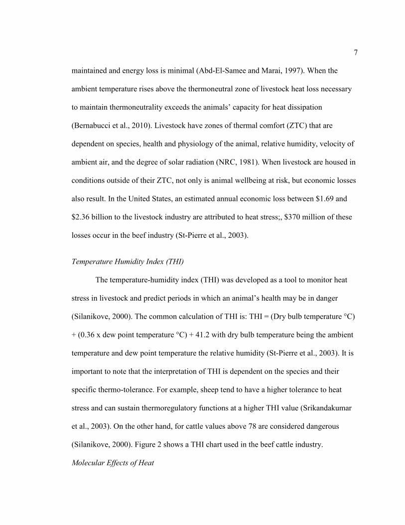
7 maintained and energy loss is minimal (Abd-El-Samee and Marai, 1997). When the
ambient temperature rises above the thermoneutral zone of livestock heat loss necessary
to maintain thermoneutrality exceeds the animals’ capacity for heat dissipation
(Bernabucci et al., 2010). Livestock have zones of thermal comfort (ZTC) that are
dependent on species, health and physiology of the animal, relative humidity, velocity of
ambient air, and the degree of solar radiation (NRC, 1981). When livestock are housed in
conditions outside of their ZTC, not only is animal wellbeing at risk, but economic losses
also result. In the United States, an estimated annual economic loss between $1.69 and
$2.36 billion to the livestock industry are attributed to heat stress;, $370 million of these
losses occur in the beef industry (St-Pierre et al., 2003).
Temperature Humidity Index (THI)
The temperature-humidity index (THI) was developed as a tool to monitor heat
stress in livestock and predict periods in which an animal’s health may be in danger
(Silanikove, 2000). The common calculation of THI is: THI = (Dry bulb temperature °C)
+ (0.36 x dew point temperature °C) + 41.2 with dry bulb temperature being the ambient
temperature and dew point temperature the relative humidity (St-Pierre et al., 2003). It is
important to note that the interpretation of THI is dependent on the species and their
specific thermo-tolerance. For example, sheep tend to have a higher tolerance to heat
stress and can sustain thermoregulatory functions at a higher THI value (Srikandakumar
et al., 2003). On the other hand, for cattle values above 78 are considered dangerous
(Silanikove, 2000). Figure 2 shows a THI chart used in the beef cattle industry.
Molecular Effects of Heat
8
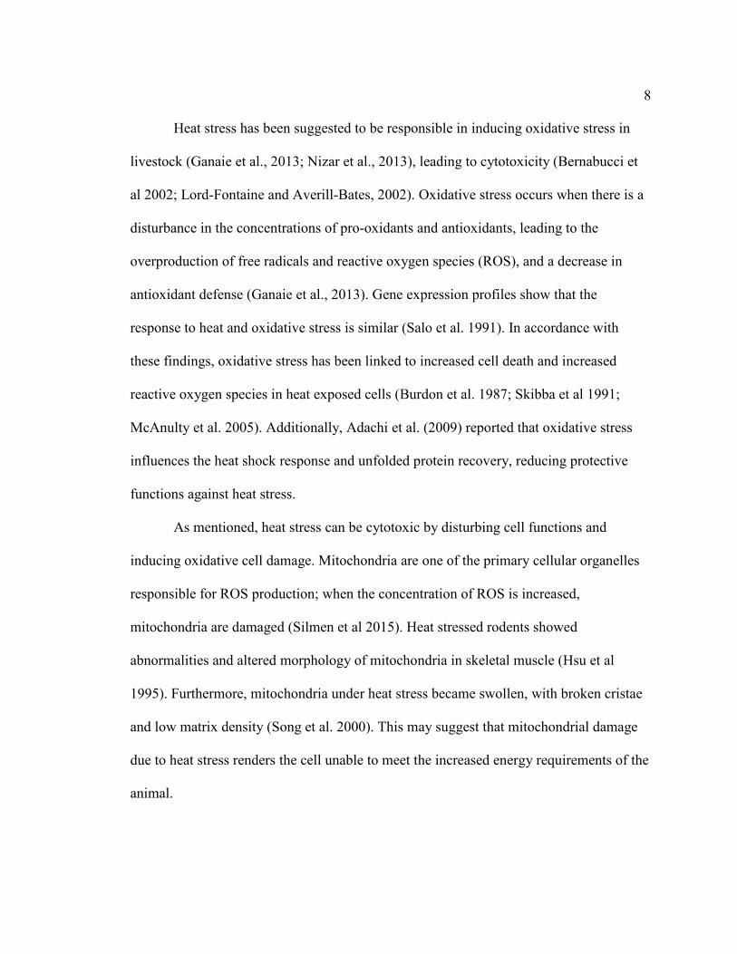
Heat stress has been suggested to be responsible in inducing oxidative stress in
livestock (Ganaie et al., 2013; Nizar et al., 2013), leading to cytotoxicity (Bernabucci et
al 2002; Lord-Fontaine and Averill-Bates, 2002). Oxidative stress occurs when there is a
disturbance in the concentrations of pro-oxidants and antioxidants, leading to the
overproduction of free radicals and reactive oxygen species (ROS), and a decrease in
antioxidant defense (Ganaie et al., 2013). Gene expression profiles show that the
response to heat and oxidative stress is similar (Salo et al. 1991). In accordance with
these findings, oxidative stress has been linked to increased cell death and increased
reactive oxygen species in heat exposed cells (Burdon et al. 1987; Skibba et al 1991;
McAnulty et al. 2005). Additionally, Adachi et al. (2009) reported that oxidative stress
influences the heat shock response and unfolded protein recovery, reducing protective
functions against heat stress.
As mentioned, heat stress can be cytotoxic by disturbing cell functions and
inducing oxidative cell damage. Mitochondria are one of the primary cellular organelles
responsible for ROS production; when the concentration of ROS is increased,
mitochondria are damaged (Silmen et al 2015). Heat stressed rodents showed
abnormalities and altered morphology of mitochondria in skeletal muscle (Hsu et al
1995). Furthermore, mitochondria under heat stress became swollen, with broken cristae
and low matrix density (Song et al. 2000). This may suggest that mitochondrial damage
due to heat stress renders the cell unable to meet the increased energy requirements of the
animal.

9
Heat shock proteins (HSP) are chaperone proteins activated by heat and other
stressors. HSP act as molecular chaperons that provide the cell with the ability to survive
injury and oxidative stress (Collier et al., 2008). HSP carry out crucial intracellular
housekeeping functions such as folding, unfolding and refolding of stress-denatured
proteins (Morimoto et al. 1990). In heat stressed sheep (Romero et al. 2013; Salces-Ortiz
et al. 2013) and cattle (Deb et al. 2014; Kishore et al. 2014), HSP-70 and HSP-90 were
increased, likely due to an increase in damaged proteins which induced HSP as a
mechanism for cellular repair (Slimen et al., 2015)). Furthermore, pretreatment with
HSP70 significantly protected intestinal epithelial barrier function impaired by chronic
stress (Yang et al., 2009). The increased expression of these proteins during heat stress
suggests their importance in cell survival to injury and stress.
Metabolic and Physiologic Impacts of Heat Stress
Physiological responses to heat stress include increased respiratory rate (Marai et
al., 2002), increased water consumption (Marai et al., 2007), decreased feed intake
(Marai et al., 2007; Guo et al., 2018; Johnson, 2018), ADG, and final weights (Mitlöhner
et al., 2002; Blaine and Nsahlai, 2011). These negative responses to heat stress have
detrimental effects on the animals’ health and wellbeing. Productivity of sheep in heat
stress inducing environments causes reduced voluntary feed intake leading to reduced
metabolizable energy (Dixon et al. 1999). In a study performed Mitlöhner et al. (2001),
heifers in heat stress had lower DMI than those provided shade. Additionally, control
heifers provided with shade reached their target BW 20 days earlier than the unshaded,
heat stressed heifers. Heat stress upregulates the secretion and expression of the receptors

10 of two adipokines: leptin and adiponectin (Bernabucci et al. 2009). Leptin stimulates the
hypothalamic axis resulting in reduced feed intake (Rabe et al. 2008) while adiponectin
regulates feeding behavior through a ‘starvation signal’ (Hoyda et al 2011). Therefore,
heat stress stimulates the hypothalamic axis by increasing the levels of these adipokines
resulting in reduced feed intake, partially impacting changes seen in ADG and DMI.
Exposure to heat results in redistribution of blood to the periphery and
compensatory reduction in the blood supply to the gut, with blood flow to the abdominal
organs, stomach and ileum is decreasing by 55%, 58%, and 32% respectively (Sakurada
and Hales 1998). The lack of blood flow to the gut damages the cell lining, allowing for
endotoxins to enter the body. The endotoxins released into the body can then cause tissue
damage and an acute phase immune response (Cronjé 2015). When exposure to heat
ceases and normal blood flow resumes, reactive oxygen species and cytokines are
released, which cause multiple organ injury (Cronjé, 2005). It is therefore possible that
the integrity of the gut lining is critical for heat stress abatement. This is especially
important in livestock species as energy–dense production diets damage the gut lining,
even in the absence of heat stress (Conlon et al., 2015), permitting endotoxins to enter the
body (Cronjé, 2005).
Beta-Adrenergic Agonists
Beta-adrenergic agonists (β-AA) are similar to endogenous catecholamines that
increase rate of gain, decrease carcass fat, and improve feed efficiency when fed before
harvest by stimulating the adrenoreceptors, which are similarly stimulated during stress
(Nelson, 1980; Vasconcelos et al., 2008; Elam et al., 2009; Montgomery et al., 2009).

11 These compounds have been an area of interest for researchers and producers for the past
two decades due to their ability to improve efficiency in livestock production
(Montgomery et al., 2009). They are commonly fed to livestock to redirect nutrients from
fat deposition towards muscle formation. However, their mechanism of action is not fully
understood and several factors such as diet, dose and duration of treatment, age, genetics,
and weight impact the efficiency of β-AA supplementation. Therefore, due to β-AA
signaling through adrenoreceptors, understanding if the mechanism by which β-AA act
will allow for a better understanding of whether their use in animal agriculture increases
stress to the animal or alters an animal’s ability to respond properly to other stressors.
Proposed mechanism of action
Beta-adrenergic receptors (β-AR), on the surface of muscle, fat, and other cells
and are part of a large family of G protein-coupled receptors (Mills and Mersmann,
1995). Subtypes of β-AR include β1-AR, β2-AR and β3-AR. The β1-AR and β2-AR are
the most well characterized and most abundant in mammalian cells (Mersmann, 1998).
The function of these receptors is similar, only differing by the ligands they bind. The
type of β-AR varies by tissue type and across species although one β-AR type generally
predominates within a tissue (Barnes, 1995). For example, bovine and ovine skeletal
muscle and adipocytes contain primarily β2-AR (Johnson et al., 2014) while rat heart
contains over 90% β1-AR and rat adipocytes contains over 90% β3-AR (Mersmann,
1998; Mersmann, 2002).
Substrates bind the ligand binding site of β-AR located at the center of the seven
transmembrane domains (Mills and Mersmann, 1995). Activation of β-AR is coupled

12 with Gs proteins and the activation of adenylate cyclase. When adenylate cyclase is
stimulated this leads to the conversion of adenosine triphosphate (ATP) to cyclic
adenosine monophosphate (cAMP) (Liggett and Raymond, 1993). Cyclic AMP is an
intracellular signaling molecule that binds to protein kinase A (PKA) to release its
catalytic subunit. PKA then phosphorylates intracellular enzymes such as serine residues
of metabolic hormones and regulates their use. The phosphorylation results in the partial
hydrolysis of triacylglycerol and inhibits de novo biosynthesis of fatty acids (Ricks et al.,
1984; Mersmann, 1998). Based on the proposed mechanism of action for β-AA, it has
been hypothesized that β-AA reduce adipose tissue accretion by inhibiting de novo
biosynthesis of fatty acids and by stimulating triacylglycerol hydrolysis (Mersmann,
2002). Furthermore, in muscle cells the activation of the β-AA signaling pathway results
in increased muscles mass by inhibiting protein turnover and promoting myofibrillar
protein synthesis (Johnson et al., 2014). Overall it is hypothesized that the binding of β-
AA induces signals in fat cells to decrease fat synthesis and increase fat degradation,
resulting in a slower rate of fat accretion while in muscle cells, binding of a β-AA results
in increased muscle protein synthesis (Anderson et al., 2005).
Β-Adrenergic Agonist Types
The β2-AA cimaterol and clenbuterol were the first β-AA investigated in
livestock production as a way to increase production efficiency in both cattle and sheep
(Warriss et al., 1989). Although they were effective in increasing overall weight gain and
gain to feed ratio (Kim et al. 1987), their overall benefit was not significant enough to
account for their cost (Warriss et al., 1989; Koohmaraie et al., 1991; Pringle et al., 1993).

13 During the 1990s and early 2000s, two β-AA were approved for use in cattle in the
United States: zilpaterol HCl (ZH) a β2 agonist, and ractopamine HCl (RAC) a β1
agonist (Delmore et al., 2010; Boler et al., 2012).
Zilpaterol HCl (ZH) is produced by Merck Animal Health and sold under the
trade name Zilmax. Zilpaterol HCl is on average fed to cattle in the last 26.6 ± 9 days on
the feedlot, having a substantial impact on live and carcass traits (Lean et al. 2014). A
meta-analysis by Lean et al (2014) reported that animals fed ZH had a BW 8kg and ADG
of 0.15kg more than non-supplemented controls, while also having reduced DMI
(0.1kg/hd/day). Additionally, supplementation with ZH improves hot carcass weight,
cool carcass weight, dressing percentage, and carcass meat percentage in heifers
(Montgomery et al. 2009). While ZH benefits livestock production by increasing body
weight, the β-AA acts as a repartitioning agent to redirect nutrient from fat to protein
deposition reducing marbling score and fat depth (Lean et al. 2014), which is often
accompanied by a decrease in tenderness (Knight, 2014)
Ractopamine HCl (RAC) is produced by Elanco Animal Health and sold under
the trade name Optaflexx for cattle, Paylean for swine and Topmax for turkeys and is fed
during the last 20 to 40 days. Animals supplemented with RAC show an increase in both
BW and ADG of 8kg and 0.19 kg/day, respectively, compared to non-supplemented
controls (Lean et al. 2014). Moreover, supplementation with RAC increases G:F ratios by
20.5% (Abney et al., 2007) and final live weight (Vogel et al., 2005; Lopez-Carlos et al.,
2011).
14
Contrary to the previously stated results, administration of either ZH or RH did
not improve carcass traits of Pelibuey x Katahdin male lambs (Estrada-Angulo et al.,
2008; Robles-Estrada et al., 2009). Additionally, control and supplemented ewe lambs,
fed 10 mg of ZH/day had similar (P > 0.10) feedlot performance and body fat deposition
(Macías-Cruz et al., 2010). These studies demonstrate a variation in response to ZH or
RH across animals that could be due genetics, species, and other variables.
Due to rapid elimination of β-AA residue from the animal, β-AA have gained
popularity in livestock systems. RAC has not been shown to leave residues after feeding
so a withdrawal period is not necessary (Elanco, 2017). Conversely, ZH has a minimum
three-day withdrawal period before slaughter to ensure residues in the muscle tissue are
eliminated before consumption (Merck, 2017). Additionally, ZH and RAC have no
antibiotic activity and therefore do not act as antibiotic growth promoters (Anderson et al.
2005). However, neither β-AA is approved for use in sheep or chickens (AMSA, 2015).
Additionally, while both are approved for use by the FDA, animal welfare concerns
related to the use of ZH in feedlot cattle resulted in the removal of ZH from production
systems, leading to RAC as the primary β-AA used in cattle production (AVMA, 2014)
Factors that influence response to β-AA
Although β-AA improve animal performance and efficiency, their effect varies
across studies (Fiems, 1987). It has thus been hypothesized, and some data have
supported that the age of the animal, weight, accessibility of β-AR, type of β-AA,
species, and genetics may impact the overall effect of the β-AA. For example, the use of
two different β-AA, metaproterenol and terbutaline, had different impact on feedlot

15 parameters and carcass characteristics of lambs fed the same diet (Nourozi et al., 2008).
Additionally, β-AA may differ in effect due to varying abundance of β-AR across cell
and tissue types (Moody et al. 2000). The β-AR available on the cell surface also may
become desensitized over time with β2-AR being more readily desensitized than β1-AR
(Lafontan 1993, Johnson et al 2014). Due to the desensitization of β-AR reports have
suggested that at high doses, RAC can bind β2-AR and elicit a biological response, but
both the cost of that level of supplementation and possible side effects keep this from
being a viable option (Vogel et al., 2005).
Potential Adverse Effects of Beta-Adrenergic Agonists
While the use of β-AA is beneficial in improving growth and animal performance,
reports suggest that these supplements have adverse effects as well. RAC improves both
growth performance through increased protein synthesis (Moody et al. 2000) and
decreased protein degradation (Mershmann 1998; Moody et al. 2000; Dunshea et al.
2005). Given that an effect attributed to β-AA supplementation is decreased protein
degradation, it is possible its use may lead to tougher meat. In a literature review,
Koohmaraie et al. (2002) concluded that improvements in animal growth could be due to
decreased protein degradation, which reduces the rate of post-mortem proteolysis in
muscle resulting in tougher meat. However, Merck Animal Health reported that although
tenderness is decreased in animals fed ZH, flavor is unaffected (Merck, 2017) and
sensory panel tasting scores showed that there were no differences between the products
of control and ZH fed cattle (Weber et al., 2013).
16
In 2013, instances of increased mobility issues and mortality were observed in
feedlots using ZH and as a result, concerns arose about the wellbeing of cattle being fed
ZH (Loneragan et al., 2014). Although no scientific data were available to support the
connection of ZH and lameness, the possible association of ZH with these issues caused
Tyson and other packing plants to stop accepting cattle fed the supplement (Sorensen,
2016). The claims against ZH led to Merck Animal Health removing ZH from the market
in the US, leading to a decrease of its use in the United States and a negative public
perception of ZH (Sorensen, 2016).
To investigate the claims that ZH diminishes animal wellbeing, studies have
compared mobility of animals supplemented ZH to controls. As time on feed increased,
mobility decreased for all animals, regardless of ZH supplementation (Boyd et al., 2015).
This work suggested that ZH was not the cause of mobility issues but instead the increase
in overall mass as cattle aged may have resulted in the issues reported. Further, transport
and flooring at the packing plants were hypothesized to contribute to mobility problems
(Boyd et al., 2015). Overall, research conducted to investigate adverse effects due to the
feeding of ZH has shown no measurable negative effect of ZH supplementation on
animal wellbeing (Hales et al., 2014; Boyd et al., 2015; Buntyn et al., 2016; Sorensen,
2016).
β-AA Impact on the Microbiome
Although β-AA improve ADG, feed efficiency and carcass weight, the potential
effect β-AA have on the ruminant ecosystem in feedlot animals is not yet understood.
Natural catecholamines, norepinephrine, epinephrine, and dopamine, stimulate bacterial

17 growth (Roberts et al. 2002), affect gut motility and the secretory response (Ruckebush,
1983; McIntyre and Thompson 1992), and also increase the amount of gram-negative
bacteria species in vitro (Lyte and Ernst 1992). The latter impact of β-AA on the
microbiome is important as many gram-negative bacteria of the rumen are vital for
fermentation (Walker and Drouillard 2012).
Walker et al. (2006) showed a reduced response to RAC when finishing heifers
were fed ruminally degraded forms of nitrogen. The reported interaction between RAC
and the nitrogen supplied suggested a direct effect of RAC on microbial populations of
the rumen. Therefore, it is possible that RAC alters proteolysis in the rumen, resulting in
less degradable protein available to the rumen microbes. Additionally, Walker et al.
(2006) demonstrated that the ratio of degradable to undegradable protein provided in diet
to the ruminal microflora is important for maximizing response to RAC.
The effect of natural catecholamines on bacteria is not fully understood; however,
it is possible that catecholamines increase the ability of bacteria to uptake iron, which is
essential for bacterial growth (Kinney et al. 2010). Catecholamines can also function to
chelate iron and thus allow bacteria to recognize and use these siderophores (Kinney et
al. 2000). RAC may therefore be providing a competitive advantage by increasing iron
availability for bacteria to utilize; this may explain the increased bacterial population
growth seen by Walker and Drouillard (2010) in an in vitro assay using RAC. Walker and
Drouillard (2010) reported that RAC increased gas production in vitro providing
evidence that the impact of RAC on an animal is not limited to the tissue response.
Because β-AR are present all along the digestive tract, the binding of β-AA directly

18 affects its motility and secretory functions (Ruckebush, 1983; McIntyre and Thompson,
1992).
Interaction between heat stress and beta-adrenergic agonist supplementation
While a lot of attention has been given to the impacts of heat stress and β-AA
separately, little attention has been paid to the interaction between the two. Heat stress
and β-AA both stimulate the β- adrenergic system, however it is unknown how their
simultaneous stimulation impacts the animal. Based upon anecdotal observations, it has
been hypothesized that the supplementation of β-AA could potentially exacerbate the
stress response caused by hyperthermia (Grandin, 2013; Loneragan et al., 2014).
Additionally, Allen et al. (2017) found an association, but no supported causation,
between cattle fed during the summer months and a decrease in feed intake compared to
animals fed ZH during cooler months. Little to no research has examined the interaction
between β-AA usage, heat stress, and their combined impact on the ruminant GI tract
ecosystem and physiology.
Marcías-Cruz et al. (2010) represents one of the few studies evaluating how heat
stress β-AA supplementation impact growth traits and carcass characteristics. In the
study, ewe lambs were fed 10 mg of ZH/ewe/day and were housed in an environment
with an average ambient temperature of 34.1°C with 50.4% humidity. No difference was
observed in final live weight, ADG, feed intake, feed conversion, and G:F between ZH
and control lambs even though previous studies have shown all these traits excepting feed
intake increase with ZH supplementation (Vasconcelos et al., 2008; Montgomery et al.,
2009; Hales et al., 2014). Therefore, Macías-Cruz et al. (2010) suggests that under heat
19 stress conditions the effect of ZH on performance traits is diminished. Additionally, ZH-
supplemented lambs showed an increase in hot and cold carcass weight, dressing
percentage, conformation and rib-eye area when compared to control (Macías-Cruz et al.,
2010). These data indicate that there is no consequential interaction between heat stress
and β-AA. However, this is a single study and additional research must be conducted to
fully understand what is occurring when β-AA are fed during heat stress conditions.
Conclusion
Much is known about the ruminant gastrointestinal tract and its importance in
nutrient absorption, utilization, and energy production. Changes in diet as well as
environmental stressors can affect rumen function impacting animal performance. Heat
stress and β-AA have been studied individually, but little is known about their
interaction. Studying the impact of heat stress and β-AA supplementation on the rumen is
important because many feedlot cattle are fed β-AA during summer month. By gaining a
better understanding of the response to each factor individually as well as in concert, we
can improve growth and efficiency along with animal wellbeing. Therefore, this research
aimed to identify how β-AA supplementation and heat stress impacts the rumen
gastrointestinal ecosystem and physiology to further improve the sustainability of animal
production.

20 References
Abd-El-Samee, A., and I. Marai. 1997. Daily body gain some related physiological and biochemical changes in dromedary camels as affected by hot climate. In: International Conference on Animal, Poultry, Rabbit Production and Health, Cairo (Egypt), 2-4 Sep 1997
Abney, C., J. Vasconcelos, J. McMeniman, S. Keyser, K. Wilson, G. Vogel, and M. Galyean. 2007. Effects of ractopamine hydrochloride on performance, rate and variation in feed intake, and acid-base balance in feedlot cattle. Journal of animal science 85(11):3090-3098.
Adachi M, Liu Y, Fujii K, Calderwood SK, Nakai A, Imai K, et al. (2009) Oxidative Stress Impairs the Heat Stress Response and Delays Unfolded Protein Recovery. PLoS ONE 4(11): e7719. https://doi.org/10.1371/journal.pone.0007719
Allen, K. 2017. Beta-agonists, The Environment and Cattle Fatigue. http://beef2live.com/story-beta-agonists-environment-cattle-fatigue-part-1-0-107020.
Ametaj BN, Zebeli Q, Saleem F, Psychogios N, Lewis MJ, Dunn SM, Xia J andWishart DS 2010. Metabolomics reveals unhealthy alterations in rumen meta-bolism with increased proportion of cereal grain in the diet of dairy cows.Metabolomics 6, 583–594
AMSA. 2015. Beta-Agonists: What are they and why do we use them in livestock production? https://www.meatscience.org/docs/default-source/publications-resources/fact-sheets/beta-agonists---dilger-20158d82e7711b766618a3fcff0000a508da.pdf?sfvrsn=69f481b3_0
Anderson, D. B., D.E Moody, D.L. Hancock. (2005). Beta adrenergic agonists.
Argenzio, R., N. Miller, and W. von Engelhardt. 1975. Effect of volatile fatty acids on water and ion absorption from the goat colon. American Journal of Physiology-Legacy Content 229:997-1002.
AVMA. 2014. Literature Review on the Welfare Implications of the Use of Beta-Adrenoreceptor Agonists https://www.avma.org/KB/Resources/LiteratureReviews/Pages/Use-of-Beta-Agonists.aspx.
Baldwin RL (1965) Pathways of carbohydrate metabolism in the rumen. In Dungherty RW (Ed.) Physiology of Digestion in the Ruminant. Butterworths. London, UK. pp. 379-389
Baldwin, R. L., and McLeod, K. R. (2000). Effects of diet forage:concentrate ratio and metabolizable energy intake on isolated rumen epithelial cell metabolism in vitro. J. Anim. Sci. 78, 771–783

21 Baldwin, R. L., Vi McLeod, K. R., Klotz, J. L., and Heitmann, R. N. (2004). Rumen
development, intestinal growth and hepatic metabolism in the preand postweaning ruminant. J. Dairy Sci. 87, E55–E65. doi: 10.3168/jds.S0022-0302(04)70061-2
Baldwin, R., S. Wu, W. Li, C. Li, B. Bequette, and R. Li. 2012. Quantification of Transcriptome Responses of the Rumen Epithelium to Butyrate Infusion using RNA-seq Technology. Gene Regulation and Systems Biology 6:GRSB.S9687.
Barnes, P.J. (1995). Beta-adrenergic receptors and their regulation. Am. J. Respir. Crit. Care Med., 152, 838-860
Bernabucci, U., B. Ronchi, N. Lacetera, and A. Nardone. 2002. Markers of oxidative status in plasma and erythrocytes of transition dairy cows during hot season. J. Dairy Sci. 85:2173–2179.
Bernabucci U, Basirico` L, Morera P, Lacetera N, Ronchi B, and Nardone A. 2009 Heat shock modulates adipokines expression in 3T3-L1 adipocytes. Journal of Molecular Endocrinology 42 139–147.
Bernabucci, U., N. Lacetera, L. H. Baumgard, R. P. Rhoads, B. Ronchi, and A. Nardone. 2010. Metabolic and hormonal acclimation to heat stress in domesticated ruminants. Animal 4(7):1167-1183.
Blaine, K., and I. V. Nsahlai. 2011. The effects of shade on performance, carcass classes and behaviour of heat-stressed feedlot cattle at the finisher phase. Tropical animal health and production 43(3):609-615.
Boler, D., A. Shreck, D. B. Faulkner, J. Killefer, F. McKeith, J. Homm, and J. Scanga. 2012. Effect of ractopamine hydrochloride (Optaflexx) dose on live animal performance, carcass characteristics and tenderness in early weaned beef steers. Meat science 92(4):458-463
Boyd, B. M., S. D. Shackelford, K. E. Hales, T. M. Brown-Brandl, M. L. Bremer, M. L. Spangler, T. L. Wheeler, D. A. King, and G. E. Erickson. 2015. Effects of shade and feeding zilpaterol hydrochloride to finishing steers on performance, carcass quality, heat stress, mobility, and body temperature. Journal of animal science 93(12):5801-5811.
Bryant, M. P., Small, N., Bouma, C. & Chu, H. Bacteroides ruminicola n. sp. and Succinimonas amylolytica; the new genus and species; species of succinic acid-producing anaerobic bacteria of the bovine rumen. J. Bacteriol. 76, 15–23 (1958).
Buntyn, J., N. Burdick Sanchez, T. Schmidt, G. Erickson, S. Sieren, S. Jones, and J. Carroll. 2016. The metabolic, stress axis, and hematology response of zilpaterol hydrochloride supplemented beef heifers when exposed to a dual corticotropin-releasing hormone and vasopressin challenge. Journal of animal science 94(7):2798-2810.

22 Burdon RH, Gill VM, Rice-Evans C (1987) Oxidative stress and heat shock protein
induction in human cells. Free Radic Res Commun 3:129–139.
Carberry, C., D. Kenny, S. Han, M. McCabe, and S. Waters. 2012. Effect of Phenotypic Residual Feed Intake and Dietary Forage Content on the Rumen Microbial Community of Beef Cattle. Applied and Environmental Microbiology 78:4949-4958.
Castillo-González, A., Burrola-Barraza, M., Domínguez-Viveros, J., & Chávez-Martínez, A. (2014). Rumen microorganisms and fermentation. Archivos De Medicina Veterinaria, 46(3), 349-361. doi: 10.4067/s0301-732x2014000300003
Chen, G., S. Song, B. Wang, Z. Zhang, Z. Peng, C. Guo, J. Zhong, and Y. Wang. 2015. Effects of Forage:Concentrate Ratio on Growth Performance, Ruminal Fermentation and Blood Metabolites in Housing-feeding Yaks. Asian-Australasian Journal of Animal Sciences 28:1736-1741.eaning Development. Applied and Environmental Microbiology 84:e02675-17
Collier, R., J. Collier, R. Rhoads, and L. Baumgard. 2008. Invited review: genes involved in the bovine heat stress response. Journal of Dairy Science 91(2):445-454.
Conlon, M., and A. Bird. 2014. The Impact of Diet and Lifestyle on Gut Microbiota and Human Health. Nutrients 7:17-44.
Cronjé PB. 2005. Heat stress in livestock - role of the gut in its aetiology and a potential role for betaine in its alleviation. Recent Adv Animal Nutr Australia. 15: 107-122.
Cummings, J.H., and Macfarlane, G.T. 1997. Role of intestinal bacteria in nutrient metabolism. J. Parenter. Enteral. Nutr. 21: 357-365.
Deb, R.; Sajjanar, B.; Singh, U.; Kumar, S.;Singh, R.; Sengar, G.; Sharma, A., 2014:Effect of heat stress on the expressionprofile of Hsp90 among Sahiwal (Bos in-dicus) and Frieswal (Bos indicus 9Bostaurus) breed of cattle: a comparativestudy. Gene 536, 435–440.
Delmore, R., J. Hodgen, and B. Johnson. 2010. Perspectives on the application of zilpaterol hydrochloride in the United States beef industry. Journal of animal science 88(8):2825- 2828.
Dias, J., M. Marcondes, S. Motta de Souza, B. Cardoso da Mata e Silva, M. Fontes Noronha, R. Tassinari Resende, F. Machado, H. Cuquetto Mantovani, K. Dill-McFarland, and G. Suen. 2018. Bacterial Community Dynamics across the Gastrointestinal Tracts of Dairy Calves during Preweaning Development. Applied and Environmental Microbiology 84:e02675-17.
Dixon, R., R. Thomas, and J. Holmes. 1999. Interactions between heat stress and nutrition in sheep fed roughage diets. The Journal of Agricultural Science 132:351-359.

23 Dunshea, F. R., D. N. D’Souza, D. W. Pethic, G. S. Harper, and R. D. Warner. 2005.
Effects of dietary factors and other metabolic modifiers on quality and nutritional value of meat. Meat Sci. 71:8–38.
Elam, N., J. Vasconcelos, G. Hilton, D. VanOverbeke, T. Lawrence, T. Montgomery, W. Nichols, M. Streeter, J. Hutcheson, and D. Yates. 2009. Effect of zilpaterol hydrochloride duration of feeding on performance and carcass characteristics of feedlot cattle. Journal of animal science 87(6):2133-2141.
Elanco. 2018. Optaflexx®. https://www.elanco.us/products-services/beef/optaflexx.
Estrada-Angulo, A., A. Barreras-Serrano, G. Contreras, J. F. Ob-regon, J. C. Robles-Estrada, A. Plascencia, and R. A. Zinn. 2008. Influence of level of zilpaterol chlorhydrate supplementa-tion on growth performance and carcass characteristics of feed-lot lambs. Small Rumin. Res. 80:107–110
Fernando, S., H. Purvis, F. Najar, L. Sukharnikov, C. Krehbiel, T. Nagaraja, B. Roe, and U. DeSilva. 2010. Rumen Microbial Population Dynamics during Adaptation to a High-Grain Diet. Applied and Environmental Microbiology 76:7482-7490.
Fiems, L.O., 1987. Effect of beta-adrenergic agonists in animal production and their mode of action. Annals Zootechnol. 36, 271-290.
Fouts DE, Szpakowski S, Purushe J, Torralba M, Waterman RC, MacNeil MD, et al. Next generation sequencing to define prokaryotic and fungal diversity in the bovine rumen. PLoS ONE. 2012;7:e48289.
Fuentes MC, S Calsamiglia, PW Cardozo, B Vlaeminck. 2009. Effect of pH and level of concentrate in the diet on the production of biohydrogenation intermediates in a dual-flow continuous culture. J Dairy Sci 92, 4456-4466.
Ganaie, A. H.; Shanker, G.; Bumla, N. A.;Ghasura, R. S.; Mir, N. A., 2013: Bio-chemical and physiological changes dur-ing thermal stress in bovines. Journal ofVeterinary and Science Technology 4, 126
Górka, P., Z. M. Kowalski, P. Pietrzak, A. Kotunia, W. Jagusiak, J. J. Holst, R. Guilloteau, and R. Zabielski. 2011a. Effect of method of delivery of sodium butyrate on rumen development in newborn calves. J. Dairy Sci. 94:5578–5588.
Grandin, T. 2018. Heat Stress and Lameness in Fed Feedlot Cattle is Detrimental to Animal Welfare. http://www.grandin.com/heat.stress.lameness.html.
Guan, L., J. Nkrumah, J. Basarab, and S. Moore. 2008. Linkage of microbial ecology to phenotype: correlation of rumen microbial ecology to cattle's feed efficiency. FEMS Microbiology Letters 288:85-91.
Guarner F. Enteric flora in health and disease. Digestion. 2006;73:5–12.
Guo, K., H. Cao, Y. Zhu, T. Wang, G. Hu, B. Kornmatitsuk, and J. Luo. 2018. Improving effects of dietary rumen protected γ‐aminobutyric acid additive on apparent
24
nutrient digestibility, growth performance and health status in heat‐stressed beef cattle. Animal Science Journal. DOI: 10.1111/asj.13053.
Hales, K., S. Shackelford, J. Wells, D. King, M. Hayes, T. Brown-Brandl, L. Kuehn, H. Freetly, and T. Wheeler. 2014. Effects of feeding dry-rolled corn-based diets with and without wet distillers grains with solubles and zilpaterol hydrochloride on performance, carcass characteristics, and heat stress in finishing beef steers. Journal of animal science 92(9):4023-4033.
Henderson, G. et al. Rumen microbial community composition varies with diet and host, but a core microbiome is found across a wide geographical range. Sci. Rep. 5, 14567; doi: 10.1038/srep14567 (2015).
Hobson P. N. Stewart C. S. 1997. The rumen microbial ecosystem. 2nd ed.Blackie Acad. Prof., New York.
Hook, S., M. Steele, K. Northwood, A. Wright, and B. McBride. 2011. Impact of High-Concentrate Feeding and Low Ruminal pH on Methanogens and Protozoa in the Rumen of Dairy Cows. Microbial Ecology 62:94-105.
Hoyda TD, Samson WK, and Ferguson AV 2011 Central nervous system roles for adiponectin in neuroendocrine and automic function. In Adipokines, pp 167–184. Eds VR Preedy & RJ Hunter. Boca Raton, FL, USA: Science Publishers, CRC Press.
Hsu YD, Chen SS, Lee WH, Lin SZ, Kao MC, Tsao WL. 1995. Mitochondrial alterations of skeletal muscle in a heat stress rat model. Proc. Natl. Sci. Counc. Repub. China B. 19:233–39
Hua C., Tian J., Tian P., Cong R., Luo Y., Geng Y., et al. . (2017). Feeding a high concentration diet induces unhealthy alterations in the composition and metabolism of ruminal microbiota and host response in a goat model. Front. Microbiol. 8:138. 10.3389/fmicb.2017.00138
Hyder, I., P. Ravi Kanth Reddy, J. Raju, P. Manjari, C. Srinivasa Prasad, K. Aswani Kumar, and V. Sejian. 2017. Alteration in Rumen Functions and Diet Digestibility During Heat Stress in Sheep. Sheep Production Adapting to Climate Change:235-265. doi: 10.1007/978-981-10-4714-5_11
Jami E., Mizrahi I. (2012). Similarity of the ruminal bacteria across individual lactating cows. Anaerobe 18, 338–343. 10.1016/j.anaerobe.2012.04.003
Jami, E., A. Israel, A. Kotser, and I. Mizrahi. 2013. Exploring the bovine rumen bacterial community from birth to adulthood. ISME J. 7:1069–1079. doi:10.1038/ismej.2013.2.
Johnson, B. J. 2014. Mechanism of action of beta adrenergic agonists and potential residue issues. American Meat Association Reference Paper:1-6.

25 Johnson, B. J., S. B. Smith, and K. Y. Chung. 2014. Historical overview of the effect of
β-adrenergic agonists on beef cattle production. Asian-Australasian journal of animal sciences 27(5):757.
Johnson, J. 2018. Impact of Heat Stress on Livestock and Mitigation Strategies to Improve Productivity and Well-Being. Journal of Animal Science 96:6-6.
Jung, H., and M. Allen. 1995. Characteristics of plant cell walls affecting intake and digestibility of forages by ruminants. Journal of Animal Science 73:2774.
Kim, Y. S., Lee, Y. B. and Dalrymple, R. H. 1987. Effect of repartitioning agent cimaterol on growth, carcass and skeletal muscle characteristics in lambs. Journal of Animal Science 65: 1392–1399
Kim M, Morrison M. Yu Z. Status of the phylogenetic diversity census of ruminal microbiomes. FEMS Microbiol Ecol. 2011;76:49–63
Kim M., Yu Z. T. 2012. Quantitative comparisons of select cultured and uncultured microbial populations in the rumen of cattle fed different diets. J. Anim. Sci. Biotechnol. 3:28. 10.1186/2049-1891-3-28
Kishore, A.; Sodhi, M.; Kumari, P.; Moh-anty, A. K.; Sadana, D. K.; Kapila, N.;Khate, K.; Shandilya, U.; Kataria, R. S.;Mukesh, M., 2014: Peripheral bloodmononuclear cells: a potential cellular system to understand differential heatshock response across native cattle (Bosindicus), exotic cattle (Bos taurus), andriverine buffaloes (Bubalus bubalis)ofIndia. Cell Stress and Chaperones 19,613–621
Knight, C. H. 2014. Understanding beef carcass reports, University of Georgia and Ft Valley State University, www.ugaext.gov.za.
Kong, R., G. Liang, Y. Chen, P. Stothard, and L. Guan. 2016. Transcriptome profiling of the rumen epithelium of beef cattle differing in residual feed intake. BMC Genomics 17.
Koohmaraie, M., S. Shackelford, T. Wheeler, S. M. Lonergan, and M. Doumit. 1995. A muscle hypertrophy condition in lamb (callipyge): characterization of effects on muscle growth and meat quality traits. Journal of animal science 73(12):3596-3607.
Koohmaraie, M., M. P. Kent, S. D. Shackelford, E. Veiseth, and T. L. Wheeler. 2002. Meat tenderness and muscle growth: is there any relationship? Meat science 62(3):345-352.
Kowalski, Z. M., P. Górka, J. Flaga, A. Barteczko, K. Burakowska, J. Oprządek, and R. Zabielski. 2015. Effect of microencapsulated sodium butyrate in the close-up diet on performance of dairy cows in the early lactation period. J. Dairy Sci. 98:3284–3291. https:// doi.org/10.3168/jds.2014-8688.
Lafontan M, Berlan M 1993 Fat cell adrenergic receptors and the control of white and brown fat cell function. J Lipid Res 34:1057–1091
26 Lean, I., J. Thompson, and F. Dunshea. 2014. A Meta-Analysis of Zilpaterol and
Ractopamine Effects on Feedlot Performance, Carcass Traits and Shear Strength of Meat in Cattle. PLoS ONE 9:e115904.
Li, F., and L. Guan. 2017. Metatranscriptomic Profiling Reveals Linkages between the Active Rumen Microbiome and Feed Efficiency in Beef Cattle. Applied and Environmental Microbiology 83.
Liggett, S. B., and J. R. Raymond. 1993. Pharmacology and molecular biology of adrenergic receptors. Baillieres Clin. Endocrinol. Metab. 7:279−305
Loneragan, G. H., D. U. Thomson, and H. M. Scott. 2014. Increased mortality in groups of cattle administered the β-adrenergic agonists ractopamine hydrochloride and zilpaterol hydrochloride. PloS one 9(3):e91177.
Lopez-Carlos, M., R. Ramírez, J. Aguilera-Soto, A. Plascencia, H. Rodríguez, C. Arechiga, R. Rincón, C. Medina-Flores, and H. Gutierrez-Bañuelos. 2011. Effect of two beta adrenergic agonists and feeding duration on feedlot performance and carcass characteristics of finishing lambs. Livestock Science 138(1):251-258.
Lord-Fontaine, S.; Averill-Bates, D. A.,2002: Heat shock inactivates cellularantioxidant defense against hydrogenperoxide: protection by glucose. FreeRadical Biology & Medicine 32, 752–765
Lyte, M., and S. Ernst. 1992. Catecholamine induced growth of gram negative bacteria. Life Sciences 50:203-212.
Maala CP, Smith DF, Hintz HF, Sack WO. Removal of the cecum, including the ileocecocolic junction, and its effects on digestibility in cattle. Am J Vet Res. 1983 Dec;44(12):2237–2243
Marai, I., A. El-Darawany, A. Fadiel, and M. Abdel-Hafez. 2007. Physiological traits as affected by heat stress in sheep—a review. Small Ruminant Research 71(1):1-12.
Marai, I., A. Habeeb, and A. Gad. 2002. Rabbits’ productive, reproductive and physiological performance traits as affected by heat stress: a review. Livestock Production Science 78(2):71-90.
Macías-Cruz, U., F. Álvarez-Valenzuela, N. Torrentera-Olivera, J. Velázquez-Morales, A. Correa-Calderón, P. Robinson, and L. Avendaño-Reyes. 2010. Effect of zilpaterol hydrochloride on feedlot performance and carcass characteristics of ewe lambs during heat-stress conditions. Animal Production Science 50(10):983-989.
McAnulty SR, McAnulty L, Pascoe DD, Gropper SS, Keith RE, et al. (2005) Hyperthermia increases exercise-induced oxidative stress. Int J Sports Med 26:188–192.
McCann, J. C., T.A. Wickersham, and J.J. Loor. 2014. High-throughput methods redefine the rumen microbiome and its relationship with nutrition and metabolism. Bioinform. Biol. Insights. 8:109–125. doi:10.4137/BBI.S15389

27 McIntyre, A., and D. Thompson. 1992. Review article: adrenergic control of motor and
secretory function in the gastrointestinal tract. Alimentary Pharmacology & Therapeutics 6:125-142.
Merck. 2017. ZILMAX®. https://www.merck-animal-health-usa.com/product/cattle/Zilmax/1.
Mersmann, H. 2002. Beta-adrenergic receptor modulation of adipocyte metabolism and growth. Journal of Animal Science 80(E-Suppl_1):E24-E29.
Mersmann, H. J. 1998. Overview of the effects of beta-adrenergic receptor agonists on animal growth including mechanisms of action. Journal of animal science 76(1):160-172.
Mills, S., and H. Mersmann. 1995. Beta-adrenergic agonists, their receptors, and growth: Special reference to the peculiarities in pigs. The biology of fat in meat animals: Current advances. p:1-34.
Mitlöhner, F., J. Morrow, J. Dailey, S. Wilson, M. Galyean, M. Miller, and J. McGlone. 2001. Shade and water misting effects on behavior, physiology, performance, and carcass traits of heat-stressed feedlot cattle. Journal of Animal Science 79(9):2327-2335.
Mitlöhner, F., M. Galyean, and J. McGlone. 2002. Shade effects on performance, carcass traits, physiology, and behavior of heat-stressed feedlot heifers. Journal of Animal Science 80(8):2043-2050.
Montgomery, J., C. Krehbiel, J. Cranston, D. Yates, J. Hutcheson, W. Nichols, M. Streeter, D. Bechtol, E. Johnson, and T. TerHune. 2009. Dietary zilpaterol hydrochloride. I. Feedlot performance and carcass traits of steers and heifers. Journal of animal science 87(4):1374-1383.
Moody D.E. Hancock D.L. Anderson D.B. 2000. Phenethanolamine repartitioning agents. Pages 65–95inFarm Animal Metabolism and Nutrition. D'Mello J.P.F. ed. CAB Int., New York, NY.
Morimoto R.I., Tissieres A., Georgopoulos C. (1990) The stress response, function of the proteins, and perspectives. in Stress proteins in biology and medicine, eds Morimoto R.I., Tissieres A., Georgopoulos C.(Cold Spring Harbor Laboratory Press, Cold Spring Harbor, NY), pp 1–36.
Muegge, B., J. Kuczynski, D. Knights, J. Clemente, A. Gonzalez, L. Fontana, B. Henrissat, R. Knight, and J. Gordon. 2011. Diet Drives Convergence in Gut Microbiome Functions Across Mammalian Phylogeny and Within Humans. Science 332:970-974.
Nagaraja, T. 2016. Microbiology of the Rumen. Rumenology:39-61.
National Research Council. (NRC). Effect of Environment on Nutrient Requirement of Domestic Animals, Mattonia Academy press. Washington, DC.1981.
28 Nelson, D. 1980. Adrenal androgens. The adrenal cortex. IN Smith L Jr (ed):
Physiological
Nizar, A. N.; Mudasir, S.; Hina, A. W.,2013: Oxidative stress –Threat to ani-mal health and production. InternationalJournal of Livestock Research 3,76–83
Nourozi M. Abazari M. Raisianzadeh M. Mohammadi M. ZareShahne A. 2008. Effect of terbutaline and metaproterenol (two beta-adrenergic agonists) on performance and carcass composition of culled Moghani ewes. Small Rumin. Res. 74:72–77.
Nozière, P., Ortigues-Marty, I., Loncke, C., & Sauvant, D. (2010). Carbohydrate quantitative digestion and absorption in ruminants: from feed starch and fibre to nutrients available for tissues. Animal, 4(07), 1057-1074. doi: 10.1017/s1751731110000844
Paz, H., K. Hales, J. Wells, L. Kuehn, H. Freetly, E. Berry, M. Flythe, M. Spangler, and S. Fernando. 2018. Rumen bacterial community structure impacts feed efficiency in beef cattle. Journal of Animal Science 96:1045-1058.
Penner, G., M. Steele, J. Aschenbach, and B. McBride. 2011. RUMINANT NUTRITION SYMPOSIUM: Molecular adaptation of ruminal epithelia to highly fermentable diets1. Journal of Animal Science 89:1108-1119.
Popova, M., E. McGovern, M. McCabe, C. Martin, M. Doreau, M. Arbre, S. Meale, D. Morgavi, and S. Waters. 2017. The Structural and Functional Capacity of Ruminal and Cecal Microbiota in Growing Cattle Was Unaffected by Dietary Supplementation of Linseed Oil and Nitrate. Frontiers in Microbiology 8.
Pringle, T., C. R. Calkins, M. Koohmaraie, and S. J. Jones. 1993. Effects over time of feeding a beta-adrenergic agonist to wether lambs on animal performance, muscle growth, endogenous muscle proteinase activities, and meat tenderness. Journal of animal science 71(3):636-644.
Rabe, K., M. Lehrke, K. Parhofer, and U. Broedl. 2008. Adipokines and Insulin Resistance. Molecular Medicine 14:741-751.
Ricks, C. A., P. K. Baker, and R. H. Dalrymple. 1984. Use of repartitioning agents to improve performance and body composition of meat animals. Reciprocal Meat Conf. Proc. 37:5−11.
Roberts, A., J. Matthews, S. Socransky, P. Freestone, P. Williams, and I. Chapple. 2002. Stress and the periodontal diseases: effects of catecholamines on the growth of periodontal bacteria in vitro. Oral Microbiology and Immunology 17:296-303.
Robles-Estrada, J., A. Barreras-Serrano, G. Contreras, A. Estrada-Angulo, J. Obregón, A. Plascencia, and F. Ríos. 2009. Effect of two β-adrenergic agonists on finishing performance and carcass characteristics in lambs fed all-concentrate diets. Journal of Applied Animal Research 36(1):33-36.

29 Roh, S. G., M. Kuno, D. Hishikawa, Y. H. Hong, K. Katoh, Y. Obara, H. Hidari, and S.
Sasaki. 2007. Identification of differentially expressed transcripts in bovine rumen and abomasum using a differential display method. J. Anim. Sci. 85:395–403. doi:10.2527/jas.2006-234
Romero, R. D.; Pardo, A. M.; Montaldo, H.H., 2013: Differences in body tempera-ture, cell viability, and HSP-70 concen-trations between Pelibuey and Suffolksheep under heat stress. Tropical Ani-mal Health and Production 45, 1691–1696.
Ruckebusch, Y. 1983. Pharmacology of reticulo-ruminal motor function. Journal of Veterinary Pharmacology and Therapeutics 6:245-272.
Salces-Ortiz, J.; Gonzalez, C.; Moreno-Sanchez, N.; Calvo, J. H.; Perez-Guzman, M. D.; Serrano, M. M., 2013:Ovine HSP90AA1 expression rate isaffected by several SNPs at the promoterunder both basal and heat stress condi-tions. PLoS ONE 8, e66641
Salo, D.; Donovan, C.; Davies, K., 1991:HSP70 and other possible heat shock oroxidative stress proteins are induced inskeletal muscle, heart, and liver duringexercise. Free Radical Biology and Medicine11, 239–246.
Silanikove, N. 2000. Effects of heat stress on the welfare of extensively managed domestic ruminants. Livestock production science 67(1):1-18.
Skibba JL, Powers RH, Stadnicka A, Cullinane DW, Almagro UA, et al. (1991) Oxidative stress as a precursor to the irreversible hepatocellular injury caused by hyperthermia. Int J Hyperthermia 7:749–761.
Srikandakumar, A., E. Johnson, and O. Mahgoub. 2003. Effect of heat stress on respiratory rate, rectal temperature and blood chemistry in Omani and Australian Merino sheep. Small Ruminant Research 49(2):193-198.
Steele, M., G. Vandervoort, O. AlZahal, S. Hook, J. Matthews, and B. McBride. 2011. Rumen epithelial adaptation to high-grain diets involves the coordinated regulation of genes involved in cholesterol homeostasis. Physiological Genomics 43:308-316.
St-Pierre, N., B. Cobanov, and G. Schnitkey. 2003. Economic losses from heat stress by US livestock industries. Journal of dairy science 86:E52-E77.
Tajima, K., R. Aminov, T. Nagamine, H. Matsui, M. Nakamura, and Y. Benno. 2001. Diet-Dependent Shifts in the Bacterial Population of the Rumen Revealed with Real-Time PCR. Applied and Environmental Microbiology 67:2766-2774.
Tajima, K., I. Nonaka, K. Higuchi, N. Takusari, M. Kurihara, A. Takenaka, M. Mitsumori, H. Kajikawa, and R. Aminov. 2007. Influence of high temperature and humidity on rumen bacterial diversity in Holstein heifers. Anaerobe 13:57-64. doi: 10.1016/j.anaerobe.2006.12.001

30 Tamminga, S. and Van Vuuren, A. M. 1988. Formation and utilization of end products of
lignocellulose degradation in ruminants. Animal Feed Science and Technology, 21, 141-151.
Tucker, R., G. Mitchell, and C. Little. 1968. Ruminal and Postruminal Starch Digestion in Sheep. Journal of Animal Science 27:824.
Ulyatt, M. J., Dellow, D. W., Reid, C. S. W. & Bau-chop, T. 1975. Strucrure and function of the large intestine of ruminants. - ln: I.W. McDonald & A.C.I. Warner (eds.). Digestion and Metabolism in the Ruminant. Adelaide, Australia.
Uyeno, Y., Y. Sekiguchi, K. Tajima, A. Takenaka, M. Kurihara, and Y. Kamagata. 2010. An rRNA-based analysis for evaluating the effect of heat stress on the rumen microbial composition of Holstein heifers. Anaerobe 16:27-33.
Sakurada, S., and J. Hales. 1998. A role for gastrointestinal endotoxins in enhancement of heat tolerance by physical fitness. Journal of Applied Physiology 84:207-214.
Sanad YM, JrG Closs, Kumar A, LeJeune JT, Rajashekara G (2013) Molecular epidemiology and public health relevance of Campylobacter isolated from dairy cattle and European starlings in Ohio, USA. Foodborne Pathoq Dis 3:229–236. doi:10.1089/fpd.2012.1293
Silanikove, N. 2000. Effects of heat stress on the welfare of extensively managed domestic ruminants. Livestock production science 67(1):1-18.
Slimen, S.I., T. Najar, A. Ghram, and M. Abdrrabba. 2015. Heat stress effects on livestock: molecular, cellular and metabolic aspects, a review. Journal of Animal Physiology and Animal Nutrition 100:401-412.
Song XL, Qian LJ, Li FZ. Injury of heat stress on rat cardiomyocytes. Chin J Appl Physiol. 2000;16:227–230.1000-6834(2000)016<0227:IOHSOR>2.0.CO;2
Sorensen, L. 2016. STUDY: Zilmax, animal welfare issues not linked. http://www.midwestproducer.com/news/livestock/study-zilmax-animal-welfare-issues-not-linked/article_48dab89e-dc0d-11e5-a549-db92d2c718d3.html.
Srikandakumar, A., E. Johnson, and O. Mahgoub. 2003. Effect of heat stress on respiratory rate, rectal temperature and blood chemistry in Omani and Australian Merino sheep. Small Ruminant Research 49(2):193-198.
Van S,PJ. Nutritional Ecology of the Ruminant. 2nded. Ithaca, New York: Cornell University Press; 1994.
Vogel, G. J., A. A. Aguilar, A. L. Schroeder, W. J. Platter, S. B. Laudert, and M. T. Van Koevering. 2005. The effects of Optaflexx® on growth, performance and carcass traits of calf-fed Holstein steers fed to harvest: A summary of four post approved studies. Elanco Animal Health.
31 Vasconcelos, J., R. Rathmann, R. Reuter, J. Leibovich, J. McMeniman, K. Hales, T.
Covey, M. Miller, W. Nichols, and M. Galyean. 2008. Effects of duration of zilpaterol hydrochloride feeding and days on the finishing diet on feedlot cattle performance and carcass traits. Journal of animal science 86(8):2005-2015.
Walker, D. K., E. C. Titgemeyer, J. S. Drouillard, E. R. Loe, B. E. Depenbusch, and A. S. Webb. 2006. Effects of ractopamine and protein source on growth performance and carcass characteristics of feedlot heifers. J. Anim. Sci. 84:2795–2800.
Walker, C., and J. Drouillard. 2010. Effects of ractopamine hydrochloride are not confined to mammalian tissue: Evidence for direct effects of ractopamine hydrochloride supplementation on fermentation by ruminal microorganisms1. Journal of Animal Science 88:697-706.
Walker, C., and J. Drouillard. 2012. Effects of Catecholamines on Gut Microflora and Potential for Beta- Adrenergic Agonists to Impact Ruminal Fermentation. The Open Agriculture Journal 6:57-66.
Wallace, R. (1996). The proteolytic systems of ruminal microorganisms. Annales De Zootechnie, 45(Suppl. 1), 301-308. doi: 10.1051/animres:19960653
Warriss, P., S. Kestin, and S. Brown. 1989. The effect of beta-adrenergic agonists on carcass and meat quality in sheep. Animal Science 48(2):385-392.
Weber, M., M. Dikeman, J. Jaeger, J. Unruh, L. Murray, and T. Houser. 2013. Effects of feeding a single or sequence of beta-adrenergic agonists on cull cow meat quality. Meat science 93(2):275-281.
Xiang R, Oddy VH, Archibald AL, Vercoe PE, Dalrymple BP. 2016. Epithelial, metabolic and innate immunity transcriptomic signatures differentiating the rumen from other sheep and mammalian gastrointestinal tract tissues. PeerJ 4:e1762
Yang PC, Tu YH, Perdue MH, Oluwole C, Struiksma S (2009) Regulatory effect of heat shock protein 70 in stress-induced rat intestinal epithelial barrier dysfunction. N Am J Med Sci 1(1):9–15
Zeng, Y., D. Zeng, X. Ni, H. Zhu, P. Jian, Y. Zhou, S. Xu, Y. Lin, Y. Li, Z. Yin, K. Pan, and B. Jing. 2017. Microbial community compositions in the gastrointestinal tract of Chinese Mongolian sheep using Illumina MiSeq sequencing revealed high microbial diversity. AMB Express 7.

32

33 Figure 1. The link between nutrition, metabolism, and the rumen microbiome (Adapted
from McCann et al. (2014) and Dairy Essentials: Babcock Institute for International
Dairy Research and Development http://bizplan-uz.com/learning/course/): Changes in
microbiome composition affect bacteria within functional niches responsible for feedstuff
degradation. Fermentation of feedstuffs results in the production of volatile fatty acids
(VFA), methane (CH4), carbon dioxide (CO2), and ammonia (NH3). CO2 and CH4 are
released as gasses into the environment while other fermentation products, VFAs and
NH3 are absorbed by the rumen to be transported to the liver. Fatty acids (FA) and
microbial crude protein are taken up by the small intestine and also taken to the liver.
NH3 is converted to urea to be recycled to the GI tract. Additionally, propionate is
converted to glucose and distributed with amino acids and lipoproteins to skeletal muscle
and adipose. These metabolic reactions all work with one another to alter animal
performance.

34
Figure 2. A diagram of the ruminant digestive system of cattle (Source: Irish Farmers
Journal www.farmersjournal.ie/)

35
Figure 2. Standard THI diagram for beef cattle. (Source: University of Nebraska -
Lincoln https://beef.unl.edu/beefwatch/heat-stress-handling-cattle-through-high-heat-
humidity-indexes)

36 Table 1. Bacterial classes and their functions
Bacterial Class Function Cellulolytic Break down cellulose; changes in activity may be caused by a shift
in ruminal pH (Nozière et al. 2010) as well as temperature (Castillo-González et al. 2014).
Fibrolytic Conduct microbial fermentation by hydrolyzing the β 1,4 bonds in plant polysaccharides which breaks down these plant fibers for nutrient utilization for both the bacteria and the host animal. Fermentation leads to the production of volatile fatty acids (VFAs). Once absorbed the VFAs are transported by the blood to the liver where they are converted to other forms of energy.
Amylolytic Starch digesters that produce enzymes such as alpha-amylase and pullulanase, a debranching enzyme (Nagaraja 2016). Higher levels of amylolytic organisms, caused by increased starch availability, induces an increase in the propionate (Nozière et al. 2010) which is processed to create glucose to be used by adipose and skeletal muscle.
Proteolytic Secrete proteases to hydrolyze peptide bonds in proteins from feedstuffs. The breakdown of these proteins results in amino acids and short peptides that can be used as potential nutrients for the growth of rumen microorganisms, but they may also be degraded to ammonia and lost from the rumen (Wallace 1996).
Lipolytic Utilize lipase enzymes for decomposing plant and animal fats. The decomposition of these fats results in the formation of glycerol, fatty acids, and water. The bacteria perform the degradation of lipids to provide themselves with carbon as an energy source, as well as for the host.

37 CHAPTER II: INVESTIGATION OF THE RUMEN MICROBIOME AND RUMINAL
VOLATILE FATTY ACID CONCENTRATIONS OF LAMBS SUPPLEMENTED
WITH BETA-ADRENERGIC AGONISTS AND SUBJECTED TO HEAT STRESS
FOR 21 DAYS
Introduction
The ruminant gastrointestinal tract and the associated microbial community is a
complex system that is vital for regulation of nutrient uptake (Baldwin et al., 2004),
metabolic activity (Baldwin and McCleod, 2000), host protection (Roh et al., 2007), and
overall host health (Guarner, 2006). Within the rumen, cellulolytic microbes break down
cellulose and fibrous compounds whereas amylolytic bacteria ferment starches to produce
volatile fatty acids (VFA). Volatile fatty acids are produced primarily from dietary
carbohydrates and provide a significant amount of energy through direct oxidation or as
precursors to hepatic gluconeogenesis or lipid biosynthesis (Dijkstra, 1994). The different
classes of bacteria play distinct roles and therefore understanding the bacterial population
of the rumen may allow for more efficient nutritional management to improve overall
health and optimize production efficiency. The development of next-generation
sequencing (NGS) technology has allowed for studies of the microbiome to be performed
independent of culture-based methodology. Studies using NGS have evaluated microbial
community diversity (Fernando et al., 2010), function (Muegge et al., 2011) and impact
on the host (Paz et al., 2017). By better understanding changes in the microbiome,
management practices can be altered to increase production efficiency and animal
wellbeing.

38 The rumen microbial community is altered by many host factors including diet
(Tajima et al., 2001; McCann et al., 2014), age (Jami et al., 2013), and physical
environment (Tajima et al., 2007; Uyeno et al., 2010). An environmental variable of
concern in livestock production is heat stress, which reduces feed intake and feed
efficiency (Hagenmaier et al., 2016; Barnes et al., 2004). In the United States beef
industry alone, the impact of heat stress results in an estimated loss of $370 million per
year (St-Pierre et al., 2003). With such substantial losses and other impacts on animal
wellbeing, it is important to understand how changes in the rumen bacterial community
impact livestock performance and health.
Heat stress may indirectly change the microbial community of the rumen by
decreasing feed intake (Marai et al., 2007). Additionally, significant reductions in VFA
levels have been reported during increased environmental temperature in both ad libitum
and limit-fed animals (Weldy et al. 1964; Kelley et al. 1967; Freestone and Lyte 2010),
which may be attributed to changes in feeding behavior and the bacterial community.
Ruminants also respond to environmental heat by redistributing blood flow from
gastrointestinal organs to their periphery (Sakurada and Hales, 1998). The gut
microbiome is important in maintaining intestinal epithelial health. Therefore, the host-
microbe interaction is critical during times of stress (Pearce et al., 2018). The reduction in
blood supply to the gut can damage its epithelial lining, permitting endotoxins to enter
the body (Cronjé, 2005). Changes in the epithelium tissue that impact this interaction
may also alter the associated microbial community.

39 Alternatively to the negative effects of heat stress, β-adrenergic agonists (β-AA),
which are fed as supplements to finishing cattle, act via specific adrenergic pathways to
increase the efficiency of muscle growth (Buntyn et al., 2016). However, these same
adrenergic pathways are associated with stress responses when stimulated by endogenous
ligands, catecholamines (Nelson, 1980). The catecholamines, norepinephrine,
epinephrine, and dopamine, stimulate bacterial growth (Roberts et al., 2002), affect gut
motility and secretory response (Ruckebush, 1983; McIntyre and Thompson 1992), and
increase gram-negative bacteria species in vitro (Lyte and Ernst 1992); the latter
potentially impact feed conversion because many gram-negative bacteria of the rumen
are starch fermenters (Walker and Drouillard 2012).
Two β-AA are approved for use in cattle in the United States: zilpaterol HCl (ZH)
a β2 agonist, and ractopamine HCl (RAC) a β1 agonist (Delmore et al., 2010; Boler et al.,
2012). These are named for the isoform of the β-adrenergic receptor (β-AR) to which
they preferentially bind. Average daily gain (ADG) was increased by 17.9% in cattle
supplemented RH (Vogel et al., 2005) and up to 74% in cattle supplemented ZH
(Montgomery et al., 2009; Hales et al., 2014). Additionally, live weights were improved
by 5% when supplementing ZH (Montgomery et al., 2009; Robles-Estrada et al., 2009).
Particular operational taxonomic units (OTUs) of the rumen have been attributed to
improved ADG and average daily feed intake (ADFI) (Paz et al., 2017), suggesting that
β-AA supplementation may change the microbial community in a means that improves
animal performance.

40 Researchers also have observed that β-AA reduce the frequency and intensity of
ruminal contractions (Ruckebusch et al. 1983; Brikas et al. 1989; Leek 2001), which are
important to digestion, and others have shown that β-AA increase absorption in the
digestive tract (McIntyre and Thompson, 1992). These changes in ruminant digestion
attributed to β-AA may lead to changes in VFA production. Additionally, β-AA may
influence the production of VFA directly to increase the efficiency of digestion and
provide more energy to the animal. Considering the impact of β-AA on growth efficiency
(Scramlin et al., 2010; Romero-Maya et al., 2013) and the noted effects of the adrenergic
system on gut activity (Ruckebush, 1983; McIntyre and Thompson 1992) and bacterial
abundance (Lyte and Ernst 1992), the role of β-AA in determining the rumen microbiome
of livestock remains of interest.
While attention has been given to the impacts of heat stress (Hagenmaier et al.,
2016) and β-AA (Romero-Maya et al., 2013) separately, fewer studies have evaluated the
interaction between the two. Heat stress and β-AA both stimulate the β- adrenergic
system, however it is unknown how their simultaneous stimulation impacts the animal.
Based upon anecdotal observations, it has been hypothesized that the supplementation of
β-AA could potentially exacerbate the stress response caused by hyperthermia (Grandin,
2013; Lonergan et al., 2014). Additionally, supplementing β-AA during warmer
conditions could potentially increase the symptoms of heat stress (Boyd et al., 2015).
Marcias-Cruz et al (2010), however, reported that lambs supplemented ZH and housed in
heat stress environments showed no difference from non-supplemented lambs in final

41 live weight, ADG, feed intake, feed conversion, and feed efficiency, although the positive
effect of ZH on performance traits was diminished by heat stress.
Because β-AA supplementation in heat-stressed animals may negatively
exacerbate stimulation of the adrenergic system, we hypothesized that the interaction
between environment and supplementation with β-AA will have a negative impact on the
rumen microbiome; resulting in decreased animal performance by altering VFA
production. Therefore, the objective of this study was to determine the impact that
supplementation with β-AA (ractopamine HCl or zilpaterol HCl) and heat stress on the
rumen bacterial community and concentration of VFAs in the rumen using lambs as a
model for ruminant livestock. With the profound role of the rumen microbiome in
nutrient utilization and animal health, it is possible that heat stress can impact the
microbiota decreasing its metabolic activity and fermentation, thus decreasing heat
produced and energy provided to the animal. Additionally, β-AA may alter the
microbiome to increase nutrient digestion providing more energy to the animal in the
form of VFAs. However, to our knowledge, the influence of heat stress and β-AA
supplementation together on the rumen microbiome and VFA concentrations has not
been investigated.
Materials and Methods
Animals and Experimental Design
The following experimental procedures were in compliance with the Guide for the
Care and Use of Agricultural Animals in Research and Teaching and approved by the
Institutional Animal and Care and Use Committee at the University of Nebraska -

42 Lincoln (IACUC protocol 1300). Animal studies were performed at the University of
Nebraska-Lincoln Animal Science Complex (Lincoln, NE). Forty-nine Suffolk x
Rambouillet crossbred lambs (53.26 ± 3.68 kg) were stratified by body weight and
divided into two groups (group one = 39.99 ±1.92 kg; group two = 37.35 ± 1.92 kg) to
accommodate for the capacity of the environmental chambers (12 stalls).
The lambs were housed in one of two environmental conditions. The
thermoneutral (TN) condition was approximately 21 °C and 25% humidity over the 21 d
study period. Heat stress (HS) was achieved by maintaining a temperature of
approximately 34 °C and 35% humidity from 0800 to 2000 h, and 29 °C between 2000
and 0800 with temperature changing over a period of 2 h at the beginning and end of
each heat cycle. In both environmental conditions, the light was from 0630 to 2045 h.
Lambs assigned to group one were transitioned to a 90% concentrate finishing
diet (49% SweetBran®; 37.8% dry-rolled corn; 8.3% chopped alfalfa hay; 4% mineral
supplementation; dosage of coccidiostat (20 g/ton)) over a period of 21 days. Lambs
assigned to group two were held on a maintenance diet (54.8% SweetBran®; 41.1%
chopped alfalfa hay; 4% mineral supplementation; dosage of coccidiostat (20 g/ton)) at
2% of body weight for 41 days prior to a 21 day transition to the 90% concentrate
finishing diet previously stated with the addition of ammonium chloride (10 mg/hd/d).
On trial, lambs were fed at 2.5% body weight and administered one of three
supplemental treatments: zilpaterol hydrochloride (ZH) supplemented at 5.44 g/ton (2.5
mg/kg), ractopamine hydrochloride (RH) supplemented at 18.14 g/ton (40 mg/kg), or no
supplement (control; C). The supplement was mixed into 200 g of fine ground corn with

43 the control receiving only the 200g of fine ground corn. β-AA supplementation began on
study day 1 and was mixed into 907.19 g of feed offered at 0800h to ensure consumption
of the supplement. The remainder of the diet was fed at 1200h.
Within group, lambs were stratified by body weight and randomly assigned to one
of six experimental groups for a 2x3 (diet/environment) factorial design: Ractopamine
HCl/thermoneutral (RHTN), Zilpaterol HCl/thermoneutral (ZHTN), control/heat stress
(CHS), Ractopamine HCl/heat stress (RHHS), and Zilpaterol HCl/heat stress (ZHHS).
All lambs were harvested on day 21 of the study via captive bolt followed by
exsanguination. Rumen contents were flash frozen in liquid nitrogen and stored at −80 °C
until use.
DNA extraction, 16S rRNA library preparation, and sequencing of the V4 Bacteria
region
Total DNA was extracted from the rumen samples (~0.25-1 g) using the
PowerMag Soil DNA Isolation Kit (Mo Bio Laboratories, Inc., Carlsbad, CA, USA)
according to the manufacturer’s protocol, with the exception of the following
modification. During cell lysis, two bead-beating steps were performed in a TissueLyser
(Qiagen Inc., Valencia, CA, USA) for 10 minutes (min) at 30 Hz and samples were
incubated in a 95 °C water bath for 5 min between the two bead-beading steps to ensure
cell lysis.
The V4 region of the 16s rRNA gene was amplified from the extracted DNA
using universal 16S primers as described previously by Kozich et al. (2013). The 25 μL
PCR reaction consisted of 0.5 μL Terra PCR Direct Polymerase Mix (0.625 Units), 10 μL

44 2× Terra PCR Direct Buffer, 1 μL of primers (10 μM), 2 μL of extracted DNA (20 to 70
ng DNA), and 7.5 μL of molecular grade water. PCR conditions for amplification of the
16S rRNA gene included: an initial denaturation of 98 °C for 3 min, followed by 25
cycles of 98 °C for 30 s, 55 °C for 30 s, and 68 °C for 45 s, and a final extension of 68 °C
for 4 min. Following amplification, 5 μL of amplicon product was run of a 1.2% agarose
gel at 120 V for 55 minutes for size and amplification verification.
Following amplification, PCR products from each sample were normalized (1 to 2
ng/µL) using the SequalPrep Normalization Plate Kit (Invitrogen, Carlsbad, CA, USA) as
described by the manufacturer, then pooled (10 µL/sample) using the Eppendorf
epMotion (M5073, Germany). The pooled libraries were purified using the MinElute
PCR Purification Kit (Qiagen, Valencia, CA, USA) according to manufacturer’s protocol.
The resulting concentrated samples were size selected using the Pippin Prep (Sage
Science, Inc., Beverly, MA, USA) automated size selection instrument with 1.5% agarose
gel cassettes. The resulting libraries were quantified using a DeNovix
Spectrophotometer/Fluorometer (Wilmington, DE, USA) and PCR product size and
quantity was verified using the Agilent BioAnalyzer 2100 (Agilent Technologies, Santa
Clara, CA, USA). The libraries were sequenced on an Illumina Miseq (Illumina, San
Diego, CA, USA) using the V2 500 cycle kit according to the manufacturer’s protocol.
Volatile fatty acid extraction
Rumen fluid and contents collected from each lamb were analyzed for VFA
concentrations according to Erwin et al. (1961), with the following modifications. Five g
of each sample was weighed into a flask and 15 ml of 0.5N H2SO4 was added. The flasks

45 were incubated overnight at 4°C. After overnight incubation, the protocol was followed
as suggested. A standard was prepared containing known amounts of VFA, and 2.0 ml of
this solution was also combined with 0.5 ml of 25% metaphosphoric acid and 25 mM 2-
ethyl butyrate solution. Samples and standards were refrigerated for 30 min and then
centrifuged at 10,000 × g for 15 min. The supernatant was filtered through a 25-mm
Whatman (GE Healthcare Life Sciences, Pittsburgh, PA) syringe filter using a 3-ml BD
(Becton, Dickinson and Company, Franklin Lakes, NJ) tuberculin syringe into a 2 ml
vial. The samples were analyzed for VFA using a Thermo Scientific Trace 1300 (Thermo
Fisher Scientific, Waltham, MA) gas chromatographer.
Microbial Community Analysis
Contigs were assembled and quality filtered to remove sequences with ambiguous
bases, incorrect length (min=245, max=275), or those improperly assembled (MOTHUR
v.1.38.1; Schloss et al., 2009). Utilizing the UPARSE pipeline (USEARCH v7.0.1090;
Edgar, 2013), sequences were then clustered into operational taxonomic units (OTUs).
Clustering steps included dereplication, sorting by cluster size (descending and not
retaining singletons), mapping sequences to OTUs at 97% identity, and filtering chimeric
sequences using UCHIME (Edgar et al., 2011) with ChimeraSlayer gold.fa as the
reference database.
Taxonomy assignments were performed as described in MOTHUR (Schloss et al.,
2009) using a Naive Bayes classifier similar to the RDP Classifier (Wang et al., 2007),
with the Greengenes database (gg_13_8_otus; McDonald et al., 2012) reference
sequences. Taxonomy was then assigned to the OTU table previously generated and the
46 resulting table was converted to a BIOM (McDonald et al., 2012) file for further analysis.
The Silva database (Quast et al., 2013) was used to generate a customized reference
database to check for sequences outside the V4 region. Outliers were removed, and
sequences aligned to the customized reference alignment. Overall OTUs were removed if
there was poor alignment, singletons, or classified within the Archaea kingdom of
Cyanobacteria phylum. The resulting alignment was used to construct a phylogenetic tree
using Clearcut (Sheneman et al., 2006).
Alpha metrics were used to describe bacterial richness (observed OTUs), diversity
(Shannon–Weiner index [logarithm base 2]; Shannon and Weaver, 1949), and dominance
(1-Simpson index). To reduce the effect of animal-to-animal variation in rumen bacterial
community composition, a core measurable microbiome (CMM) was defined as OTUs
that were present in all animals split by environment (HS, TN), supplementation (ZH,
RH, C), and group (G1, G2). After filtering out OTUs that were not found in all animals
in each group, the filtered OTU tables were then merged back together. Once the CMM
was defined, OTUs were filtered from the original OTU table that were not to be
included. Once unwanted OTUs were filtered from the original OUT table, the CMM
OTU table was formed.
Statistical Analysis of the Microbial Community
The OTU table was rarefied to lowest sequencing depth of 10,943 reads using
QIIME v.1.9.1 (Caporaso et al., 2010) implementing the Mersenne Twister pseudo-
random number generator. Alpha and beta diversity indices and statistical comparisons of
the total microbial community and of the core microbial community were also conducted

47 with QIIME v.1.9.1. Alpha diversity metrics were compared using a nonparametric two-
sample t-test with multiple comparisons that were corrected for false discovery rate
(Benjamini and Hochberg, 1995). Good’s coverage (Good, 1953) was calculated to
evaluate adequate sampling depth after rarefaction. Differences between total and core
microbial composition were evaluated in R (R Core Team, 2018) with the adonis function
of the vegan package (Oksanen et al., 2017). The weighted UniFrac distance matrix was
used as an input for PERMANOVA using environment, supplement, group as the main
effects and testing for an interaction between environment and supplement. Significance
was declared for alpha and beta diversity metrics at P≤0.10.
Pairwise comparisons of total and core microbial communities across main effects
were tested with the linear discriminatory analysis (LDA) effect size (LefSe; Segata et
al., 2011) to identify differentially abundant OTUs/bacterial features. LefSe was
implemented using default parameters with a significance threshold of P < 0.05 for the
factorial Kruskal-Wallis test among classes and a threshold of 2.0 for the LDA score for
discriminative features.
Phylogenetic Investigation of Communities by Reconstruction of Unobserved
States (PICRUSt) was used to predict metagenome functional content based of 16S rRNA
gene data (Langille et al 2013). To utilize the PICRUSt software a biom-formatted OTU
table was generated by close reference picking against the Greengenes database
(gg_13_8) followed by normalization by copy number and metagenome predictions using
the KEGG Orthologs option. After identification of metagenomic functional profiles,
statistical analysis of taxonomic and functional profiles (STAMP; Parks et al., 2014) was

48 used to analyze and determine biological significance of taxonomic and metabolic
profiles determined by PICRUSt. Differences were evaluated in STAMP using Welch’s t-
test with a cut off P < 0.10.
Statistical Analysis of Volatile Fatty Acids
Data were analyzed using the GLM procedure of SAS (SAS Institute INC., Cary,
NC). The model for measurement of VFA included the effects of supplement,
environment, group, and the interaction between diet and environment. Differences were
declared significant at P<0.05, and tendencies noted at P<0.10. If significant differences
were determined, Tukey’s Honest Significant Difference test was used to evaluate
pairwise comparisons for significance. Data are presented as means ± SEM.
Results
Total Microbial Community
Approximately 1.3 million quality filtered sequences were generated from the 48
samples. Before rarefaction, 1,630 OTUs were identified; after rarefaction to 10,943
sequences per sample, 1,447 OTUs remained. The Good’s coverage index demonstrated
that the sampling depth obtained represented on average 99.3% of the total bacterial
community.
The Shannon (diversity) and Simpson (dominance) metrics indicated no
difference (P > 0.10) among any groups evaluated (the interaction between environment
and supplement, environment, supplement, or group). Species richness measured by
observed OTUs was decreased (P = 0.08) in HS compared to TN lambs. Beta diversity,
calculated using the weighted unifrac distance matrix as input for a permutational

49 multivariate analysis of variance (PERMANOVA), suggested that environment had an
impact (P = 0.06) on the overall composition of the bacterial community. However,
group (P = 0.25), supplement (P = 0.60), and the interaction between environment and
supplement (P = 0.33), did not affect the overall rumen microbial composition.
Core Measurable Microbiome
The core OTU table had 194 OTUs in total. PERMANOVA analysis of only the
core OTUs showed no effect of environment (P = 0.14), rep (P = 0.27), supplement (P =
0.64), or environment x supplement (P = 0.24) on the core rumen microbial composition.
Alpha diversity metrics revealed that ZH (P = 0.10) and RH-supplemented (P = 0.10)
lambs had greater OTUs from the control. All other comparisons for the alpha diversity
metrics were not affected.
A majority (95%) of the CMM were represented by 5 phyla (Figure 1), primarily
composed of Firmicutes (56%) and Bacteroidetes (26%). The major family members in
the CMM included Lachnospiraceae (15.01%), Prevotellaceae (14.84%),
Erysipelotrichaceae (13.47%), Veillonellaceae (8.85%), Ruminococcaceae (8.07%),
Paraprevotellaceae (4.33%), Coriobacteriaceae (3.46%), and S24-7 (Figure 2).
LEfSe analyses of differentially abundant OTUs found 16 OTUs that differed (P
< 0.05) in abundance between HS and TN lambs (Figure 3), equating to 8.25% of the
core microbiome. Four OTUs in RH and 12 OTUs in ZH-supplemented lambs were
differentially abundant (P < 0.05; Figure 4a & 4b) when compared to control lambs and
27 OTUs were differentially abundant (P < 0.05; Figure 5) between group one and two
(Table 2). When evaluating differentially abundant families of the core microbial

50 community, Bacillaceae and Mycoplasmataceae were more abundant (P < 0.05) in HS
animals. Lachnospiraceae and Pirellulaceae were more abundant (P < 0.05) in ZH
supplemented lambs compared to the controls, while RH supplemented lambs were not
different from the controls.
LEfSe identified that the bacterial OTU 52, belonging to the family
Veillonellaceae, was more abundant (P < 0.05) in ZH supplemented lambs compared to
control supplemented lambs. PICRUSt predicted Veillonellaceae to have functional
categories related to biosynthesis of secondary metabolites, carbon metabolism,
glycolysis and gluconeogenesis, the TCA cycle, and propionate and butyrate metabolism.
Predicted functionality was not available for any other differentially abundant OTUs.
STAMP identified 14 features that differed (P < 0.10) between the CMM of the HS and
TN lamb from the PICRUSt output (Figure 6a). RH supplemented lambs had 0 and ZH
supplemented lambs had 19 different features (P < 0.10) from the control (Figure 6b). No
features differed between group one and group two.
Volatile Fatty Acids
Due to low detection of VFA, data from 7 animals were removed leaving that of
6-8 lambs per treatment for analysis. Group had no impact of VFA concentrations (P >
0.05) and was therefore removed from analyses. The ratio of acetate, propionate, and
butyrate remained constant among experimental groups, with an average of 75:15:10.
Total VFA concentrations did not differ (P = 0.65) between TN and HS lambs
(11.88±1.12 mM and 12.6±1.12 mM, respectively). Total VFA concentrations were
decreased (P < 0.05) in RH- (10.0±1.37 mM) and ZH-supplemented lambs (11.6±1.42

51 mM) compared to non-supplemented lambs (15.12±1.32 mM). RH-supplemented lambs
had reduced (P = 0.05) concentrations of isobutyrate compared to non-supplemented
lambs.
A significant supplement-environment interaction (P < 0.05) was found for
acetate, propionate, and total VFA concentration. RH-supplemented HS lambs produced
less (P < 0.05) acetate than non-supplemented HS lambs (Figure 7). The concentration of
propionate was less (P < 0.05) in HS lambs supplemented with RH or ZH and in non-
supplemented TN lambs compared to non-supplemented HS lambs (Figure 7).
Additionally, non-supplemented TN lambs produced less propionate than non-
supplemented HS lambs (P = 0.02). Finally, HS lambs supplemented with RH or ZH
produced less (P < 0.05) total VFA than non-supplemented HS lambs. There was no
difference in total VFA concentrations between non-supplemented TN and non-
supplemented HS lambs.
Discussion
In this study, we have reported that β-AA supplementation and heat stress each
affect specific bacterial OTUs and family members, which may explain changes in
overall lamb performance of feedlot animals. Notably, there were no interactions between
environment and supplementation on the microbial community, suggesting that β-AA in
heat-stressed animals does not change their rumen microbial population differently than
in thermoneutral animals. However, there were interactions between environment and
supplementation on the concentration of acetate, propionate, and total VFA in the rumen.
Although heat stress and β-AA supplementation each altered specific members of the

52 rumen bacterial composition, the overall microbial population was not altered by
experimental group. Similar to previous rumen bacterial studies (Petri et al., 2013;
Henderson et al., 2015), most bacteria of the ruminant communities in our lambs in the
current study were classified in the phyla Bacteroidetes and Firmicutes. However, the
compositions of these phyla in our study were represented by different OTUs, which
varied by among our experimental group. Overall, these results indicate that the changing
physiology of the lambs under heat stress or supplemented a β-AA only minimally
selected for specific groups of bacteria.
Effects of Heat Stress
The core measurable microbiome was analyzed rather than the total microbial
community to reduce noise and to focus on differences that were consistent within our
experimental groups. In the core microbial community, heat stress altered the relative
abundance of 16 OTUs and 2 familial taxa. Although Tajima et al (2007) and Uyeno et al
(2010) previously reported that heat and humidity had notable effects on the rumen
microbial community, our results did not reveal changes in the same taxa reported by
these studies. The discrepancy between the present and previous observations could be
attributed to the differences in housing, diet, and species.
Differentially Abundant Bacterial Taxa in Heat Stress
The present study identified the family Mycoplasmataceae as increased by heat
stress. Mycoplasmataceae are obligate parasites that need to obtain nutrients from the
environment to survive and multiply (Jarquin, 2011). The prevalence of Mycoplasma, a
genus of the family of Mycoplasmataceae, was also increased in response to heats stress.

53 Mycoplasma attach to the surface of epithelial cells, causing damage that can interfere
with membrane receptors and alter transport mechanisms of the host cells (Jarquin,
2011). No known toxins are released by Mycoplasma, but STAMP results from our study
indicate that bacterial features involved in bacterial toxins were increased in HS lambs.
Although the abundance of Mycoplasmataceae was relatively low (0.085% in HS lambs),
the increase in members of this family may have compromised the epithelial lining of the
HS lambs and thus nutrient uptake and fermentation capacities. The greater abundance of
the Bacillaceae family in response to heat stress us understandable, as most form
endospores that are resistant to heat allowing for prolonged survival in adverse conditions
(Setlow, 2006; Baril et al., 2012). Therefore, the increase in relative abundance of
Bacillaceae, as indicated by LEfSe, may have resulted from the ability to protect
themselves against heat stress.
Conversely, abundance of OTU87, Ruminococcus albus, a fiber fermenting,
cellulolytic bacteria was reduced by heat stress. Ruiping et al. (2013) reported that in
mid- and late-lactation heifers, R. albus was similarly decreased by heat stress.
Cellulolytic bacteria often rely on other microbes to meet their nutrient requirements
(Walker and Drouillard 2012). Therefore, their reduction indicates that shifts in the
bacterial community during heat stress resulted not providing nutrients for R. albus to
survive.
β-AA Supplementation
Although studies have reported that natural catecholamines impact bacterial growth, to
our knowledge there is limited literature available discussing the impact of ZH and RH

54 on bacterial communities. Similar to Walker and Drouillard (2010), who reported that RH
added directly to buffered ruminal fluid had no impact microbial growth in culture, no
significant effect of RH was observed on the overall bacterial community. Therefore,
these data conclude that RH is not changing or interacting with the microbial community,
and thus is not impacting microbial fermentation.
Differential Taxa Due to β-AA
Although there were no changes in family abundances in RH-supplemented
animals, Lachnospiraceae were more abundant in ZH-supplemented lambs compared to
those on the control diet. Lachnospiraceae are important members of the GI tract because
of their ability to produce butyrate (Bryant 1986) which promotes microbial growth (Liu
et al., 1999), host epithelial cell growth (Roediger 1980; McIntyre et al., 1993; Hague et
al., 1996; Pryde et al., 2002), and is utilized as an energy source (Bugaut,1987). An
increase in the production of butyrate due in lambs supplemented with ZH due to
Lachnospiraceae may help to explain increased ADG in animals supplemented with ZH
(Montgomery et al., 2009; Hales et al., 2014).
OTU 52, which was of greater abundance in ZH supplemented lambs by LEfSe
was identified to belong to the family Veillonellaceae, which produce propionate as a
major fermentation product (Kishimoto et al., 2006). Additionally, PICRUSt identified
OTU 52 to be involved in in butyrate and propionate metabolism. Propionate is the only
major VFA that contributes to gluconeogenesis (Young, 1977). Increased production of
propionate may lead to changes in glucose metabolism potentially increasing feed
efficiency and promoting growth (Nafikov and Beitz, 2007). Myer et al., (2015) reported

55 that Veillonellaceae were decreased in abundance in cattle that had decreased ADG and
increased average daily feed intake. The decreased abundance of Veillonellaceae
observed by Myer et al. (2015) may have impacted cattle performance resulting in low
ADG. Furthermore, STAMP results identified a feature increased in ZH supplemented
lambs to be pentose and glucuronate interconvention, which is involved in carbohydrate
metabolism and thus may also contribute to a mechanisms by which ZH increases animal
efficiency. The differences in relative abundance in particular OTUs may play a part in
the increase in carbohydrate metabolism in ZH supplemented lambs, suggested by
STAMP.
Ruminal VFA Concentrations
Previous reports (Weldy et al. 1964; Kelley et al. 1967; Freestone and Lyte
2010) have shown heat stress reduces VFA levels; while our study revealed a significant
interaction between environment and supplement on acetate, propionate, and total VFA
in the rumen. Control lambs in HS tended to have a higher level of VFAs compared to the
others. HS events may impact rumen functionality by altering blood flow to the rumen
(Crandall et al., 2008), which may consequently affect rumen epithelium and its ability to
absorb nutrients (Storm et al., 2012). Therefore, we hypothesize that non-supplemented
lambs in the HS environment were not producing more totals VFAs but rather the
environment altered the ability of the rumen to uptake VFAs produced, leaving them
unabsorbed. Reduced absorption of VFAs leads to reduced energy uptake by the animal
which can reduce overall efficiency impacting livestock production systems (Bergman
1990; Doreau et al., 1997).
56
Changes in VFA attributed to heat stress have been hypothesized to be caused by
changes in the bacterial composition in response to heat stress itself or due to decreased
feed intake and rumination, in turn decreasing buffering agent entering the rumen (Hyder
et al., 2017). Additionally, temperature and humidity have a notable effect on the rumen
microbial composition which may lead to changes in VFA production (Tajima et al.,
2007). There was a difference in microbial taxa present in our lambs comparing HS to
TN conditions, therefore we expected to see a clear difference in VFA as well. However,
these results suggest that the response due to the interaction between environment and
supplementation is more complex. Our results could be attributed to a change in the
metabolic properties of microbial fermentation. By the microbes changing their metabolic
function they were able to combat heat stress and maintain adequate ruminal
fermentation.
A study observing the effect of ZH on ruminal fermentation in finishing steers
showed no impact of the supplement on VFA production (Romero et al., 2008).
Zilpaterol hydrochloride, however, increases muscle glucose oxidation (Barnes et al.
2017; Cadaret et al. 2017), suggesting its mode of action is elsewhere rather than in the
GI tract. We did see a significant interaction in the production of propionate and total
VFA in ZH supplemented lambs in the HS environment, suggesting that ZH together
with HS cause a negative impact on propionate and total VFA. Additionally, the
interaction between environment and RH supplement for production of acetate and
propionate and the effect of RH on total VFA production was results were similar to
Walker et al. (2007) who attributed decreased VFA production to RH. These results

57 suggest that the interaction between supplementation with β-AA and the environment
fails to support efficient ruminal fermentation, which can decrease animal performance.
Changes in ruminal fermentation may be caused by poor ruminal contractions which can
cause feed to pass through the rumen quickly, not allowing for microbes to come into
contact with feedstuffs. Conversely, the decreased amount of VFA in ruminal contents
attributed to the interaction of environment and supplement in this study may cause
VFAs to be metabolized and absorbed more rapidly, leaving less unabsorbed in the
contents collected.
The observed change in total VFA is contrary to Walker and Drouillard (2010) in
which RH added directly to buffered ruminal fluid had no impact on VFA levels,
showing that RH does not impact microbial growth or fermentation end products when
grown in pure cultures. We have found that RH supplementation did not change bacterial
composition, however RH and the interaction of RH with environment impacted
production of VFAs. This implicates another mechanism, besides the change in the
bacterial community itself in altered VFA production. The relative reduction in total
VFAs when supplemented with β-AA may therefore be suggestive of decreased
microbial activity. Additionally, the reduction in VFA production in this study suggests
that production depends on the overall anatomy and function of the rumen in vivo which
would explain why in vitro studies saw no reduction in the production of VFA.
Conclusion
Overall, within a heat stress environment and supplementation of a β-AA did not
have a strong effect on the overall core microbial community. However, changes in the
58 abundance of particular OTUs did occur. The underlying causes for shifts in bacterial
communities is mostly unknown, however it is possible the changes in environmental
temperature may be an indirect cause, via changes in the physiology of animals in
response to stress. Additionally, the present study found the environment-
supplementation interaction had a significant impact on the concentration of acetate,
propionate, and total VFA found in the rumen. Heat stress decreases feed intake (Lu et
al., 2007; Padua et al., 1997; Marai et al., 1997a), which could impact the microbial
composition. As the HS and TN lambs were not pair fed, the effect of reduced feed intake
on the microbiome cannot be elucidated from these data.
59 References
Abney, C., J. Vasconcelos, J. McMeniman, S. Keyser, K. Wilson, G. Vogel, and M. Galyean. 2007. Effects of ractopamine hydrochloride on performance, rate and variation in feed intake, and acid-base balance in feedlot cattle1. Journal of Animal Science 85:3090-3098.
Ames, D. 1980. Thermal Environment Affects Production Efficiency of Livestock. BioScience 30:457-460. doi:10.2307/1307947
Baldwin, R. L., Vi McLeod, K. R., Klotz, J. L., and Heitmann, R. N. (2004). Rumen development, intestinal growth and hepatic metabolism in the preand postweaning ruminant. J. Dairy Sci. 87, E55–E65. doi: 10.3168/jds.S0022-0302(04)70061-2
Baldwin, R. L., and McLeod, K. R. (2000). Effects of diet forage:concentrate ratio and metabolizable energy intake on isolated rumen epithelial cell metabolism in vitro. J. Anim. Sci. 78, 771–783
Baril E, Coroller L, Couvert O, El Jabri M, Leguerinel I, Postollec F,Boulais C, Carlin F, Mafart P. 2012. Sporulation boundaries and sporeformation kinetics of Bacillus spp. as a function of temperature, pH and a(w). Food Microbiol 32:79–86
Barnes, A., Beatty, D., Taylor, E., Stockman, C., Maloney, S., McCarthy, M. (2004). Physiology of heat stress in cattle and sheep. Project number LIVE.209, Australia. Meat and Livestock Australia Limited, 35 pp.
Barnes, T. L., R. M. Kubik, C. N. Cadaret, et al. 2017. WS Identifying hyperthermia in heat-stressed lambs and its effects on β agonist–stimulated glucose oxidation in muscle. Proc. West. Sec. Amer. Soc. Anim. Sci. 63:25-29. doi:10.2527/asasws.2017.0038
Benjamini, Y., and Y. Hochberg. 1995. Controlling the false discovery rate: a practical and powerful approach to multiple testing. J. R. Stat. Soc. Ser. B 57:289–300.
Bergman, E. 1990. Energy contributions of volatile fatty acids from the gastrointestinal tract in various species. Physiological Reviews 70:567-590. doi: 10.1152/physrev.1990.70.2.567
Bohmanova, J., I. Misztal, and J. Cole. 2007. Temperature-Humidity Indices as Indicators of Milk Production Losses due to Heat Stress. Journal of Dairy Science 90:1947-1956.
Boler, D., A. Shreck, D. B. Faulkner, J. Killefer, F. McKeith, J. Homm, and J. Scanga. 2012. Effect of ractopamine hydrochloride (Optaflexx) dose on live animal performance, carcass characteristics and tenderness in early weaned beef steers. Meat science 92(4):458-463
Boyd, B. M., S. D. Shackelford, K. E. Hales, T. M. Brown-Brandl, M. L. Bremer, M. L. Spangler, T. L. Wheeler, D. A. King, and G. E. Erickson. 2015. Effects of shade
60
and feeding zilpaterol hydrochloride to finishing steers on performance, carcass quality, heat stress, mobility, and body temperature. J. Anim. Sci. 93(12):5801–5811. doi:10.2527/jas.2015-9613
Brikas, P., L. Bueno, and J. Fioramonti. 1989. Central and peripheral β-adrenergic influences on reticulo-rumen and upper-gut myoelectrical activity in sheep. J. Vet. Pharmacol. Ther. 12:430-437. doi:10.1111/j.1365-2885.1989.tb00694.x
Bryant G, Haberern C, Rao CN, Liotta LA: Butyrate induced reduction of tumor cell laminin receptors. Cancer Res 46:807–811, 1986
Bugaut M (1987) Occurrence, absorption and metabolism of short chain fatty acids in the digestive tract of mammals. Comparative biochemistry and physiology B, Comparative biochemistry 86: 439–472.
Buntyn, J., N. Burdick Sanchez, T. Schmidt, G. Erickson, S. Sieren, S. Jones, and J. Carroll. 2016. The metabolic, stress axis, and hematology response of zilpaterol hydrochloride supplemented beef heifers when exposed to a dual corticotropin-releasing hormone and vasopressin challenge. Journal of animal science 94(7):2798-2810.
Cadaret, C. N., K. A. Beede, H. E. Riley, and D. T. Yates. 2017. Acute exposure of primary rat soleus muscle to zilpaterol HCl (β2 adrenergic agonist), TNFα, or IL-6 in culture increases glucose oxidation rates independent of the impact on insulin signaling or glucose uptake. Cytokine. 96:107–113. doi:10.1016/j.cyto.2017.03.014
Caporaso, J. G., J. Kuczynski, J. Stombaugh, K. Bittinger, F. D. Bushman, E. K. Costello, N. Fierer, A. G. Pena. 2010. Qiime allows analysis through of high-throughput community sequencing data. Nat. Methods. 7:335-336. doi:10.1038/nmeth.f.303
Carberry, C., D. Kenny, S. Han, M. McCabe, and S. Waters. 2012. Effect of Phenotypic Residual Feed Intake and Dietary Forage Content on the Rumen Microbial Community of Beef Cattle. Appl. Environ. Microbiol. 78:4949-4958. doi:10.1128/aem.07759-11
Crandall, C., T. Wilson, J. Marving, T. Vogelsang, A. Kjaer, B. Hesse, and N. Secher. 2008. Effects of passive heating on central blood volume and ventricular dimensions in humans. The J. Physiol. 586(1):293-301
Cronjé PB. 2005. Heat stress in livestock - role of the gut in its aetiology and a potential role for betaine in its alleviation. Recent Adv Animal Nutr Australia. 15: 107-122.
Dehority, B. A. 2003. Starch digesters, other less numerous species, and facultative anaerobes in the rumen Rumen Microbiology. p 243-264. Nottingham Univ. Press, UK.
61 Delmore, R., J. Hodgen, and B. Johnson. 2010. Perspectives on the application of
zilpaterol hydrochloride in the United States beef industry. Journal of animal science 88(8):2825- 2828.
Dijkstra, J. 1994. Production and absorption of volatile fatty acids in the rumen. Livestock Production Science 39:61-69.
Doreau, M., E. Ferchal, and Y. Beckers. 1997. Effects of level of intake and of available volatile fatty acids on the absorptive capacity of sheep rumen. Small Rumin. Res. 25:99-105. doi: 10.1016/s0921-4488(96)00982-0
Duffy, E., S. Tietze, A. Knoell, N. Aluthge, S. Fernando, T. Schmidt, D. Yates, and J. Petersen. 2018. Rumen bacterial composition in lambs is affected by β-adrenergic agonist supplementation and heat stress at the phylum level1. Translational Animal Science 2:S145-S148. doi: https://doi.org/10.1093/tas/txy052
Edgar, R. C., B.J. Haas, J.C. Clemente, C. Quince, and R. Knight. 2011. UCHIME improves sensitivity and speed of chimera detection. Bioinformatics. 27:2194–2200. doi:10.1093/bioinformatics/btr381
Edgar, R. C. 2013. UPARSE: highly accurate OTU sequences form microbial amplicon reads. Nat. Methods. 10:996-998. doi:10.1038/nmeth.2604
Erwin, E., G. Marco, and E. Emery. 1961. Volatile Fatty Acid Analyses of Blood and Rumen Fluid by Gas Chromatography. J. Dairy Sci. 44:1768-1771. doi:10.3168/jds.s0022-0302(61)89956-6
Fernando, S., H. Purvis, F. Najar, L. Sukharnikov, C. Krehbiel, T. Nagaraja, B. Roe, and U. DeSilva. 2010. Rumen Microbial Population Dynamics during Adaptation to a High-Grain Diet. Applied and Environmental Microbiology 76:7482-7490.
Freestone, P., and M. Lyte. 2010. Stress and microbial endocrinology: prospects for ruminant nutrition. animal 4:1248-1257. doi: 10.1017/s1751731110000674
Gall, L., W. Burroughs, P. Gerlaugh, and B. Edgington. 1949. Rumen Bacteria in Cattle and Sheep on Practical Farm Rations. Journal of Animal Science 8:441-449.
Good, I. J. 1953. The Population Frequencies of Species and the Estimation of Population Parameters. Biometrika 40:237-264.
Grandin, T. 2013. Temple Grandin explains animal welfare problems with beta-agonists. Beef Magazine 9Good, I.J. 1953. The population frequencies of species and the estimation of population parameters. Biometrika 40:237–264. doi:10.1093/biomet/40.3-4.237
Guarner F. Enteric flora in health and disease. Digestion. 2006;73:5–12.
Hagenmaier, J.A., C.D. Reinhardt, S.J. Bartle, and D.U. Thomson. 2016. Effect of shade on animal welfare, growth performance, and carcass characteristics in large pens

62
of beef cattle fed a beta agonist in a commercial feedlot. J. Anim. Sci. 94:5064-5076.
Hague A, Butt AJ, Paraskeva C. 1996. The role of butyrate in human colonic epithelial cells: an energy source or inducer of differentiation and apoptosis? Proc Nutr Soc. 55:937–943.
Hales, K., S. Shackelford, J. Wells, D. King, M. Hayes, T. Brown-Brandl, L. Kuehn, H. Freetly, and T. Wheeler. 2014. Effects of feeding dry-rolled corn-based diets with and without wet distillers grains with solubles and zilpaterol hydrochloride on performance, carcass characteristics, and heat stress in finishing beef steers. Journal of animal science 92(9):4023-4033.
Hausdorff W.P., Caron M.G. & Lefkowitz R.J. (1990) Turning off the signal: desensitization of beta-adrenergic receptor function. Faseb Journal 4, 2881-9.
Henderson, G. et al. Rumen microbial community composition varies with diet and host, but a core microbiome is found across a wide geographical range. Sci. Rep. 5, 14567; doi: 10.1038/srep14567 (2015).
Hyder, I., P. Ravi Kanth Reddy, J. Raju, P. Manjari, C. Srinivasa Prasad, K. Aswani Kumar, and V. Sejian. 2017. Alteration in Rumen Functions and Diet Digestibility During Heat Stress in Sheep. Sheep Production Adapting to Climate Change:235-265. doi: 10.1007/978-981-10-4714-5_11
Jami, E., A. Israel, A. Kotser, and I. Mizrahi. 2013. Exploring the bovine rumen bacterial community from birth to adulthood. ISME J. 7:1069–1079. doi:10.1038/ismej.2013.2.
Jarquin, Robin Levi, "Detection of Two Pathogens of High Importance to The National Poultry Improvement Plan: Salmonella spp. and Mycoplasma spp." (2011). Theses and Disertations.
Kelley, R., F. Martz, and H. Johnson. 1967. Effect of Environmental Temperature on Ruminal Volatile Fatty Acid Levels with Controlled Feed Intake. J. Dairy Sci. 50:531-533. doi:10.3168/jds.s0022-0302(67)87460-5
Kishimoto A Ushida K Phillips GO Ogasawara T Sasaki Y (2006) Identification of intestinal bacteria responsible for fermentation of gum arabic in pig model. Curr Microbiol 53: 173–177.
Kozich, J. J., Westcott, S. L., Baxter, N. T., Highlander, S. K., Schloss, P. D. (2013) Development of a dual-index sequencing strategy and curation pipeline for analyzing amplicon sequence data on the MiSeq Illumina sequencing platform. Appl Environ Microbiol 79:17 5112-20
Langille, M.G.I., J. Zaneveld, J.G. Caporaso, D. McDonald, D. Knights, J.A. Reyes, J.C. Clemente, D.E. Burkepile, R.L. Vega Thurber, R. Knight,et al. 2013. Predictive

63
functional profiling of microbial communities using 16S rRNA marker gene sequences. Nat. Biotech . 31:814–821. doi:10.1038/nbt.2676
Liu Y, Balkwill DL, Aldrich HC, Drake GR, Boone DR. 1999. Characterization of the anaerobic propionate-degrading syntrophs Smithella propionica gen. nov., sp. nov and Syntrophobacter wolinii. Int J Syst Bacteriol. 49(Pt 2):545–556
Lu, Q.; Wen, J.; Zhang, H., 2007: Effect of chronic heat exposure on fat deposition and meat quality in two genetic types of chicken. Poultry Science 86, 1059–1064.
Lyte, M., and S. Ernst. 1992. Catecholamine induced growth of gram negative bacteria. Life Sciences 50:203-212.
Leek, B. 2001. Pharmacology: Review: Reticuloruminal motility‐a pharmacological target with a difference?. Vet. Q. 23:26-31. doi: 10.1080/01652176.2001.9695071
Loneragan, G. H., D. U. Thomson, and H. M. Scott. 2014. Increased mortality in groups of cattle administered the β-adrenergic agonists ractopamine hydrochloride and zilpaterol hydrochloride. PloS one 9(3):e91177.
Macías-Cruz, U., F. Álvarez-Valenzuela, N. Torrentera-Olivera, J. Velázquez-Morales, A. Correa-Calderón, P. Robinson, and L. Avendaño-Reyes. 2010. Effect of zilpaterol hydrochloride on feedlot performance and carcass characteristics of ewe lambs during heat-stress conditions. Animal Production Science 50(10):983-989.
Marai, I., A. El-Darawany, A. Fadiel, and M. Abdel-Hafez. 2007. Physiological traits as affected by heat stress in sheep—a review. Small Ruminant Research 71(1):1-12.
McCann, J. C., T.A. Wickersham, and J.J. Loor. 2014. High-throughput methods redefine the rumen microbiome and its relationship with nutrition and metabolism. Bioinform. Biol. Insights. 8:109–125. doi:10.4137/BBI.S15389
McDonald, D., M.N. Price, J. Goodrich, E.P. Nawrocki, T.Z. DeSantis, A. Probst, G.L. Andersen, R. Knight, and P. Hugenholtz. 2012. An improved Greengenes taxonomy with explicit ranks for ecological and evolutionary analyses of bacteria and archaea. ISME J . 6:610–618. doi:10.1038/ismej.2011.139
McIntyre A, Gibson PR, Young GP. 1993. Butyrate production from dietary fibre and protection against large bowel cancer in a rat model. Gut 34:386–391.
McIntyre, A., and D. Thompson. 1992. Review article: adrenergic control of motor and secretory function in the gastrointestinal tract. Alimentary Pharmacology & Therapeutics 6:125-142.
Mohammed R, Stevenson DM, Weimer PJ, Penner GB, Beauchemin KA (2012) Individual animal variability in ruminal bacterial communities and ruminal acidosis in primiparous Holstein cows during the periparturient period. Journal of Dairy Science.
64 Montgomery, J., C. Krehbiel, J. Cranston, D. Yates, J. Hutcheson, W. Nichols, M.
Streeter, D. Bechtol, E. Johnson, and T. TerHune. 2009. Dietary zilpaterol hydrochloride. I. Feedlot performance and carcass traits of steers and heifers. Journal of animal science 87(4):1374-1383.
Muegge, B., J. Kuczynski, D. Knights, J. Clemente, A. Gonzalez, L. Fontana, B. Henrissat, R. Knight, and J. Gordon. 2011. Diet Drives Convergence in Gut Microbiome Functions Across Mammalian Phylogeny and Within Humans. Science 332:970-974.
Myer PR, Smith TPL, Wells JE, Kuehn LA, Freetly HC (2015) Rumen Microbiome from Steers Differing in Feed Efficiency. PLoS ONE 10(6): e0129174. https://doi.org/10.1371/journal.pone.0129174
Nafikov, R., and D. Beitz. 2007. Carbohydrate and Lipid Metabolism in Farm Animals. The Journal of Nutrition 137:702-705.
Nelson, D. 1980. Adrenal androgens. The adrenal cortex. IN Smith L Jr (ed): Physiological Function and Disease 18:102
Oksanen, J., F.G. Blanchet, M. Friendly, R. Kindt, P. Legendre, D. McGlinn, P.R. Minchin, R.B. O’Hara, G.L. Simpson, P. Solymos, M.H.H. Stevens, E. Szoecs, and H. Wagner. 2017. vegan: Community Ecology Package. R package version 2.4-5 . https://CRAN.R-project.org/package=vegan
Padua, J.T., R.G. Dasilva, R.W. Bottcher, S.J. Hoff, Effect of high environmental temperature on weight gain and food intake of Suffolk lambs reared in a tropical environment. Proceedings of 5th International Symposium, Bloomington, Minnesota, USA (1997), pp. 809-815
Parks, D., G. Tyson, P. Hugenholtz, and R. Beiko. 2014. STAMP: statistical analysis of taxonomic and functional profiles. Bioinformatics 30:3123-3124.
Paz, H., K. Hales, J. Wells, L. Kuehn, H. Freetly, E. Berry, M. Flythe, M. Spangler, and S. Fernando. 2018. Rumen bacterial community structure impacts feed efficiency in beef cattle. Journal of Animal Science 96:1045-1058.
Pearce, S., H. Coia, J. Karl, I. Pantoja-Feliciano, N. Zachos, and K. Racicot. 2018. Intestinal in vitro and ex vivo Models to Study Host-Microbiome Interactions and Acute Stressors. Frontiers in Physiology 9.
Petri RM, Schwaiger T, Penner GB, Beauchemin KA, Forster RJ, McKinnon JJ, et al. (2013) Characterization of the Core Rumen Microbiome in Cattle during Transition from Forage to Concentrate as Well as during and after an Acidotic Challenge. PLoS ONE 8(12): e83424. https://doi.org/10.1371/journal.pone.0083424
Pryde SE, Duncan SH, Hold GL, Stewart CS, Flint HJ. 2002. The microbiology of butyrate formation in the human colon. FEMS Microbiol Lett. 217:133–139

65 Quast, C., E. Pruesse, P. Yilmaz, J. Gerken, T. Schweer, P. Yarza, J. Peplies, and F.
Glöckner. 2012. The SILVA ribosomal RNA gene database project: improved data processing and web-based tools. Nucleic Acids Research 41:D590-D596.
Roberts, A., J. Matthews, S. Socransky, P. Freestone, P. Williams, and I. Chapple. 2002. Stress and the periodontal diseases: effects of catecholamines on the growth of periodontal bacteria in vitro. Oral Microbiology and Immunology 17:296-303.
Robles-Estrada, J., A. Barreras-Serrano, G. Contreras, A. Estrada-Angulo, J. Obregón, A. Plascencia, and F. Ríos. 2009. Effect of two β-adrenergic agonists on finishing performance and carcass characteristics in lambs fed all-concentrate diets. Journal of Applied Animal Research 36(1):33-36.
Roediger WE. 1980. Role of anaerobic bacteria in the metabolic welfare of the colonic mucosa in man. Gut 21:793–798
Roh, S. G., M. Kuno, D. Hishikawa, Y. H. Hong, K. Katoh, Y. Obara, H. Hidari, and S. Sasaki. 2007. Identification of differentially expressed transcripts in bovine rumen and abomasum using a differential display method. J. Anim. Sci. 85:395–403. doi:10.2527/jas.2006-234
Romero-Maya, Á. M., J. G. Herrera-Haro, J. M. Pinos-Rodríguez, J. C. García-López, R. Bárcena-Gama, and S. S. González-Muñoz. 2013. Effects of ractopamine hydrochloride on growth performance and carcass characteristics in wool and hair lambs. Italian Journal of Animal Science 12(2):e32.
Romero, M., J. Pinos-Rodríguez, J. Herrera, J. García, A. Salem, R. Bárcena, and G. Alvarez. 2009. Influence of Zilpaterol and Mineral-Yeast Mixture on Ruminal Fermentation and Growth Performance in Finishing Steers. J. Appl. Anim. Res. 35:77-81. doi: 10.1080/09712119.2009.9706989
Ruckebusch, Y. 1983. Pharmacology of reticulo-ruminal motor function. Journal of Veterinary Pharmacology and Therapeutics 6:245-272.
Ruiping, D.U., W. Yali, Y. Yanchu, S. Jiang, M. Gao. 2013. Effects of heat stress on microbial number in rumen fluid of dairy cows. Chinese Journal of Animal Nutrition 2013-02.
Sakurada, S., and J. Hales. 1998. A role for gastrointestinal endotoxins in enhancement of heat tolerance by physical fitness. Journal of Applied Physiology 84:207-214.
Schloss, P. D., S. L. Westcott, T. Ryabin, J. R. Hall, M. Hartmann, E. B. Hollister, R. A. Lesniewski, and B. B. Oakley. 2009. Introducing mothur: open-source, platform independent, community-supported software for describing and comparing microbial communities. Appl. Environ. Microbiol. 75:7537-7541. doi:0.1128/aem.01541-09
Scramlin, S., W. Platter, R. Gomez, W. Choat, F. McKeith, and J. Killefer. 2010. Comparative effects of ractopamine hydrochloride and zilpaterol hydrochloride

66
on growth performance, carcass traits, and longissimus tenderness of finishing steers. Journal of animal science 88(5):1823-1829.
Segata, N., J. Izard, L. Waldron, D. Gevers, L. Miropolsky, W.S. Garrett, and C. Huttenhower. 2011. Metagenomic biomarker discovery and explanation. Genome Biol . 12:R60. doi:10.1186/gb-2011-12-6-r60.
Setlow P. 2006. Spores of Bacillus subtilis: their resistance to and killing by radiation, heat and chemicals. J Appl Microbiol 101:514–525.
Shannon, C.E., and W. Weaver. 1949. The mathematical theory of communication . Urbana (IL): University of Illinois Press.
Sheneman, L., J. Evans, and J.A. Foster. 2006. Clearcut: a fast implementation of relaxed neighbor joining. Bioinformatics 22:2823–2824. doi:10.1093/bioinformatics/btl478
St-Pierre, N., B. Cobanov, and G. Schnitkey. 2003. Economic losses from heat stress by US livestock industries. Journal of dairy science 86:E52-E77.
Storm, A., N. Kristensen, and M. Hanigan. 2012. A model of ruminal volatile fatty acid absorption kinetics and rumen epithelial blood flow in lactating Holstein cows. Journal of Dairy Science 95:2919-2934. doi: 10.3168/jds.2011-4239
Tajima, K., R. Aminov, T. Nagamine, H. Matsui, M. Nakamura, and Y. Benno. 2001. Diet-Dependent Shifts in the Bacterial Population of the Rumen Revealed with Real-Time PCR. Applied and Environmental Microbiology 67:2766-2774.
Tajima, K., I. Nonaka, K. Higuchi, N. Takusari, M. Kurihara, A. Takenaka, M. Mitsumori, H. Kajikawa, and R. Aminov. 2007. Influence of high temperature and humidity on rumen bacterial diversity in Holstein heifers. Anaerobe 13:57-64. doi: 10.1016/j.anaerobe.2006.12.001
Uyeno, Y., Y. Sekiguchi, K. Tajima, A. Takenaka, M. Kurihara, and Y. Kamagata. 2010. An rRNA-based analysis for evaluating the effect of heat stress on the rumen microbial composition of Holstein heifers. Anaerobe 16:27-33.
Vasconcelos, J., R. Rathmann, R. Reuter, J. Leibovich, J. McMeniman, K. Hales, T. Covey, M. Miller, W. Nichols, and M. Galyean. 2008. Effects of duration of zilpaterol hydrochloride feeding and days on the finishing diet on feedlot cattle performance and carcass traits1. Journal of Animal Science 86:2005-2015.
Vogel, G. J., A. A. Aguilar, A. L. Schroeder, W. J. Platter, S. B. Laudert, and M. T. Van Koevering. 2005. The effects of Optaflexx® on growth, performance and carcass traits of calf-fed Holstein steers fed to harvest: A summary of four post approved studies. Elanco Animal Health
Walker, C., J. Drouillard, and T. Nagaraja. 2007. Optaflexx1 affects rumen fermentation. Kansas Agricultural Experiment Station Research Reports:88-90.. https://doi.org/10.4148/2378-5977.1536

67 Walker, C., and J. Drouillard. 2010. Effects of Ractopamine hydrochloride are not
confined to mammalian tissue: Evidence for direct effects of Ractopamine hydrochloride supplementation on fermentation by ruminal microorganisms. J. Anim. Sci. 88:697-706. doi:10.2527/jas.2009-1999.
Walker, C., and J. Drouillard. 2012. Effects of Catecholamines on Gut Microflora and Potential for Beta- Adrenergic Agonists to Impact Ruminal Fermentation. The Open Agriculture Journal 6:57-66.
Wang, Q., G.M. Garrity, J.M. Tiedje, and J.R. Cole. 2007. Naive Bayesian classifier for rapid assignment of rRNA sequences into the new bacterial taxonomy. Appl. Environ. Microbiol. 73:5261–5267. doi:10.1128/AEM.00062-07
Weimer PJ, Stevenson DM, Mantovani HC, Man SLC (2010) Host specificity of the ruminal bacterial community in the dairy cow following near-total exchange of ruminal contents. Journal of Dairy Science 93: 5902–5912.
Weldy, J., R. Mcdowell, P. Van Soest, and J. Bond. 1964. Influence of Heat Stress on Rumen Acid Levels and Some Blood Constituents in Cattle. J. Anim. Sci. 23:147-153. doi: 10.2527/jas1964.231147x
West, J. 2003. Effects of Heat-Stress on Production in Dairy Cattle. Journal of Dairy Science 86:2131-2144.
Young, J. 1977. Gluconeogenesis in Cattle: Significance and Methodology. Journal of Dairy Science 60:1-15.

68
Figure 1. Ruminal bacterial community taxonomy at the phylum level for either a).
thermoneutral environment or b). heat stress environment

69
Figu
re 2
. Hea
tmap
show
ing
diff
erin
g fa
mily
abu
ndan
ces f
or d
ieta
ry su
pple
men
t (co
ntro
l = C
, zilp
ater
ol H
Cl =
ZH
, and
ract
opam
ine
HC
l = R
H) a
nd e
nviro
nmen
tal c
ondi
tion
(ther
mon
eutra
l = T
N a
nd h
eat s
tress
= H
S). T
he g
radi
ent f
rom
red
to y
ello
w re
pres
ents
low
to h
igh
rela
tive
abun
danc
e of
the
eval
uate
fam
ily ta
xa.

70
Figure 3. Differential analysis of microbiota community among environmental
conditions. The Linear discrimination analysis (LDA) coupled with effect size (LEfSe)
was used to identify the most differentially abundant taxa among heat stressed (HS) and
thermoneutral (TN) lambs. Only the results meeting an LDA significant threshold of >2
are shown.

71
Figure 4. Differential analysis of microbiota community among dietary supplement. The
Linear discrimination analysis (LDA) coupled with effect size (LEfSe) was used to
identify the most differentially abundant taxa among RH-supplemented and non-
supplemented controls (a), ZH-Supplemented and non-supplemented controls (b).

72
Figure 5. Differential analysis of microbiota community among group. The Linear
discrimination analysis (LDA) coupled with effect size (LEfSe) was used to identify the
most differentially abundant taxa among group one (1, red) and group (2, green). Only
the results meeting an LDA significant threshold of >2 are shown

73
Figu
re 6
. STA
MP
prof
iles s
how
ing
diff
erin
g fe
atur
es fo
r a).
envi
ronm
enta
l con
ditio
n an
d b)
. die
tary
supp
lem
ent (
cont
rol v
s. ZH
)

74
Figure 7. Production of (A.) acetate, (B.) propionate, (C.) isobutyrate, (D.) butyrate, (E.)
isovalerate, (F.) valerate, and (G.) total VFA in lambs in one of six treatments in a 3 x 2
factorial of diet: no supplement (C), ractopamine HCl (RH), zilpaterol HCl (ZH), and
environment: heat stress (HS), thermoneutral (TN). P-values are shown for the effects of
diet (D), environment (E) or the interaction (D*E) when significant.
75
CHAPTER III: CHARACTERIZATION OF THE CECAL MICROBIAL
COMMUNITY IN LAMBS SUPPLEMENTED WITH BETA AGONISTS AND
SUBJECTED TO HEAT STRESS FOR 21 DAYS
Introduction
An animal’s ability to utilize feedstuffs for energy to promote increases in growth
and efficiency are critical aspects of the livestock industry. While it is known that the
rumen of the ruminant gastrointestinal tract and the associated microbial community
participate in nutrient uptake (Baldwin et al., 2004), metabolic activity (Baldwin and
McCleod, 2000), and overall host health (Guarner, 2006), little attention has been given
to the cecal microbiome and associated functions. Similar to ruminal fermentation, cecal
digestion of feed is also performed by specialized microbiota. However, fermentable
substrates arriving in the cecum are different from those fermented in the rumen, which
may result in compositional or structural differences in the microbial communities of the
two digestive compartments.
Although the cecum of the ruminant serves little function compared to hind-gut
fermenters such as the horse, the large intestine is the second site of fermentation and
where water and end-products from passing digesta are absorbed (Tucker et al., 1968). In
ruminants, the cecum provides an extra energy source, up to 8.6% of metabolizable
energy, in addition to energy that is produced from the rumen which is the main
fermentative component (Siciliano-Jones and Murphy, 1989). Furthermore, surgical
removal of the cecum in 7-month-old steers showed decreased digestibility of dry matter,

76 neutral-detergent fiber, acid-detergent fiber, and cellulose (Maala et al. 1983), suggesting
the importance of the cecum in the digestion of feed that has passed through the rumen.
In dairy cattle, the cecum had the lowest number of observed phyla (12 phyla)
compared to other compartments of the gastrointestinal tract (Popova et al., 2017).
Similar to the rumen, Firmicutes were the dominant phyla with proteobacteria being the
second predominant phyla. This differs from rumen bacterial communities where
Bacteroidetes are the second most prevalent phyla. Additionally, diversity indices
indicate that microbial diversity is lower in the cecum relative to other portions of the GI
tract (Popova et al., 2017). Nutrient availability has been shown to have selective
pressure on the biodiversity of microbiota (Mello et al., 2016), supporting studies
observing lower microbial diversity in the cecum than in the rumen. Furthermore,
microbial genes involved in carbohydrate metabolism in the microbiota were
significantly higher in forestomach than cecum samples (Popova et al., 2017). Other
factors such as host genetics, diet, physical environment or immune status may also have
a stronger influence on shaping cecal microbial community than for the rumen (Popova et
al. 2017).
An environmental stressor that may impact the cecal microbial community is heat
stress. Little information is available on the effects of heat stress on the cecum microbial
community in ruminants A previous study evaluating heat stress induced changes in the
cecum of ducks reported that the order Rickettsiales, which contains species pathogenic
to humans and animals (Darby et al., 2007), was significantly increased in heat stressed
animals’ cecum (He et al., 2019). However, it is important to note that ducks are nor

77 ruminant species, therefore their cecum has different function in digestion. Because the
cecum of the ruminant is important for additionally digestion of feed that passed through
the rumen it is critical to understand how heat stress may allow for pathogenic organisms
to invade healthy animals’ gastrointestinal tract.
Contrary to the negative effects of heat stress, the supplementation of beta-
adrenergic agonists is common in livestock production systems to promote growth
efficiency. While there are no known studies evaluating the impact of β-AA on the cecal
microbiota of ruminants, previous studies using a porcine model observed that
norepinephrine altered the adherence of pathogenic E.coli O157:H7 (EHEC) to porcine
cecal-colonic mucosa (Chen et al., 2006). Norepinephrine, an endogenous catecholamine
similar to the supplemented β-AA, promotes EHEC adherence, which appears to be
mediated by alpha2-adrenergic receptors that are linked to decreased cyclic AMP and
protein kinase A in colonic epithelial cells (Green et al. 2004; Chen et al. 2006). Given
the impact of β-AA on growth efficiency (Scramlin et al., 2010; Romero-Maya et al.,
2013), and with noted effects of the adrenergic system on gut activity (Ruckebush, 1983;
McIntyre and Thompson 1992) and bacterial abundance (Lyte and Ernst 1992), the role
of β-AA in post ruminal fermentation is of interest in livestock production.
Given that there has been anecdotal suggestions that heat stress in conjunction
with β-AA supplementation may negatively exacerbate the adrenergic system (Grandin,
2013; Lonergan et al., 2014), it is hypothesized that the interaction between environment
and supplementation with β-AA may have a negative impact on the cecal microbiome
resulting in changes in post-ruminal fermentation. Therefore, the objective of this study

78 was to determine the impact that supplementation with β-AA (ractopamine HCl or
zilpaterol HCl) and heat stress on the cecal bacterial community of lambs fed a high
concentrate diet for 21 days.
Material and Methods
Animals and Experimental Design
The following experimental procedures were in compliance with the Guide for the
Care and Use of Agricultural Animals in Research and Teaching and approved by the
Institutional Animal and Care and Use Committee at the University of Nebraska -
Lincoln (IACUC protocol 1300). Animal studies were performed at the University of
Nebraska-Lincoln Animal Science Complex (Lincoln, NE). Forty-nine Suffolk x
Rambouillet crossbred lambs (53.26 ± 3.68 kg) were utilized in a 2x3 factorial design.
Lambs were stratified by body weight and divided into two groups (group one = 39.99
±1.92 kg; group two = 37.35 ± 1.92 kg) to accommodate for the capacity of the
environmental chambers (12 stalls).
Environmental Conditions
Lambs were housed in one of two environmental conditions. The thermo neutral
(TN) condition was approximately 21 ⁰C and 25% humidity during both study periods.
Heat stress (HS) was achieved by maintaining a temperature of approximately 34 °C and
35% humidity from 0800 to 2000 h, and 29 °C between 2000 and 0800 with temperature
changing over a period of 2 h at the beginning and end of each heat cycle. In both
environmental conditions, the light was on from 0630 to 2045 h.

79 Supplementation and Feed
Lambs assigned to group one were transitioned to a 90% concentrate finishing
diet (49% SweetBran®; 37.8% dry-rolled corn; 8.3% chopped alfalfa hay; 4% mineral
supplementation; dosage of coccidiostat (20 g/ton)) over a period of 21 d. Lambs
assigned to group two were held on a maintenance diet (54.8% SweetBran®; 41.1%
chopped alfalfa hay; 4% mineral supplementation; dosage of coccidiostat (20 g/ton)) at
2% of BW for 41 d prior to 21 d transition to the 90% concentrate finishing diet
previously stated with the addition of ammonium chloride (10 mg/hd/d).
On trial, lambs were fed at 2.5% body weight and administered one of three
supplemental treatments: zilpaterol hydrochloride (ZH) supplemented at 5.44 g/ton (2.5
mg/kg), ractopamine hydrochloride (RH) supplemented at 18.14 g/ton (40 mg/kg), or no
supplement (control; C). The supplement was mixed into 200 g of fine ground corn with
the control receiving only the 200g of fine ground corn. β-AA supplementation began on
study day 1 and was mixed into 907.19 g of feed offered at 0800h to ensure consumption
of the supplement. The remainder of the diet was fed at 1200h.
Within group, lambs were stratified by body weight and randomly assigned to one
of six treatment groups for a 2x3 (diet/environment) factorial design:
control/thermoneutral (CTN), Ractopamine HCl/thermoneutral (RHTN), Zilpaterol
HCl/thermoneutral (ZHTN), control/heat stress (CHS), Ractopamine HCl/heat stress
(RHHS), and Zilpaterol HCl/heat stress (ZHHS). Orts, feed left over from the day before,
were collected daily and recorded to evaluate average daily gain (ADG), feed to gain
ratio (G:F), and feed intake. β-adrenergic agonists were supplemented into the diet daily

80 beginning on study d1 of each group, mixed into 907.19 g of feed. The remaining daily
allotment of feed was given at approximately 1400 hours.
Necropsy and Sample Collection
Lambs were harvested on d 21 of the trial. The harvest order was randomly
assigned and 12 were harvested each d via captive bolt followed by exsanguination.
Cecum and rumen contents were collected, and flash frozen for microbial community
analysis.
DNA extraction, 16S rRNA library preparation, and sequencing of the V4 Bacteria
region
Total DNA was extracted from the cecum samples (~0.25-1 g) using the
PowerMag Soil DNA Isolation Kit (Mo Bio Laboratories, Inc., Carlsbad, CA, USA)
according to the manufacturer’s protocol, with the exception of the following
modification. During cell lysis, two bead-beating steps were performed in a TissueLyser
(Qiagen Inc., Valencia, CA, USA) for 10 min at 30 Hz and samples were incubated in a
95 °C water bath for 5 min between the two bead-beading steps to ensure cell lysis.
The V4 region of the 16s rRNA gene was amplified from the extracted DNA
using universal 16S primers (Kozich et al., 2013). The 25 μL PCR reaction consisted of
0.5 μL Terra PCR Direct Polymerase Mix (0.625 Units), 10 μL 2× Terra PCR Direct
Buffer, 1 μL of primers (10 μM), 2 μL of extracted DNA (20 to 70 ng DNA), and 7.5 μL
of molecular grade water. PCR conditions for amplification of the 16S rRNA gene
included: an initial denaturation of 98 °C for 3 min, followed by 25 cycles of 98 °C for 30
s, 55 °C for 30 s, and 68 °C for 45 s, and a final extension of 68 °C for 4 min. Following

81 amplification, 5 μL of amplicon product was run of a 1.2% agarose gel at 120 V for 55
minutes for size and amplification verification.
Following amplification, PCR products from each sample were normalized (1 to 2
ng/µL) using the SequalPrep Normalization Plate Kit (Invitrogen, Carlsbad, CA, USA) as
described by the manufacturer, then pooled (10 µL/sample) using the Eppendorf
epMotion (M5073, Germany). The pooled libraries were purified using the MinElute
PCR Purification Kit (Qiagen, Valencia, CA, USA) according to manufacturer’s protocol.
The resulting concentrated samples were size selected using the Pippin Prep (Sage
Science, Inc., Beverly, MA, USA) automated size selection instrument with 1.5% agarose
gel cassettes. The resulting libraries were quantified using a DeNovix
Spectrophotometer/Fluorometer (Wilmington, DE, USA) and PCR product size and
quantity was verified using the Agilent BioAnalyzer 2100 (Agilent Technologies, Santa
Clara, CA, USA). The libraries were sequenced on an Illumina Miseq (Illumina, San
Diego, CA, USA) using the V2 500 cycle kit according to the manufacturer’s protocol.
Microbial Community Analysis
Contigs were assembled and quality filtered to remove sequences with ambiguous
bases, incorrect length (min=245, max=275), or those improperly assembled (MOTHUR
v.1.38.1; Schloss et al., 2009). Utilizing the UPARSE pipeline (USEARCH v7.0.1090;
Edgar, 2013), sequences were then clustered into operational taxonomic units (OTUs).
Clustering steps included dereplicate, sorting by cluster size (descending and not
retaining singletons), mapping sequences to OTUs at 97% identity, and filtering chimeric

82 sequences using UCHIME (Edgar et al., 2011) with ChimeraSlayer gold.fa as the
reference database.
Taxonomy assignments were performed as described in MOTHUR (Schloss et al.,
2009) using a Naive Bayes classifier similar to the RDP Classifier (Wang et al., 2007),
with the Greengenes database (gg_13_8_otus; McDonald et al., 2012) reference
sequences. Taxonomy was then assigned to the OTU table previously generated and the
resulting table was converted to a BIOM (McDonald et al., 2012) file for further analysis.
The Silva database (Quast et al., 2013) was used to generate a customized reference
database to check for sequences outside the V4 region. Outliers were removed, and
sequences aligned to the customized reference alignment. Overall OTUs were removed if
they had poor alignment, singletons, or classified within the Archaea kingdom of
Cyanobacteria phylum. The resulting alignment was used to construct a phylogenetic tree
using Clearcut (Sheneman et al., 2006).
Alpha metrics were used to describe bacterial richness (observed OTUs), diversity
(Shannon–Weiner index [logarithm base 2]; Shannon and Weaver, 1949), and dominance
(1-Simpson index). Asa measure of β-diversity, PCoA plots were generated by utilizing
unweighted unifrac matrices (Lozupone et al., 2011). To reduce the effect of animal-to-
animal variation in cecum bacterial community composition, a core measurable
microbiome (CMM) was defined as OTUs that were present in all animals split by
environment (HS, TN), supplementation (ZH, RH, C), and group (G1, G2). After filtering
out OTUs that were not found in all animals in each group, the filtered OTU tables were
then merged back together. Once the CMM was defined, OTUs were filtered from the

83 original OTU table that were not to be included. Once unwanted OTUs were filtered from
the original OTU table, the CMM OTU table was formed.
Statistical Analysis
The OTU table was rarefied to lowest sequencing depth of 8,148 reads using
QIIME v.1.9.1 (Caporaso et al., 2010) implementing the Mersenne Twister pseudo-
random number generator. Alpha and beta diversity indices and statistical comparisons of
the total microbial community and of the core microbial community were also conducted
with QIIME v.1.9.1. Alpha diversity metrics were compared using a nonparametric two-
sample t-test with multiple comparisons that were corrected for false discovery rate
(Benjamini and Hochberg, 1995). Good’s coverage (Good, 1953) was calculated to
evaluate adequate sampling depth after rarefaction. Differences between total and core
microbial composition were evaluated in R (R Core Team, 2018) with the adonis function
of the vegan package (Oksanen et al., 2017). The weighted UniFrac distance matrix was
used as an input for PERMANOVA using environment, supplement, group as the main
effects and testing for an interaction between environment and supplement. Significance
was declared for alpha and beta diversity metrics at P ≤ 0.10.
Pairwise comparisons of total and core microbial communities across main effects
were tested with the linear discriminatory analysis (LDA) effect size (LefSe; Segata et
al., 2011) to identify differentially abundant OTUs/bacterial features. LefSe was
implemented using default parameters with a significance threshold of P < 0.05 for the
factorial Kruskal-Wallis test among classes and a threshold of 2.0 or greater for the LDA
score for discriminative features.

84
Multivariate Association with Linear Models (MaAsLin), a statistical tool
developed by the The Huttenhower Lab (Department of Biostatistics, Harvard School of
Public Health, Boston, MA), was used to evaluate associations between metadata and the
microbial community abundancy, utilizing OUT distribution in samples. The metadata
used in the analysis included the environment, supplement, group, ADG, and average
daily feed intake (ADFI). For any association with a q-value ≤ 0.05 (the minimum false
discovery rate), the MaAsLin tool generated knotched box plots for factor data found in
the metadata file. At the top of the plots, the coefficient effect size (r) is listed, followed
by the standard deviation, P-value, and q-value. In addition to the knotched box plots, a
complementary partial residual plot was generated. In the partial residual plot, the
residuals are plotted against the independent variable, which is useful in order to detect
outliers or assess the presence or absence of inhomogeneity of variance (Larsen and
Mccleary, 1972).
DNA sequences associated with OTUs associated with the metadata from the
MaAsLin results were then submitted to the NCBI Standard Nucleotide BLAST tool to
identify the specific bacterial microorganisms represented. The resulting list of potential
matches to microorganisms in the database were output, along with query cover
percentage, E-value, and maximum identity percentage. The query cover percentage
represents the proportion of the input sequence that overlaps the potentially matching
microorganism’s sequence. The E-value represents the number of expected hits that occur
by chance when searching the database; the smaller the E-value, the more significant the

85 match. The identity percentage is that between the query and the hit in a nucleotide-to-
nucleotide alignment (Agostino, 2013a).
Results
Total Microbial Composition
1.3 million quality filtered sequences were generated from 47 samples. Before
rarefaction 2,109 OTUs were identified; after rarefaction to 8,148 sequences per sample,
1,866 OTUs remained representing 88% of the original OTU table. Good’s coverage
demonstrated that the sampling depth obtained represented on average 98.6% of the total
bacterial community.
The Shannon (diversity) and observed OTUs (species richness) metrics identified
no difference (P > 0.10) between all categories evaluated (supplement, environment,
group, and the supplement-environment interaction). There was a significant difference
(P < 0.10) for environment and supplement as determined by the Simpson (dominance)
metric. Beta diversity, calculated using calculated using the weighted unifrac distance
matrix as input for a permutational multivariate analysis of variance (PERMANOVA),
differed (P = 0.005) based upon groip. However, environment (P = 0.25), supplement (P
= 0.33), and the environment-supplement interaction (P = 0.34) , did not have an effect
on the overall cecum microbial community. To better establish a visual model of the
community structure and its influence by group, Principle Coordinate Analyses was
performed using rarefied OTU tables to account for unequal read depth (Figure 1).
Core Microbial Composition

86
The core OTU table had 302 OTUs in total. PERMANOVA analysis of only the
core OTUs showed no effect of environment (P = 0.20), supplement (P = 0.33), or
environment x supplement (P = 0.31), on the core cecal microbial composition. However,
group had a significant (P = 0.002) effect on the core microbial composition.
The CMM was primarily composed of Firmicutes (70%), Bacteroidetes (9%), and
Proteobacteria (4%), with 5% of the phyla classified as unknown. Proteobacteria were
more abundant in group one (6.48%) compared to group two (2.13%). The major family
members in the CMM included Ruminococcaceae (19.10%), and Veillonellaceae
(10.18%), Lachnospiraceae (8.86%).
LEfSe analyses of differentially abundant OTUs found 27 differed in abundance
(P < 0.05, LDA scores ≥ 2) between HS and TN lambs (Figure 2). Fourteen OTUs in RH
(Figure 3) and 17 OTUs in ZH-supplemented (Figure 4) lambs were differentially
abundant (P < 0.05, LDA scores ≥ 2) when compared to control lambs and 80 OTUs
were differentially abundant (P < 0.05, LDA scores ≥ 2) between group one and two
(Figure 5a, Table5), equating to 24.10% of the core microbiome. When evaluating
differentially abundant families of the core microbial community, Verrucomicrobiaceae
and Veillonellaceae were more abundant (P < 0.05, LDA scores ≥ 2) in HS animals,
while Ruminococcaceae were more abundant (P < 0.05, LDA scores ≥ 2) in TN
animals. Spirochaetaceae were more abundant (P < 0.05, LDA scores ≥ 2) in ZH-
supplemented lambs compared to the controls, while family taxa found in RH-
supplemented lambs were not different from the controls. When evaluating the effect of
group on family abundance it was observed that Desulfovibrionaceae,

87 Peptostreptococcaceae, Pasteurellaceae, and WCB1_25 were more abundant (P < 0.05,
LDA scores ≥ 2) in group one.
Results generated by the MaAsLin analysis identified significant (P ≤ 0.05)
associations between 2 OTUs and group. OTU 296 (Figure 6a), belonging to the genus
SMB53, was associated with group one (Figure 3a), while OTU 55 (Figure 6b),
belonging to the family Ruminococcaceae, was associated with group two. The NCBI
BLAST function was able to further classify OTU 296 as Papillibacter
cinnamivorans(Identity,% = 93.70%), and OTU 55 as Holdemania massiliensis (Identity,
% = 86.96%).
Discussion
In this study, we found that β-AA supplementation and heat stress each affect
specific bacterial OTUs and taxa, which may cause changes in post-ruminal fermentation.
Notably there was no interaction between environment and supplementation on the
microbial community, suggesting that β-AA in conjuncture with heat stress does not
change the cecal microbial composition. Moreover, group had a significant impact on the
overall bacterial community indicating that feeding of ammonium chloride to group two
of the study impacted the cecal microbiota and potentially digestive function.
Heat Stress
In the core microbial community of the cecum, family Ruminococcaceae were
more abundant in thermoneutral lambs than in heat stressed lambs. This is similar to
studies performed in broilers where Ruminococcaceae were in higher abundance in the
cecum of thermoneutral compared to heat-stress animals (Sohail et al., 2015).

88 Ruminococcaceae produce acetate and butyrate and are involved in the first step of
microbiome-assisted carbohydrate metabolism by degrading polysaccharides (Flint et al.,
2008), indicating their importance in energy provided to the animal. An increased
abundance of Ruminococcaceae in our thermoneutral lambs compared to the lambs under
heat stressed conditions indicates a possible impact on cecum carbohydrate metabolism.
Bacteria of the family Verrucomicrobiaceae were more abundant in heat stressed
compared to thermoneutral lambs. In a previous study, Verrucomicrobiaceae populations
were increased in corals undergoing thermal stress, similar to our lambs. The authors
suggested that increased abundance of Verrucomicrobiaceae was due to invading
microbes of the family taking advantage of the stressed corals (Lee et al., 2015).
Additionally, after dehorning and castration of dairy calves, Verrucomicrobiaceae were in
higher abundance in the light-weight, compared to heavier calves (Mir et al., 2019). This
further indicates that Verrucomicrobiaceae may take advantage of stressed environments,
allowing for their growth and survival.
β-AA Supplementation
Natural catecholamines such as norepinephrine and epinephrine promote bacterial
growth in vitro (Lyte and Ernst, 1992), however, there is no known information available
regarding the impact of ZH and RH on cecal bacterial communities. Similarly to Walker
and Drouillard (2010), and our previous study evaluating β-AA supplementation on the
rumen bacterial population (Chapter II), ZH and RH supplementation had little impact on
the CMM. Moreover, when evaluating differentially abundant families, only one was
differentially abundant in supplemented compared to control lambs. Spirochaetaceae

89 families, more abundant in ZH-supplemented lambs, have been associated with a
decrease in G:F in beef cattle when isolated from ruminal contents. (Paz et al., 2018).
However, current literature is not available that would explain the increase of
Spirochaetaceae we saw in our lambs supplemented with ZH, which increases G:F
(Vasconcelos et al., 2008; Montgomery et al., 2009; Hales et al., 2014).
When evaluating differentially abundant OTUs, Harryflintia acetispora (OTU
243) and Prevotella ruminciola (OTU 299) were in higher abundance in control lambs
compared to both ZH- and RH-supplemented lambs. This suggests that β-AA may impact
the ability for these species to survive causing their populations to decrease.
H. acetispora is a novel species that has been isolated from the cecum of chickens
(Petzoldt et al 2016). H. acetispora are non-motile, gram-negative, anaerobes that can
form endospores (Petzoldt et al 2016). Given that H. acetispora is a novel species there is
no available literature that would provide an explanation as to why it was more abundant
in control lambs compared to those supplemented with β-AA. Additionally, Prevotella
ruminicola were more abundant in non-supplemented lambs. The genus Prevotella is one
of the most predominant genera in the rumen, with major roles in carbohydrate and
nitrogen metabolism (Bladen et al. 1961; Gasparic et al., 1995). Specifically, P.
ruminicola are saccharolytic species of gram-negative bacteria that ferment a wide range
of carbohydrates. (Bryant et al.,1958). The increase of P. ruminicola in the control lambs
compared to lambs supplemented with β-AA suggests that the use of β-AA impacts post-
ruminal carbohydrate metabolism.
Group

90 The addition of ammonium chloride to the diet in group two caused changes to
the cecal microbial community. Beta diversity indices of both the total and core microbial
community showed group one and group two were significantly different. The single
difference in treatment between group was the addition of ammonium chloride to the diet,
conducted due to the formation of bladder stones in lambs of group one. The addition of
ammonium chloride reduces urinary pH and thus prevents the formation of urinary
calculi. Ammonium chloride has been suggested to be considered as a digestive tract
irritant (Scientific Opinion on ammonium chloride for lambs for fattening, 2012),
therefore the changes in the microbial community due to the addition of ammonium
chloride are not surprising. However, the addition of ammonium chloride was not
expected to have a greater impact on the cecal microbiome than that of the main study
variables (environment and supplement).
Proteobacteria were higher abundance in group one than in group two, after
addition of ammonium chloride. Proteobacteria is a major phylum of gram-negative
bacteria consisting of a variety of pathogens such as Escherichia and Salmonella, as well
as, bacteria responsible for ammonia and nitrogen oxidizing (Head et al. 1993; Teske et
al., 1994). In humans, Proteobacteria are low abundance in healthy individuals but
increased Proteobacteria can result in gut dysbiosis and disease (Shin et al., 2015).
Therefore, the increase in Proteobacteria in group one is interesting due to the addition of
ammonium chloride in group two. Additionally, the family Pasteurellaceae and species
Pasteurella aerogenes, found within the phyla Proteobacteria, were more abundant in
group one. Pasteurella aerogenes has been observed in individuals with ulcers and

91 wounds due to pig bites or handling (Ejlertsen et al.,1996). Additionally, P. aerogenes
has the ability to hydrolyze urea, produce oxidase and catalase, decarboxylate ornithine
and, to produce gas from glucose (Ejlertsen et al.,1996). Lambs from group one were not
supplemented ammonium chloride, therefore it is possible that these lambs underwent
damage to their digestive tract allowing for the growth of harmful P. aerogenes.
Additionally, the family Peptostreptococcaceae was observed to be more abundant in
group one. Peptostreptococcaceae were over-represented in the guts of colorectal cancer
patients (Ahn et al., 2013). This finding further suggests that lambs in group one were
experiencing dysbiosis in their cecum.
Papillibacter cinnamivorans, identified by the NCBI BLAST tool, was associated
with animals in group one. Papillibacter cinnamivorans is a strictly anaerobic, Gram-
positive, non-spore forming, mesophilic bacterium (Defnoun et al. 2000); the reason for
its association with group one lambs remains unknown. OTU 55, which was associated
with group two, was identified by NCBI BLAST as Holdemania massiliensis. However,
due to identification parameters being low in confidence, the Greengenes classification
given by QIIME1 was used for further evaluation. Greengenes classified OTU 55
belonging to the family Ruminococcaceae. As previously mentioned, Ruminococcaceae
are involved in microbe-assisted carbohydrate metabolism (Flint et al., 2008), indicating
their importance in energy production. Therefore, Ruminococcaceae being associated
with group two may indicate a change in cecum carbohydrate metabolism associated with
the administration of ammonium chloride.

92 Conclusion
Overall, group had a strong effect on the overall microbial community. This can
be attributed to the addition of ammonium chloride to the diet of animals in group two.
Interestingly supplementation with β-AA and heat stress conditions did not have a strong
effect on the overall microbial community, but changes in particular OTUs did occur.
The finding of different taxa in group one that have been associated with epithelial
damage and gut dysbiosis indicates that animals in group one was affected by an agent
that was negatively impacting the cecal microbiota. Treatment with ammonium chloride
in group two, in addition to mediating the development of bladder stones, appeared to
prevent these harmful taxa from invading the cecum. The observation of a significant
difference between group one and two is interesting as similar analysis of the rumen
microbiome from the same lambs did not have similar results. This suggests that the
cecum microbial community may be more susceptible to infections compared to the
rumen microbial community.

93 References
Agostino, M. 2013. Practical bioinformatics. Garland Science, Taylor & Francis Group, LLC, New York, NY.
Ahn, J., Sinha, R., Pei, Z., Dominianni, C., Wu, J., Shi, J., Goedert, J., Hayes, R. and Yang, L. (2013). Human Gut Microbiome and Risk for Colorectal Cancer. JNCI: Journal of the National Cancer Institute, 105(24), pp.1907-1911.
Baldwin, R. L., Vi McLeod, K. R., Klotz, J. L., and Heitmann, R. N. (2004). Rumen development, intestinal growth and hepatic metabolism in the preand postweaning ruminant. J. Dairy Sci. 87, E55–E65. doi: 10.3168/jds.S0022-0302(04)70061-2
Baldwin, R. L., and McLeod, K. R. (2000). Effects of diet forage:concentrate ratio and metabolizable energy intake on isolated rumen epithelial cell metabolism in vitro. J. Anim. Sci. 78, 771–783
Benjamini, Y., and Y. Hochberg. 1995. Controlling the false discovery rate: a practical and powerful approach to multiple testing. J. R. Stat. Soc. Ser. B 57:289–300.
Bladen, H. A., Bryant, M. P. & Doetsch, R. N. A study of bacterial species from the rumen which produce ammonia from protein hydrolyzate. Appl. Microbiol. 9, 175–180 (1961).
Bryant, M. P., Small, N., Bouma, C. & Chu, H. Bacteroides ruminicola n. sp. and Succinimonas amylolytica; the new genus and species; species of succinic acid-producing anaerobic bacteria of the bovine rumen. J. Bacteriol. 76, 15–23 (1958).
Caporaso, J. G., J. Kuczynski, J. Stombaugh, K. Bittinger, F. D. Bushman, E. K. Costello, N. Fierer, A. G. Pena. 2010. Qiime allows analysis through of high-throughput community sequencing data. Nat. Methods. 7:335-336. doi:10.1038/nmeth.f.303
Chen C, Lyte M, Stevens MP, Vulchanova L, Brown DR (2006) Mucosally-directed adrenergic nerves and sympathomimetic drugs enhance non-intimate adherence of Escherichia coli O157:H7 to porcine cecum and colon. Eur J Pharmacol 539:116–124
Darby, A. C., Cho, N. H., Fuxelius, H. H., Westberg, J., Andersson, S. G. Intracellular pathogens go extreme: genome evolution in the Rickettsiales. Trends Genet. 23, 511-520 (2007).
Defnoun, S., M. Labat, M. Ambrosio, J. Garcia, and B. Patel. 2000. Papillibacter cinnamivorans gen. nov., sp. nov., a cinnamate-transforming bacterium from a shea cake digester. International Journal of Systematic and Evolutionary Microbiology 50:1221-1228.
Edgar, R. C., B.J. Haas, J.C. Clemente, C. Quince, and R. Knight. 2011. UCHIME improves sensitivity and speed of chimera detection. Bioinformatics. 27:2194–2200. doi:10.1093/bioinformatics/btr381

94
Edgar, R. C. 2013. UPARSE: highly accurate OTU sequences form microbial amplicon reads. Nat. Methods. 10:996-998. doi:10.1038/nmeth.2604
Ejlertsen, T., B. Gahrn-Hansen, P. SØgaard, O. Heltberg, and W. Frederiksen. 1996. Pasteurella aerogenes Isolated from Ulcers or Wounds in Humans with Occupational Exposure to Pigs: A Report of 7 Danish Cases. Scandinavian Journal of Infectious Diseases 28:567-570.
Flint, H. J., Bayer, E. A., Rincon, M. T., Lamed, R., and White, B. A. (2008). Polysaccharide utilization by gut bacteria: potential for new insights from genomic analysis. Nat. Rev. Microbiol. 6, 121–131. doi: 10.1038/ nrmicro1817
Gasparic A, Marinsek-Logar R, Martin J, Wallace RJ, Nekrep FV, Flint HJ. Isolation of genes encoding beta-D-xylanase, beta-D-xylosidase and alpha-L-arabinofuranosidase activities from the rumen bacterium Prevotella ruminicola B(1)4. FEMS Microbiol Lett. 1995b;125:135–141.
Grandin, T. 2013. Temple Grandin explains animal welfare problems with beta-agonists. Beef Magazine 9Good, I.J. 1953. The population frequencies of species and the estimation of population parameters. Biometrika 40:237–264. doi:10.1093/biomet/40.3-4.237
Green BT, Lyte M, Chen C, Xie Y, Casey MA, Kulkarni-Narla A, Vulchanova L, Brown DR (2004) Adrenergic modulation of Escherichia coli O157:H7 adherence to the colonic mucosa. Am J Physiol Gastrointest Liver Physiol 287:G1238–G1246
Good, I. J. 1953. The Population Frequencies of Species and the Estimation of Population Parameters. Biometrika 40:237-264.
Guarner F. Enteric flora in health and disease. Digestion. 2006;73:5–12.
Hales, K., S. Shackelford, J. Wells, D. King, M. Hayes, T. Brown-Brandl, L. Kuehn, H. Freetly, and T. Wheeler. 2014. Effects of feeding dry-rolled corn-based diets with and without wet distillers grains with solubles and zilpaterol hydrochloride on performance, carcass characteristics, and heat stress in finishing beef steers. Journal of animal science 92(9):4023-4033.
He, J., Y. He, D. Pan, J. Cao, Y. Sun, and X. Zeng. 2019. Associations of gut microbiota with heat stress-induced changes of growth, fat deposition, intestinal morphology, and antioxidant capacity in ducks. Frontiers in Microbiology 10.
Head, I. M., W. D. Hiorns, T. M. Embley, A. J. McCarthy, and J. R. Saunders. 1993. The phylogeny of autotrophic ammonia-oxidizing bacteria as determined by analysis of 16S ribosomal RNA gene-sequences. J. Gen. Microbiol. 139:1147–1153.
Kozich, J. J., Westcott, S. L., Baxter, N. T., Highlander, S. K., Schloss, P. D. (2013) Development of a dual-index sequencing strategy and curation pipeline for

95
analyzing amplicon sequence data on the MiSeq Illumina sequencing platform. Appl Environ Microbiol 79:17 5112-20
Larsen, W., and S. McCleary. 1972. The Use of Partial Residual Plots in Regression Analysis. Technometrics 14:781.
Lee, S., S. Davy, S. Tang, T. Fan, and P. Kench. 2015. Successive shifts in the microbial community of the surface mucus layer and tissues of the coralAcropora muricataunder thermal stress. FEMS Microbiology Ecology 91:fiv142.
Lehtovirta-Morley, L. (2018). Ammonia oxidation: Ecology, physiology, biochemistry and why they must all come together. FEMS Microbiology Letters, 365(9).
Loneragan, G. H., D. U. Thomson, and H. M. Scott. 2014. Increased mortality in groups of cattle administered the β-adrenergic agonists ractopamine hydrochloride and zilpaterol hydrochloride. PloS one 9(3):e91177
Lozupone, C., M. E. Lladser, D. Knights, J. Stombaugh, and R. Knight. 2011. UniFrac: an effective distance metric for microbial community comparison. The ISME Journal 5: 169-172.
Lyte, M., and S. Ernst. 1992. Catecholamine induced growth of gram negative bacteria. Life Sciences 50:203-212.
Maala CP, Smith DF, Hintz HF, Sack WO. Removal of the cecum, including the ileocecocolic junction, and its effects on digestibility in cattle. Am J Vet Res. 1983 Dec;44(12):2237–2243
Mao, S. et al. Characterising the bacterial microbiota across the gastrointestinal tracts of dairy cattle: membership and potential function. Sci. Rep. 5, 16116; doi: 10.1038/srep16116 (2015).
McDonald, D., M.N. Price, J. Goodrich, E.P. Nawrocki, T.Z. DeSantis, A. Probst, G.L. Andersen, R. Knight, and P. Hugenholtz. 2012. An improved Greengenes taxonomy with explicit ranks for ecological and evolutionary analyses of bacteria and archaea. ISME J . 6:610–618. doi:10.1038/ismej.2011.139
McIntyre, A., and D. Thompson. 1992. Review article: adrenergic control of motor and secretory function in the gastrointestinal tract. Alimentary Pharmacology & Therapeutics 6:125-142.
Mello, B. L., Alessi, A. M., McQueen-Mason, S., Bruce, N. C., and Polikarpov, I. (2016). Nutrient availability shapes the microbial community structure in sugarcane bagasse compost-derived consortia. Sci. Rep. 6:38781. doi: 10.1038/srep38781
Mir RA, Kleinhenz MD, Coetzee JF, Allen HK, Kudva IT (2019) Fecal microbiota changes associated with dehorning and castration stress primarily affects light-weight dairy calves. PLoS ONE 14(1): e0210203. doi.org/10.1371/journal.pone.0210203
96 Montgomery, J., C. Krehbiel, J. Cranston, D. Yates, J. Hutcheson, W. Nichols, M.
Streeter, D. Bechtol, E. Johnson, and T. TerHune. 2009. Dietary zilpaterol hydrochloride. I. Feedlot performance and carcass traits of steers and heifers. Journal of animal science 87(4):1374-1383.
Morotomi, M., F. nagai, H. Sakon and R. Tanaka. 2008. Dialister succinatiphilus sp. nov. and Baresiella intestinihominis sp. nov., isolated from human faeces. Int. J. Syst. Evol. Microbiol. 58: 2716-2720.
Myer, P., H. Freetly, J. Wells, T. Smith, and L. Kuehn. 2017. Analysis of the gut bacterial communities in beef cattle and their association with feed intake, growth, and efficiency1,2,3. Journal of Animal Science 95:3215-3224.
Oksanen, J., F.G. Blanchet, M. Friendly, R. Kindt, P. Legendre, D. McGlinn, P.R. Minchin, R.B. O’Hara, G.L. Simpson, P. Solymos, M.H.H. Stevens, E. Szoecs, and H. Wagner. 2017. vegan: Community Ecology Package. R package version 2.4-5 . https://CRAN.R-project.org/package=vegan
Paz, H., K. Hales, J. Wells, L. Kuehn, H. Freetly, E. Berry, M. Flythe, M. Spangler, and S. Fernando. 2018. Rumen bacterial community structure impacts feed efficiency in beef cattle. Journal of Animal Science 96:1045-1058.
Petzoldt, D., Breves, G., Rautenschlein, S. and Taras, D. (2016). Harryflintia acetispora gen. nov., sp. nov., isolated from chicken caecum. International Journal of Systematic and Evolutionary Microbiology, 66(10), pp.4099-4104.
Popova, M., E. McGovern, M. McCabe, C. Martin, M. Doreau, M. Arbre, S. Meale, D. Morgavi, and S. Waters. 2017. The Structural and Functional Capacity of Ruminal and Cecal Microbiota in Growing Cattle Was Unaffected by Dietary Supplementation of Linseed Oil and Nitrate. Frontiers in Microbiology 8.
Quast, C., E. Pruesse, P. Yilmaz, J. Gerken, T. Schweer, P. Yarza, J. Peplies, and F. Glöckner. 2012. The SILVA ribosomal RNA gene database project: improved data processing and web-based tools. Nucleic Acids Research 41:D590-D596.
Romero-Maya, Á. M., J. G. Herrera-Haro, J. M. Pinos-Rodríguez, J. C. García-López, R. Bárcena-Gama, and S. S. González-Muñoz. 2013. Effects of ractopamine hydrochloride on growth performance and carcass characteristics in wool and hair lambs. Italian Journal of Animal Science 12(2):e32.
Ruckebusch, Y. 1983. Pharmacology of reticulo-ruminal motor function. Journal of Veterinary Pharmacology and Therapeutics 6:245-272.
Schloss, P. D., S. L. Westcott, T. Ryabin, J. R. Hall, M. Hartmann, E. B. Hollister, R. A. Lesniewski, and B. B. Oakley. 2009. Introducing mothur: open-source, platform independent, community-supported software for describing and comparing microbial communities. Appl. Environ. Microbiol. 75:7537-7541. doi:0.1128/aem.01541-09

97 Scientific Opinion on ammonium chloride (Amoklor) for lambs for fattening. (2012).
EFSA Journal, 10(2), p.2569.
Scramlin, S., W. Platter, R. Gomez, W. Choat, F. McKeith, and J. Killefer. 2010. Comparative effects of ractopamine hydrochloride and zilpaterol hydrochloride on growth performance, carcass traits, and longissimus tenderness of finishing steers. Journal of animal science 88(5):1823-1829.
Segata, N., J. Izard, L. Waldron, D. Gevers, L. Miropolsky, W.S. Garrett, and C. Huttenhower. 2011. Metagenomic biomarker discovery and explanation. Genome Biol . 12:R60. doi:10.1186/gb-2011-12-6-r60.
Shannon, C.E., and W. Weaver. 1949. The mathematical theory of communication . Urbana (IL): University of Illinois Press.
Sheneman, L., J. Evans, and J.A. Foster. 2006. Clearcut: a fast implementation of relaxed neighbor joining. Bioinformatics 22:2823–2824. doi:10.1093/bioinformatics/btl478
Shin, N., T. Whon, and J. Bae. 2015. Proteobacteria: microbial signature of dysbiosis in gut microbiota. Trends in Biotechnology 33:496-503.
Siciliano-Jones, J., and Murphy, M. R. (1989). Production of volatile fatty acids in the rumen and cecum-colon of steers as affected by forage: concentrate and forage physical form. J. Dairy Sci. 72, 485–492. doi: 10.3168/jds.S0022-0302(89)79130-X
Sohail, M., Hume, M., Byrd, J., Nisbet, D., Shabbir, M., Ijaz, A. and Rehman, H. (2019). Molecular analysis of the caecal and tracheal microbiome of heat-stressed broilers supplemented with prebiotic and probiotic.
Tao, S., Tian, P., Luo, Y., Tian, J., Hua, C., Geng, Y., Cong, R., Ni, Y. and Zhao, R. (2017). Microbiome-Metabolome Responses to a High-Grain Diet Associated with the Hind-Gut Health of Goats. Frontiers in Microbiology, 8.
Teske, A., Alm, E., Regan, J. M., Toze, S., Rittmann, B. E. & Stahl, D. A. (1994). Evolutionary relationships among ammonia- and nitrite-oxidizing bacteria, J Bacteriol 176, 6623–6630.
Tucker, R., G. Mitchell, and C. Little. 1968. Ruminal and Postruminal Starch Digestion in Sheep. Journal of Animal Science 27:824.
Vasconcelos, J., R. Rathmann, R. Reuter, J. Leibovich, J. McMeniman, K. Hales, T. Covey, M. Miller, W. Nichols, and M. Galyean. 2008. Effects of duration of zilpaterol hydrochloride feeding and days on the finishing diet on feedlot cattle performance and carcass traits. Journal of animal science 86(8):2005-2015.
Walker, C., and J. Drouillard. 2010. Effects of Ractopamine hydrochloride are not confined to mammalian tissue: Evidence for direct effects of Ractopamine

98
hydrochloride supplementation on fermentation by ruminal microorganisms. J. Anim. Sci. 88:697-706. doi:10.2527/jas.2009-1999.
Wang, Q., G.M. Garrity, J.M. Tiedje, and J.R. Cole. 2007. Naive Bayesian classifier for rapid assignment of rRNA sequences into the new bacterial taxonomy. Appl. Environ. Microbiol. 73:5261–5267. doi:10.1128/AEM.00062-07

99
Figure 1. Principle Coordinate Analysis of the cecal bacterial community structuring
between group one and group two. Each dot (black square or blue circle) represents a
community from animal. The two axes provide a 2D visualization with variance between
the axes measured. This figure shows slight clustering of group two towards the left
portion of the figure.
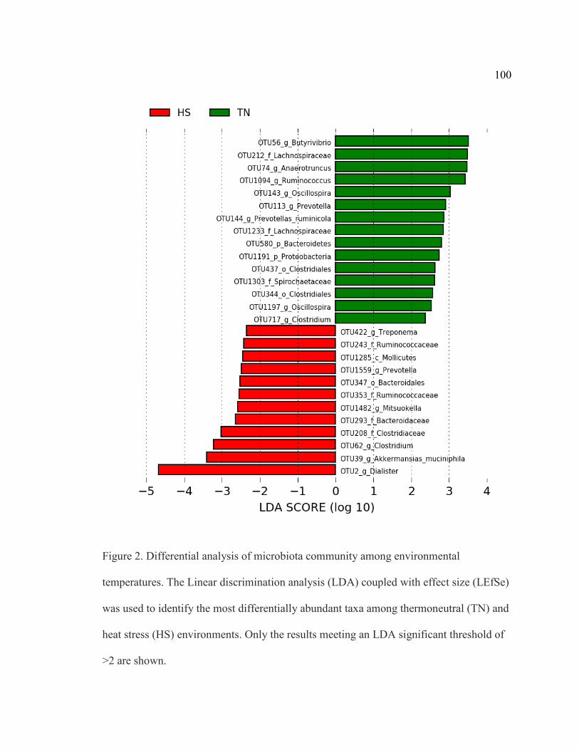
100
Figure 2. Differential analysis of microbiota community among environmental
temperatures. The Linear discrimination analysis (LDA) coupled with effect size (LEfSe)
was used to identify the most differentially abundant taxa among thermoneutral (TN) and
heat stress (HS) environments. Only the results meeting an LDA significant threshold of
>2 are shown.

101
Figure 3. Differential analysis of microbiota community among dietary supplements. The
Linear discrimination analysis (LDA) coupled with effect size (LEfSe) was used to
identify the most differentially abundant taxa among non-supplemented (C) and ZH-
supplements (ZH). Only the results meeting an LDA significant threshold of >2 are
shown.

102
Figure 4. Differential analysis of microbiota community among dietary supplements. The
Linear discrimination analysis (LDA) coupled with effect size (LEfSe) was used to
identify the most differentially abundant taxa among non-supplemented (C) and RH-
supplements (RH). Only the results meeting an LDA significant threshold of >2 are
shown.

103
Figure 5. Differential analysis of microbiota community among groups. The Linear
discrimination analysis (LDA) coupled with effect size (LEfSe) was used to identify the
most differentially abundant taxa among group one (1, red) and group two (2, green).
Only the results meeting an LDA significant threshold of >2 are shown.

104
OTU 296 Strain Query cover % E value Identity, % Papillibacter
cinnamivorans
CIN1 100% 1e-105 93.70%
Clostridium
Viride
T2-7 100% 2e-102 92.89%
Intestinimonas
butyriciproducens
SRB-521-5-I 100% 4e-95 91.34%
Figure 6a. The association of the relative abundance of OTU 296 and group one (G1) or
group two (G2), along with the complementary partial residual plot. The r (sd) coefficient
effect size, and P and q value tests for significance, are shown above the figure. Below
the figure, the top three microorganisms matching the OTU as identified by NCBI
Standard Nucleotide BLAST (Bethesda, MD) are listed.

105
OTU 55 Strain Query cover % E value Identity, % Holdemania massiliensis AP2 100% 3e-76 86.96%
Holdemania filiformis J1-31B-1 100% 3e-76 86.96%
Anaerorhabdus furcosa T-301-A2 100% 1e-69 85.38%
Figure 6b. The association of the relative abundance of OTU 55 and group one (G1) or
group two (G2), along with the complementary partial residual plot. The r (sd) coefficient
effect size, and P and q value tests for significance, are shown above the figure. Below
the figure, the top three microorganisms matching the OTU as identified by NCBI
Standard Nucleotide BLAST (Bethesda, MD) are listed.
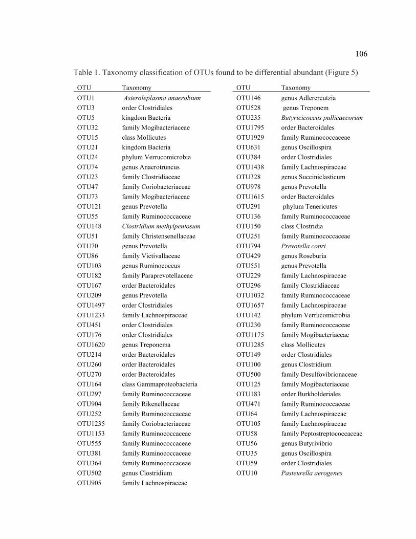
106 Table 1. Taxonomy classification of OTUs found to be differential abundant (Figure 5)
OTU Taxonomy OTU1 Asteroleplasma anaerobium OTU3 order Clostridiales OTU5 kingdom Bacteria OTU32 family Mogibacteriaceae OTU15 class Mollicutes OTU21 kingdom Bacteria OTU24 phylum Verrucomicrobia OTU74 genus Anaerotruncus OTU23 family Clostridiaceae OTU47 family Coriobacteriaceae OTU73 family Mogibacteriaceae OTU121 genus Prevotella OTU55 family Ruminococcaceae OTU148 Clostridium methylpentosum OTU51 family Christensenellaceae OTU70 genus Prevotella OTU86 family Victivallaceae OTU103 genus Ruminococcus OTU182 family Paraprevotellaceae OTU167 order Bacteroidales OTU209 genus Prevotella OTU1497 order Clostridiales OTU1233 family Lachnospiraceae OTU451 order Clostridiales OTU176 order Clostridiales OTU1620 genus Treponema OTU214 order Bacteroidales OTU260 order Bacteroidales OTU270 order Bacteroidales OTU164 class Gammaproteobacteria OTU297 family Ruminococcaceae OTU904 family Rikenellaceae OTU252 family Ruminococcaceae OTU1235 family Coriobacteriaceae OTU1153 family Ruminococcaceae OTU555 family Ruminococcaceae OTU381 family Ruminococcaceae OTU364 family Ruminococcaceae OTU502 genus Clostridium OTU905 family Lachnospiraceae
OTU Taxonomy OTU146 genus Adlercreutzia OTU528 genus Treponem OTU235 Butyricicoccus pullicaecorum OTU1795 order Bacteroidales OTU1929 family Ruminococcaceae OTU631 genus Oscillospira OTU384 order Clostridiales OTU1438 family Lachnospiraceae OTU328 genus Succiniclasticum OTU978 genus Prevotella OTU1615 order Bacteroidales OTU291 phylum Tenericutes OTU136 family Ruminococcaceae OTU150 class Clostridia OTU251 family Ruminococcaceae OTU794 Prevotella copri OTU429 genus Roseburia OTU551 genus Prevotella OTU229 family Lachnospiraceae OTU296 family Clostridiaceae OTU1032 family Ruminococcaceae OTU1657 family Lachnospiraceae OTU142 phylum Verrucomicrobia OTU230 family Ruminococcaceae OTU1175 family Mogibacteriaceae OTU1285 class Mollicutes OTU149 order Clostridiales OTU100 genus Clostridium OTU500 family Desulfovibrionaceae OTU125 family Mogibacteriaceae OTU183 order Burkholderiales OTU471 family Ruminococcaceae OTU64 family Lachnospiraceae OTU105 family Lachnospiraceae OTU58 family Peptostreptococcaceae OTU56 genus Butyrivibrio OTU35 genus Oscillospira OTU59 order Clostridiales OTU10 Pasteurella aerogenes

107
CHAPTER IV: INVESTIGATION OF THE RUMEN EPITHELIUM
TRANSCRIPTOME IN LAMBS FED BETA-ADRENERGIC AGONISTS AND
SUBJECTED TO HEAT STRESS
Introduction
The primary functions of the gastrointestinal epithelium are to provide the host
protection from microorganisms, toxins and chemicals (Gäbel et al., 2002), and to aid in
nutrient absorption, metabolism, and delivery of nutrients to other body tissues (Furness
et al., 2013). The gastrointestinal tract plays a significant role in energetics by utilizing
20% of the oxygen in ruminants and accounting for 30% of metabolic and protein
synthesis activities (Cant et al., 1996). Improving animal growth and efficiency are
critical points of research as the world’s population and demand for agriculture products
increase. Therefore, adaptions or changes in the gut can are of interest to maximize
growth efficiency and wellbeing of livestock.
Supplementation with dietary β-adrenergic agonists (β-AA) is one strategy used
in the livestock industry to increase efficiency of growth in livestock; these supplements
do so by decreasing adipose deposition while increasing protein accretion (Johnson et al.,
2014). However, there is no current literature available that addresses the impact
supplementation with β-AA has on the rumen epithelium. Although β-AA provide a
positive impact on animal agriculture, it has been suggested that feeding supplement
while animals are subjected to stressors such as heat stress can increase mortality
(Grandin, 2008). Conversely, however, Dávila-Ramírez et al. (2014) reported feeding ZH
to rams under heat stress had no negative impact on animal health. The differing reports

108
regarding heat stress and β-AA indicates a gap in the present knowledge of how these
variables impact animal well-being.
Heat stress occurs when an animal’s body temperature rises above its
thermoneutral zone, at which point the heat load exceeds the animal’s capacity for heat
dissipation (Bernabucci et al., 2010) resulting in decreased feed intake and poor
performance (Marai et al., 2007; Guo et al., 2018; Johnson, 2018). Additionally, feed
intake ADG, and final body weight decrease due to heat stress (Beatty et al., 2006;
Mitlöhner et al., 2002; Blaine and Nsahlai, 2011). With respect to the digestive system,
animals also respond to heat by redistributing blood flow from gastrointestinal organs to
the periphery (Sakurada and Hales, 1998). The reduction in blood supply to the gut can
damage its epithelial lining impacting nutrient absorption and permitting endotoxin to
enter the body (Cronjé, 2005). The decrease in animal performance and wellbeing
attributed to heat stress affects economically important carcass and performance traits
leading to millions of dollars in losses every year (Renaudeau et al., 2012).
Heat stress and β-AA supplementation have antagonistic effects on animal
performance, however, there is a gap in knowledge in the understanding of how the two
impact rumen function. Specifically, how these variables individually, and in concert,
impact animal performance and wellbeing in regard to the rumen has yet to be elucidated.
Therefore, the purpose of this study was to investigate the effects of β-AA
supplementation, heat stress, and their interaction on the rumen epithelium using
transcriptomic analyses

109
Materials and Methods
The following experimental procedures were in compliance with the Guide for the
Care and Use of Agricultural Animals in Research and Teaching and approved by the
Institutional Animal and Care and Use Committee at the University of Nebraska -
Lincoln (IACUC protocol #1300). Animal studies were performed at the University of
Nebraska-Lincoln Animal Science Complex.
Experiment One
Forty-nine Suffolk x Rambouillet crossbred lambs (53.26 ± 3.68 kg) were utilized
in a 2X3 factorial design. Upon arrival, lambs were weighed, rectal temperatures
recorded, ear tagged with individual ID’s, metaphylacticaly treated [Ivomec®; 10 mL/hd
(Merial, Duluth, GA) and Draxxin®; 1 mL/hd (Zoetis, Parsippany, NJ)]. Lambs were
stratified by body weight and divided into two groups (group one = 39.99 ±1.92 kg;
group two = 37.35 ± 1.92 kg) to accommodate for the availability of thermal chambers
(12 stalls).
Lambs were housed in one of two environmental conditions. The thermoneutral
(TN) condition was approximately 21 °C and 25% humidity during both study periods.
Heat stress (HS) was achieved by maintaining a temperature of approximately 34 °C and
35% humidity from 0800 to 2000 h, and 29 °C between 2000 and 0800 with temperature
changing over a period of 2 h at the beginning and end of each heat cycle. In both
environmental conditions, the light was on from 0630 to 2045 h.
Lambs assigned to group one were transitioned to a 90% concentrate finishing
diet (49% SweetBran®; 37.8% dry-rolled corn; 8.3% chopped alfalfa hay; 4% mineral

110
supplementation; dosage of coccidiostat (20 g/ton)) over a period of 21 d. Lambs
assigned to group two were held on a maintained diet (54.8% SweetBran®; 41.1%
chopped alfalfa hay; 4% mineral supplementation; dosage of coccidiostat (20 g/ton)) at
2% of BW for 41 d prior to 21 d transition to the 90% concentrate finishing diet
previously stated with the addition of ammonium chloride (10 mg/hd/d).
Lambs were supplemented with one of three supplemental treatments. Zilpaterol
hydrochloride (ZH) was supplemented at 5.44 g/ton (2.5 mg/kg) and ractopamine
hydrochloride (RH) was supplemented at 18.14 g/ton (40 mg/kg). The supplement was
mixed into 200 g of fine ground corn with the control (C) receiving no supplement in
200g of fine ground corn. β-AA were supplemented into the diet beginning on d1 and
mixed into 907.19 g of feed. The remaining amount of regular feed was given around
1400 h.
Within group, lambs were stratified by body weight and randomly assigned to one
of six treatment groups: control/thermoneutral (CTN), Ractopamine HCl/thermoneutral
(RHTN), Zilpaterol HCl/thermoneutral (ZHTN), control/heat stress (CHS), Ractopamine
HCl/heat stress (RHHS), and Zilpaterol HCl/heat stress (ZHHS).
Lambs were harvested on d 21 of the trial. The harvest order was randomly
assigned and 12 were harvested each d via captive bolt followed by exsanguination.
Tissue samples were taken from full thickness sections of the rumen epithelium. All
tissue samples collected were placed in 2 mL collection tubes and flash frozen in liquid
nitrogen. During RNA isolation the rumen tissue samples were dissected from the muscle
so that only the epithelium was isolated.

111
Experiment Two
Twenty-five crossbred Rambouillet lambs (44kg) were utilized in 2x2 factorial
design. Upon arrival, lambs were weighed, rectal temperatures recorded, ear tagged with
individual ID’s, metaphylacticaly treated [Ivomec®; 10 mL/hd (Merial, Duluth, GA) and
Draxxin®; 1 mL/hd (Zoetis, Parsippany, NJ)]. Lambs were housed in group pens and
acclimated for 21-d. After 21 d, lambs were stratified by body weight and randomly
assigned to one of four treatment groups: control/thermoneutral (CTN), Ractopamine
HCl/thermoneutral (RHTN), control/heat stress (CHS), and Ractopamine HCl/heat stress
(RHHS). Additionally, lambs were parsed into four groups (HS: A & B; TN: C & D) to
allow for pair feeding (starting on d -5).
Lambs were housed in one of two environmental conditions; thermoneutral
(controls; 25°C, 15% RH) or heat stress (40°C, 35% RH) for 30 d. Heat stress was
achieved by maintaining a temperature of approximately 40°C and 35% humidity from
0800 to 2000 h, and 29°C between 2000 and 0800 with temperature changing over a
period of 2 h at the beginning and end of each heat cycle. In both environmental
conditions, the light was on from 0630 to 2045 h.
Lambs were fed a 16% protein, 2% fat diet (16% crude protein, 2% crude fat,
16% crude fiber; Lamb Grower-Finisher Complete B30 (Purina)) beginning on d -5 of
the study. Orts, feed left over from the day before, were collected daily and recorded to
evaluate average performance statistics (e.g., average daily gain, gain:feed), to determine
the amount of feed to offer the following day as well as that to offer the pair fed
counterparts. Supplemental treatment included either ractopamine hydrochloride given

112
via oral bolus at 60mg/d/animal, or no supplement (control), for which only an empty
capsule was administered.
Lambs were harvested on d 30 of the study by an overdose of pentobarbital. The
harvest order was randomly assigned and 6 were harvested each day, with the exception
harvest d 1 and d 4 with 5 and 9 lambs being harvested, respectively. Rumen muscle and
epithelium tissue samples were dissected out from each other, placed in 2 mL collection
tubes, and flash frozen in liquid nitrogen.
RNA Extraction from Rumen Epithelium and Sequencing
Total RNA was isolated from the rumen epithelium from the 49 wethers in study
one and 25 wethers in study two using the Direct-zol RNA MiniPrep Plus kit (Zymo
Research) with modifications. Roughly 100 mg of tissue was weighed and minced
quickly before being placed in 600uLof Trizol. The tissue was homogenized in Trizol
using five bead-beating steps performed in a TissueLyser (Qiagen Inc., Valencia, CA,
USA) for 45 sec at 30 Hz with a two-minute incubation in ice between bead-beating
steps.
Following this bead beading step, tissue was further homogenized with the
addition of 400 uL of Trizol. The homogenized tissue was incubated at room temperature
for 5 min after which the samples were spun down to remove any excess tissue. After
transferring the supernatant from this step to new tubes, a chloroform precipitation was
performed where 200 uL of chloroform was added, vortexed for 30 sec and incubated at
room temperature for 3 min. Following this incubation, samples were centrifuged for 15
min at 11,900 rpm and 4 ⁰C. The top aqueous layer was removed and placed in 600 uL of

113
100% ethanol and mixed. This mixture was placed on a spin column and washed twice
using 400 uL of RNA pre-wash buffer. A 15 min DNase treatment was then performed
using 75 uL of DNA digestion buffer and 5 uL of 6U/uL of DNase per sample. This
treatment was followed by three washes with 700 uL of RNA wash buffer. RNA was
eluted in 100 uL of RNase free water.
Once isolated, RNA was quantified, and integrity analyzed using the Agilent
Bioanalyzer. RNA Integrity Number (RIN) scores above 8 were considered adequate.
Samples from study one were sent to the University of California Davis’ Genome Center
(Davis, CA) for QuantSeq 3’ mRNA library prep and 100 bp, single-end sequencing on
an Illumina HiSeq 4000. Samples from study two were sent to the Oklahoma Medical
Research Foundation (Oklahoma City, OK) for standard poly-A+ mRNA prep and
paired-end sequencing was performed on an Illumina NovaSeq S1.
Bioinformatics Analyses
HTStream (Ver. htstream/e919d21) was used for preprocessing and quality
control of sequence data by removing adapter sequences, trim 11 bp and remove 5’ poly-
A/T repeats. Trimmed reads were aligned to the Ovine Oar_v3.1 with STAR (ver2.5)
alignment (Dobin & Gingeras, 2015). Differential expression analysis was performed
comparing samples in HS to TN environments and supplement fed animals to the control
using limma-voom (Law et al. 2014; Ritchie et al. 2015). Lowly-expressed transcripts
(zero expression in half or more of the individuals in each treatment, or with <1 count per
million in n libraries where n=number of individuals in the analysis) were removed.
Transcripts with an adjusted P ≤ 0.10 for study one and adjusted P ≤ 0.05 for study two
were considered differentially expressed (DE). DE transcripts were then analyzed using

114
Ingenuity Pathway Analysis (QIAGEN Inc.; IPA) to predict pathways altered. For genes
without annotated gene IDs in the ovine genome, human or mouse orthologs were input
when possible.
Results
Study One
An average of 4.7 million reads were obtained per sample with 58.49% of the
reads uniquely mapping to the Ovine Oar_v3.1 genome. Of the 27,054 annotated
transcripts, 5,632 were observed in these data; 3,623 remained after removing those
lowly expressed. No interaction was found between temperature and supplement,
therefore only the main effects were evaluated. Analyses identified 607 transcripts with
P(raw)<0.05, with 7 DE (Adj. P < 0.10; Table 1) between TN and HS lambs. No genes
were differentially expressed due to supplement.
Study Two
An average of 26.3 million reads were obtained per sample with 83% mapping
uniquely to the genome. Data from one lamb (L15) identified him as an outlier and thus,
that observation was removed from the analysis. Of the 27,054 transcripts evaluated,
9,690 remained after removing those that were lowly expressed. No interaction between
environment and supplement was observed. Evaluation of the MDS plot generated
showed that samples clustered by group as well as environment (Figure 1), therefore the
model from which DE was identified included the pair fed groups with group A/C
considered batch one and group B/D considered batch two. Resulting analyses identified
733 DE transcripts (Adj. P ≤ 0.05) between TN and HS lambs (Appendix A). From these

115
results, 106 pathways were predicted to be altered due to environment (Table 2). No
transcripts were differentially expressed in the control vs. RH comparison.
Discussion
In this study, we report that heat stress, but not β-AA supplementation, affects the
transcriptome profile of the rumen epithelium. Additionally, no supplement-environment
interaction was present suggesting that supplementation of β-AA during heat stress
conditions does not exacerbate the effects of heat stress on the rumen epithelium.
Interestingly, neither RH nor ZH had an effect on the rumen epithelial transcriptome,
which was unexpected given the presence of β-AR in the epithelium. These results could
suggest that β-AA does not affect the rumen epithelium and nutrient absorption, which
would indicate that these supplements are acting elsewhere in the animal to achieve the
desirable changes in carcass traits. Skeletal muscle strips from animals in study one to
measure ex vivo glucose oxidation under basal and insulin-stimulated conditions.
Supplementation of β2-AA increased muscle glucose oxidation by ~15% and addition of
insulin to the media increased glucose oxidation by ~25%, while supplementation of β1-
AA had no effects on muscle glucose oxidation (Barnes et al. 2017). Taken together,
these data suggest B2-AA impact carcass traits through binding BAR in skeletal muscle
and not through altering rumen epithelial characteristics. Semitendinosus muscle taken
from the lambs in study one also found an impact of ZH; supplement with the B2-AA
altered the expression of 8 transcripts at harvest, and 42 at biopsy (Kubik et al., 2018).
However, RH again had no measurable impact on the skeletal muscle. These findings,
especially the RH results, were surprising due to the known role β-AA play in improving
carcass composition. Adipose tissue was taken from lambs in both studies but has yet to

116
be analyzed. Upon analysis, it could provide an insight into where RH may be working in
the body if not in the skeletal muscle or rumen epithelium.
Seven transcripts from study one and 733 from study two were DE due to heat
stress. The seven transcripts from study one include SLC25A21, LIAS, CCDC17,
HRNNPA3, ZBTB44, ACOX1, and ENSOARG00000008612. Interestingly none of those
DE transcripts overlapped between studies. In study two we utilized full length, paired
end, traditional RNA sequencing while it study one we utilized 3’Tag-Seq which
generates a single library molecule per transcript that is complementary to 3’end
sequences. We speculate that some of the difference in results may be attributed to
missing annotation of the 3’ end of some transcripts in the current ovine reference
genome; poor annotation of the 3’ end of the gene would cause these transcripts to be
absent in analysis while assaying the entire transcript as performed in study two allows
for alignment across the larger genic region. Additionally, it is also possible that
expression in transcripts associated with heat stress and β-AA supplementation in study
one were altered during the period of time the lambs were held at ambient condition in
the abattoir awaiting harvest. Moreover, animals were not given supplementation the day
of harvest in study one which could have also altered expression of transcripts. Due to the
minimal significance observed in study one, the remainder of the discussion will focus on
the results from study two.
Protein Ubiquitination
Protein ubiquitination and the SUMOylation pathways were predicted to be
impacted due to heat stress. During protein ubiquitination, ubiquitin is attached to the
lysine residue of a protein, marking it for degradation and/or preventing its activity in

117
various interactions (Lecker et al., 2006). Heat stress causes protein damage (Schröder et
al., 1993), therefore an increase in ubiquitination is expected. Although there was no
directionality of activity identified by IPA (z-score = NaN), 23 of the 271 genes
annotated as being involved in this pathway were upregulated, while 5 out of the 271
were downregulated; this could be interpreted as suggesting an increase in protein
degradation. Heat shock induces an initially rapid increase in protein degradation in
mammalian cells (Parag et al., 1987; Gropper et al., 1991), which would also explain why
many transcripts involved with the protein ubiquitination pathway were upregulated.
Under the heat stressed condition our lambs were undergoing increased protein damage
which led to an upregulation in transcripts involved in protein ubiquitination to mark
damaged protein for degradation.
Post-translational modification of proteins by members of the small ubiquitin-
related modifier (SUMO) protein family regulates cellular processes, including
transcription, replication, and DNA repair (Johnson, 2004; Geiss-Friedlander and
Melchior, 2007). Modulation of SUMO conjugation is impacted by various
environmental stressors such as osmotic, heat and oxidative stress (Saitoh and Hinchey,
2000). Oxidative and ethanol stress have also been reported to increase SUMOylation in
yeast (Zhou et al., 2004), however these results are not simple as changes in
SUMOylation depends on stress intensity and duration (Tempé et al., 2008). Low to
moderate oxidative stress inhibits global SUMOylation with exposure to 1 mM H2O2
leading to deSUMOylation of cellular proteins within one hour (Bossis et al., 2006). In
our study, the SUMOylation pathway was downregulated (z-score = -0.707), suggesting
oxidative stress in the rumen was causing a delay in the heat stress response (Adachi et
118
al., 2009). Therefore, it is possible that by suppressing the heat stress response, cells
experiencing DNA damage were not able to undergo DNA repair by SUMO proteins,
leading to apoptosis. Alternatively, a downregulation in the SUMO pathway could be due
to the lambs becoming acclimated to heat stress environment, resulting in a
desensitization to oxidative stress and a decreased need for the response.
Oxidative Stress
Heat stress is a cause of oxidative stress (Akbarian et al., 2016; Alemu et al.,
2018). The production of reactive oxygen species (ROS) is a key event of many
inflammatory disorders, including disorders of the gastrointestinal tract. In the gut,
disruption of the mucosal barrier will rapidly activate the innate immune system and
induce an acute inflammatory response (Aviello et al, 2017). The production of ROS in
the gastrointestinal tract due to the immune response may aid in the resolution of
inflammation (Campbell et al., 2014). However, uncontrolled and persistent oxidative
stress leading to an overproduction of reactive oxygen species will cause tissue injury
(Rezaie et al., 2007).
The NRF2-mediated oxidative stress response was predicted to be upregulated (z-
score = 0.905) in the lambs housed in a heat stress environment compared to the
thermoneutral controls. This pathway is activated when oxidative stress is present and the
genes involved act to eliminate and detoxify reactive oxidants (Nguyen et al., 2009).
Similarly to the tissue samples in this study, when bovine granulosa cells were exposed to
heat stress, there was an increase in NRF2-mediated signaling (Alemu et al., 2018).
Additionally, specific genes expected to be involved in heat stress and oxidative stress,
including DNAJ genes, NQO1, ACTG2, and GAB1, were upregulated in these lambs after

119
30 d of heat exposure. These lambs, therefore, may have responded to heat by excessive
production of ROS, leading to an upregulation of this pathway to eliminate reactive
oxidants that were causing intestinal injury. Increased intestinal injury in our lambs due
to the overproduction of ROS, could allow for endotoxin to escape from the gut, leading
to increase in the inflammation response. Additionally, intestinal tissue damage would
have major impacts on an animal’s ability to absorb nutrients, leading to a decline in
animal growth and performance.
Another pathway that was altered by heat stress in the study was colanic acid
building blocks biosynthesis (P = 2.7E-06). Colanic acid (CA) is an exopolysaccharide
structure, produced under stress that is associated with the surface of a variety of bacterial
species (Grant et al., 1969). Colanic acid is associated with Enterobacteriaceae,
specifically Escherichia coli, and acts as a protective agent against environmental
stressors (Navasa et al., 2009). Colanic acid is normally lowly produced, however, large
amounts can be synthesized in response to specific mutation or environmental factors.
The biosynthesis of colonic acid is up regulated in response to several environmental
factors that modulate or damage cell structure including temperature (Navasa et al., 2009)
and oxidative stress (Chen et al., 2004). Additionally, CA regulates mitochondrial
dynamics of the host and unfolded protein response (Han et al., 2017). Because heat
stress impacts mitochondrial function (Huang et al., 2015) it is possible that increased
secretion of CA by bacteria in the gut worked to combat damage host mitochondria
underwent. Therefore, the upregulation of this pathway in our study is evidence that the
bacteria of the rumen microbiome were producing increased levels of CA to combat heat
stress.

120
Additionally, SAPK/JNK signaling (P = 4.17E-03; z-score = 1.667; Figure. 2)
and ERK/MAPK (P = 3.28E-03; z-score = 0.577; Figure. 3) were identified by IPA as
likely to have been altered by heat stress. c-Jun N-terminal kinases (JNKs), also known as
stress-activated protein kinases (SAPKs), are characterized by their activation in response
to cell stress by playing a vital role in cell proliferation, apoptosis, and differentiation
(Dhanasekaran et al., 2008). Whether the activation of JNKs leads to apoptosis or cell
differentiation is dependent on the stimuli and the cell-type involved (Lin and Dibling,
2002; Liu and Lin, 2005). The extracellular-signal-regulated kinase (ERK) pathway is
one of the major signaling cassettes of the mitogen activated protein kinase (MAPK)
signaling pathway. The ERK cascade can be activated by cellular stressors to induce cell
growth, proliferation, differentiation and apoptosis (Lu et al., 2006).
The upregulation of these two pathways in our study are consistent with other
reports evaluating the ERK and JNK pathways. Considering the effect of oxidative stress
on intestinal epithelial cell apoptosis, Zhou et al. (2005), suggested that activation of
ERK and JNK may be involved in the H2O2-induced intestinal epithelial cell apoptosis
since the inhibition of either pathway resulted in reduction of the apoptotic response. Due
to heat stress being associated with oxidative stress the lambs in our study may have been
undergoing rumen epithelial cell apoptosis due to overproduction of ROS which is
suggested by these two pathways being upregulated. In terms of heat stress, cells respond
to elevated temperatures by activation of p38 MAPK, ERK1/2, and JNK, as well as by
increasing the production of heat shock proteins (Mearow et al., 2002; Gourgou et al.,
2010; Murai et al., 2010). Although, upregulation of HSP transcripts involved in these
pathways were not observed, RAS and MAPK transcripts were predicted to be

121
upregulated. Both RAS and MAPK can induce apoptosis (Cox and Der, 2003; Wada and
Penninger; 2004); additionally, RAS can also stimulate the production of ROS (Ozaki et
al., 2000). Upregulation of these transcripts in response to heat stress further suggests
increased apoptosis of cells damaged due to heat and oxidative stress.
Immune Response
Animals exposed to heat stress have a suppressed immune response, which could
include serious health implications because an effective immune response is critical for
maintenance of health and productive function (Ajuwon and Hua, 2015). Nuclear factor-
κB (NF-κB) is an important intracellular signaling protein that controls the transcription
of genes involved in cell growth, survival, apoptosis, and inflammatory responses
(Baldwin, 2001). Interestingly the NF-κB signaling pathway was downregulated (z-score
= -0.58) in the lambs of this current study. Similar results were observed in the
semitendinosus of female pigs subjected to heat stress in which it was shown that heat
stress did not increase inflammatory signaling through the NF-κB pathway, suggesting
NF-κB pathway quiescence (Montilla et al., 2014). Additionally, a variety of heat shock
response inducers are reported to prevent activation of NF-κB (Rossi et al.,1997). A study
utilizing HS-Sultan cells showed that heat stress was able to inhibit constitutive NF-κB
DNA-binding activity, resulting in apoptosis (Belardo et al., 2009). The downregulation
of NF-κB signaling due to heat stress seen in the current study could indicate increased
cell apoptosis as well as decreased immune function which could have negative impacts
on animal health and viability in a livestock production system.
122
Cell Cycle and DNA Damage
Heat stress induces DNA damage responses in S, G1, and G2-phase cells
(Velichko et al., 2012). Additionally, severe heat stress may lead to cell death through
apoptosis (Velichko et al., 2013), downregulate DNA repair genes (Rockett et al., 2001),
and induce senescence-like cell cycle arrest (Velichko et al., 2013). In the lambs of study
two, both the G2/M DNA damage checkpoint regulation (P = 4.81E-03) and nucleotide
excision repair (NER; P = 1.86E-03) pathways were projected to be upregulated by heat
stress. DNA damage arrests the cell cycle to allow for DNA repair to occur before DNA
replication or mitosis begins (Harper and Elledge, 2007; Ciccia and Elledge, 2010; Hu et
al., 2016). In our lambs the DNA checkpoint pathway was upregulated (z-score = 0.45),
suggesting an increase in cell cycle arrest to repair damaged DNA. Although previous
pathways discussed regarding this study suggest an increase in apoptosis, the
upregulation in DNA repair in our lambs may indicate that epithelial cells were trying to
preserve cell viability through DNA repair. The NER pathway was also upregulated (z-
score = 1.667) in our lambs. The NER pathway recognizes DNA helix-distorting lesions
(Marteijn et al. 2014). The upregulation of the NER pathway further indicates DNA
damage that was occurring due to heat stress and DNA repair was increased through NER
and DNA damage regulation.
Conclusion
Feedlot cattle finished in the summer months are often adversely affected by heat
stress which can impact production, costing the industry and impacting animal well-
being. Further, β-AA supplementation is common in livestock production to improve
animal performance. Although no interaction between environment and supplement was

123
identified, nor were any transcripts differentially expressed due to β-AA, transcripts
found to be differentially expressed due to heat stress revealed potential mechanisms
through which heat stressed ruminants respond to environmental stressors.
No impact of either β-AA supplement, RH or ZH, was observed. β-AAs therefore
appear to have little effect on the epithelial lining of the rumen; thus, they likely have an
alternate mode of action to achieve the desirable change in carcass weight and
composition observed. ZH appears to be acting on the skeletal muscle (Barnes et al,
2017; Kubik et al., 2018), however mechanisms behind the RH mode of action are still
unclear, reinforcing the need for additional molecular studies to elucidate the specific
impacts and mode of action of β-AA. Many of the transcripts and pathways identified to
be altered due to heat stress were associated with oxidative stress and apoptosis.
Therefore, understanding how these pathways impact rumen function and the ability to
derive and absorb nutrients will be critical in improving animal wellbeing and
performance while under heat-stressed conditions.

124
References
Adachi M, Liu Y, Fujii K, Calderwood SK, Nakai A, Imai K, et al. (2009) Oxidative Stress Impairs the Heat Stress Response and Delays Unfolded Protein Recovery. PLoS ONE 4(11): e7719. https://doi.org/10.1371/journal.pone.0007719
Ajuwon, K. and Hua, Z. (2015). Heat stress and LPS activate multiple signaling pathways to regulate cytokine expression in macrophages. The Federation of American Societies for Experimental Biology, 29(1).
Akbarian, A., J. Michiels, J. Degroote, M. Majdeddin, A. Golian, and S. Smet. 2016. Association between heat stress and oxidative stress in poultry; mitochondrial dysfunction and dietary interventions with phytochemicals. Journal of animal science and biotechnology 7(1):37.
Alemu, T. W., H. O. Pandey, D. S. Wondim, S. Gebremedhn, C. Neuhof, E. Tholen, M. Holker, K. Schellander, and D. Tesfaye. 2018. Oxidative and endoplasmic reticulum stress defense mechanisms of bovine granulosa cells exposed to heat stress. Theriogenology 110:130-141.
Argan Oil as a Model Transposable to Aging. Oxidative Medicine and Cellular Longevity 2018:1-13.
Aviello, G., and U. Knaus. 2016. ROS in gastrointestinal inflammation: Rescue Or Sabotage?. British Journal of Pharmacology 174:1704-1718.
Baldwin, A. S. Control of oncogenesis and cancer therapy resistance by the transcription factor NF-kappaB. J Clin Invest 107, 241–246 (2001)
Barnes, T. L., R. Kubik, C. Cadaret, K. Beede, E. Merrick, S. Chung, T. Schmidt, J. Petersen, and D. Yates. 2017. Identifying hyperthermia in heat-stressed lambs and its effects on β agonist–stimulated glucose oxidation in muscle. Translational Animal Science 1(supplement1):100-104. 11671.
Beatty, D., A. Barnes, E. Taylor, D. Pethick, M. McCarthy, and S. Maloney. 2006. Physiological responses of and cattle to prolonged, continuous heat and humidity. Journal of Animal Science 84(4):972-985.
Belardo, G., R. Piva, and M. Santoro. 2009. Heat stress triggers apoptosis by impairing NF-κB survival signaling in malignant B cells. Leukemia 24:187-196.
Bernabucci, U., N. Lacetera, L. H. Baumgard, R. P. Rhoads, B. Ronchi, and A. Nardone. 2010. Metabolic and hormonal acclimation to heat stress in domesticated ruminants. Animal 4(7):1167-1183.
Blaine, K., and I. V. Nsahlai. 2011. The effects of shade on performance, carcass classes and behaviour of heat-stressed feedlot cattle at the finisher phase. Tropical animal health and production 43(3):609-615.
Bossis G., Melchior F. (2006) Regulation of SUMOylation by reversible oxidation of SUMO conjugating enzymes. Mol. Cell 21:349–357.

125
Campbell EL, Bruyninckx WJ, Kelly CJ, Glover LE, McNamee EN, Bowers BE, et al. (2014). Transmigrating neutrophils shape the mucosal microenvironment through localized oxygen depletion to influence resolution of inflammation. Immunity 40: 66–77.
Cant, J., B. McBride, and W. Croom. 1996. The regulation of intestinal metabolism and its impact on whole animal energetics. Journal of Animal Science 74:2541.
Chen J, Lee SM, Mao Y. 2004. Protective effect of exopolysaccharide colanic acid of Escherichia coli O157:H7 to osmotic and oxidative stress. Int. J. Food Microbiol. 93:281–286.
Ciccia, A., and S. Elledge. 2010. The DNA Damage Response: Making It Safe to Play with Knives. Molecular Cell 40:179-204.
Cox AD, Der CJ. The dark side of Ras: regulation of apoptosis. Oncogene. 2003;22:8999–9006.
Cronjé PB. 2005. Heat stress in livestock - role of the gut in its aetiology and a potential role for betaine in its alleviation. Recent Adv Animal Nutr Australia. 15: 107-122.
Dávila-Ramírez, J., U. Macías-Cruz, N. Torrentera-Olivera, H. González-Ríos, S. Soto-Navarro, R. Rojo-Rubio, and L. Avendaño-Reyes. 2014. Effects of zilpaterol hydrochloride and soybean oil supplementation on feedlot performance and carcass characteristics of hair-breed ram lambs under heat stress conditions. Journal of animal science 92(3):1184-1192.
Dhanasekaran, D., and E. Reddy. 2008. JNK signaling in apoptosis. Oncogene 27:6245-6251.
Dobin A. & Gingeras T.R. (2015) Mapping RNA-seq Reads with STAR. Curr Protoc Bioinformatics 51, 11 4 1-9.
El Hajj HI, et al. The inflammatory response in acyl-CoA oxidase 1 deficiency (pseudoneonatal adrenoleukodystrophy) Endocrinology. 2012;153(6):2568–2575. doi: 10.1210/en.2012-1137.
Flohr C, Burkle A, Radicella JP, Epe B. Poly(ADP-ribosyl)ation accelerates DNA repair in a pathway dependent on Cockayne syndrome B protein. Nucleic Acids Res 2003;31:5332–5337.
Furness, J., L. Rivera, H. Cho, D. Bravo, and B. Callaghan. 2019. The gut as a sensory organ. Nat. Rev. Gastroenterol. Hepatol., 10 (2013), pp. 729-740
Gäbel, G., J. R. Aschenbach, and F. Muller. 2002. Transfer of energy substrates across the ruminal epithelium: Implications and limitations. Anim. Health Res. Rev. 3:15– 30.
Geiss-Friedlander, R. and Melchior, F. (2007). Concepts in sumoylation: a decade on. Nat. Rev Mol. Cell. Biol. 8, 947-956.
126
Gourgou E., Aggeli I.-K., Beis I., Gaitanaki C. Hyperthermia-induced Hsp70 and MT20 transcriptional upregulation are mediated by p38-MAPK and JNKs in Mytilus galloprovincialis (Lamarck); a pro-survival response. Journal of Experimental Biology. 2010;213(2):347–357. doi: 10.1242/jeb.036277.
Grandin, T. 2013. Temple Grandin explains animal welfare problems with beta-agonists. Beef
Magazine 9Good, I.J. 1953. The population frequencies of species and the estimation of population parameters. Biometrika 40:237–264. doi:10.1093/biomet/40.3-4.237
Grant WD, Sutherland IW, Wilkinson JF. 1969. Exopolysaccharide colanic acid and its
occurrence in the Enterobacteriaceae. J Bacteriol 100:1187–1193. Gropper, R., R. A. Bran&, S. Elias, C. F. Bearer, A. Mayer, A. L. Schwartz, and A.
Ciechanover. 1991. The ubiquitin-activating enzyme, El, is required for stress-induced tysosomal degradation of celtular proteins. J. Biol. Chem. 266:3602-3610.
Guo, K., H. Cao, Y. Zhu, T. Wang, G. Hu, B. Kornmatitsuk, and J. Luo. 2018. Improving
effects of dietary rumen protected γ‐aminobutyric acid additive on apparent nutrient digestibility, growth performance and health status in heat‐stressed beef cattle. Animal Science Journal. doi: 10.1111/asj.13053.
Han, B., P. Sivaramakrishnan, C. Lin, I. Neve, J. He, L. Tay, J. Sowa, A. Sizovs, G. Du, J. Wang, C. Herman, and M. Wang. 2017. Microbial Genetic Composition Tunes Host Longevity. Cell 169:1249-1262.e13.
Harper, J., and S. Elledge. 2007. The DNA Damage Response: Ten Years After. Molecular Cell 28:739-745.
Hong Y., Rogers R., Matunis M.J., Mayhew C.N., Goodson M.L., Park-Sarge O.K., Sarge K.D. (2001) Regulation of heat shock transcription factor 1 by stress-induced SUMO-1 modification. J. Biol. Chem. 276:40263–40267.
Hu, Z., T. Cools, and L. De Veylder. 2016. Mechanisms Used by Plants to Cope with DNA Damage. Annual Review of Plant Biology 67:439-462.
Huang, C., H. Jiao, Z. Song, J. Zhao, X. Wang, and H. Lin. 2015. Heat stress impairs mitochondria functions and induces oxidative injury in broiler chickens1. Journal of Animal Science 93:2144-2153.
Johnson, E. S. (2004). Protein modification by SUMO. Annu. Rev. Biochem. 73, 355-382.
Johnson, B. J. 2014. Mechanism of action of beta adrenergic agonists and potential residue issues. American Meat Association Reference Paper:1-6.
Johnson, B. J., S. B. Smith, and K. Y. Chung. 2014. Historical overview of the effect of β-adrenergic agonists on beef cattle production. Asian-Australasian journal of animal sciences 27(5):757.

127
Johnson, J. 2018. Impact of Heat Stress on Livestock and Mitigation Strategies to Improve Productivity and Well-Being. Journal of Animal Science 96:6-6.
Kubik, R., S. Tietze, T. Schmidt, D. Yates, and J. Petersen. 2018. Investigation of the skeletal muscle transcriptome in lambs fed β adrenergic agonists and subjected to heat stress for 21 d1. Translational Animal Science 2:S53-S56.
Kurepa, J., J. Walker, J. Smalle, M. Gosink, S. Davis, T. Durham, D. Sung, and R. Vierstra. 2002. The Small Ubiquitin-like Modifier (SUMO) Protein Modification System inArabidopsis. Journal of Biological Chemistry 278:6862-6872.
Law C.W., Chen Y., Shi W. & Smyth G.K. (2014) voom: Precision weights unlock linear model analysis tools for RNA-seq read counts. Genome Biol 15, R29.
Lecker, S., A. Goldberg, and W. Mitch. 2006. Protein Degradation by the Ubiquitin–Proteasome Pathway in Normal and Disease States. Journal of the American Society of Nephrology 17:1807-1819.
Li Y, Arita Y, Koo HC, Davis JM, Kazzaz JA 2003 Inhibition of c-Jun N-terminal kinase pathway improves cell viability in response to oxidant injury. Am J Respir Cell Mol Biol 29: 779–783
Lin A, Dibling B. The true face of JNK activation in apoptosis. Aging Cell. 2002;1:112–116.
Liu J, Lin A. Role of JNK activation in apoptosis: a double-edged sword. Cell Res. 2005;15:36–42.
Lu Z, Xu S. ERK1/2 MAP kinases in cell survival and apoptosis. IUBMB Life. 2006. 58(11):621–31. pmid:17085381
Marteijn JA, Lans H, Vermeulen W, Hoeijmakers JH (2014) Understanding nucleotide excision repair and its roles in cancer and ageing. Nat Rev Mol Cell Biol 15:465–481. https://doi.org/10. 1038/nrm3822
Mearow K. M., Dodge M. E., Rahimtula M., Yegappan C. Stress-mediated signaling in PC12 cells: the role of the small heat shock protein, Hsp27, and Akt in protecting cells from heat stress and nerve growth factor withdrawal. Journal of Neurochemistry. 2002;83(2):452–462. doi: 10.1046/j.1471-4159.2002.01151.x.
Montilla, S., T. Johnson, S. Pearce, D. Gardan-Salmon, N. Gabler, J. Ross, R. Rhoads, L. Baumgard, S. Lonergan, and J. Selsby. 2014. Heat stress causes oxidative stress but not inflammatory signaling in porcine skeletal muscle. Temperature 1:42-50.
Marai, I., A. El-Darawany, A. Fadiel, and M. Abdel-Hafez. 2007. Physiological traits as affected by heat stress in sheep—a review. Small Ruminant Research 71(1):1-12.
Mitlöhner, F., M. Galyean, and J. McGlone. 2002. Shade effects on performance, carcass traits, physiology, and behavior of heat-stressed feedlot heifers. Journal of Animal Science 80(8):2043-2050.

128
Murai H., Hiragami F., Kawamura K., et al. Differential response of heat-shock-induced p38 MAPK and JNK activity in PC12 mutant and PC12 parental cells for differentiation and apoptosis. Acta Medica Okayama. 2010;64(1):55–62.
Navasa N, Rodríguez-Aparicio L, Martínez-Blanco H, Arcos M, Ferrero MA. 2009. Temperature has reciprocal effects on colanic acid and polysialic acid biosynthesis in E. coli K92. Appl. Microbiol. Biotechnol. 82:721–729.
Nguyen, T., P. Nioi, and C. Pickett. 2009. The Nrf2-Antioxidant Response Element Signaling Pathway and Its Activation by Oxidative Stress. Journal of Biological Chemistry 284:13291-13295.
Ozaki M., Deshpande S. S., Angkeow P., Bellan J., Lowenstein C. J., Dinauer M. C., Goldschmidt-Clermont P. J., Irani K. (2000): Inhibition of the Rac1 GTPase protects against nonlethal ischemia/reperfusion-induced necrosis and apoptosis in vivo. FASEB J. 14, 418—429
Parag HA, Raboy B, Kulka RG (1987) Effect of heat shock on protein degradation in mammalian cells: Involvement of the ubiquitin system. EMBO J 6:55-61.
Renaudeau, D., A. Collin, S. Yahav, V. De Basilio, J. Gourdine, and R. Collier. 2012. Adaptation to hot climate and strategies to alleviate heat stress in livestock production. Animal 6(5):707-728.
Rezaie A, Parker RD, Abdollahi M (2007). Oxidative stress and pathogenesis of inflammatory bowel disease: an epiphenomenon or the cause? Dig Dis Sci 52: 2015–2021.
Ritchie M.E., Phipson B., Wu D., Hu Y., Law C.W., Shi W. & Smyth G.K. (2015) limma powers differential expression analyses for RNA-sequencing and microarray studies. Nucleic Acids Research 43, e47.
Rockett JC, Mapp FL, Garges JB, Luft JC, Mori C, Dix DJ. Effects of hyperthermia on spermatogenesis, apoptosis, gene expression, and fertility in adult male mice. Biol Reprod 2001;65:229–239
Rossi A, Elia G, Santoro MG. Inhibition of nuclear factor kappa B by prostaglandin A1: an effect associated with heat shock transcription factor activation. Proc Natl Acad Sci USA 1997; 94: 746–750.
Saitoh, H. and Hinchey, J. (2000). Functional heterogeneity of small ubiquitin-related protein modifiers SUMO-1 versus SUMO-2/3. J. Biol. Chem. 275, 6252-6258.
Sakurada, S., and J. Hales. 1998. A role for gastrointestinal endotoxins in enhancement of heat tolerance by physical fitness. Journal of Applied Physiology 84:207-214.
Schreiber V, Ame JC, Dolle P, Schultz I, Rinaldi B, Fraulob V, Menissier-de Murcia J, de Murcia G. Poly(ADP-ribose) polymerase-2 (PARP-2) is required for efficient base excision DNA repair in association with PARP-1 and XRCC1. J Biol Chem 2002;277:23028– 23036.
129
Schröder H., Langer,T., Hartl,F.-U. and Bukau,B. (1993) DnaK, DnaJ and GrpE form a cellular chaperone machinery capable of repairing heat-induced protein damage. EMBO J., 12, 4137–4144
Stanciu M, DeFranco DB 2002 Prolonged nuclear retention of activated extracellular signal-regulated protein kinase promotes cell death generated by oxidative toxicity or proteasome inhibition in a neuronal cell line. J Biol Chem 277: 4010–4017
Tempé, D., M. Piechaczyk, and G. Bossis. 2008. SUMO under stress: Figure 1. Biochemical Society Transactions 36:874-878.
Tramontano F, Malanga M, Farina B, Jones R, Quesada P. Heat stress reduces poly(ADPR)polymerase expression in rat testis. Mol Hum Reprod 2000;6:575– 581.
Vamecq, J., P. Andreoletti, R. El Kebbaj, F. Saih, N. Latruffe, M. El Kebbaj, G. Lizard, B. Nasser, and M. Cherkaoui-Malki. 2018. Peroxisomal Acyl-CoA Oxidase Type 1: Anti-Inflammatory and Anti-Aging Properties with a Special Emphasis on Studies with LPS and
Velichko,A.K., Petrova,N.V., Kantidze,O.L. and Razin,S.V. (2012) Dual effect of heat shock on DNA replication and genome integrity. Mol. Biol. Cell, 23, 3450–3460.
Velichko,A.K., Markova,E.N., Petrova,N.V., Razin,S.V. and Kantidze,O.L. (2013) Mechanisms of heat shock response in mammals. Cell. Mol. Life Sci., 70, 4229–4241.
Wada, T., and J. Penninger. 2004. Mitogen-activated protein kinases in apoptosis regulation. Oncogene 23:2838-2849.
Zhou W., Ryan J.J., Zhou H. (2004) Global analyses of sumoylated proteins in Saccharomyces cerevisiae. Induction of protein sumoylation by cellular stresses. J. Biol. Chem. 279:32262–32268
Zhou, Y., Q. Wang, B. Evers, and D. Chung. 2005. Signal Transduction Pathways Involved in Oxidative Stress-Induced Intestinal Epithelial Cell Apoptosis. Pediatric Research 58:1192-1197.
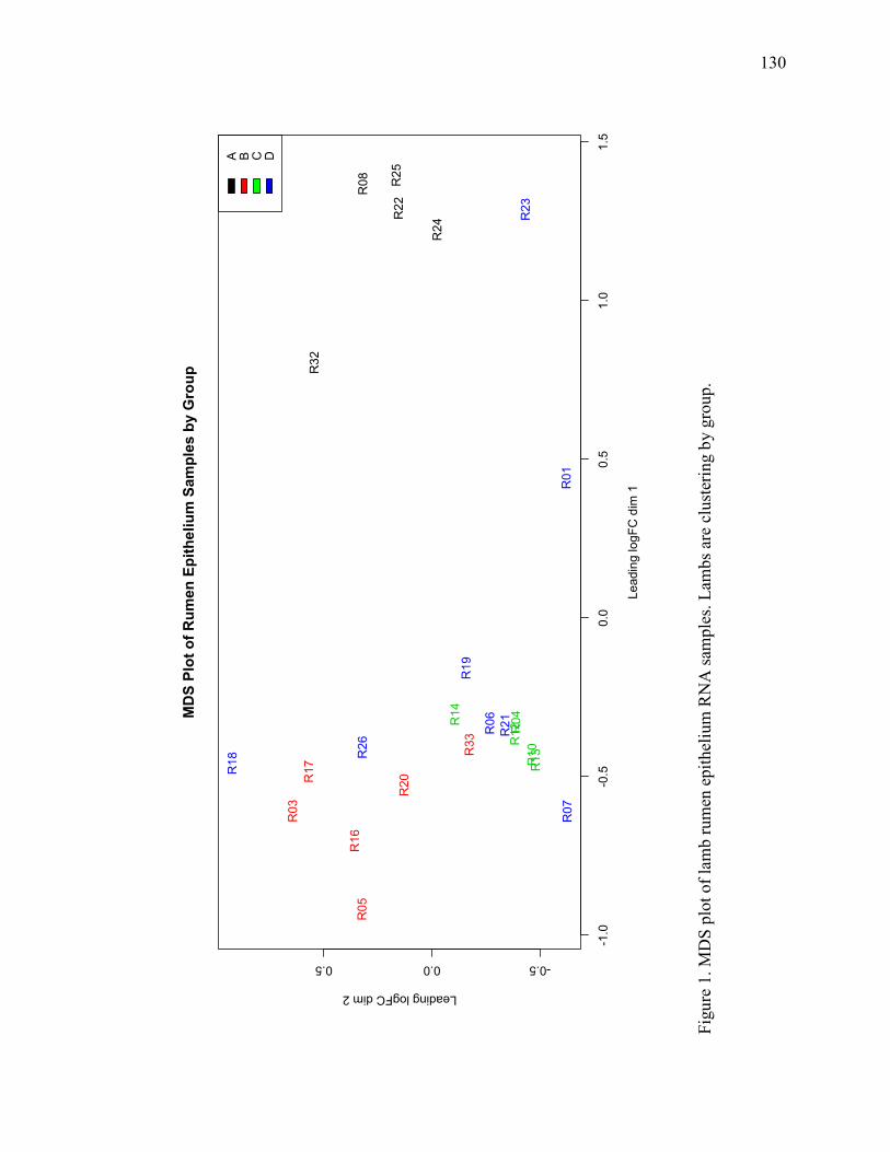
130
Figu
re 1
. MD
S pl
ot o
f lam
b ru
men
epi
thel
ium
RN
A sa
mpl
es. L
ambs
are
clu
ster
ing
by g
roup
.

131
Figu
re 2
. SA
PK/J
NK
Sig
nalin
g (IP
A):
The
JNK
sign
alin
g pa
thw
ay w
as p
redi
cted
to b
e up
regu
late
d in
anim
als i
n th
e he
at st
ress
env
ironm
ent.
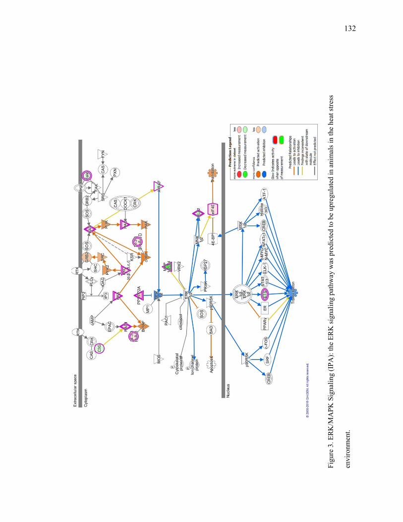
132
Figu
re 3
. ERK
/MA
PK S
igna
ling
(IPA
): th
e ER
K si
gnal
ing
path
way
was
pre
dict
ed to
be
upre
gula
ted
in a
nim
als i
n th
e he
at st
ress
envi
ronm
ent.

133
Table 1. Transcripts differentially expressed in study one due to heat stress (adj P-value <
0.1) in the rumen epithelium.
Gene Name logFC Adj. P.Value
SLC25A21 -1.5126 0.04 LIAS 0.947 0.05 CCDC17 0.7333 0.06 HNRNPA3 1.1341 0.06 ZBTB44 0.7533 0.06 ACOX1 0.6781 0.08 ENSOARG00000008612 0.6725 0.08

134
Table 2. Top canonical pathways altered in the rumen epithelium due to heat stress (P-
value < 0.05, -log(p-value) > 1.3).
Canonical Pathways P-value z-score Protein Ubiquitination Pathway 7.00E-08 NaN NRF2-mediated Oxidative Stress Response 1.00E-06 0.905 Colanic Acid Building Blocks Biosynthesis 2.70E-06 2.449 Sumoylation Pathway 2.81E-05 -0.707 Sirtuin Signaling Pathway 3.07E-05 -0.258 Heme Biosynthesis II 1.24E-04 1 LPS/IL-1 Mediated Inhibition of RXR Function 1.33E-04 0 Germ Cell-Sertoli Cell Junction Signaling 1.37E-04 NaN Tetrapyrrole Biosynthesis II 3.21E-04 NaN Acyl-CoA Hydrolysis 4.37E-04 1 Bile Acid Biosynthesis, Neutral Pathway 6.15E-04 1 GDP-mannose Biosynthesis 6.27E-04 NaN Adenine and Adenosine Salvage I 1.05E-03 NaN Integrin Signaling 1.33E-03 1.807 Mismatch Repair in Eukaryotes 1.45E-03 NaN Vitamin-C Transport 1.45E-03 NaN Estrogen Receptor Signaling 1.68-03 NaN Adenosine Nucleotides Degradation II 1.85E-03 -1 NER Pathway 1.86-03 1.667 Virus Entry via Endocytic Pathways 1.92E-03 NaN Xenobiotic Metabolism Signaling 2.17E-03 NaN Glucocorticoid Receptor Signaling 2.47E-03 NaN ILK Signaling 2.86E-03 1.897 Macropinocytosis Signaling 2.94E-03 1.633 Sertoli Cell-Sertoli Cell Junction Signaling 3.01E-03 NaN Semaphorin Signaling in Neurons 3.14E-03 NaN Renal Cell Carcinoma Signaling 3.16E-03 0 ERK/MAPK Signaling 3.28E-03 0.577 Regulation of Actin-based Motility by Rho 3.40E-03 2.121 Fcγ Receptor-mediated Phagocytosis in Macrophages and Monocytes 3.40E-03 0.333
Granzyme A Signaling 3.48E-03 NaN Purine Nucleotides Degradation II (Aerobic) 3.48E-03 -1 Cardiac Hypertrophy Signaling 3.92E-03 1.941 Actin Cytoskeleton Signaling 4.11E-03 1.941 SAPK/JNK Signaling 4.17E-03 1.667 Androgen Signaling 4.74E-03 0.816 Signaling by Rho Family GTPases 4.76E-03 1.155

135
Cell Cycle: G2/M DNA Damage Checkpoint Regulation 4.81E-03 0.447 Cholecystokinin/Gastrin-mediated Signaling 5.31E-03 1.89 IL-6 Signaling 5.58E-03 -0.333 RhoGDI Signaling 5.89E-03 -1.265 Pyrimidine Deoxyribonucleotides De Novo Biosynthesis I 5.89E-03 2 Guanosine Nucleotides Degradation III 5.96E-03 NaN BER pathway 5.96E-03 NaN Arsenate Detoxification I (Glutaredoxin) 6.05E-03 NaN CXCR4 Signaling 6.74E-03 2.828 HGF Signaling 7.07E-03 0 Paxillin Signaling 7.07E-03 1.89 Urate Biosynthesis/Inosine 5'-phosphate Degradation 7.57E-03 NaN Caveolar-mediated Endocytosis Signaling 7.99E-03 NaN tRNA Charging 8.10E-03 2.236 Glioma Invasiveness Signaling 8.72E-03 2 Oxidative Phosphorylation 8.92E-03 3 NF-κB Signaling 9.01E-03 -0.577 Cell Cycle Control of Chromosomal Replication 9.24E-03 NaN Androgen Biosynthesis 9.40E-03 NaN Galactose Degradation I (Leloir Pathway) 9.80E-03 NaN Estrogen Biosynthesis 1.00E-02 0.447 Breast Cancer Regulation by Stathmin1 1.05E-02 NaN Epithelial Adherens Junction Signaling 1.12E-02 NaN Hereditary Breast Cancer Signaling 1.22E-02 NaN Agrin Interactions at Neuromuscular Junction 1.31E-02 1.89 Stearate Biosynthesis I (Animals) 1.34E-02 1.342 Thrombin Signaling 1.41E-02 2.449 Tec Kinase Signaling 1.44E-02 1.134 HMGB1 Signaling 1.50E-02 0.378 LXR/RXR Activation 1.69E-02 0 Glutathione-mediated Detoxification 1.90E-02 2 Thioredoxin Pathway 1.97E-02 NaN Phospholipase C Signaling 2.07E-02 2.111 IL-8 Signaling 2.32E-02 1.897 MIF-mediated Glucocorticoid Regulation 2.34E-02 0 Retinoate Biosynthesis I 2.34E-02 1 Mitochondrial Dysfunction 2.36E-02 NaN Remodeling of Epithelial Adherens Junctions 2.41E-02 NaN The Visual Cycle 2.55E-02 NaN Gαq Signaling 2.55E-02 1.134 Inflammasome pathway 2.55E-02 NaN Adenine and Adenosine Salvage III 2.57E-02 NaN Nucleotide Excision Repair Pathway 2.57E-02 NaN

136
Actin Nucleation by ARP-WASP Complex 2.89E-02 2 PAK Signaling 2.91E-02 1.633 Acute Phase Response Signaling 3.16E-02 -0.333 VEGF Signaling 3.19E-02 1.633 Glutamine Biosynthesis I 3.24E-02 NaN Purine Ribonucleosides Degradation to Ribose-1-phosphate 3.24E-02 NaN Xanthine and Xanthosine Salvage 3.24E-02 NaN Asparagine Biosynthesis I 3.24E-02 NaN D-mannose Degradation 3.24E-02 NaN Prolactin Signaling 3.28E-02 NaN Production of Nitric Oxide and Reactive Oxygen Species in Macrophages 3.41E-02 -1.134
Protein Kinase A Signaling 3.71E-02 1 CCR3 Signaling in Eosinophils 3.85E-02 1.342 LPS-stimulated MAPK Signaling 3.98E-02 1.633 Hepatic Fibrosis / Hepatic Stellate Cell Activation 4.00E-02 NaN VDR/RXR Activation 4.06E-02 0 Adrenomedullin signaling pathway 4.11E-02 1.265 Mechanisms of Viral Exit from Host Cells 4.28E-02 NaN Aryl Hydrocarbon Receptor Signaling 4.31E-02 -1 TR/RXR Activation 4.36E-02 NaN PPARα/RXRα Activation 4.40E-02 1.667 Leukocyte Extravasation Signaling 4.50E-02 2.714 RAR Activation 4.50E-02 NaN Retinol Biosynthesis 4.61E-02 1 RhoA Signaling 4.66E-02 2.121 Gα12/13 Signaling 4.81E-02 2.121

137
APPENDIX A: DIFFERENTIALLY EXPRESSED RUMEN EPITHELIUM
TRANSCRIPTS IN LAMBS FED BETA-ADRENERGIC AGONISTS AND
SUBJECTED TO HEAT STRESS FOR 30 DAYS
Table 1. Transcripts differentially expressed in study two due to heat stress (adj P-value <
0.05) in the rumen epithelium.
Gene Name logFC Adj.P.Val DES 8.768 0.000 SORBS2 4.687 0.000 FLNC 3.207 0.001 SH3BGR 3.027 0.001 MAP1A 3.122 0.001 PFDN1 0.432 0.001 KCNMA1 3.943 0.001 CETN2 0.513 0.001 LBHD1 0.923 0.001 CNN1 4.559 0.001 SYNPO2 3.319 0.001 WFIKKN2 3.912 0.002 NEXN 2.183 0.002 MYCBP 0.578 0.002 PARK7 0.419 0.002 REXO2 0.553 0.002 JPH2 2.797 0.003 COX17 0.502 0.003 PCP4 6.621 0.003 CELSR1 -0.477 0.003 RBPMS2 2.770 0.003 DUOXA1 0.746 0.003 ACTN2 2.180 0.003 LMOD1 2.162 0.003 TNFRSF6B 1.265 0.003 NEK1 -0.435 0.004 USP14 0.319 0.004 CLMP 2.815 0.004 ACTA1 5.849 0.004 PLAT -0.727 0.004
Gene Name logFC Adj.P.Val GFER 0.431 0.004 RDH11 0.529 0.004 PPP1R14A 1.484 0.004 TXN 0.524 0.004 TMEM86A -1.505 0.004 ECI2 0.610 0.005 SAMD8 -1.330 0.005 MRGPRF 2.510 0.005 PLIN4 3.237 0.005 DNASE2 0.535 0.005 ASTN2 -0.986 0.005 RNF44 -0.521 0.005 THEM6 0.789 0.005 ARHGEF25 1.961 0.005 FAM13B -0.407 0.005 NDUFB3 0.355 0.005 UBE2S 0.349 0.005 NQO1 0.812 0.005 GALK1 0.638 0.005 Tomm5 0.506 0.005 PSMA7 0.427 0.005 HAGH 0.280 0.005 CALML4 1.641 0.005 DNPH1 0.574 0.005 ITGA7 2.004 0.005 VEZF1 -0.377 0.005 GREB1 -1.047 0.005 MIEN1 0.446 0.006 DDX17 -0.304 0.006 TIMM50 0.338 0.006

138
YIF1B 0.421 0.006 DST -0.652 0.006 MAP2K7 -0.396 0.006 PLXND1 -0.777 0.007 DTYMK 0.499 0.007 GNG11 1.498 0.007 SWI5 0.381 0.007 HIST1H2BN 0.426 0.007 SMYD1 5.029 0.007 PDLIM3 2.048 0.007 JMJD7 0.593 0.007 MFAP5 1.712 0.007 NT5C2 -0.403 0.007 ITPRIPL2 -0.568 0.007 RAB34 0.431 0.007 Eloc 0.338 0.007 ACOT1 -0.919 0.007 SIGMAR1 0.419 0.007 ACTG2 3.831 0.007 MCUR1 0.401 0.007 RAC3 0.624 0.007 SLITRK2 2.778 0.008 HSPB6 1.752 0.008 SCEL -0.803 0.008 ARHGAP21 -0.345 0.008 EIF2S2 0.338 0.008 DELE1 -0.286 0.008 ENAH 0.476 0.008 CYP2B6 -2.411 0.008 LDHB 0.485 0.008 H2AFZ 0.399 0.009 MYO9B -0.299 0.009 PTGIS 2.197 0.009 STK36 -0.347 0.009 PDF 0.378 0.009 ZACN -1.234 0.009 MYL12B 0.336 0.009 PATL1 -0.477 0.009 SMPX 4.493 0.009 GPX2 1.134 0.009
STK10 -0.376 0.009 CUL7 -0.274 0.009 RAPGEF1 -0.307 0.009 SPCS3 0.361 0.009 SLC7A5 0.713 0.009 ANXA1 0.332 0.009 CMC2 0.547 0.010 ID1 0.747 0.010 FAM20C 0.639 0.010 ESD 0.673 0.010 SYNM 2.587 0.010 MELK 0.562 0.010 LAMB3 -0.643 0.010 APRT 0.574 0.010 SLC25A5 0.385 0.010 COL17A1 -0.365 0.010 AL162171.1 -0.507 0.010 MTHFD2 0.525 0.010 DNAJB5 1.155 0.010 IL1R2 -0.542 0.010 KPNA2 0.306 0.010 PAPLN -0.721 0.010 AGTPBP1 0.349 0.010 ELP1 -0.272 0.010 DHX57 -0.251 0.010 MOXD1 1.258 0.011 MRPL40 0.402 0.011 RRAS 0.766 0.011 GNMT 0.625 0.011 ATP8B1 -0.738 0.011 LY6D 1.010 0.011 FOXA1 -0.321 0.011 ANP32E 0.362 0.011 SLC20A1 -0.463 0.011 SAE1 0.269 0.011 PLS1 -0.987 0.011 PLCB4 1.376 0.011 CAP2 0.899 0.011 NR2C2 -0.323 0.011 SPC24 0.520 0.011

139
KIAA0930 -0.535 0.011 NCBP2 0.301 0.011 HYPK 0.404 0.012 GLCE -0.467 0.012 GSTM3 1.356 0.012 ELL2 0.565 0.012 HMOX1 -1.116 0.012 RNASEH2C 0.523 0.012 TP63 -0.357 0.013 CBY1 0.358 0.013 KLHL41 1.268 0.013 SH2B1 -0.318 0.013 SPTBN1 -0.267 0.013 SSNA1 0.418 0.013 TGFB1I1 1.439 0.013 NDUFAF5 0.418 0.013 ENPP2 -0.772 0.013 SLC23A1 -0.647 0.013 SERPINB3 0.975 0.013 PGP 0.412 0.013 ADA -1.070 0.014 OTUD1 -0.414 0.014 RNF20 -0.219 0.014 SCD 0.950 0.014 PCLAF 0.583 0.014 MBD6 -0.415 0.014 TMEM126A 0.366 0.014 N4BP3 0.403 0.015 PLEKHA4 0.429 0.015 H1FX 0.331 0.015 IL36G 0.810 0.015 DUSP4 -1.615 0.015 GGPS1 0.355 0.015 ILVBL -0.357 0.015 IGF2R -0.370 0.015 GAB2 -0.455 0.015 ASF1B 0.387 0.015 NFU1 0.366 0.015 ITGB4 -0.408 0.015 CNIH4 0.302 0.015
TSTA3 0.296 0.015 LSM4 0.416 0.016 TK1 0.605 0.016 CKB 0.339 0.016 CFAP36 0.388 0.016 CNTNAP1 1.499 0.016 MYBL2 0.504 0.016 POFUT2 -0.262 0.016 TUBB3 1.018 0.016 RRM2 0.582 0.016 USP36 -0.302 0.016 TP53I13 0.584 0.016 NDST2 -0.308 0.016 POLR2G 0.301 0.016 DNASE1L1 0.318 0.016 LSM7 0.367 0.016 SMDT1 0.328 0.016 BEX3 0.822 0.017 MRPS26 0.423 0.017 TEF -0.325 0.017 FIZ1 -0.386 0.017 KIF3B -0.317 0.017 SLC15A4 -0.359 0.017 HSD17B8 0.327 0.017 TIMM17B 0.410 0.017 DENND1B -0.503 0.017 RHOBTB2 -0.393 0.017 SUGT1 0.294 0.018 GMPPB 0.403 0.018 PTTG2 0.369 0.018 GLUL -0.886 0.018 STARD10 0.572 0.018 GFPT1 0.468 0.018 LAMC2 -0.438 0.018 ST6GALNAC2 0.640 0.018 USP54 -0.622 0.018 SIPA1L3 -0.415 0.018 CFL2 1.179 0.019 NUAK2 0.393 0.019 FABP7 2.553 0.019

140
TMEM184B -0.286 0.019 CIAO2B 0.306 0.019 Akr1c18 0.554 0.019 MYEF2 0.373 0.019 BAHCC1 -0.438 0.019 RFC2 0.378 0.020 RF00100 0.772 0.020 COL16A1 -0.474 0.020 Akr1c14 0.790 0.020 ATP6V1F 0.370 0.020 VBP1 0.318 0.020 TXNDC17 0.481 0.021 CEBPB 0.551 0.021 GALT 0.577 0.021 CACNA1C 1.737 0.021 POLR1A -0.259 0.023 GLCCI1 -1.020 0.023 CRCP 0.317 0.023 PITPNA 0.279 0.023 AC118754.1 -0.738 0.023 CENPU 0.481 0.023 PRRC2B -0.407 0.023 TSR3 0.302 0.023 F8 -0.652 0.023 SMIM11A 0.460 0.023 CD14 0.600 0.023 FAM114A2 0.210 0.023 TUBA4B 0.313 0.024 CDC42 0.226 0.024 POMT2 -0.289 0.024 C11orf71 0.655 0.024 TIMM8B 0.384 0.024 POP5 0.367 0.024 SPRYD3 0.206 0.024 SMG6 -0.259 0.024 CMPK1 0.291 0.024 DAAM2 1.262 0.025 COX20 0.380 0.025 MCM6 0.369 0.025 ARHGEF40 1.170 0.025
TRIT1 0.314 0.025 SAV1 -0.297 0.025 DCXR 0.410 0.025 ALOXE3 -1.161 0.025 CDCA3 0.442 0.025 RAB25 0.344 0.026 TRIO -0.278 0.026 ERCC6L 0.617 0.026 PTPRF -0.344 0.026 KCP 0.730 0.026 CTDSPL2 -0.270 0.026 UBTD2 -0.428 0.026 NCAPG 0.519 0.026 TSHZ1 -0.283 0.026 CCDC149 -0.466 0.026 PSMD14 0.270 0.026 TRMT112 0.390 0.026 BZW2 0.456 0.026 Akr1c21 0.567 0.026 RGL2 -0.344 0.026 PPOX 0.360 0.026 PRR14L -0.300 0.026 RGP1 -0.348 0.026 SNRPF 0.435 0.026 NEK7 -0.379 0.026 ADHFE1 -0.459 0.026 SRA1 0.336 0.026 NDUFB4 0.287 0.026 CYP2D6 -0.443 0.026 APOE -0.651 0.026 DLG5 -0.441 0.026 MFSD3 -0.534 0.026 STARD5 0.385 0.026 YWHAZ 0.310 0.026 AMACR -0.479 0.026 POMP 0.280 0.026 ETS1 -0.423 0.026 GTF2F2 0.238 0.027 TTC17 -0.275 0.027 PLXNB1 -0.285 0.027

141
SLC35E3 0.295 0.027 TRAP1 0.240 0.027 POLB 0.305 0.027 LFNG -0.628 0.027 TBC1D13 -0.258 0.027 PSPC1 0.394 0.027 TPP1 -0.534 0.027 CNPY4 0.345 0.027 PTMA 0.341 0.027 ACOT8 0.610 0.027 ASAP1 -0.561 0.027 E2F1 0.431 0.027 GIGYF2 -0.250 0.027 VPS26B -0.297 0.028 AKAP12 1.382 0.028 RPP25L 0.280 0.028 DHRS9 -1.541 0.028 KBTBD4 -0.614 0.028 CENPW 0.561 0.028 FKBP4 0.456 0.028 IMMP1L 0.412 0.028 THEM5 0.796 0.028 LRP4 -0.611 0.028 MRPL20 0.416 0.028 MRPS21 0.351 0.028 UBR4 -0.289 0.029 CD93 -0.730 0.029 MKLN1 0.217 0.029 CRIM1 -0.256 0.029 PYCR1 0.707 0.029 BOLA3 0.353 0.029 DYNC2LI1 0.459 0.029 SNX3 0.247 0.029 KLC3 0.383 0.030 OXLD1 0.367 0.030 UCHL5 0.306 0.030 FBXO32 -0.944 0.030 ZNF318 -0.347 0.030 PNP 0.477 0.030 TMEM69 0.345 0.030
B4GALT1 -0.300 0.030 IGFBP3 -0.938 0.030 ANKRD28 -0.253 0.030 DPH1 -0.244 0.030 FAM120B -0.213 0.030 MYL6 0.316 0.030 GTF3C2 -0.214 0.030 POLR3H 0.329 0.030 LYNX1 0.557 0.030 GPAT3 -0.938 0.030 Mxra7 0.842 0.030 LIMS2 1.282 0.030 KANK2 1.151 0.030 ANP32A 0.450 0.030 NFIL3 0.384 0.031 ERMARD -0.378 0.031 BCL7C 0.371 0.031 Akr1c20 0.527 0.031 PDXK 0.301 0.031 RAPGEF5 -0.450 0.031 CHCHD7 0.312 0.031 ATG3 0.237 0.031 RAP1A 0.214 0.031 SURF6 0.364 0.031 DESI1 0.282 0.031 ARHGAP35 -0.313 0.031 DNAJB4 0.414 0.031 UBE3B -0.200 0.031 P3H4 0.471 0.031 MRPS15 0.208 0.031 CCT6A 0.422 0.031 SARNP 0.295 0.031 GSTA1 0.766 0.031 Cenpx 0.354 0.031 RANBP1 0.351 0.031 GMNN 0.379 0.032 TXNRD1 0.695 0.032 CYP2D6 -0.407 0.032 CD164L2 0.381 0.032 ZBTB38 -0.320 0.032

142
TLN2 -0.312 0.032 RFC3 0.342 0.032 MRPL19 0.246 0.032 ELMO2 -0.285 0.032 AP3D1 -0.221 0.032 HSPD1 0.262 0.032 CHDH -0.754 0.032 PDIA6 0.278 0.032 TACC1 0.694 0.032 MMP9 1.129 0.032 SLC27A4 0.384 0.032 AR -0.323 0.032 MFN2 -0.338 0.032 APOO 0.261 0.033 ANP32D 0.552 0.033 SF3B5 0.409 0.033 MYLK 1.800 0.033 ZNF862 -0.294 0.033 PROSER1 -0.311 0.033 HIKESHI 0.313 0.034 NUDT22 0.350 0.034 AGO2 0.267 0.034 FAM204A 0.258 0.034 RPS4X 0.424 0.034 FERMT1 -0.277 0.034 TLR4 -0.585 0.034 PPP2R3C 0.249 0.034 Akr1c6 0.594 0.034 AK1 0.547 0.034 PSMG3 0.247 0.034 CACTIN -0.176 0.034 EP300 -0.389 0.034 PEX26 -0.340 0.034 WWC1 -0.324 0.034 NUBP2 0.274 0.035 NUP58 -0.172 0.035 IL1B -0.744 0.035 RPA3 0.320 0.035 COPG2 -0.269 0.035 HCFC1 -0.573 0.035
SLC20A2 -0.492 0.035 DBP -0.447 0.035 PCNA 0.381 0.035 TMEM231 -0.329 0.035 NCBP2-AS2 0.361 0.035 CTSO -0.368 0.035 PRDM2 -0.385 0.035 AP1M2 0.271 0.035 MYO19 0.279 0.035 PSMD1 0.249 0.035 AGRN -0.423 0.035 EBP 0.421 0.035 LAMA3 -0.299 0.035 TNFRSF12A 0.767 0.036 DMPK 1.116 0.036 LTBP3 0.357 0.036 CHERP -0.253 0.036 SKP1 0.249 0.036 IFT22 0.409 0.036 MRPL42 0.240 0.036 GLRX5 0.390 0.036 TRIP13 0.422 0.036 GRID1 1.881 0.036 CRY2 -0.333 0.036 RNF216 -0.279 0.036 DPP3 0.244 0.036 LUZP1 -0.268 0.036 COMTD1 0.388 0.036 BNIP3L -0.404 0.036 RAPGEFL1 -0.313 0.036 AP002956.1 1.209 0.036 PDSS1 0.297 0.036 PSMA5 0.344 0.036 KALRN 0.590 0.036 BCL9 -0.304 0.036 ENDOG 0.376 0.036 PLA2G6 -0.544 0.036 AARSD1 0.250 0.036 CPSF7 -0.263 0.036 TIMM10B 0.379 0.036

143
SEC14L1 -0.450 0.036 TRAPPC12 -0.201 0.037 NDUFA4 0.262 0.037 IL27RA 0.486 0.037 CEP78 0.335 0.037 DRG1 0.278 0.037 DUSP14 -0.662 0.037 FAM83A 0.474 0.037 ETV4 0.714 0.037 ARL3 0.327 0.037 SLC50A1 0.514 0.037 ZNRF1 0.329 0.037 COPA -0.188 0.037 ZNF592 -0.302 0.037 USP11 -0.220 0.037 RAD51 0.365 0.037 NR3C1 -0.247 0.038 PSMD6 0.227 0.038 NDUFB2 0.295 0.038 NMRAL1 0.297 0.038 SDF2L1 0.475 0.038 CCSER2 -0.428 0.038 PEX11G 0.346 0.038 ADGRF4 0.283 0.038 LTA4H 0.358 0.038 KIAA1211L -0.211 0.038 DRAM2 -0.276 0.038 CKS2 0.363 0.038 USP2 -0.458 0.038 PHTF2 -0.630 0.038 Anapc11 0.261 0.038 HAUS1 0.281 0.039 MND1 0.422 0.039 MRTFB -0.380 0.039 IL6ST -0.306 0.039 JAG2 -0.338 0.039 ZC3H12C -0.497 0.039 TEC 0.247 0.039 PRKCB 1.024 0.039 DYRK2 -0.241 0.039
WDHD1 0.390 0.039 FARSB 0.280 0.039 C1QBP 0.200 0.039 IMPA2 0.216 0.039 PNKP 0.239 0.039 MED16 -0.206 0.039 PKMYT1 0.673 0.039 NDUFA12 0.237 0.039 THRB -0.339 0.039 TBC1D22B -0.233 0.039 DPH7 0.256 0.039 ABO 1.261 0.039 SLC29A1 0.526 0.039 LGALS7 0.399 0.039 DSG2 -0.490 0.040 ATP5MC1 0.276 0.040 MIB1 0.184 0.040 ENOSF1 -0.360 0.040 EMC9 0.356 0.040 A2M -0.609 0.040 CHST2 -0.858 0.040 MRPL24 0.332 0.040 MAP3K9 -0.582 0.040 FCHO2 -0.361 0.040 FAM98C 0.343 0.040 IL19 -1.837 0.040 CARS2 0.255 0.040 GTF2IRD2 -0.432 0.040 PET100 0.346 0.040 ALAD 0.285 0.040 SNRPA1 0.353 0.040 DRAP1 0.265 0.040 ISOC2 0.329 0.040 PSMB10 0.378 0.040 NRDE2 -0.396 0.040 ZFHX3 -0.370 0.040 UBL4A 0.284 0.040 PEPD -0.332 0.040 ME2 0.362 0.040 DDX54 0.190 0.040

144
RERE -0.431 0.040 GOLGA3 -0.238 0.040 MTMR4 -0.264 0.040 Akr1c18 0.503 0.040 RBM3 -0.551 0.040 ACOT7 0.754 0.040 ARAF -0.272 0.040 CYB5R4 0.249 0.040 TMEM258 0.216 0.040 BBOX1 -0.581 0.040 CMTR1 -0.288 0.040 CCNH 0.217 0.040 MRPL32 0.261 0.040 ZZEF1 -0.347 0.040 HARS2 0.166 0.040 NRBP1 -0.194 0.040 YEATS2 -0.246 0.040 ZNF512B -0.388 0.040 ACPP -0.522 0.040 B9D1 0.414 0.040 HEATR4 -0.665 0.040 BCOR -0.363 0.040 UBN1 -0.217 0.040 AXL -0.570 0.040 OGDH -0.199 0.040 RAPH1 -0.506 0.040 PECAM1 -0.596 0.040 LRIF1 0.264 0.040 CHMP6 0.284 0.041 P2RX4 0.371 0.041 SF1 -0.211 0.041 TSTD1 0.314 0.041 SPAG9 -0.372 0.041 CRYAB 1.314 0.041 MRPL41 0.352 0.041 BRAT1 -0.480 0.041 LTB4R 0.490 0.041 MAGED1 -0.312 0.041 BTF3L4 0.204 0.041 HIVEP2 -0.301 0.041
BUD23 0.226 0.041 PTPMT1 0.379 0.042 LRRC24 -0.304 0.042 HECTD1 -0.271 0.042 ATG5 0.192 0.042 ANAPC13 0.304 0.042 TPP2 -0.334 0.042 RB1CC1 -0.238 0.042 CSRP1 0.998 0.042 SEL1L -0.339 0.043 KNTC1 0.372 0.043 MAP4K2 0.266 0.043 HMBS 0.223 0.043 CASC3 -0.205 0.043 PRPF8 -0.233 0.043 MICOS10 0.282 0.043 TIMM22 0.285 0.043 PIH1D1 0.235 0.043 PIGF 0.312 0.043 WWC3 -0.466 0.043 HMGXB3 -0.245 0.043 PRKACA 0.197 0.043 ARID1B -0.278 0.043 RRNAD1 -0.268 0.044 MCM2 0.349 0.044 PRDX2 0.278 0.044 HEBP2 0.272 0.044 SIPA1L2 -0.287 0.044 DPM3 0.408 0.044 ENHO 0.825 0.044 ATP5MD 0.304 0.044 PLEKHA8 0.256 0.044 RAB19 0.336 0.044 RYK 0.217 0.044 MPI 0.363 0.044 MXD3 0.353 0.044 SRRM1 -0.225 0.044 CDCP1 -0.241 0.044 DENND4B -0.366 0.044 UBE2C 0.336 0.044

145
FTSJ1 -0.458 0.044 C8orf33 0.267 0.044 CHAMP1 -0.224 0.044 FOXN1 0.499 0.044 MET -0.289 0.045 MRPL55 0.361 0.045 APCDD1 -0.299 0.045 RUSC1 0.280 0.045 NDUFA2 0.306 0.045 TPX2 0.249 0.045 SMARCD1 -0.228 0.045 MRPL57 0.302 0.045 TBCB 0.279 0.045 EIF4E 0.293 0.045 WDR81 -0.390 0.045 SEPT7 0.250 0.045 OSBPL1A -0.297 0.045 WDR83 0.346 0.045 SEC22B 0.202 0.045 MLF2 0.277 0.045 SUSD1 -0.456 0.045 ZEB1 0.734 0.045 HIST1H1D 0.535 0.046 BUB3 0.336 0.046 RGS2 0.615 0.046 TGIF2 0.239 0.046 CNST -0.496 0.046 DAP3 0.215 0.046 TMEM94 -0.237 0.046 CDK5RAP3 0.164 0.046 SIVA1 0.394 0.046 HMGCS1 0.306 0.046 EXOSC1 0.276 0.046 TRAPPC2B 0.249 0.046 POLR2A -0.435 0.046 ATRAID 0.308 0.046 CDC20 0.387 0.046 FRMD4A -0.309 0.047 SEC61G 0.258 0.047 TGFBRAP1 -0.233 0.047
MBNL1 0.351 0.047 TAF4 -0.278 0.047 ANKMY2 0.286 0.047 DNAJB6 0.278 0.047 ATPAF2 0.223 0.047 UBAP2L -0.229 0.047 NUP98 -0.207 0.047 EWSR1 0.195 0.047 ANKS3 0.234 0.047 BNIP1 0.301 0.047 ATP9B -0.199 0.047 BAG2 0.621 0.047 CAMK1 0.500 0.047 ALAS1 -0.373 0.047 EXO1 0.566 0.047 STAT6 -0.310 0.047 PLIN3 0.298 0.047 FBP2 -0.407 0.047 CDH13 -0.430 0.047 Akr1cl 0.468 0.047 PDLIM2 0.295 0.047 NCOA6 -0.328 0.047 ABCG1 -0.824 0.047 MRPL58 0.262 0.047 TARS 0.215 0.047 SUB1 0.374 0.047 CNNM4 -0.219 0.047 CCT5 0.327 0.047 MTRR -0.681 0.047 ESCO2 0.496 0.047 KIF13B -0.488 0.047 IMP4 0.294 0.047 CLUH -0.224 0.047 EEF1E1 0.347 0.047 SMARCAD1 -0.265 0.047 PMM1 0.303 0.047 CD276 -0.275 0.047 EMILIN1 1.253 0.047 PSMD4 0.218 0.047 MGST2 0.353 0.047

146
VPS29 0.268 0.048 TNRC6C -0.397 0.048 DACT3 0.921 0.048 PARP4 -0.274 0.048 NEO1 -0.199 0.048 SGSH -0.305 0.048 TTK 0.462 0.048 CCDC88C -0.301 0.048 EPRS 0.236 0.048 KLHDC8B 0.584 0.048 CDC6 0.537 0.048 CBS 0.366 0.048 ICE1 -0.207 0.048 ACSS1 0.773 0.048 SMCR8 -0.345 0.048 NCOA2 -0.376 0.049 TAPBPL 0.226 0.049 RHOD 0.472 0.049
SLC9A8 -0.352 0.049 NCCRP1 1.464 0.049 NR2C2AP 0.337 0.049 AOX1 -0.345 0.049 ASB8 -0.226 0.049 C6orf132 -0.344 0.050 ASNS 0.487 0.050 MXRA5 -0.664 0.050 GSTO1 0.345 0.050 TUBA1B 0.260 0.050 SAP18 0.196 0.050 RPL26L1 0.264 0.050 DBNDD2 0.327 0.050 TPM4 0.420 0.050 SBF1 -0.215 0.050