Immunotherapy in Multiple Myeloma - Remedy Publications · Journal of Hematology & Multiple...
9
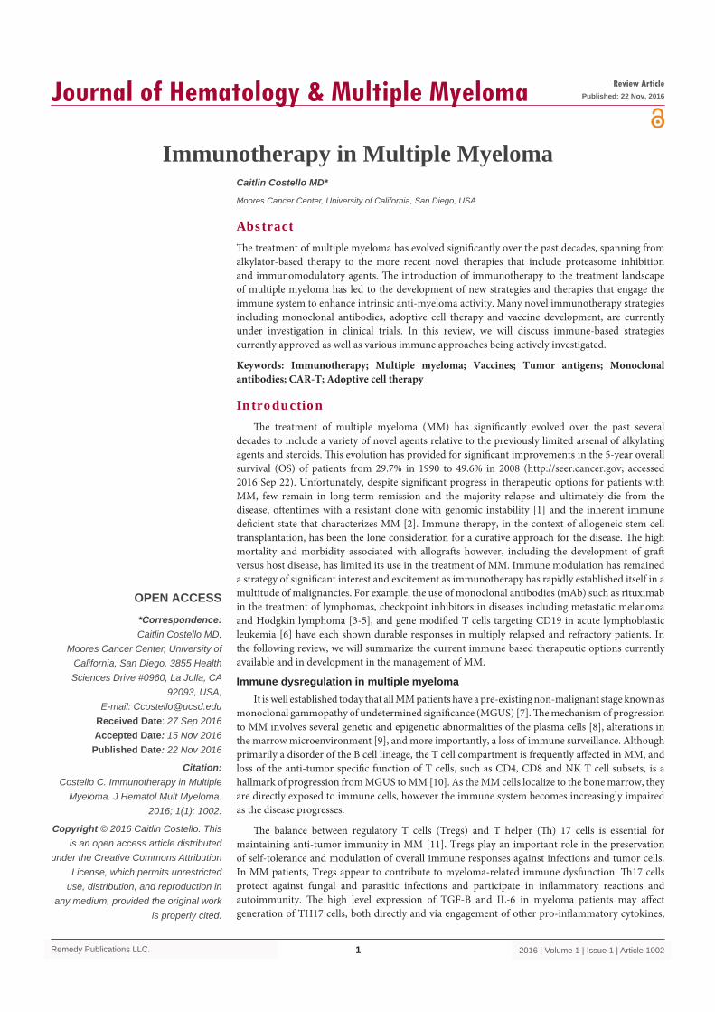
Remedy Publications LLC. Journal of Hematology & Multiple Myeloma 2016 | Volume 1 | Issue 1 | Article 1002 1 Introduction e treatment of multiple myeloma (MM) has significantly evolved over the past several decades to include a variety of novel agents relative to the previously limited arsenal of alkylating agents and steroids. is evolution has provided for significant improvements in the 5-year overall survival (OS) of patients from 29.7% in 1990 to 49.6% in 2008 (http://seer.cancer.gov; accessed 2016 Sep 22). Unfortunately, despite significant progress in therapeutic options for patients with MM, few remain in long-term remission and the majority relapse and ultimately die from the disease, oſtentimes with a resistant clone with genomic instability [1] and the inherent immune deficient state that characterizes MM [2]. Immune therapy, in the context of allogeneic stem cell transplantation, has been the lone consideration for a curative approach for the disease. e high mortality and morbidity associated with allograſts however, including the development of graſt versus host disease, has limited its use in the treatment of MM. Immune modulation has remained a strategy of significant interest and excitement as immunotherapy has rapidly established itself in a multitude of malignancies. For example, the use of monoclonal antibodies (mAb) such as rituximab in the treatment of lymphomas, checkpoint inhibitors in diseases including metastatic melanoma and Hodgkin lymphoma [3-5], and gene modified T cells targeting CD19 in acute lymphoblastic leukemia [6] have each shown durable responses in multiply relapsed and refractory patients. In the following review, we will summarize the current immune based therapeutic options currently available and in development in the management of MM. Immune dysregulation in multiple myeloma It is well established today that all MM patients have a pre-existing non-malignant stage known as monoclonal gammopathy of undetermined significance (MGUS) [7]. e mechanism of progression to MM involves several genetic and epigenetic abnormalities of the plasma cells [8], alterations in the marrow microenvironment [9], and more importantly, a loss of immune surveillance. Although primarily a disorder of the B cell lineage, the T cell compartment is frequently affected in MM, and loss of the anti-tumor specific function of T cells, such as CD4, CD8 and NK T cell subsets, is a hallmark of progression from MGUS to MM [10]. As the MM cells localize to the bone marrow, they are directly exposed to immune cells, however the immune system becomes increasingly impaired as the disease progresses. e balance between regulatory T cells (Tregs) and T helper () 17 cells is essential for maintaining anti-tumor immunity in MM [11]. Tregs play an important role in the preservation of self-tolerance and modulation of overall immune responses against infections and tumor cells. In MM patients, Tregs appear to contribute to myeloma-related immune dysfunction. 17 cells protect against fungal and parasitic infections and participate in inflammatory reactions and autoimmunity. e high level expression of TGF-B and IL-6 in myeloma patients may affect generation of TH17 cells, both directly and via engagement of other pro-inflammatory cytokines, Immunotherapy in Multiple Myeloma OPEN ACCESS *Correspondence: Caitlin Costello MD, Moores Cancer Center, University of California, San Diego, 3855 Health Sciences Drive #0960, La Jolla, CA 92093, USA, E-mail: [email protected] Received Date: 27 Sep 2016 Accepted Date: 15 Nov 2016 Published Date: 22 Nov 2016 Citation: Costello C. Immunotherapy in Multiple Myeloma. J Hematol Mult Myeloma. 2016; 1(1): 1002. Copyright © 2016 Caitlin Costello. This is an open access article distributed under the Creative Commons Attribution License, which permits unrestricted use, distribution, and reproduction in any medium, provided the original work is properly cited. Review Article Published: 22 Nov, 2016 Abstract e treatment of multiple myeloma has evolved significantly over the past decades, spanning from alkylator-based therapy to the more recent novel therapies that include proteasome inhibition and immunomodulatory agents. e introduction of immunotherapy to the treatment landscape of multiple myeloma has led to the development of new strategies and therapies that engage the immune system to enhance intrinsic anti-myeloma activity. Many novel immunotherapy strategies including monoclonal antibodies, adoptive cell therapy and vaccine development, are currently under investigation in clinical trials. In this review, we will discuss immune-based strategies currently approved as well as various immune approaches being actively investigated. Keywords: Immunotherapy; Multiple myeloma; Vaccines; Tumor antigens; Monoclonal antibodies; CAR-T; Adoptive cell therapy Caitlin Costello MD* Moores Cancer Center, University of California, San Diego, USA
Transcript of Immunotherapy in Multiple Myeloma - Remedy Publications · Journal of Hematology & Multiple...
Remedy Publications LLC.
Journal of Hematology & Multiple Myeloma
2016 | Volume 1 | Issue 1 | Article 10021
IntroductionThe treatment of multiple myeloma (MM) has significantly evolved over the past several
decades to include a variety of novel agents relative to the previously limited arsenal of alkylating agents and steroids. This evolution has provided for significant improvements in the 5-year overall survival (OS) of patients from 29.7% in 1990 to 49.6% in 2008 (http://seer.cancer.gov; accessed 2016 Sep 22). Unfortunately, despite significant progress in therapeutic options for patients with MM, few remain in long-term remission and the majority relapse and ultimately die from the disease, oftentimes with a resistant clone with genomic instability [1] and the inherent immune deficient state that characterizes MM [2]. Immune therapy, in the context of allogeneic stem cell transplantation, has been the lone consideration for a curative approach for the disease. The high mortality and morbidity associated with allografts however, including the development of graft versus host disease, has limited its use in the treatment of MM. Immune modulation has remained a strategy of significant interest and excitement as immunotherapy has rapidly established itself in a multitude of malignancies. For example, the use of monoclonal antibodies (mAb) such as rituximab in the treatment of lymphomas, checkpoint inhibitors in diseases including metastatic melanoma and Hodgkin lymphoma [3-5], and gene modified T cells targeting CD19 in acute lymphoblastic leukemia [6] have each shown durable responses in multiply relapsed and refractory patients. In the following review, we will summarize the current immune based therapeutic options currently available and in development in the management of MM.
Immune dysregulation in multiple myelomaIt is well established today that all MM patients have a pre-existing non-malignant stage known as
monoclonal gammopathy of undetermined significance (MGUS) [7]. The mechanism of progression to MM involves several genetic and epigenetic abnormalities of the plasma cells [8], alterations in the marrow microenvironment [9], and more importantly, a loss of immune surveillance. Although primarily a disorder of the B cell lineage, the T cell compartment is frequently affected in MM, and loss of the anti-tumor specific function of T cells, such as CD4, CD8 and NK T cell subsets, is a hallmark of progression from MGUS to MM [10]. As the MM cells localize to the bone marrow, they are directly exposed to immune cells, however the immune system becomes increasingly impaired as the disease progresses.
The balance between regulatory T cells (Tregs) and T helper (Th) 17 cells is essential for maintaining anti-tumor immunity in MM [11]. Tregs play an important role in the preservation of self-tolerance and modulation of overall immune responses against infections and tumor cells. In MM patients, Tregs appear to contribute to myeloma-related immune dysfunction. Th17 cells protect against fungal and parasitic infections and participate in inflammatory reactions and autoimmunity. The high level expression of TGF-B and IL-6 in myeloma patients may affect generation of TH17 cells, both directly and via engagement of other pro-inflammatory cytokines,
Immunotherapy in Multiple Myeloma
OPEN ACCESS
*Correspondence:Caitlin Costello MD,
Moores Cancer Center, University of California, San Diego, 3855 Health Sciences Drive #0960, La Jolla, CA
92093, USA,E-mail: [email protected]
Received Date: 27 Sep 2016Accepted Date: 15 Nov 2016Published Date: 22 Nov 2016
Citation: Costello C. Immunotherapy in Multiple
Myeloma. J Hematol Mult Myeloma. 2016; 1(1): 1002.
Copyright © 2016 Caitlin Costello. This is an open access article distributed
under the Creative Commons Attribution License, which permits unrestricted
use, distribution, and reproduction in any medium, provided the original work
is properly cited.
Review ArticlePublished: 22 Nov, 2016
AbstractThe treatment of multiple myeloma has evolved significantly over the past decades, spanning from alkylator-based therapy to the more recent novel therapies that include proteasome inhibition and immunomodulatory agents. The introduction of immunotherapy to the treatment landscape of multiple myeloma has led to the development of new strategies and therapies that engage the immune system to enhance intrinsic anti-myeloma activity. Many novel immunotherapy strategies including monoclonal antibodies, adoptive cell therapy and vaccine development, are currently under investigation in clinical trials. In this review, we will discuss immune-based strategies currently approved as well as various immune approaches being actively investigated.
Keywords: Immunotherapy; Multiple myeloma; Vaccines; Tumor antigens; Monoclonal antibodies; CAR-T; Adoptive cell therapy
Caitlin Costello MD*
Moores Cancer Center, University of California, San Diego, USA

Caitlin Costello MD Journal of Hematology & Multiple Myeloma
Remedy Publications LLC. 2016 | Volume 1 | Issue 1 | Article 10022
and thereby modulate anti-tumor responses. As a result, the balance between Tregs and Th17 cells seems to be skewed towards Th17 cells [12]. This imbalance results in significant immune deficiency in MM, leading to suboptimal tumor-specific immune responses and thereby allowing for disease progression [13].
The PD-1 receptor-ligand interaction is a major pathway hijacked by tumors to suppress immune control. Cancer immune escape due to tumor-induced NK- and T-cell anergy/exhaustion has emerged as an important determinant of cancer progression and/or recurrence [14]. PD-1, an Ig super family member related to CD28 and CTLA-4, has been shown to negatively regulate antigen receptor signaling upon engagement of its ligands (PD-L1 and/or PD-L2). The normal function of PD-1, expressed on the cell surface of activated T-cells under healthy conditions, is to down-regulate unwanted or excessive immune responses, including autoimmune reactions. PD-1 is upregulated on activated T cells, B cells, NK cells, NK/T cells, activated macrophages, and dendritic cells (DCs) [15]. Subsequent activation of phosphatases that deactivate signals emanating from the T cell receptor and coinciding impairment of T cell proliferation and cytokine secretion results in severe inhibition of T cell function [16,17]. In MM, tumor cells, pDCs and myeloid-derived suppressor cells (MDSCs) all express PD-L1, whereas BM-resident cytotoxic T, NK, and NKT cells express PD-1.
The PD-1/PD-L1 pathway is upregulated in MM, and PD-L1 over expression is seen in advanced disease and in therapy resistance, enhances MM invasiveness and renders tumor cells less susceptible to cytotoxic T lymphocytes, establishing the basis for therapeutic intervention with PD-1/PD-L1 blockade [18,19].
Regulatory T cells (Tregs) are responsible for the suppression of immune responding cells, such that accumulation of Tregs will suppress the immune response and play an important role in self-tolerance. Currently, there is significant disagreement in the literature concerning Treg numbers and function in MM. Studies from Prabhala, et al. and Gupta, et al. [20-22] reported that Tregs are elevated in patients with MM, leading to the suppression of function of naïve T cells, and are potentially associated with adverse clinical features and may predict progression of disease. Other research however has reported conflicting data demonstrating increased frequency of peripheral blood Treg cells in MM patients when compared to normal controls, with varying frequency of CTLA-4 expression, for example [23,24]. Tregs were associated with strong inhibitory function with increased levels of FoxP3 and inhibition of proliferation and IFN-y production, similar to normal controls. A study investigating the role of Treg cells after allogeneic transplantation in myeloma also supported this finding, showing no difference in Treg numbers in patients with myeloma versus controls [25]. This discordance may be due to differences in Treg cell identification strategies, but ultimately no strong conclusion can thus far be made regarding Treg cell frequency or function in MM.
Myeloid derived suppressor cells (MDSC), a heterogeneous population of immature myeloid cells that accumulate in MM, have been shown to play a key role in tumor-induced immunosuppression, and also have the antigen-presenting ability that contributes to immune evasion of cancer cells. While rare in healthy patients, MDSCs play a prominent role in limiting the inflammatory response due to infections [26], and the development of immunotolerance to different types of malignancies. Accumulation of MDSCs is seen in the bone marrow and peripheral blood of MM patients, and has been
shown to increase with disease progression. MDSCs have emerged as a main regulatory population, playing an essential role in disease progression through the orchestration of immune suppression [26-28].
Current treatment options for multiple myelomaImmunotherapy in MM can be divided into several categories
including: pharmacologic immunomodulation; mAbs targeting surface molecules present on the myeloma cell; mAbs targeting checkpoint inhibitors on immune cells; vaccines; and adoptive T cell therapy.
Pharmacologic immunomodulation: The increased understanding of the interactions between malignant plasma cells and the bone marrow microenvironment has led to the identification of several new treatment paradigms [29]. Novel therapeutic agents, including proteasome inhibitors (PIs) and immunomodulatory agents (IMiDs), have dramatically changed the outcomes of patients with MM. PIs, such as bortezomib, carfilzomib, and ixazomib are designed to disrupt normal degradation of intracellular proteins by the proteasome. Inhibition of the proteasome leads to the accumulation of misfolded and ubiquitinated intracellular debris thereby preventing the degradation of pro-apoptotic factors, thus promoting programmed cell death [30,31]. Malignant cells, which depend heavily on the suppression of apoptotic pathways, are particularly sensitive to the interruption of routine proteolysis. The proteasome has been shown to regulate intracellular levels of the anti-apoptotic protein NF-kB that is constitutively present in the cytosol and inactivated by the IkB family inhibitors. When phosphorylated, IkB is targeted for degradation by the 26S proteasome, allowing translocation of NF-kB into the nucleus. Proteasome inhibition increases the availability of IkB within the cytosol, thus inhibiting NF-kB and impairing one of the anti-apoptotic mechanisms of NF-kB-dependent tumor clones [32,33].
IMiDs, such as thalidomide, lenalidomide and pomalidomide, represent a class of drugs with versatile therapeutic properties. These properties include: anti-proliferative effects on the malignant clone; immunomodulatory effects such as costimatulation of T cells, suppression of Tregs, and activation of NK cells; and disruption of plasma cell microenvironment interactions with their ability to stimulate apoptosis of established neovasculature, inhibit angiogenesis, and downregulate adhesion molecules, thereby counteracting the overall protective effect of the bone marrow milieu [34]. Cereblon (CRBN) has been identified as a direct target protein responsible for thalidomide teratogenicity [35], and evidence has demonstrated that wild-type CRBN expression is required for the anti-myeloma activity of IMiDs [36]. A recent study demonstrated that CRBN forms an E3 ubiquitin ligase complex with damaged DNA binding protein 1, cullin 4A, and regulator of cullin 1, resulting in ubiquitination and proteolysis of target proteins such as Ikaros family zinc finger protein 1 (IKZF1) and IKZF3, which are important transcription factors for B-cell differentiation [37]. These potent costimulators of primary human T cells synergize via the T cell receptor complex to increase IL-2 mediated T cell proliferation and IFN-y production which in turn increase the number of NK cells, improve their function, and mediate lysis of MM cells by the increase of activator protein 1 transcriptional activity. These data suggest in part that IMiDs may mediate their anti-MM effect at least partially by modulating NK cell number and function [38,39]. Further, recent data suggests that lenalidomide and pomalidomide enhance
Caitlin Costello MD Journal of Hematology & Multiple Myeloma
Remedy Publications LLC. 2016 | Volume 1 | Issue 1 | Article 10023
tumor antigen uptake by DCs with an increased efficacy of antigen presentation [40].
Histone deacetylases (HDAC) are a class of multifunction enzymes with distinct structure and target specificities that mediate epigenetic silencing of gene expression, thereby modulating key cellular processes, including proliferation, migration, and survival [41]. HDAC inhibitors offer a rationale to disrupt the aggresomal protein degradation in MM. Simultaneous proteasome and HDAC inhibition allows duel blockade of the proteasome and aggresome, triggering synergistic cytotoxicity and overcoming bortezomib resistance in preclinical studies [42]. Phase 3 clinical trial data of bortezomib with or without the pan-HDAC inhibitor, panobinostat, achieved a 4-month prolongation of PFS with combination vs. bortezomib-alone treatment (12 vs. 8 months; P< 0.001). In addition, 28% vs. 16% of patients achieved nCR or better with the combination therapy (P=0.00006). These data led to FDA approval of panobinostat in combination with bortezomib and dexamethasone as third-line therapy for MM patients.
Hematopoietic stem cell transplantation has long been considered an anti-MM treatment strategy. Autologous stem cell transplantation (ASCT) with high dose chemotherapy is widely considered the standard-of-care for newly diagnosed and transplant-eligible MM patients [43,44]. Use of PIs and IMiDs prior to ASCT as induction therapy has been shown to improve the rate of complete response and event-free survival [45]. Immunotherapy, in the form of allogeneic hematopoietic stem cell transplantation, offers the opportunity to introduce an immune system that has not been negatively influenced by the presence of tumor cells. This strategy has historically provided the only treatment modality associated with long-term complete remissions and possible cures in MM [46-48]. The “graft-versus-myeloma” effect, a principle that the immune system can eradicate a malignant clone, has unfortunately provided limited clinical success primarily due to a lack of a predictable uniform and potent response as well as to complications of the procedure including high treatment related mortality [49].
Monoclonal antibody therapy in MM: The introduction of the mAb to the armamentarium for multiple myeloma has provided promising results, particularly in patients with advanced and refractory disease. The use of mAbs harbors the potential of the immune system when activated through a variety of mechanisms in order to exert clinical efficacy. mAbs can be used against surface targets on the malignant tumor cell itself via antibody-dependent cellular cytotoxicity (ADCC) or complement-dependent cytotoxicity (CDC). The identification of specific antigens that can serve as immunotherapeutic targets has provided for the development of novel mAbs that may diminish tumor cell survival, proliferation, angiogenesis and interactions between the MM and the BM cells. The identification of tumor-specific targets is critical to establish novel mAbs that can be used to target malignant plasma cells in MM.
Tumor-specific targets CD38: CD38 is a type II transmembrane glycoprotein that combines adhesion, receptor and enzymatic functions [50-52]. While the expression on normal lymphoid and myeloid cells is relatively low, CD38 is highly and uniformly expressed on myeloma cells. mAbs against target antigens, such as CD38 expressed on MM cells, can induce tumor cell killing via a variety of mechanisms including CDC, ADCC, and antibody-dependent cellular phagocytosis (ADCP) [53-55]. A more direct effect of mAbs can be through direct binding and subsequent activation of a cell
surface receptor on tumor cells that triggers apoptosis.
Daratumumab, a human anti-CD38 IgG1k antibody was generated by immunization of transgenic mice possessing human immunoglobulin gene with recombinant CD38 protein and NIH 353 cells until CD38-specific serum titer development. Among the proposed mechanisms of action of daratumumab, in addition to the well described CDC and ADCC, are ADCP, induction of autophagy/apoptosis, as well as loss of enzymatic activity. Immune profiling and assessments of the functional activity have recently been performed in patients with RRMM who received daratumumab monotherapy [56]. This analysis demonstrated significant increases in broad T cell populations, including both CD4+ and CD8+, within the peripheral blood and bone marrow. Daratumumab elicited significant decreases in naïve T cells but also significant increases in effector memory CD8+ T cells, shifting towards effector T cells with an antigenic experienced phenotype that retained immunologic memory and may be reactive against tumor antigens. Ratios of CD8+:CD4+ and CD8+:Tregs also increased significantly with treatment, demonstrating a shift in positive versus negative immune regulators. Interestingly, patients with a partial response or greater to daratumumab showed greater maximum effector and helper T cell increases, elevated antiviral and alloreactive functional responses, and significantly greater increases in T cell clonality as measured by T cell receptor sequencing. A novel subpopulation of peripheral Tregs were also found to express high levels of CD38 and exquisite sensitivity to daratumumab.
Preclinical studies have shown synergy in inducing ADCC between both lenalidomide or bortezomib and daratumumab. This synergistic effect was also seen in lenalidomide/bortezomib-resistant multiple myeloma cell lines and primary MM cells from bone marrow mononuclear cells derived from lenalidomide- and/or bortezomib-refractory patients [57].
Recently published phase I and phase II studies of daratumumab showed encouraging anti-myeloma clinical activity and good tolerance in heavily pretreated patients with refractory myeloma. As a single agent, it yielded a 36% overall response rate, and in the responder group, 65% remained progression-free over a period of 12 months [58]. More recently, larger phase III trials have demonstrated the significant effect of daratumumab in combination with standard myeloma therapies, lenalidomide [59] and bortezomib [60]. The addition of daratumumab to standard doublet therapies provided significantly increased overall response rates and progression free survival times in patients with relapsed/refractory (RRMM) who had received a median of one prior line of therapy (Table 1). Future clinical trials with novel daratumumab combinations are planned and will explore its use and efficacy further in the newly-diagnosed setting.
SAR650984 (isatuximab) and MOR03087 (MOR202) are two additional anti-CD38 mAbs that have shown potent anti-myeloma activity in vitro and in vivo [61] and are currently under clinical development as monotherapy or in combination therapy.
SLAMF7 (CS1): SLAMF7 (Signaling lymphocyte activation molecular family member 7), previously known as CS1 (CD2 subset 1), is a transmembrane glycoprotein with high expression on malignant plasma cells in most MM patients. It is similarly expressed on normal plasma cells, NK cells, CD8+ cells and NK-like T cells, although at lower levels. While the exact role of SLAMF7 is not entirely clear, evidence suggests that it promotes the adhesion of MM

Caitlin Costello MD Journal of Hematology & Multiple Myeloma
Remedy Publications LLC. 2016 | Volume 1 | Issue 1 | Article 10024
cells to BM stromal cells and mediates tumor cell proliferation and survival [62]. An IgG1 antibody targeting SLAMF7, elotuzumab, has shown significant in vitro activity against myeloma cells, with direct activation of NK cells by SLAMF7 binding, and activation of NK cells via CD16, enabling ADCC [63,64]. Phase I clinical trials of elotuzumab demonstrated acceptable toxicities, but minimal single agent clinical activity in patients with RRMM [65]. The combination of lenalidomide, dexamethasone and elotuzumab in a larger phase III study (ELOQUENT-2) of 646 patients showed a significantly higher overall response rate in the study arm when compared to lenalidomide and dexamethasone alone in patients with RRMM. Progression-free survival was 68% and 41% at 1 and 2 years, respectively, compared with 57% and 27% in controls [66] (Table 1). This response was importantly seen in disease with high risk cytogenetic features, such at t(4;14) and del(17p). Several studies are currently evaluating additional novel combinations with elotuzumab, including PIs, anti-KIR antibodies and anti-CD137.
CD138: CD138 is a membrane protein and member of the
syndecan family of heparin sulfate proteoglycans. It functions as an adhesion molecule, binding to the extracellular matrix molecules collagen and fibronectin [67]. In hematopoietic tissues, CD138 expression is restricted to malignant and differentiated plasma cells, and due to its expression in 100% of MM patients, CD138 is used as a primary diagnostic marker [68,69]. As CD138 has been shown to promote survival and invasion of malignant plasma cells in vivo, it has been identified as an antigen for anti-based therapy [70].
Maytansinoid, a microtubule toxin, has been used as an immunoconjugate to target CD138 (BT062; Indatuximab), as it showed cytotoxic activity against MM cells using in vitro and in vivo models, however phase I/II clinical studies showed modest single agent activity [71,72]. Combination therapy of this antibody-drug conjugate with lenalidomide and decadron achieved a 78% ORR, including responses in bortezomib- and lenalidomide-refractory MM [72]. Although targeting CD138 appears to be an attractive approach for MM therapy, it may provide a mechanism for tumor escape due to the existence of CD138-negative MM cells. Although these cells
Table 1: Clinical trials of anti-CD38 monoclonal antibodies in multiple myeloma.
R: Lenalidomide; d: Dexamethasone; NDMM: Newly Diagnosed Multiple Myeloma; VMP: Bortezomib, Melphalan, Prednisone; VTD: Bortezomib, Thalidomide, Dexamethasone; RRMM: Relapsed/refractory Multiple Myeloma; MM: Multiple Myeloma; P: Pomalidomide; NR: Not Reached; ORR: Overall Response Rate; PFS: Progression-free survivala15 evaluable patients
Study Phase N Regimen ORR PFS rate Median PFS (months)
GEN501Lokhorst et al. [58] 1/2
Part 1, 32 Daratumumab (4-24 mg/kg)
Part 2, 72 Daratumumab (16mg/kg) 36% - 5.6
SIRIUSLonial et al. [97] 2
Part 1, 18 Daratumumab (8mg/kg; 16mg/kg) 29% - -
Part 2, 106 Daratumumab 16mg/kg - 3.7GEN 501/ SIRIUSUsmani et al. [98] 148 Daratumumab 16mg/kg 31% 1-year 46% -
GEN503Plesner et al. [99] 1/2
Part 1, 13 Daratumumab (2-16mg/kg) + Rd 100% - -
Part 2, 32 Daratumumab 16mg/kg + Rd 81% 18mo 72% -
Mateos et al. [100] 1bNDMM, 25
Daratumumab 16mg/kg + Vd or VMP or VTD100% - -
RRMM, 24 54.5% - -
Chari et al. [101] 1b 98 Daratumumab 16mg/kg + Pd 71% 6mo 66% -
CASTORPalumbo et al. [60] 3
251 Daratumumab 16mg/kg + Vd 83% 1-year 60% NR
247 Vd 63% 1-year 27% 7.2
POLLUXDimopolous et al. [59] 3
286 Daratumumab 16mg/kg + Rd 93% 1-year 83% NR
283 Rd 76% 1-year 60% 18.4
Zonder et al. [65] 1 35 Elotuzumab (0.5 – 20 mg/kg) 0 - -
Lonial et al. [102] 1b 29 Elotuzumab (5-20mg/kg) + Rd 82% - 32.9
Richardson et al. [103] 236 Elotuzumab 10mg/kg + Rd 92% - 32.5
37 Elotuzumab 20mg/kg + Rd 76% - 25.0
ELOQUENT-2Lonial et al. [66] 3
321 Elotuzumab 10mg/kg + Rd 79% 1-year 68 %,2-year 41 % 19.4
325 Rd 66% 1-year 57 %,2-year 27 % 14.9
Jakubowiak et al. [104] 1 28 Elotuzumab (2.5-20mg/kg) + V 48% - 9.5
Palumbo [105]
Jakubowiak et al. [106]1/2
77 Elotuzumab (10mg/kg) +Vd 66% 1-year 39 %,2-year 18 % 9.7
75 Vd 63% 1-year 33 %2-year 11 % 6.9
Martin [107] 2 97 Isatuximab (3-10mg/kg) 24% - 3.6
Vij et al. [108] 1b 26 Isatuximab (10-20mg/kg) + Rd 57% - -
Raab et al. [109] I/2a7 MOR202 (4-16mg/kg) + Rd
40%a - -21 MOR 202 (4-16mg/kg) +/- Pd

Caitlin Costello MD Journal of Hematology & Multiple Myeloma
Remedy Publications LLC. 2016 | Volume 1 | Issue 1 | Article 10025
are found at low frequency, they have been shown to be drug resistant and may possess tumor-propagating activity [73]. Accordingly, anti-CD138 therapies likely will need to be used as combination therapy in order to target the full spectrum of MM cells.
Immune checkpoint inhibitors (PD-1/PD-L1): Immune checkpoint inhibitors targeting PD-1, pidilizumab, pembrolizumab and nivolumab, on T cells or its cognate ligand, PD-L1 on tumor cells, have been established in a variety of malignancies [74]. Although the primary mechanism of PD-1/PD-L1 inhibitors is via improved survival of T cells, increasing evidence has supported the role of antigen presenting cells and activation of NK cells in the activity of checkpoint inhibitors in MM [19]. Evaluation of single agent use of nivolumab and pidilizumab in RRMM has demonstrated disappointing results without objective responses but has yielded stable disease [75,76]. Early phase trials that have evaluated the combination of pembrolizumab with IMiDs reported a 50% objective response rate, including near complete and very good partial responses in a double refractory RRMM population [77]. With this result, a variety of selected immune checkpoint inhibitors are currently undergoing clinical investigation in early and later phase trials evaluating combination strategies with IMiDs both in the relapsed/refractory and newly diagnosed settings (NCT02036502, NCT02289222, NCT02576977, NCT02077959, NCT02331368 and NCT02431208). Future trials will combine checkpoint inhibitors, mAbs, vaccinations, and/or IMiDs in an attempt to further enhance autologous, selective anti-MM memory immunity and achieve durable clinical responses.
Bispecific T cell engagers (BiTEs): Bispecific T cell engagers (BiTEs) combine specificities of two antibodies by simultaneously binding to multiple epitopes, one of which involves the engagement and activation of T cells via CDs, and is a new area of antibody research in MM [78]. A promising bispecific engager that is currently under clinical investigation targets BCMA via a defucosylated antibody that is conjugated to the monomethyl aurastatin F (MMAF, GSK2857916). This antibody is undergoing open-label dose escalation in a phase I study in RRMM [79].
Adoptive T cell therapies: Adoptive cell therapy (ACT) involves enrichment, ex vivo expansion, and/or modification of autologous or allogeneic lymphocytes followed by infusion into the patient. Success of ACT is determined by the ability of the infused lymphocytes to traffic to the tumor site and to mediate tumor destruction. Host lymphodepletion to facilitate homeostatic lymphocytic expansion and persistence of the transferred T cells is common [80,81].
The adoptive transfer of tumor-infiltrating lymphocytes (TILs) includes the harvesting and activation of bone marrow-infiltrating cells (MILs). MILs expanded ex vivo demonstrate greater antitumor specificity than similarly manipulated peripheral blood-derived lymphocytes [82]. An initial trial evaluating the therapeutic use of marrow-infiltrating lymphocytes (MILs) in MM included 25 patients who had not achieved a CR prior to planned myeloablative ASCT produced promising results [83]. MILs were harvested from 25 patients with NDMM or RRMM, activated and expanded with anti-CD3/CD28 beads and interleukin 2, and reinfused on the third day post-ASCT. Partial responses or better were seen in 54% with increased progression free survival (25.1 months vs. 11.8 months; p=0.01) when compared to those who did not respond as well. Persistence of myeloma-specific immunity in the BM at one-year post-ACT was observed in all enrolled patients, but to a higher level
in those patients who achieved complete remission. A randomized phase 2 study (NCT01858558) comparing auto-ASCT +/- activated MILs in patients with high-risk myeloma is currently ongoing to begin to clarify the clinical relevance of the transfused MILs.
Adoptive NK cell therapy has also been tested in MM with infusion of haplo-identical NK cells followed by ASCT, demonstrating 50% near complete remissions in patients with RRMM [84]. Limited availability of suitable donors and limited quantities of NK cells for infusion present major challenges to this approach.
CAR-T: Chimeric antigen receptor-modified (CAR) T-cells has revolutionized immunotherapy and cancer treatment, as remarkable responses in CD-19 related disease including relapsed chronic lymphocytic leukemia, non-Hodgkin lymphoma and acute lymphoblastic leukemia have been achieved [85-87]. CARs are artificial fusion proteins linking the specificity of an antibody-binding domain to the signaling subunits of activating T cell proteins. When readministered, autologous T-cells transduced with these constructs exhibit high, HLA-independent target specificity and cytotoxicity. The development of clinically effective CAR therapies for MM is inherent upon the identification of appropriate target structures that are expressed on the surface of MM cells and have minimal off-target expression.
While CD19 is not generally considered to be a valid immunotherapeutic target in multiple myeloma, the efficacy of CLT019 cells, autogolous T-cells expressing a CD3 zeta/CD137-based anti-CD19 CAR from a lentiviral vector, was seen in a patient with highly refractory MM who achieved remission [88]. This promising result has led to the search for and identification of alternative targets for CAR T-cell therapy for MM.
While CD38 and SLAMF7 are attractive targets for CAR development and are in preclinical investigation, CAR T cells directed against B cell maturation antigen (BCMA) are currently being evaluated in early phase clinical trials. BCMA, a TNF receptor member that is expressed on terminally differentiated B cells, is also found on MM cells. mAbs targeting BCMA [89] and BCMA-specific CAR T-cells [90] are currently under development and preliminary data on 12 patients who were treated on a dose-escalation trial have been reported [91]. While 8 of the 12 patients were reported to have stable disease, a partial response or better was seen in 4 patients, with one stringent CR. Further investigation of CAR T-cell therapy is needed and the consideration for post-CAR T-cell therapy with IMiDs and/or checkpoint inhibitors may allow for persistence of cancer immune surveillance by avoiding T-cell exhaustion.
Vaccines: Vaccines aim to increase the precursor frequency of antigen specific T cells or antibodies through in vivo priming, and are of particular interest in settings of early-stage or minimal residual disease. Tumor vaccines are designed from a multitude of antigens from live attenuated or killed organisms. Their efficacy however is often strapped by the inherent immune dysfunction of the host, its use in the presence of disease burden, and the limited antigens the vaccines often target.
Idiotype vaccines initially took advantage of the unique expression of the somatically mutated variable regions of a specific immunoglobulin by malignant plasma cells. This idiotype provided a specific antigen to be expressed on malignant plasma cells and allowed for patient-specific tumor associated antigens. Unfortunately,
Caitlin Costello MD Journal of Hematology & Multiple Myeloma
Remedy Publications LLC. 2016 | Volume 1 | Issue 1 | Article 10026
vaccines using the idiotype were only weakly immunogenic and did not elicit a response with measurable clinical benefit [92,93].
Ongoing trials are evaluating the use of vaccination in patients with smoldering multiple myeloma as a means to delay the progression to active disease. The vaccine PVX-410 consists of a variety of HLA-A2-specific peptides derived from X-box binding protein 1, SLAMF-7 and CD138 antigens, which can trigger HLA-restricted expansion and activation of MM-specific T cells. The use of PVX-401 in combination with lenalidomide and an anti-PD-1 mAb may further enhance MM-specific immune responses (NCT01718899) [94].
Dendritic cell vaccines have incorporated patient-derived myeloma cells that have been fused with autologous dendritic cells (DCs), such that a broad array of myeloma antigens is presented in the context of DC-mediated costimulation. Early phase trials in RRMM demonstrated that MM-DC fusion vaccination triggers both humoral and cellular anti-MM responses, associated with 70% stable disease [95]. MM-DC vaccination in the post-ASCT setting achieved a 78% very good partial response rate and a 47% CR or nCR rate, with responses improving from PR to CR/nCR after 100 days post-transplant in 24% of patients, suggesting its utility to treat minimal residual disease [96]. A phase 2 randomized clinical trial of post-ASCT maintenance using lenalidomide with or without MM-DC vaccination is ongoing (NCT02728102).
Summary and Future DirectionsThe loss of immune surveillance in multiple myeloma has
presented an attractive rationale for the development of novel immunotherapeutics designed to reverse immune suppression and enhance intrinsic anti-myeloma activity. Various preliminary immunotherapeutic approaches, including IMiDs, hematopoietic stem cell transplantation, mAbs, vaccines and adoptive T cell therapy have already dramatically advanced the treatment of MM with significant improvements in the median overall survival. Combination strategies of these novel agents have provided synergistic opportunities to overcome disease that has otherwise been refractory to standard approaches. The development of ideal immunotherapy however has been limited by the heterogeneity of disease and difficulties in identifying an ideal target that is expressed exclusively by the malignant cells and limits off-target effects. Ongoing preclinical and clinical trials are critical to the development of immunotherapeutic strategies that will ultimately be effective for all stages within the spectrum of disease.
References1. Neri P, Bahlis NJ. Genomic instability in multiple myeloma: mechanisms
and therapeutic implications. Expert Opin Biol Ther. 2013; 13: S69-82.
2. Pratt G, Goodyear O, Moss P. Immunodeficiency and immunotherapy in multiple myeloma. Br J Haematol. 2007; 138: 563-579.
3. Bennani-Baiti N, Thanarajasingam G, Ansell S. Checkpoint Inhibitors for the Treatment of Hodgkin Lymphoma. Expert Rev Clin Immunol. 2016; 12: 673-679.
4. Ansell SM, Lesokhin AM, Borrello I, Halwani A, Scott EC, Gutierrez M, eta al. PD-1 blockade with nivolumab in relapsed or refractory Hodgkin's lymphoma. N Engl J Med. 2015; 372: 311-319.
5. Topalian SL, Hodi FS, Brahmer JR, Gettinger SN, Smith DC, McDermott DF, et al. Safety, activity, and immune correlates of anti-PD-1 antibody in cancer. N Engl J Med. 2012; 366: 2443-2454.
6. Gill S, June CH. Going viral: chimeric antigen receptor T-cell therapy for
hematological malignancies. Immunol Rev. 2015; 263: 68-89.
7. Korde N, Kristinsson SY, Landgren O. Monoclonal gammopathy of undetermined significance (MGUS) and smoldering multiple myeloma (SMM): novel biological insights and development of early treatment strategies. Blood. 2011; 117: 5573-5581.
8. Kuehl WM, Bergsagel PL. Multiple myeloma: evolving genetic events and host interactions. Nat Rev Cancer. 2002; 2: 175-187.
9. Morgan GJ, Walker BA, Davies FE. The genetic architecture of multiple myeloma. Nat Rev Cancer. 2012; 12: 335-348.
10. Dhodapkar MV, Krasovsky J, Osman K, Geller MD. Vigorous premalignancy-specific effector T cell response in the bone marrow of patients with monoclonal gammopathy. J Exp Med. 2003; 198: 1753-1757.
11. Prabhala RH, Pelluru D, Fulciniti M, Prabhala HK, Nanjappa P, Song W, et al. Elevated IL-17 produced by TH17 cells promotes myeloma cell growth and inhibits immune function in multiple myeloma. Blood. 2010; 115: 5385-5392.
12. Noonan K, Borrello I. The immune microenvironment of myeloma. Cancer Microenviron. 2011; 4: 313-323.
13. Schütt P, Brandhorst D, Stellberg W, Poser M, Ebeling P, Müller S, et al. Immune parameters in multiple myeloma patients: influence of treatment and correlation with opportunistic infections. Leuk Lymphoma. 2006; 47: 1570-1582.
14. Pardoll DM. The blockade of immune checkpoints in cancer immunotherapy. Nat Rev Cancer. 2012; 12: 252-264.
15. Keir ME, Butte MJ, Freeman GJ, Sharpe AH. PD-1 and its ligands in tolerance and immunity. Annu Rev Immunol. 2008; 26: 677-704.
16. Riley JL. PD-1 signaling in primary T cells. Immunol Rev. 2009; 229: 114-125.
17. Quigley M, Pereyra F, Nilsson B, Porichis F, Fonseca C, Eichbaum Q, et al. Transcriptional analysis of HIV-specific CD8+ T cells shows that PD-1 inhibits T cell function by upregulating BATF. Nat Med. 2010; 16: 1147-1151.
18. Paiva B, Azpilikueta A, Puig N, Ocio EM, Sharma R. PD-L1/PD-1 presence in the tumor microenvironment and activity of PD-1 blockade in multiple myeloma. Leukemia. 2015; 29: 2110-2113.
19. Benson DM Jr, Bakan CE, Mishra A, Hofmeister CC, Efebera Y, Becknell B, et al. The PD-1/PD-L1 axis modulates the natural killer cell versus multiple myeloma effect: a therapeutic target for CT-011, a novel monoclonal anti-PD-1 antibody. Blood. 2010; 116: 2286-2294.
20. Muthu Raja KR, Kovárová L, Stossová J, Hájek R. Flow cytometric phenotyping and analysis of T regulatory cells in multiple myeloma patients. Klin Onkol. 2011; 24: S30-33.
21. Gupta R, Ganeshan P, Hakim M, Verma R, Sharma A, Kumar L. Significantly reduced regulatory T cell population in patients with untreated multiple myeloma. Leuk Res. 2011; 35: 874-878.
22. Prabhala RH, Neri P, Bae JE, Tassone P, Shammas MA, Allam CK, et al. Dysfunctional T regulatory cells in multiple myeloma. Blood. 2006; 107: 301-304.
23. Beyer M, Kochanek M, Giese T, Endl E, Weihrauch MR, Knolle PA, et al. In vivo peripheral expansion of naive CD4+CD25 high FoxP3+ regulatory T cells in patients with multiple myeloma. Blood. 2006; 107: 3940-3949.
24. Feyler S, von Lilienfeld-Toal M, Jarmin S, Marles L, Rawstron A, Ashcroft AJ, et al. CD4(+)CD25(+)FoxP3(+) regulatory T cells are increased whilst CD3(+)CD4(-)CD8(-)alphabetaTCR(+) Double Negative T cells are decreased in the peripheral blood of patients with multiple myeloma which correlates with disease burden. Br J Haematol. 2009; 144: 686-695.
25. Atanackovic D, Cao Y, Luetkens T, Panse J, Faltz C, Arfsten J, et al. CD4+CD25+FOXP3+ T regulatory cells reconstitute and accumulate in

Caitlin Costello MD Journal of Hematology & Multiple Myeloma
Remedy Publications LLC. 2016 | Volume 1 | Issue 1 | Article 10027
the bone marrow of patients with multiple myeloma following allogeneic stem cell transplantation. Haematologica. 2008; 93: 423-430.
26. Tebartz C, Horst SA, Sparwasser T, Huehn J, Beineke A, Peters G, et al. A major role for myeloid-derived suppressor cells and a minor role for regulatory T cells in immunosuppression during Staphylococcus aureus infection. J Immunol. 2015; 194: 1100-1111.
27. Görgün GT, Whitehill G, Anderson JL, Hideshima T, Maguire C, Laubach J, et al. Tumor-promoting immune-suppressive myeloid-derived suppressor cells in the multiple myeloma microenvironment in humans. Blood. 2013; 121: 2975-2987.
28. Condamine T, Gabrilovich DI. Molecular mechanisms regulating myeloid-derived suppressor cell differentiation and function. Trends Immunol. 2011; 32: 19-25.
29. Hideshima T, Mitsiades C, Tonon G, Richardson PG, Anderson KC. Understanding multiple myeloma pathogenesis in the bone marrow to identify new therapeutic targets. Nat Rev Cancer. 2007; 7: 585-598.
30. Adams J. The proteasome: a suitable antineoplastic target. Nat Rev Cancer. 2004; 4: 349-360.
31. Mitsiades N, Mitsiades CS, Poulaki V, Chauhan D, Fanourakis G, Gu X, et al. Molecular sequelae of proteasome inhibition in human multiple myeloma cells. Proc Natl Acad Sci USA. 2002; 99: 14374-14379.
32. Siegel DS, Martin T, Wang M, Vij R, Jakubowiak AJ, Lonial S, et al. A phase 2 study of single-agent carfilzomib (PX-171-003-A1) in patients with relapsed and refractory multiple myeloma. Blood. 2012; 120: 2817-2825.
33. Hideshima T, Chauhan D, Richardson P, Mitsiades C, Mitsiades N, Hayashi T, et al. NF-kappa B as a therapeutic target in multiple myeloma. J Biol Chem. 2002; 277: 16639-16647.
34. Quach H, Ritchie D, Stewart AK, Neeson P, Harrison S, Smyth MJ, et al. Mechanism of action of immunomodulatory drugs (IMiDS) in multiple myeloma. Leukemia. 2010; 24: 22-32.
35. Ito T, Ando H, Suzuki T, Ogura T, Hotta K, Imamura Y, et al. Identification of a primary target of thalidomide teratogenicity. Science. 2010; 327: 1345-1350.
36. Zhu YX, Braggio E, Shi CX, Bruins LA, Schmidt JE, Van Wier S, et al. Cereblon expression is required for the antimyeloma activity of lenalidomide and pomalidomide. Blood. 2011; 118: 4771-4779.
37. Krönke J, Udeshi ND, Narla A, Grauman P, Hurst SN, McConkey M, et al. Lenalidomide causes selective degradation of IKZF1 and IKZF3 in multiple myeloma cells. Science. 2014; 343: 301-305.
38. Davies FE, Raje N, Hideshima T, Lentzsch S, Young G, Tai YT, et al. Thalidomide and immunomodulatory derivatives augment natural killer cell cytotoxicity in multiple myeloma. Blood. 2001; 98: 210-216.
39. LeBlanc R, Hideshima T, Catley LP, Shringarpure R, Burger R, Mitsiades N, et al. Immunomodulatory drug costimulates T cells via the B7-CD28 pathway. Blood. 2004; 103: 1787-1790.
40. Henry JY, Labarthe MC, Meyer B, Dasgupta P, Dalgleish AG, Galustian C. Enhanced cross-priming of naive CD8+ T cells by dendritic cells treated by the IMiDsŽ immunomodulatory compounds lenalidomide and pomalidomide. Immunology. 2013; 139: 377-385.
41. Bolden JE, Peart MJ, Johnstone RW. Anticancer activities of histone deacetylase inhibitors. Nat Rev Drug Discov. 2006; 5: 769-784.
42. Hideshima T, Bradner JE, Wong J, Chauhan D, Richardson P, Schreiber SL, et al. Small-molecule inhibition of proteasome and aggresome function induces synergistic antitumor activity in multiple myeloma. Proc Natl Acad Sci USA. 2005; 102: 8567-8572.
43. Hussein M. Role of high-dose chemotherapy with hematopoietic stem-cell rescue for multiple myeloma. Leukemia. 2004; 18: 893.
44. Shah N, Callander N, Ganguly S, Gul Z, Hamadani M, Costa L, et al. Hematopoietic Stem Cell Transplantation for Multiple Myeloma: Guidelines from the American Society for Blood and Marrow Transplantation. Biol Blood Marrow Transplant. 2015; 21: 1155-1166.
45. Barlogie B, Tricot G, Anaissie E, Shaughnessy J, Rasmussen E, van Rhee F, et al. Thalidomide and hematopoietic-cell transplantation for multiple myeloma. N Engl J Med. 2006; 354: 1021-1030.
46. Gahrton G, Tura S, Ljungman P, Belanger B, Brandt L, Cavo M, et al. Allogeneic bone marrow transplantation in multiple myeloma using HLA-compatible sibling donors--an EBMT Registry Study. Bone Marrow Transplant. 1991; 7: 32.
47. Gahrton G, Tura S, Ljungman P, Belanger C, Brandt L, Cavo M, et al. Allogeneic bone marrow transplantation in multiple myeloma. European Group for Bone Marrow Transplantation. N Engl J Med. 1991; 325: 1267-1273.
48. Huff CA, Fuchs EJ, Noga SJ, O'Donnell PV, Ambinder RF, Diehl L, et al. Long-term follow-up of T cell-depleted allogeneic bone marrow transplantation in refractory multiple myeloma: importance of allogeneic T cells. Biol Blood Marrow Transplant. 2003; 9: 312-319.
49. Tricot G, Vesole DH, Jagannath S, Hilton J, Munshi N, Barlogie B. Graft-versus-myeloma effect: proof of principle. Blood. 1996; 87: 1196-1198.
50. Funaro A, Malavasi F. Human CD38, a surface receptor, an enzyme, an adhesion molecule and not a simple marker. J Biol Regul Homeost Agents. 1999; 13: 54-61.
51. Mehta K, Shahid U, Malavasi F. Human CD38, a cell-surface protein with multiple functions. FASEB J. 1996; 10: 1408-1417.
52. Malavasi F, Deaglio S, Funaro A, Ferrero E, Horenstein AL, Ortolan E, et al. Evolution and function of the ADP ribosyl cyclase/CD38 gene family in physiology and pathology. Physiol Rev. 2008; 88: 841-886.
53. Ferris RL, Jaffee EM, Ferrone S. Tumor antigen-targeted, monoclonal antibody-based immunotherapy: clinical response, cellular immunity, and immunoescape. J Clin Oncol. 2010; 28: 4390-4399.
54. Weiner LM, Surana R, Wang S. Monoclonal antibodies: versatile platforms for cancer immunotherapy. Nat Rev Immunol. 2010; 10: 317-327.
55. Overdijk MB, Verploegen S, Bögels M, van Egmond M, Lammerts van Bueren JJ, Mutis T, et al. Antibody-mediated phagocytosis contributes to the anti-tumor activity of the therapeutic antibody daratumumab in lymphoma and multiple myeloma. MAbs. 2015; 7: 311-321.
56. Krejcik J, Casneuf T, Nijhof I, Verist B, Bald J, Plesner T, et al. Immunomodulatory Effects and Adaptive Immune Response to Daratumumab in Multiple Myeloma. American Society of Hematology 57th Annual Meeting and Exposition; Orlando, FL: Blood. 2015; 3037.
57. Nijhof IS, Groen RW, Noort WA, van Kessel B, de Jong-Korlaar R, Bakker J, et al. Preclinical Evidence for the Therapeutic Potential of CD38-Targeted Immuno-Chemotherapy in Multiple Myeloma Patients Refractory to Lenalidomide and Bortezomib. Clin Cancer Res. 2015; 21: 2802-2810.
58. Lokhorst HM, Plesner T, Laubach JP, Nahi H, Gimsing P, Hansson M, et al. Targeting CD38 with Daratumumab Monotherapy in Multiple Myeloma. N Engl J Med. 2015; 373: 1207-1219.
59. Dimopoulos M, Oriol A, Nah G, San Miguel J, Bails NJ, Rabin N, et al. An open-label, randomized phase 3 study of daratumumab, lenalidomide, and dexamethasone versus lenalidomide and dexamethasone in relapsed or refractory multiple myeloma: POLLUX. [Abstract LB2238]. Presentation at 21st Congress of the European Hematology Association (EHA); Copenhagen, Denmark. 2016; 135349.
60. Palumbo A, Chanan-Khan A, Weisel K, Nooka AK, Masszi T, Beksac M, et al. Daratumumab, Bortezomib, and Dexamethasone for Multiple Myeloma. N Engl J Med. 2016; 375: 754-766.

Caitlin Costello MD Journal of Hematology & Multiple Myeloma
Remedy Publications LLC. 2016 | Volume 1 | Issue 1 | Article 10028
61. Deckert J, Wetzel MC, Bartle LM, Skaletskaya A, Goldmacher VS, Vallée F, et al. SAR650984, a novel humanized CD38-targeting antibody, demonstrates potent antitumor activity in models of multiple myeloma and other CD38+ hematologic malignancies. Clin Cancer Res. 2014; 20: 4574-4583.
62. Tai YT, Dillon M, Song W, Leiba M, Li XF, Burger P, et al. Anti-CS1 humanized monoclonal antibody HuLuc63 inhibits myeloma cell adhesion and induces antibody-dependent cellular cytotoxicity in the bone marrow milieu. Blood. 2008; 112: 1329-1337.
63. Hsi ED, Steinle R, Balasa B, Szmania S, Draksharapu A, Shum BP, et al. CS1, a potential new therapeutic antibody target for the treatment of multiple myeloma. Clin Cancer Res. 2008; 14: 2775-2784.
64. Collins SM, Bakan CE, Swartzel GD, Hofmeister CC, Efebera YA, Kwon H, et al. Elotuzumab directly enhances NK cell cytotoxicity against myeloma via CS1 ligation: evidence for augmented NK cell function complementing ADCC. Cancer Immunol Immunother. 2013; 62: 1841-1849.
65. Zonder JA, Mohrbacher AF, Singhal S, van Rhee F, Bensinger WI, Ding H, et al. A phase 1, multicenter, open-label, dose escalation study of elotuzumab in patients with advanced multiple myeloma. Blood. 2012; 120: 552-559.
66. Lonial S, Dimopoulos M, Palumbo A, White D, Grosicki S, Spicka I, et al. Elotuzumab Therapy for Relapsed or Refractory Multiple Myeloma. N Engl J Med. 2015; 373: 621-631.
67. Sanderson RD, Turnbull JE, Gallagher JT, Lander AD. Fine structure of heparan sulfate regulates syndecan-1 function and cell behavior. J Biol Chem. 1994; 269: 13100-13106.
68. Kambham N, Kong C, Longacre TA, Natkunam Y. Utility of syndecan-1 (CD138) expression in the diagnosis of undifferentiated malignant neoplasms: a tissue microarray study of 1,754 cases. Appl Immunohistochem Mol Morphol. 2005; 13: 304-310.
69. Lin P, Owens R, Tricot G, Wilson CS. Flow cytometric immunophenotypic analysis of 306 cases of multiple myeloma. Am J Clin Pathol. 2004; 121: 482-488.
70. Yang Y, MacLeod V, Dai Y, Khotskaya-Sample Y, Shriver Z, Venkataraman G, et al. The syndecan-1 heparan sulfate proteoglycan is a viable target for myeloma therapy. Blood. 2007; 110: 2041-2048.
71. Heffner L, Jagganath S, Zimmerman T, Lee K, Rosenblatt J, Lonial S, et al. BT062, an antibody-drug conjugate directed against CD138, given weekly for 3 weeks in each 4 week cycle: Safety and further evidence of clinical activity. Blood. 2012; 4042.
72. Kelly K, Hefner L, Somali G, Siegel D, Zimmerman T, Karnak A, et al. Indatuximab ravtansine (BT062) in combination with lenalidomide and low-dose dexamethasone in patients with relapsed and/or refractory multiple myeloma: Clinical activity in patients already exposed to lenalidomide and bortezomib. Blood. 2014; 4736.
73. Chaidos A, Barnes CP, Cowan G, May PC, Melo V, Hatjiharissi E, et al. Clinical drug resistance linked to interconvertible phenotypic and functional states of tumor-propagating cells in multiple myeloma. Blood. 2013; 121: 318-328.
74. Lawrence MS, Stojanov P, Polak P, Kryukov GV, Cibulskis K, Sivachenko A, et al. Mutational heterogeneity in cancer and the search for new cancer-associated genes. Nature. 2013; 499: 214-218.
75. Lesokhin A, Angel S, Armand P, Scott E, Halwani A, Gutierrez M, et al. Preliminary results of a phase 1 study of nivolumab (BMS-936558(in patients with relapsed or refractory lymphoid malignancies. Blood. 2014; 291.
76. Berger R, Rotem-Yehudar R, Slama G, Landes S, Kneller A, Leiba M, et al. Phase I safety and pharmacokinetic study of CT-011, a humanized antibody interacting with PD-1, in patients with advanced hematologic
malignancies. Clin Cancer Res. 2008; 14: 3044-3051.
77. Badros A, Kocoglu M, Ma N, Rapport A, Lederer E, Philip S, et al. A phase 2 study of anti-PD-1 antibody pembrolizumab, pomalidomide and dexamethasone in patients with relapsed/refractory multiple myeloma. Blood. 2015; 126: 506.
78. Kontermann RE, Brinkmann U. Bispecific antibodies. Drug Discov Today. 2015; 20: 838-847.
79. Tai YT, Anderson KC. Targeting B-cell maturation antigen in multiple myeloma. Immunotherapy. 2015; 7: 1187-1199.
80. Rosenberg SA, Restifo NP, Yang JC, Morgan RA, Dudley ME. Adoptive cell transfer: a clinical path to effective cancer immunotherapy. Nat Rev Cancer. 2008; 8: 299-308.
81. Restifo NP, Dudley ME, Rosenberg SA. Adoptive immunotherapy for cancer: harnessing the T cell response. Nat Rev Immunol. 2012; 12: 269-281.
82. Noonan K, Matsui W, Serafini P, Carbley R, Tan G, Khalili J, et al. Activated marrow-infiltrating lymphocytes effectively target plasma cells and their clonogenic precursors. Cancer Res. 2005; 65: 2026-2034.
83. Noonan KA, Huff CA, Davis J, Lemas MV, Fiorino S, Bitzan J, et al. Adoptive transfer of activated marrow-infiltrating lymphocytes induces measurable antitumor immunity in the bone marrow in multiple myeloma. Sci Transl Med. 2015; 7: 288ra78.
84. Shi J, Tricot G, Szmania S, Rosen N, Garg TK, Malaviarachchi PA, et al. Infusion of haplo-identical killer immunoglobulin-like receptor ligand mismatched NK cells for relapsed myeloma in the setting of autologous stem cell transplantation. Br J Haematol. 2008; 143: 641-653.
85. Maude SL, Frey N, Shaw PA, Aplenc R, Barrett DM, Bunin NJ, et al. Chimeric antigen receptor T cells for sustained remissions in leukemia. N Engl J Med. 2014; 371: 1507-1517.
86. Porter DL, Kalos M, Zheng Z, Levine B, June C. Chimeric Antigen Receptor Therapy for B-cell Malignancies. J Cancer. 2011; 2: 331-332.
87. Porter DL, Levine BL, Kalos M, Bagg A, June CH. Chimeric antigen receptor-modified T cells in chronic lymphoid leukemia. N Engl J Med. 2011; 365: 725-733.
88. Garfall AL, Maus MV, Hwang WT, Lacey SF, Mahnke YD, Melenhorst JJ, et al. Chimeric Antigen Receptor T Cells against CD19 for Multiple Myeloma. N Engl J Med. 2015; 373: 1040-1047.
89. Tai YT, Mayes PA, Acharya C, Zhong MY, Cea M, Cagnetta A, et al. Novel anti-B-cell maturation antigen antibody-drug conjugate (GSK2857916) selectively induces killing of multiple myeloma. Blood. 2014; 123: 3128-3138.
90. Carpenter RO, Evbuomwan MO, Pittaluga S, Rose JJ, Raffeld M, Yang S, et al. B-cell maturation antigen is a promising target for adoptive T-cell therapy of multiple myeloma. Clin Cancer Res. 2013; 19: 2048-2060.
91. Ali SA, Shi V, Maric I, Wang M, Stroncek DF, Rose JJ, et al. T cells expressing an anti-B-cell maturation antigen chimeric antigen receptor cause remissions of multiple myeloma. Blood. 2016; 128: 1688-1700.
92. Weiss S, Bogen B. B-lymphoma cells process and present their endogenous immunoglobulin to major histocompatibility complex-restricted T cells. Proc Natl Acad Sci USA. 1989; 86: 282-286.
93. Yi Q, Dabadghao S, Osterborg A, Bergenbrant S, Holm G. Myeloma bone marrow plasma cells: evidence for their capacity as antigen-presenting cells. Blood. 1997; 90: 1960-1967.
94. Bae J, Prabhala R, Voskertchian A, Brown A, Maguire C, Richardson P, et al. A multiepitope of XBP1, CD138 and CS1 peptides induces myeloma-specific cytotoxic T lymphocytes in T cells of smoldering myeloma patients. Leukemia. 2015; 29: 218-229.
95. Rosenblatt J, Vasir B, Uhl L, Blotta S, Macnamara C, Somaiya P, et al.

Caitlin Costello MD Journal of Hematology & Multiple Myeloma
Remedy Publications LLC. 2016 | Volume 1 | Issue 1 | Article 10029
Vaccination with dendritic cell/tumor fusion cells results in cellular and humoral antitumor immune responses in patients with multiple myeloma. Blood. 2011; 117: 393-402.
96. Rosenblatt J, Avivi I, Vasir B, Uhl L, Munshi NC, Katz T, et al. Vaccination with dendritic cell/tumor fusions following autologous stem cell transplant induces immunologic and clinical responses in multiple myeloma patients. Clin Cancer Res. 2013; 19: 3640-3648.
97. Lonial S, Weiss BM, Usmani SZ, Singhal S, Chari A, Bahlis NJ, et al. Daratumumab monotherapy in patients with treatment-refractory multiple myeloma (SIRIUS): an open-label, randomised, phase 2 trial. Lancet. 2016; 387: 1551-1560.
98. Usmani SZ, Weiss BM, Plesner T, Bahlis NJ, Belch A, Lonial S, et al. Clinical efficacy of daratumumab monotherapy in patients with heavily pretreated relapsed or refractory multiple myeloma. Blood. 2016; 128: 37-44.
99. Plesner T, Arkenau H, Gimsing P, Krejcik J, Leech C, Minnema M, et al. Daratumumab in combination with lenalidomide and dexamethasone in patients with relapsed or relapsed and refractory multiple myeloma: updated results of a phase 1/2 study (GEN503). American Society of Hematology 57th annual Meeting and Exposition, 5-8 Dec 2015; Orlando (FL). Blood. 2015; 507.
100. Mateos M, Moreau P, Comenzo R, Blade J, Benboubker L, de la Rubia J, et al. An open-label, multicenter, phase 1b study of daratumumab in combination with pomalidomide-dexamethasone and with backbone regimens in patients with multiple myeloma. European Hematology Association 20th Congress. 11-14 Jun 2015; Vienna. 2016.
101. Chari A, Lonial S, Suvannasankha A, Fay J, Arnulf B, Ifthikharuddin J, et al. Open-label multicenter, phase 1b study of daratumumab in combination with pomalidomide and dexamethasone in patients with at least 2 lines of prior therapy and relapsed or relapsed and refractory multiple myeloma. American Society of Hematology 57th Annual Meeting and Exposition, Orlando; FL. 2015.
102. Lonial S, Vij R, Harousseau JL, Facon T, Moreau P, Mazumder A, et al. Elotuzumab in combination with lenalidomide and low-dose
dexamethasone in relapsed or refractory multiple myeloma. J Clin Oncol. 2012; 30: 1953-1959.
103. Richardson PG, Jagannath S, Moreau P, Jakubowiak AJ, Raab MS, Facon T, et al. Elotuzumab in combination with lenalidomide and dexamethasone in patients with relapsed multiple myeloma: final phase 2 results from the randomised, open-label, phase 1b-2 dose-escalation study. Lancet Haematol. 2015; 2: e516-527.
104. Jakubowiak AJ, Benson DM, Bensinger W, Siegel DS, Zimmerman TM, Mohrbacher A, et al. Phase I trial of anti-CS1 monoclonal antibody elotuzumab in combination with bortezomib in the treatment of relapsed/refractory multiple myeloma. J Clin Oncol. 2012; 30: 1960-1965.
105. Palumbo A, Sonneveld P. Preclinical and clinical evaluation of elotuzumab, a SLAMF7-targeted humanized monoclonal antibody in development for multiple myeloma. Expert Rev Hematol. 2015; 8: 481-491.
106. Jakubowiak A, Offidani M, Pégourie B, De La Rubia J, Garderet L, Laribi K, et al. Randomized phase 2 study: elotuzumab plus bortezomib/dexamethasone vs bortezomib/dexamethasone for relapsed/refractory MM. Blood. 2016; 127: 2833-2840.
107. Richter J, Martin T, Vij R, Cole C, Atanackovic D, Zonder J. Updated data from a phase II dose finding trial of single agent isatuximab (SAR650984, anti-CD38 mAb) in relapsed/refractory multiple myeloma (RRMM). American Society of Clinical Oncology; Chicago, IL: J Clin Oncol. 2016.
108. Vij R, Lendvai N, Martin T, Baz R, Campana F, Mazuir F, et al. A phase Ib dose escalation trial of isatuximab (SAR650984, anti-CD38 mAb) plus lenalidomide and dexamethasone (Len/Dex) in relapsed/refractory multiple myeloma (RRMM): Interim results from two new dose cohorts. American Society of Clinical Oncology; Chicago, IL: J Clin Oncol. 2016.
109. Raab M, Chatterjee M, Goldschmidt H, Agis H, Blau I, Einsele H, et al. MOR202 alone and in combination with pomalidomide or lenalidomide in relapsed or refractory multiple myeloma: Data from clinically relevant cohorts from a phase I/IIa study. American Society of Clinical Oncology; Chicago, IL: J Clin Oncol. 2016.