Immunology
30
Immunology Chapter 8
-
Upload
amber-ryan -
Category
Documents
-
view
34 -
download
1
description
Immunology. Chapter 8. Immunity and the Immune Response. Innate immunity Molecules that pre-exist in the body that recognize common microbial motifs Adaptive Immunity Passive Administration of antibodies specific to a pathogen Antibodies neutralize pathogen before disease onset Active - PowerPoint PPT Presentation
Transcript of Immunology
ImmunologyChapter 8

Immunity and the Immune Response
• Innate immunity
• Molecules that pre-exist in the body that recognize common microbial motifs
• Adaptive Immunity
• Passive
• Administration of antibodies specific to a pathogen
• Antibodies neutralize pathogen before disease onset
• Active
• T and B lymphocytes recognize pathogen and specifically target it for destruction

Mechanisms of Innate Immunity
• Physiologic barriers
• Skin
• Mucous membranes
• Innate immunologic mechanisms
• Organs and tissues involved in recognizing foreign substances
• Phagocytic cells are strategically located in these organs, adjacent to blood and lymphatic vessels
• Cells• Neutrophils - phagocytic and possess inflammatory granules (e.g.,
histamine)
• Macrophages - phagocytic cells that exist in nearly all tissues that monitor for infectious agents
• Inflammation• Mediated by cytokines, which are small soluble proteins secreted by
many cells
• Fever - caused by interleukin-1 that acts upon hypothalamus
• Interferons
• Natural killer cells - cause lots of collateral damage

Mechanisms of Specific Host Defense• Adaptive response
• Antibody (humoral) response• Now referred to as Type 2 immunity
• Prominent antibody response
• But cells are involved, too!
• Cell-Mediated response • Now referred to as Type 1 immunity
• Prominent cellular response
• But antibodies are involved, too!
• Cells are lymphocytes• B cells
• Restricted to lymphoid tissues (e.g., lymph nodes)
• Secrete antibodies specific for the pathogen
• T cells - circulate through blood and lymph
• Helper T cells (Th)
• Direct the actions of other cells by secreting cytokines that signal and coordinate such activities
• Cytotoxic T cells (CTL)
• Recognize cells infected by viruses and kills them

Mechanisms of Specific Host Defense
• Antigens (Ag)
• Any substance that can elicit an immune response in an animal
• The body can distinguish self molecules from nonself molecules
• Failure of this system can result in autoimmune diseases
• Most antigens are large proteins• Some carbohydrates can be antigenic, but they are generally poor
antigens because T cells cannot recognize them
• Antigenic determinants (epitopes) are the parts of an antigen that are recognized by lymphocytes• B cells recognize parts of the 3D structure of an antigen
• T cells recognize linear peptides of 8-14 amino acids of a protein antigen
• Genetic constitution of the host contributes to disease susceptibility
• Dosage, route and time of antigen administration influence immune responses• Adjuvants are usually required to elicit an effective immune response
to non-living antigens
• These are usually irritants that induces localized inflammation

Mechanisms of Specific Host Defense• Cellular basis of the immune response
• B cells• Develop from stem cells in the bone marrow
• The B cell repertoire from the development results in billions of B cells specific for different antigenic determinants
• The express an antigen-specific B cell receptor (BCR), which is simply a membrane-bound antibody
• T cells• Early development is in the bone marrow
• Final development is in the thymus and results in billions of T cells specific for different antigenic determinants
• Possess an antigen-specific T cell receptor (TCR) that has substantial sequence similarity to antibodies
• Two types of T cells
• Helper T cells (Th)
• Respond to extracellular protein antigens
• Secrete cytokines that mediate local immune responses
• Cytotoxic T cells (CTL)
• Respond to intracellular protein antigens
• Kill cells that are infected

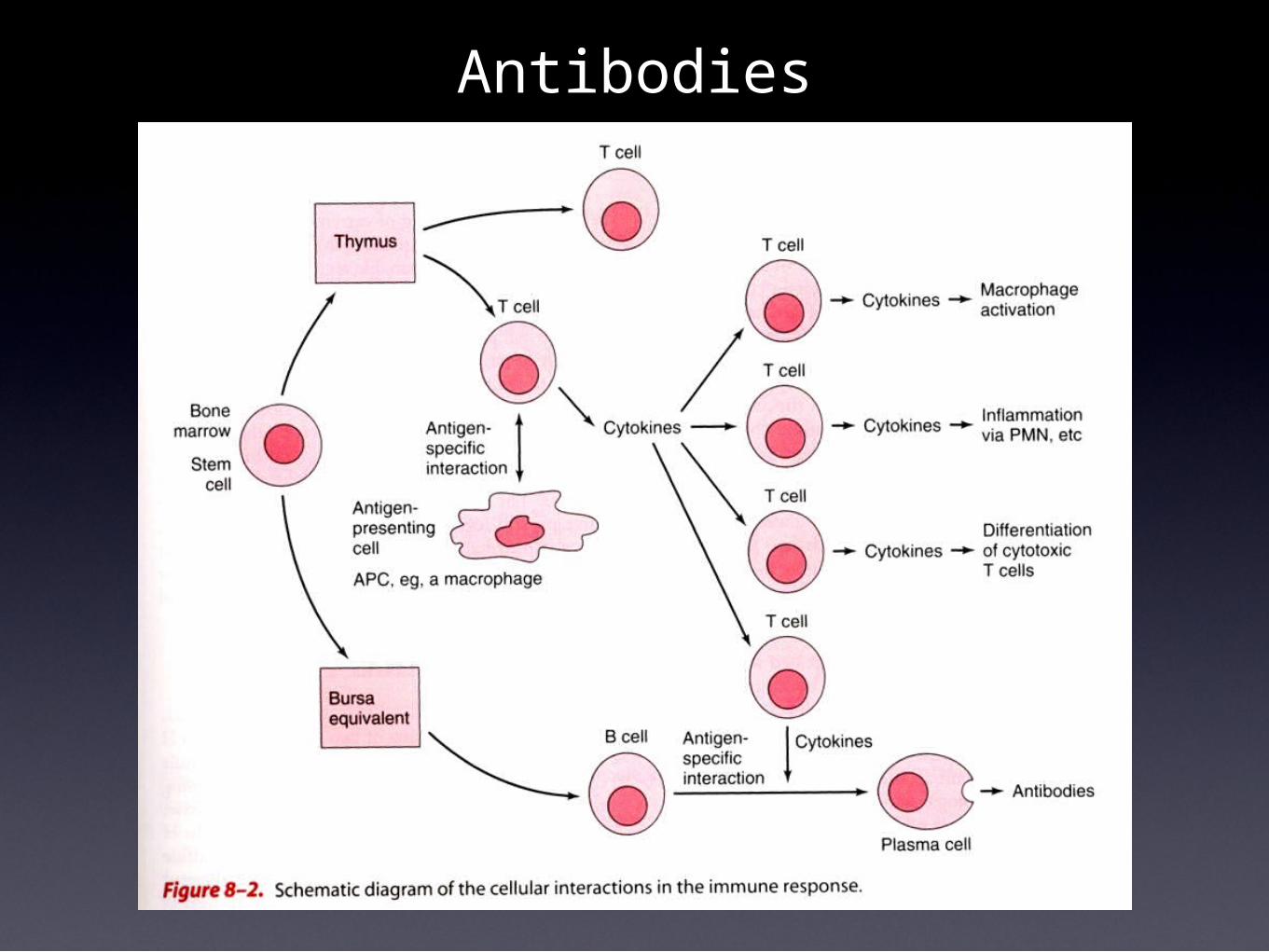
Antibodies• Antibodies (Ab) are immunoglobulins that recognize antigens
• All antibodies are immunoglobulins, but not all immunoglobulins are antibodies
• They are proteins
• B cells express BCR specific for an antigen• Each B cell possesses thousands of identical BCRs on their surfaces
• When the antigen enters the body, it must find the few B cells that possess a BCR capable of binding to it
• This can take several days
• When recognition occurs, the B cell, with the help of Th cell cytokines, begins to secrete antibodies in soluble form
• The B cell also undergoes clonal expansion; repeated rounds of cell division
• The process includes mutations in the antibody genes of the daughter cells that leads to antibodies with greater affinity for antigen; termed affinity maturation
• The antibodies also switch to other classes of Ig, each with distinct biological activities
• A plasma cell is a B cell that is committed to secreting antibodies for 2 or 3 days, then it dies
Antibodies
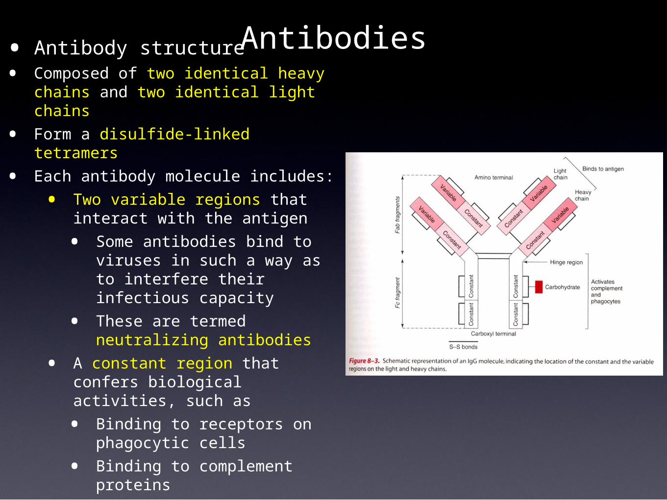
Antibodies• Antibody structure• Composed of two identical heavy
chains and two identical light chains
• Form a disulfide-linked tetramers
• Each antibody molecule includes:
• Two variable regions that interact with the antigen
• Some antibodies bind to viruses in such a way as to interfere their infectious capacity
• These are termed neutralizing antibodies
• A constant region that confers biological activities, such as
• Binding to receptors on phagocytic cells
• Binding to complement proteins

Antibodies
• Immunoglobulin classes
• IgM• First to be made during a primary (initial) response
• Often secreted as a pentamer of 5 IgM antibodies covalently-linked to one another (Fig 8-4)
• Low affinity, but high avidity
• IgG• Most common serum antibody
• Occurs after the IgM response
• High affinity
• Persist for years in the blood
• Four subclasses encoded by different genes
• IgG1
• IgG2
• IgG3
• IgG4

Antibodies• Immunoglobulin classes (cont.)
• IgA• Most common antibody produced by the body
• Secreted into mucosal areas
• Often found as a dimer of two covalently-linked IgA molecules
• IgE• Associated with parasitic and helminth infections, and with
hypersensitivities
• IgD• Expressed as a BCR only
• Immunoglobulin class switching and affinity maturation
• All secreted antibodies are initially IgM
• As the immune response ensues, the class switches to IgG, IgA or IgE
• This phenomenon is also accompanied by affinity maturation
• Both of these events require T cell help

Cell Surface Receptors for Antigen• B cell receptor for antigen
• Naive B cells express IgM or IgD as their surface receptor (sIg)• The antibody receptor possesses transmembrane domains at the C-
terminus of the H chains
• This domain anchors the H-chain in the membrane of the rough ER when the polypeptides are synthesized
• Thus, the antibody receptor protrudes from the B cell’s plasma membrane
• This receptor is usually of low affinity because it’s only a close-match to its antigenic epitope (10-7 KD)
• This receptor is used to capture the antigen, which is then internalized by receptor-mediated endocytosis
• When a B cell is activated (i.e., immune stimulus), transcriptional processing of the antibody mRNA results in a transcript free of the codons for the transmembrane domain, thus the antibody is secreted in a vesicle
• Memory B cells have IgG, IgA or IgE sIg• They have undergone class switching
• These receptors are high affinity because they have undergone affinity maturation (up to 10-11 KD)
• They are extremely efficient at capturing antigen
• All B cells have a sIg-associated Igα/Igβ signal transduction molecule

Cell Surface Receptors for Antigen

Cell Surface Receptors for Antigen
• T cell receptor
• Transmembrane heterodimer (not secreted)
• Structure is similar to the V/H domain of an antibody
• Two types (only one type found on a given T cell)• αβ (alpha-beta)
• γδ (gamma-delta)
• Two cells• Helper T cells (Th) express CD4 on their surfaces
• CD4 interacts with MHC class II proteins on antigen presenting cells (APC)
• Cytotoxic T cells (CTL) express CD8 on their surfaces
• CD8 interacts with MHC class I proteins on APC
• All T cells have CD3 and ζ-chain (zeta-chain), signal transduction components

Cell Surface Receptors for Antigen

Cell Surface Receptors for Antigen• The Major Histocompatibility Complex
• Large contiguous region of mammalian genomes that encode about 100 proteins involved in antigen processing and presentation
• Name is a misnomer; it was first characterized as controlling tissue rejection in mice in the 1930s-1950s
• Two principal classes• Class I molecules (3 loci in humans)
• Expressed by all nucleated cells, which act as APC• Responsible for presenting peptides from endogenously-synthesized polypeptides
(i.e., made on cellular ribosomes)• At rough ER, viral polypeptides are clipped into 8-10 amino acid peptides by the
proteosome and then transported into the lumen of the ER• Class I molecules bind viral peptides at the ER, then traffic to the cell surface• Marks the cell for death by CTL (via CD8 interaction with class I molecule)
• Class II molecules (3 loci in humans)• Expressed by professional APC
• B cells• Macrophages• Dendritic cells• A few others
• For antigens captured by receptor-mediated endocytosis or phagocytosis (i.e., exogenous)
• Fusion of phagosome with class II-bearing endosome results phagolysosome• Peptides bind to class II proteins• The class II molecules traffic to the cell surface where they are engaged by helper
T cells• CD4 binds to the class II molecule, thus restricting the interaction to Th cells

Cell Surface Receptors for Antigen

Type II (Antibody-Mediated) Immunity• Principally for exogenous (extracellular) antigens
• The primary response
• Within days after infection, antigen-specific IgM appears
• Within a week, antigen-specific IgG appears• Class switching and affinity maturation improve the efficiency of the
antibody response
• As antigen becomes limiting (i.e., the immune system clears the infection) the immune response wanes
• Most (98%) responding B cells die, but a few remain as memory cells• Unlike naive B cells with IgM, memory cells possess high-affinity sIgG or
sIgA
• The secondary (anamnestic) response
• In the event the pathogen invades again, the immune response will be of greater speed and magnitude because of the memory cells• No need for class switching
• No need for affinity maturation
• Antibody response is quicker and of higher affinity
• There are usually no clinical symptoms of infection

Type II (Antibody-Mediated) Immunity
• The role of helper T cells in Type II immunity
• Exogenous antigens (such as free virus) are captured by professional APC• B cell uses its sIg receptor
• Macrophages are phagocytic
• Dendritic cells are pinocytotic
• The antigen is imported into a lysosome (phagolysosome) that has more than 60 types of molecules, including digestive enzymes
• Proteins are digested into peptides of varying length
• The phagolysosome is fused with another endosome containing nascent MHC class II proteins• Packaged with the class II endosome is the invarient chain, which occludes
the peptide binding cleft to prevent premature peptide binding
• The peptides bind to the class II proteins and additional protease activity occurs until the peptides are about 11-15 amino acids
• The vesicle traffics to the cell surface where it fuses with the plasma membrane (i.e., class II/peptide complex is outside of the cell)
• The Th cell must recognize both the class II protein and peptide before the T cell can become activated (proliferation and cytokine synthesis)

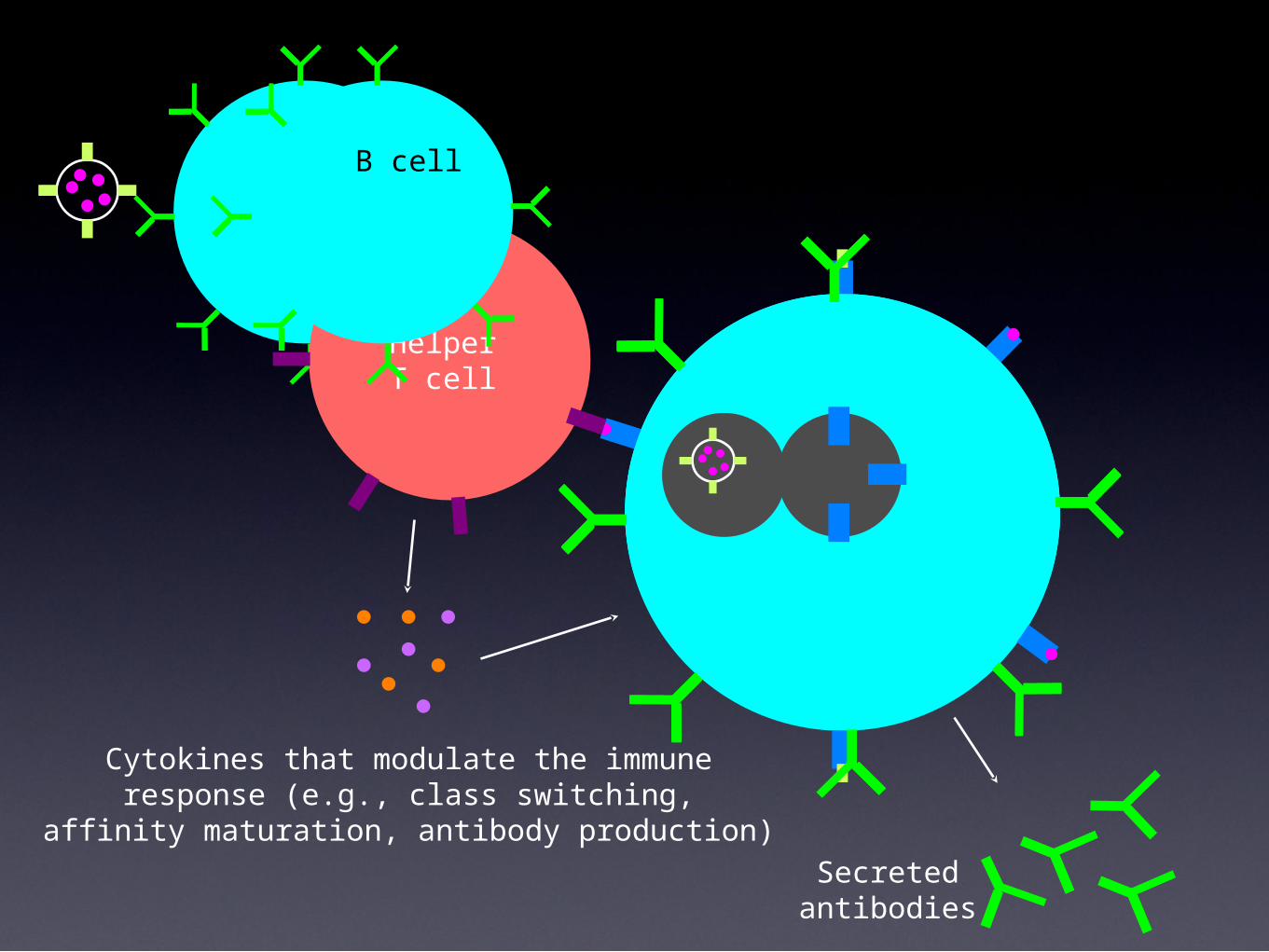
HelperT cell
Cytokines that modulate the immuneresponse (e.g., class switching,
affinity maturation, antibody production)
B cell
Secretedantibodies

Type I (Cell Mediated) Immunity• Principally for containment of endogenously-produced (intracellular)
antigens
• Viruses rely upon cellular ribosomes for the biosynthesis of their polypeptides
• A small proportion of all polypeptides (self and non-self) synthesized in a cell are re-directed to the proteosome complex in the cytoplasm• The proteosome is a tubular protein structure that has protease activity
• Polypeptides enter the proteosome, which can hold about 8-10 amino acids, then its protease activity results in clipping into peptides
• The peptides are ejected from the other end of the proteosome
• The transporter associated with antigen processing (TAP) translocates the peptides into the lumen of the ER• This event requires ATP
• Inside the ER, nascent MHC class I molecules bind the peptides
• After budding from the ER, the class I/peptide traffic to the Golgi, then the cell surface
• The class I/peptide complex is displayed on the outside of the cells for scanning by the CTL repertoire• The CTL kills the cell by sending it a death signal (apoptosis) or by secreting
perforin, a protein that polymerizes to form channels in the membrane of the target cell


The Complement System
• The complement system is composed of several pre-existing blood proteins that puncture membranes
• There are three pathways, one of which is important to viral infections, the classical pathway
• The classical pathway is antibody-dependent
• When IgM or IgG bind to envelope viruses, the complement cascade ensues
• This cascade is a series of enzymatic reactions carried out by the complement proteins
• Two final outcomes occur• Complement proteins attack the envelope, thus compromising
it
• The complement-bound viruses are opsonized, that is, they are more efficiently phagocytosed

Cytokines
• Cytokines are hormone-like proteins
• There are more than 80 known
• Interleukins - secreted by leukocytes
• Interferons - potent antivirals
• Chemokines - mediate chemotaxis
• Inflammation/immunosuppression
• Potent, often active at picomolar concentrations
• Bind to receptors on target cells and modulate gene expression
• Some are used therapeutically (recombinant)
• Some are associated with T cell subsets
• Th1 cells secrete...• IFNγ
• TNF
• LT
• Th2 cells secrete...• IL-4
• IL-5

Overly Aggressive Immune Responses
• Often results in immunopathology
• There are no good clinical strategies for managing pathologic immune responses

• The use of blood to detect antigen-specific antibodies in testing is termed serology
• Relies upon the fact that serum antibodies to specific pathogens cannot be detected until after infection (or immunization)
• Provides diagnostic indicator of infection
• IgM - current/recent infection
• IgG - current or past infection (decades)
• Common serological tests
• Enzyme-linked immunosorbant assay (ELISA)• Rapid
• High throughput
• Inexpensive
• Immunofluorescent antibody (IFA)
• Immunoblotting
Immunologic Diagnostic Tests

Immunologic Diagnostic Tests

• Immunoblotting
• More specific and sensitive than ELISA
• Also more work and more expensive
• Most common is western blot
• First step is electrophoresis of antigen (SDS-PAGE)
• Second is transfer of antigen from gel to solid phase, such as nitrocellulose membrane
• Steps for western blot• Block membrane with irrelevant protein (nonfat powdered milk
solution)
• Incubate serum sample
• Incubate anti-antibody, enzyme conjugate
• Incubate substrate
• Look for bands appearing on membrane
Immunologic Diagnostic Tests
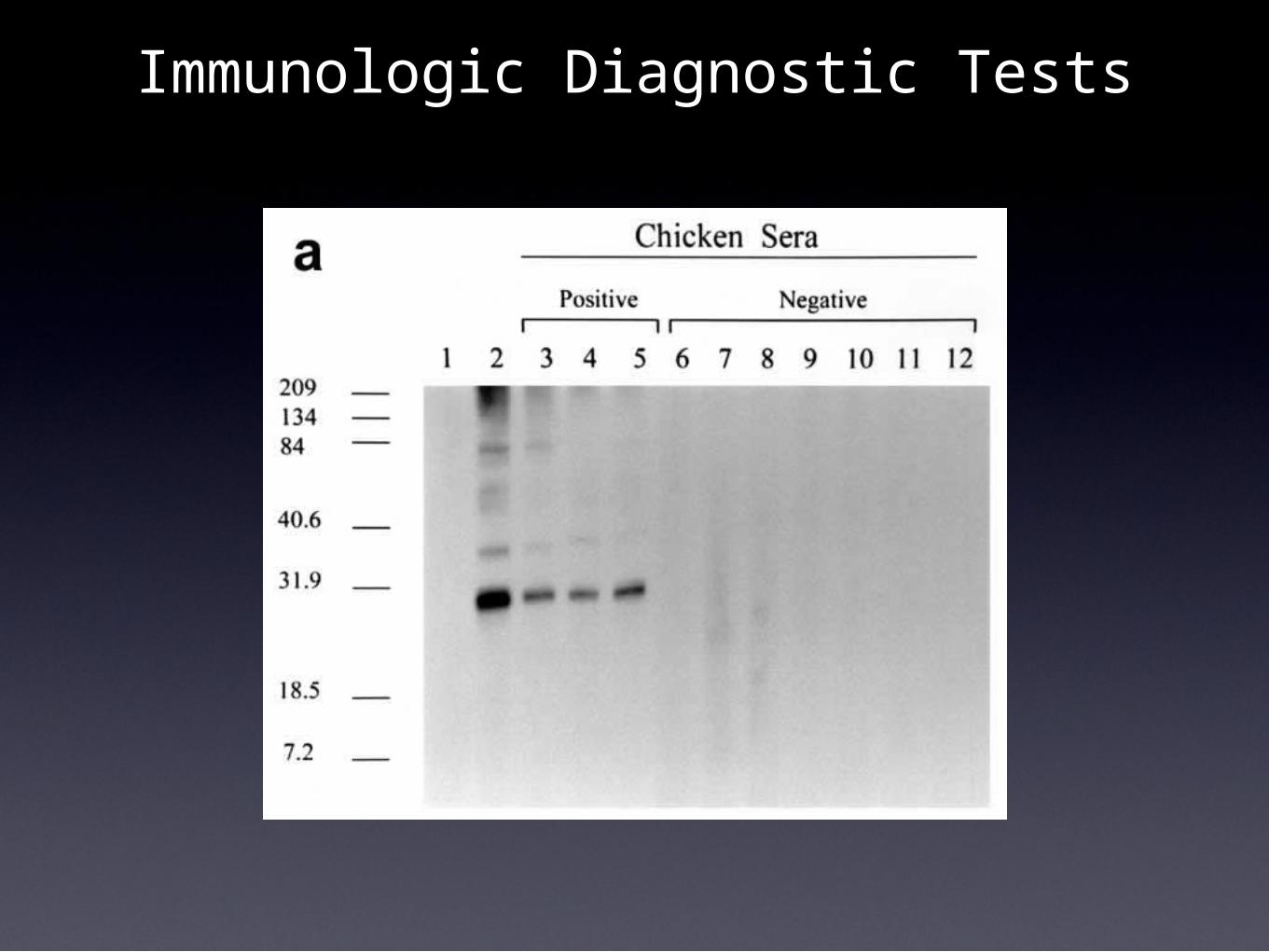
Immunologic Diagnostic Tests