Immunological Basis of Membranous Glomerulonephritis
14
2 Immunological Basis of Membranous Glomerulonephritis Gian Marco Ghiggeri 1 et al. * 1 Division of Nephrology and Laboratory on Pathophysiology of Uremia, G. Gaslini Children Hospital, Genoa Italy 1. Introduction Primary membranous glomerulonephritis (MGN) is a major glomerular disease causing proteinuria in humans (Jones, 1957). It is the prototype of an autoimmune disease (Couser, et al., 1978) characterized by sub-epithelial immune deposits within glomeruli. Its pathogenesis remains still unknown. Immune deposits are formed by IgG 4 , their respective antigen and complement. The definition of the immune deposit architecture has been a main focus of the pathology research for years but advances were restricted, until recently, to animal models of the disease, in particular to Heymann nephritis (HN) (Heymann, et al., 1959; Heymann, et al., 1952; Van Damme, et al., 1978). Unfortunately, results from experimental HN cannot be readily exported to human MGN since the major antigen of immune deposits in rat is not present in human glomeruli. Therefore different podocyte antigens are involved in human MGN and their identification is a fundamental step in understanding human pathology. Technology problems, mainly concerning dissection of glomeruli and purification/characterization of glomerular antibodies from human biopsies, have limited the experimental approach in humans for years. In the last 5 years, human MGN has become the topic of renewed nephrologic research. Debiec et al. (Debiec, et al., 2002, 2004) first showed that neutral endopeptidase (NEP) emerges as podocyte antigen in a rare form of congenital MGN due to maternal NEP deficiency and alloimmunization during pregnancy. More recently, the existence of three new glomerular auto-antigens, i.e. phospholipase A2 receptor (PLA2R), aldose reductase (AR) and superoxide dismutase 2 (SOD2) have been proposed by independent groups (Beck, et al., 2009; Prunotto, et al., 2010). Technology evolution in the field of laser capture and proteomics allowed a direct experimental approach in humans, a crucial step for a direct analysis ‘in vivo’. It is now clear that IgG 4 eluted from glomeruli of MGN patients recognize a panel of podocyte proteins * Corrado Murtas 1 , Maria Luisa Carnevali 2 , Giovanni Candiano 1 , Maurizio Bruschi 1 , Marco Prunotto 1 , Riccardo Magistroni 3 and Landino Allegri 2 . 1 Division of Nephrology and Laboratory on Pathophysiology of Uremia, G. Gaslini Children Hospital, Genoa, Italy, 2 Department of Clinical Medicine, Nephrology and Health Sciences, University of Parma, Parma, Italy, 3 Chair of Nephrology, University of Modena, Italy. www.intechopen.com
Transcript of Immunological Basis of Membranous Glomerulonephritis
2
Immunological Basis of
Membranous Glomerulonephritis
Gian Marco Ghiggeri1 et al.* 1Division of Nephrology and Laboratory on Pathophysiology of Uremia,
G. Gaslini Children Hospital, Genoa
Italy
1. Introduction
Primary membranous glomerulonephritis (MGN) is a major glomerular disease causing
proteinuria in humans (Jones, 1957). It is the prototype of an autoimmune disease (Couser,
et al., 1978) characterized by sub-epithelial immune deposits within glomeruli. Its
pathogenesis remains still unknown. Immune deposits are formed by IgG4, their respective
antigen and complement. The definition of the immune deposit architecture has been a main
focus of the pathology research for years but advances were restricted, until recently, to
animal models of the disease, in particular to Heymann nephritis (HN) (Heymann, et al.,
1959; Heymann, et al., 1952; Van Damme, et al., 1978). Unfortunately, results from
experimental HN cannot be readily exported to human MGN since the major antigen of
immune deposits in rat is not present in human glomeruli. Therefore different podocyte
antigens are involved in human MGN and their identification is a fundamental step in
understanding human pathology. Technology problems, mainly concerning dissection of
glomeruli and purification/characterization of glomerular antibodies from human biopsies,
have limited the experimental approach in humans for years.
In the last 5 years, human MGN has become the topic of renewed nephrologic research.
Debiec et al. (Debiec, et al., 2002, 2004) first showed that neutral endopeptidase (NEP)
emerges as podocyte antigen in a rare form of congenital MGN due to maternal NEP
deficiency and alloimmunization during pregnancy. More recently, the existence of three
new glomerular auto-antigens, i.e. phospholipase A2 receptor (PLA2R), aldose reductase
(AR) and superoxide dismutase 2 (SOD2) have been proposed by independent groups
(Beck, et al., 2009; Prunotto, et al., 2010).
Technology evolution in the field of laser capture and proteomics allowed a direct
experimental approach in humans, a crucial step for a direct analysis ‘in vivo’. It is now clear
that IgG4 eluted from glomeruli of MGN patients recognize a panel of podocyte proteins
* Corrado Murtas1, Maria Luisa Carnevali2, Giovanni Candiano1, Maurizio Bruschi1, Marco Prunotto1, Riccardo Magistroni3 and Landino Allegri2. 1 Division of Nephrology and Laboratory on Pathophysiology of Uremia, G. Gaslini Children Hospital, Genoa, Italy, 2 Department of Clinical Medicine, Nephrology and Health Sciences, University of Parma, Parma, Italy, 3 Chair of Nephrology, University of Modena, Italy.
www.intechopen.com
An Update on Glomerulopathies – Etiology and Pathogenesis
20
that have been only in part characterized and that represent good candidates for being
involved in the pathogenesis of MGN as “auto-antigens”.
The same approach might be extended to different renal auto-immune pathologies. However, before proceeding, the scientific community needs a consensus on criteria and technologies that should be utilized for recognizing and validating auto-antigens.
2. Animal models
Heymann nephritis (Heymann, et al., 1952, 1959) is the animal model most frequently used for studying mechanisms implicated in the process of deposition of antibodies within glomeruli. Passive Heymann nephritis is induced in susceptible rat strains by injection of heterologous antisera from sheep or rabbit immunized with a crude extract of rat proximal tubular antigens known as Fx1A. This model is highly similar to human MGN: renal pathology in HN is constantly charachterized by the presence of glomerular sub-epithelial immune deposits. Heymann nephritis is quite simple to be produced and has offered for years the unique chance to study the structure of immune deposits and the mechanisms involved in their formation. This goal has been completed many years ago. Several studies, between 1980 and 1990, demonstrated that megalin is the target antigen of sub-epithelial IgG. Complement factors, mainly C5b-9, and other ancillary proteins such as receptor associated protein (RAP) and anti-RAP IgG complete the structure of immune deposits (Couser, et al., 1978; Kerjaschki, 2004; Kerjaschki & Farquhar, 1982; Kerjaschki, et al., 1987, 1992; Saito, et al., 1994; Salant, et al., 1980a, 1980b; Van Damme, et al., 1978). The presence of IgG directed against a podocyte antigen triggers the complement cascade that ends with the formation of C5b-9, the key mediator of podocyte damage in MGN. C5b-9 aggregation on the podocyte membrane causes the activation of the epithelial cell. The podocyte starts the production of a cascade of mediators with phosphorylation of PKC and formation of free oxygen radicals (Cybulsky, et al., 2000). The result is a rearrangement of the cytoskeleton and the consequent loss of cell junction that leads to a pathologic alteration of the glomerular filter and to the appearance of proteinuria. Immune deposits also contain clusterin (Ghiggeri, et al., 2002; Rastaldi, et al., 2006), a natural binder of megalin, that probably represents an endogenous inhibitor of C5b-9. The definition of pathology features and physiopathology events in Heymann nephritis represented a breakthrough in research of renal autoimmunity. Unfortunately, in spite some of the components above have been detected in human MGN (i.e. C5b-9, clusterin), Heymann nephritis could not be utilized as a direct model of human MGN because megalin is not present in human glomeruli. Moreover, many years later, it was described that megalin structural homolog, the LDL-receptor, is not recognized by circulating IgG4 in human MGN (Bruschi, et al., 2009). Diverse animal models of membranous nephropathy have been developed and different podocyte antigens have been identified such as neutral endopeptidase in rabbit, and dipeptidyl peptidase IV in mouse (Table 1) (Assmann, et al., 1992; Ronco, et al., 1989). These proteins are present in human glomeruli; however they were not identified in immune deposits and their involvement in idiopathic MGN was never proved (Allegri, 1997). After all, animal models showed the heterogeneity of potential podocyte antigens in MGN and straightened the concept of ‘in situ’ formation of immune deposit. Problems related to the translation of the lesson from animal models to human beings slowed progression in understanding of mechanisms of human MGN. After twenty years, however, new insights
www.intechopen.com
Immunological Basis of Membranous Glomerulonephritis
21
were generated by the observation of familial models of MGN and from the characterization of antigens involved in their pathogenesis.
Animal Models Ref.
Megalin rat Kerjaschki & Farquhar, 1982
Dipeptidyl peptidase IV rat, mouse, rabbit Assmann, et al., 1992
Neutral endopeptidase rabbit Ronco, et al., 1989
Neonatal MGN
Neutral endopeptidase humans Debiec, et al., 2002
Idiopathic MGN
Phospholipase A2 receptor humans Beck, et al., 2009
Aldose reductase humans Prunotto, et al., 2010
Mn-superoxide dismutase humans Prunotto, et al., 2010
Alpha enolase humans Bruschi, et al., 2011
Others humans Bruschi, et al., 2011
Table 1. Glomerular antigens in experimental models and in human MGN
3. Human MGN
3.1 Neonatal MGN
Seminal studies by Debiec and Ronco (Debiec, et al., 2002; Debiec, et al., 2004) represented a fundamental passage on the road of comprehension of human MGN many years after the definition of HN. These authors’ contributions led to the definition of neutral endopeptidase as the auto-antigen in rare forms of familial congenital MGN (Table 1). Antibodies against neutral endopeptidase was first recognized in a newborn presenting with congenital nephrotic syndrome. Renal histology demonstrated MGN. The basis of the pathogenesis was the mother, carrying a genetic deficiency of neutral endopeptidase because of an homozygous deletion in MME, the corresponding gene. She became alloimmunized against neutral endopeptidase during a prior pregnancy, ended with miscarriage. Anti- neutral endopeptidase antibodies were then transferred to the fetus during the successive pregnancy and congenital MGN developed in the newborn while disappearing thereafter.
www.intechopen.com
An Update on Glomerulopathies – Etiology and Pathogenesis
22
This elegant model could therefore demonstrate, for the first time, the pathogenic role of a podocyte antigen in human MGN. Similar mutations of MME were found in three further families that had in common at least one case of congenital MGN. Even if the presence and the involvement of antibodies against neutral endopeptidase was clearly demonstrated, the prevalence of this form of MGN is probably very low and these results cannot be translated directly in idiopathic MGN. There is, in fact, now much debate about considering neutral endopeptidase an auto-antigen in non-familial MGN. In fact, circulating anti- neutral endopeptidase IgG4 can be detected in a minor part of patients with primary MGN but this antigen is not recognized by IgG4 eluted from micro-dissected glomeruli (Ghiggeri, personal observation). Further studies are required to rule out the involvement of neutral endopeptidase in idiopathic MGN.
3.2 Technology advances for ‘in vivo’ studies
New technologies in the fields of tissue micro-dissection and protein characterization have
recently played an essential role in discovering new auto-antigen in human MGN. They
allowed for the first time the analysis of minute amounts of human tissue from a diagnostic
renal biopsy. In fact, micro-dissection and proteomics are of fresh development but have
been rapidly adapted to renal tissue studies (Murtas, et al., 2011).
The basic approach starts with the laser dissection of glomeruli from cryo-sections of renal
tissue. This technique is highly specific and reproducible. It permits to obtain purified
glomeruli from which antibodies can be eluted utilizing osmotic gradients and further
characterizated.
Western blot analysis coupled with mass spectrometry is the second basic technique for
characterizing auto-antibodies. In this case, podocyte proteins are first separated with
mono-dimensional electrophoresis in both denaturing and non-denaturing conditions (a few
proteins are not recognized after mild denaturing treatment) or with two-dimensional gel
electrophoresis and they are then incubated with glomerular eluates. The binding of specific
IgG4 with podocyte antigens is detected with anti-IgG4 antibodies and revealed with
chemiluminescence. Finally, podocyte proteins recognized by IgG4 are characterized by
mass spectrometry.
Western-blot/mass spectrometry are also utilized for the characterization of circulating antibodies: serum replaces eluted antibodies.
3.3 Glomerular antigens in human MGN
In terms of general discussion it is rational to propose at least three basic characteristics that should be fulfilled by a protein when considered as an auto-antigen in idiopathic MGN. The first one is that the candidate auto-antigen is expressed by podocytes at least in pathological conditions and that it is recognized by IgG4 eluted from micro-dissected glomeruli. The second criterion is that the candidate antigen co-localizes with IgG4 (and possibly with C5b-9) in glomerular sub-epithelial immune deposits. The final condition is that IgG4 against the protein must be detected in MGN patients sera at least during the overt phase of the disease. A question apart is specificity, that is a major issue of the consensus debate. Therefore, characterizing an auto-antigen requires a multistep approach: first of all immunoglobulins are eluted from MGN glomeruli. Then their target is evaluated by
www.intechopen.com
Immunological Basis of Membranous Glomerulonephritis
23
western-blot against podocyte proteins. The evaluation of co-localization within the immune deposits, isotype analysis and titration of circulating antibodies are further steps of the same project. Following this scheme three podocyte proteins have been characterized as putative auto-antigens: phospholipase A2 receptor (PLA2r), Mn-superoxide dismutase (SOD2), aldose reductase (AR). Alpha enolase is a recent finding (Table 1 and Figure 1). Few others are still in progress. PLA2r is a member of the mannose-receptor family that is normally expressed on the
podocyte membrane in humans (East & Isacke, 2002; Lambeau & Lazdunski, 1999). It binds
circulating phospholipase A2 but its physiological role is still uncertain. In MGN auto-
antibodies recognize PLA2r only in its native configuration that is dependent on disulphide
bonds and this is the reason why this protein requires stringent conditions of analysis
rigorously in non-denaturing environment. It is reasonable that conformation of PLA2r
plays a key role in auto-antibodies formation and binding. Studies (Beck, et al., 2009;
Hofstra, et al., 2011) in limited groups of patients with idiopathic MGN revealed high
circulating levels in a significant portion of patients and their absence in cases of secondary
MGN suggesting a direct implication as causative factor.
SOD2 is a key anti-oxidant mitochondrial enzyme implicated in transformation of
superoxide ions into hydrogen peroxide and diatomic oxygen (Son, et al.,2008). In the
kidney, SOD2 is widely expressed in tubular epithelial cells, especially in the cortex, where
it plays a central role in preserving the kidney during ischemia/reperfusion events but it has
never been reported in normal glomeruli. In fact, SOD2 is neo-expressed in podocytes and
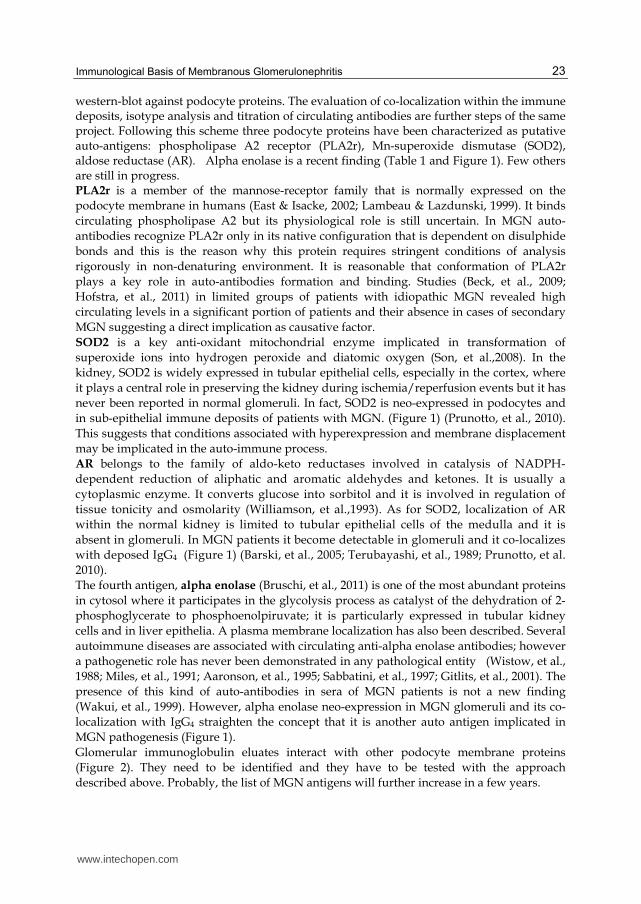
in sub-epithelial immune deposits of patients with MGN. (Figure 1) (Prunotto, et al., 2010).
This suggests that conditions associated with hyperexpression and membrane displacement
may be implicated in the auto-immune process.
AR belongs to the family of aldo-keto reductases involved in catalysis of NADPH-
dependent reduction of aliphatic and aromatic aldehydes and ketones. It is usually a
cytoplasmic enzyme. It converts glucose into sorbitol and it is involved in regulation of
tissue tonicity and osmolarity (Williamson, et al.,1993). As for SOD2, localization of AR
within the normal kidney is limited to tubular epithelial cells of the medulla and it is
absent in glomeruli. In MGN patients it become detectable in glomeruli and it co-localizes
with deposed IgG4 (Figure 1) (Barski, et al., 2005; Terubayashi, et al., 1989; Prunotto, et al.
2010).
The fourth antigen, alpha enolase (Bruschi, et al., 2011) is one of the most abundant proteins
in cytosol where it participates in the glycolysis process as catalyst of the dehydration of 2-
phosphoglycerate to phosphoenolpiruvate; it is particularly expressed in tubular kidney
cells and in liver epithelia. A plasma membrane localization has also been described. Several
autoimmune diseases are associated with circulating anti-alpha enolase antibodies; however
a pathogenetic role has never been demonstrated in any pathological entity (Wistow, et al.,
1988; Miles, et al., 1991; Aaronson, et al., 1995; Sabbatini, et al., 1997; Gitlits, et al., 2001). The
presence of this kind of auto-antibodies in sera of MGN patients is not a new finding
(Wakui, et al., 1999). However, alpha enolase neo-expression in MGN glomeruli and its co-
localization with IgG4 straighten the concept that it is another auto antigen implicated in
MGN pathogenesis (Figure 1).
Glomerular immunoglobulin eluates interact with other podocyte membrane proteins
(Figure 2). They need to be identified and they have to be tested with the approach
described above. Probably, the list of MGN antigens will further increase in a few years.
www.intechopen.com
An Update on Glomerulopathies – Etiology and Pathogenesis
24
Fig. 1. Glomerular expression of aldose reductase, Mn-superoxide dismutase and alpha enolase in MGN patients. Immunofluorescence on renal cryo-sections analyzed with confocal microscopy. Merged images show co-localization between antigens and IgG4.
3.4 Antibody isotypes and clinical correlations
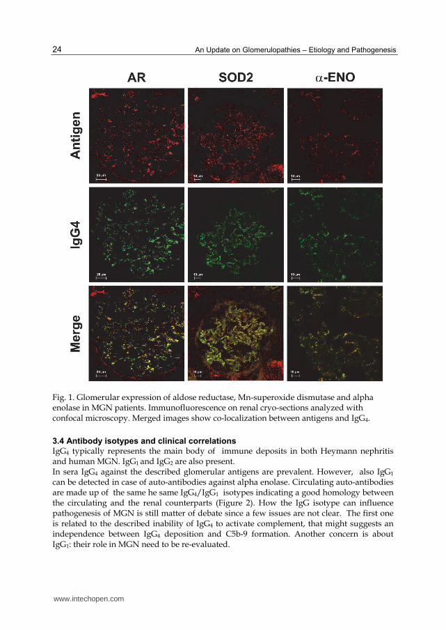
IgG4 typically represents the main body of immune deposits in both Heymann nephritis and human MGN. IgG1 and IgG2 are also present. In sera IgG4 against the described glomerular antigens are prevalent. However, also IgG1 can be detected in case of auto-antibodies against alpha enolase. Circulating auto-antibodies are made up of the same he same IgG4/IgG1 isotypes indicating a good homology between the circulating and the renal counterparts (Figure 2). How the IgG isotype can influence pathogenesis of MGN is still matter of debate since a few issues are not clear. The first one is related to the described inability of IgG4 to activate complement, that might suggests an independence between IgG4 deposition and C5b-9 formation. Another concern is about IgG1: their role in MGN need to be re-evaluated.
www.intechopen.com
Immunological Basis of Membranous Glomerulonephritis
25
Fig. 2. (a) Circulating immunoglobulin isotypes referred to autoantibodies againsts AR, SOD2 and alpha enolase. In the former two cases the predominant isotype is IgG4, in the case of alpha enolase both IgG1 and IgG4 are detectable. (b) Western-blot with IgG4 eluted from glomeruli of patients with MGN. A few podocyte proteins are recognized . Three of them have been characterized as aldose reductase (AR), Mn-superoxide dismutase (SOD2) and alpha enolase.
The determination of circulating levels of antibodies against auto-antigens and their clinical correlation is now in progress. Only small cohorts of patients have been evaluated. Moreover, in each population only one antigen have been tested (Beck, et al., 2009; Prunotto, et al., 2010; Debiec & Ronco, 2011; Hofstra, et al. 2011). A correlation of autoantibodies level with proteinuria has been proposed, suggesting that high circulating titers are in relationship with phases of immunological activity of the disease. A strong association with clinical outcome is still lacking. It is also an important issue to establish a correlation among antibodies against different antigens; such stuidies are on the way. Probably they will help in establishing a hierarchy between antigens, if it exists. Another key aspect would be to find out a correlation between glomerular deposits and serum levels or clinical outome. Surrogate biomarkers are needed for guiding treatment and long-term follow-up in MGN patients.
3.5 Predisposing factors
Many immunological or infectious diseases, neoplasms and toxic or farmacological agents have been associated with MGN. In these situations MGN is considered “secondary” to the
www.intechopen.com
An Update on Glomerulopathies – Etiology and Pathogenesis
26
respective pathological entity, even if the presumptive antigen has never been isolated from the affected glomeruli. A classical pathogenetic theory affirms that the causative agent can initiate the pathological process in genetically susceptible individuals. In primary MGN some association has been proved with HLA alleles: in particular HLA-B8, HLA-B18 and HLA-DR3. The latter has been described probably confering a threefold increased risk of the disease (Dyer, et al., 1992; Klouda, et al., 1979) More recently a genomewide association study conducted in three white European population reported an evident association of MGN with single nucleotide polymorphism (SNP) in PLA2R gene and, more strongly, in HLA-DQ alpha chain 1 (HLA-DQA1). The authors do not report any association in AR and SOD2 genes. The MGN risk in relation with SNPs in PLA2R gene is proposed also in two other Asian studies (Stanescu, et al., 2011). Even these data are not conclusive, they might suggest a “two hits” model also for the pathogenesis of MN. In fact, a variation in some component of the immune system (HLA-DQ variation) might confer an autoimmunity predisposition. Successively, an alteration, congenital or acquired, of the localization or of the structure of an antigen (PLA2R or others), can make the antigen itself became the target of the disregulated immune system.
4. Conclusions
Research on MGN pathogenesis restarted recently, after many years of frustrating findings. Technology evolution was the major incentive to new stuidies. The finding of at least four auto-antigens implicated in the pathogenesis of MGN, needs an explanation in a complex pathogenetic theory. Nowadays, it seems reasonable that mechanisms related to the formation of auto-antibodies against membrane proteins, such as PLA2r, are different from auto-antibodies against antigens typically localized inside the cell, such as SOD2 (mitochondrial), and AR or alpha enolase (cytosolic). Antibody promiscuity can be in some way justified by mimicry in case of membrane proteins whereas, for molecules such as SOD2, AR or alpha enolase, mechanisms of de-localization should play a role. Anyway, before a complete elucidation of MGN pathogenesis several studies have to be performed: more clinical data, new insights on cellular localization of antigens and , maybe, new animal models are needed. There is a some kind of hierarchy between different targets of autoimmunity? The production of a first autoantibody stimulates podocyte expression of other auto-antigens and a new wave of immunization? There are types of autoantibody not correlated with clinical evolution that are only some kind of “epiphenomenon”? There are different clusters of MGN patients with different autoantibody profile? Many questions raise when considering recent discoveries on MGN. They will be correctly answered only if scientific community proceeds with shared approach in discovering and testing candidate antigens. Exciting times are coming for the researches involved in studies on MGN and other renal auto-immune diseases.
5. Acknowledgements
This work was supported by the Italian Ministry of Health and by the Renal Child Foundation. Authors also acknowledge Fondazione Mara Wilma e Bianca Querci for financial support to the project ”Ruolo dello stress reticolare nella progressione del danno renale e
www.intechopen.com
Immunological Basis of Membranous Glomerulonephritis
27
tumorale”, Fondazione La Nuova Speranza for supporting the project ‘Progetto integrato per la definizione dei meccanismi implicati nella glomerulo sclerosi focale. Dalla predisposizione genetica alla regolazione della produzione di fattori cellulari e circolanti’ and Fondazione Compagnia di San Paolo for the project ‘Nefropatia cronica del rene trapiantato’ Data were critically discussed with Prof. Rosanna Gusmano before her death and this chapter is dedicated to her memory.
6. References
Aaronson, RM; Graven, KK; Tucci, M; McDonald, RJ & Farber, HW. (1995). Non-neuronal enolase is an endothelial hypoxic stress protein. J Biol Chem, Vol. 270, No. 46, (Nov 1995), pp. 27752-27757, ISSN 0021-9258
Allegri, L. (1997). Antigens in experimental models of membranous nephropathy: are they involved in human disease? Nephrol Dial Transplant, Vol. 12, No. 9, (Sep 1997), pp. 1801-1804, ISSN 0931-0509
Assmann, KJ; van Son, JP; Dijkman, HB & Koene, RA. (1992). A nephritogenic rat monoclonal antibody to mouse aminopeptidase A. Induction of massive albuminuria after a single intravenous injection. J Exp Med, Vol. 175, No. 3, (Mar 1992), pp. 623-635, ISSN 0022-1007
Barski, OA; Papusha, VZ; Ivanova, MM; Rudman, DM & Finegold, MJ. (2005). Developmental expression and function of aldehyde reductase in proximal tubules of the kidney. Am J Physiol Renal Physiol, Vol. 289, No. 1, (Jul 2005), pp. F200-207, ISSN 0363-6127
Beck, LH, Jr; Bonegio, RG; Lambeau, G; Beck, DM; Powell, DW; Cummins, TD; Klein, JB & Salant, DJ. (2009). M-type phospholipase A2 receptor as target antigen in idiopathic membranous nephropathy. N Engl J Med, Vol. 361, No. 1, (Jul 2009), pp. 11-21, ISSN 1533-4406
Bruschi, M; Candiano, G; Murtas, C; Prunotto, M; Santucci, L; Carnevali, ML; Scolari, F; Allegri, L & Ghiggeri, GM. (2009). Patients with primary membranous nephropathy lack auto-antibodies against LDL receptor, the homologue of megalin in human glomeruli. NDT Plus, (Jan 2009), 10.1093/ndtplus/sfp002
Bruschi, M; Carnevali, ML; Murtas, C; Candiano, G; Petretto, A; Prunotto, M; Gatti, R; Argentiero, L; Magistroni, R; Garibotto, G; Scolari, F; Ravani, P; Gesualdo, L; Allegri, L & Ghiggeri GM. (2011). Direct characterization of target podocyte antigens and auto-antibodies in human membranous glomerulonephritis: Alfa-enolase and borderline antigens. J Proteomics, (n.d.), 10.1016/j.jprot.2011.05.021
Couser, WG; Steinmuller, DR; Stilmant, MM; Salant, DJ & Lowenstein, LM. (1978). Experimental glomerulonephritis in the isolated perfused rat kidney. J Clin Invest, Vol. 62, No. 6, (Dec 1978), pp. 1275-1287, ISSN 0021-9738
Cybulsky, AV; Takano, T; Papillon, J & McTavish, AJ. (2000). Complement-induced phospholipase A2 activation in experimental membranous nephropathy. Kidney Int, Vol. 57, No. 3, (Mar 2000), pp. 1052-1062, ISSN 0085-2538
Debiec, H; Guigonis, V; Mougenot, B; Decobert, F; Haymann, JP; Bensman, A; Deschenes, G & Ronco, PM. (2002). Antenatal membranous glomerulonephritis due to anti-neutral endopeptidase antibodies. N Engl J Med, Vol. 346, No. 26, (Jun 2002), pp. 2053-2060, ISSN 0028-4793
www.intechopen.com
An Update on Glomerulopathies – Etiology and Pathogenesis
28
Debiec, H; Nauta, J; Coulet, F; van der Burg, M; Guigonis, V; Schurmans, T; de Heer, E; Soubrier, F; Janssen, F & Ronco, P. (2004). Role of truncating mutations in MME gene in fetomaternal alloimmunisation and antenatal glomerulopathies. Lancet, Vol. 364, No. 9441, (Oct 2004), pp. 1252-1259, ISSN 0140-6736
Debiec, H & Ronco, P. (2011). PLA2R autoantibodies and PLA2R glomerular deposits in membranous nephropathy. N Engl J Med, Vol. 364, No. 7, (Feb 2011), pp. 689-690, ISSN 1533-4406
Dyer, PA; Short, CD; Clarke, EA & Mallick, NP. (1992). HLA antigen and gene polymorphisms and haplotypes established by family studies in membranous nephropathy. Nephrol Dial Transplant, Vol. 7 Suppl 1, pp. 42-47, ISSN 0931-0509
East, L & Isacke, CM. (2002). The mannose receptor family. Biochim Biophys Acta, Vol. 1572, No. 2-3, (Sep 2002), pp. 364-386, ISSN 0006-3002
Ghiggeri, GM; Bruschi, M; Candiano, G; Rastaldi, MP; Scolari, F; Passerini, P; Musante, L; Pertica, N; Caridi, G; Ferrario, F; Perfumo, F & Ponticelli, C. (2002). Depletion of clusterin in renal diseases causing nephrotic syndrome. Kidney Int, Vol. 62, No. 6, (Dec 2002), pp. 2184-2194, ISSN 0083-2538
Gitlits, VM; Toh, BH & Sentry, JW. (2001). Disease association, origin, and clinical relevance of autoantibodies to the glycolytic enzyme enolase. J Investig Med, Vol. 49, No. 2, (Mar 2001), pp. 138-45, ISSN 1081-5589
Heymann, W; Hackel, DB; Harwood, S; Wilson, SG & Hunter, JL. (1959). Production of nephrotic syndrome in rats by Freund's adjuvants and rat kidney suspensions. Proc Soc Exp Biol Med, Vol. 100, No. 4, (Apr 1959), pp. 660-664, ISSN 0037-9727
Heymann, W; Lund, HZ & Hackel, DB. (1952). The nephrotic syndrome in rats; with special reference to the progression of the glomerular lesion and to the use of nephrotoxic sera obtained from ducks. J Lab Clin Med, Vol. 39, No. 2, (Feb 1952), pp. 218-224, ISSN 0022-2143
Hofstra, JM; Beck, LH Jr; Beck, DM; Wetzels, JF & Salant, DJ. (2011). Anti-Phospholipase A2 Receptor Antibodies Correlate with Clinical Status in Idiopathic Membranous Nephropathy. Clin J Am Soc Nephrol, (n.d.), ISSN 1555-905X (Electronic)
Jones, DB. (1957). Nephrotic glomerulonephritis. Am J Pathol, Vol. 33, No. 2, (Mar-Apr 1957), pp. 313-329, ISSN 0002-9440
Kerjaschki, D. (2004). Pathomechanisms and molecular basis of membranous glomerulopathy. Lancet, Vol. 364, No. 9441, (Oct 2004), pp. 1194-1196, ISSN 0140-6736
Kerjaschki, D & Farquhar, MG. (1982). The pathogenic antigen of Heymann nephritis is a membrane glycoprotein of the renal proximal tubule brush border. Proc Natl Acad Sci U S A, Vol. 79, No. 18, (Sep 1982), pp. 5557-5561, ISSN 0027-8424
Kerjaschki, D; Horvat, R; Binder, S; Susani, M; Dekan, G; Ojha, PP; Hillemanns, P; Ulrich, W & Donini, U. (1987). Identification of a 400-kd protein in the brush borders of human kidney tubules that is similar to gp330, the nephritogenic antigen of rat Heymann nephritis. Am J Pathol, Vol. 129, No. 1, (Oct 1987), pp. 183-191, ISSN 0002-9440
Kerjaschki, D; Ullrich, R; Diem, K; Pietromonaco, S; Orlando, RA & Farquhar, MG. (1992). Identification of a pathogenic epitope involved in initiation of Heymann nephritis. Proc Natl Acad Sci U S A, Vol. 89, No. 23, (Dec 1992), pp. 11179-11183, ISSN 0027-8424
www.intechopen.com
Immunological Basis of Membranous Glomerulonephritis
29
Klouda, PT; Manos, J; Acheson, EJ; Dyer, PA; Goldby, FS; Harris, R; Lawler, W; Mallick, NP & Williams, G. (1979). Strong association between idiopathic membranous nephropathy and HLA-DRW3. Lancet, Vol. 2, No. 8146, (Oct 1979), pp. 770-771, ISSN 0140-6736
Lambeau, G & Lazdunski, M. (1999). Receptors for a growing family of secreted phospholipases A2. Trends Pharmacol Sci, Vol. 20, No. 4, (Apr 1999), pp. 162-170, ISSN 0165-6147
Miles, LA: Dahlberg, CM; Plescia, J; Felez, J; Kato, K & Plow, EF. (1991). Role of cell-surface lysines in plasminogen binding to cells: identification of alpha-enolase as a candidate plasminogen receptor. Biochemistry, Vol. 30, No. 6, (Feb 1991), pp. 1682-1691, ISSN 0006-2960
Murtas, C; Bruschi, M; Carnevali, ML; Petretto, A; Corradini, E; Prunotto, M; Candiano, G; Degl'innocenti, ML; Ghiggeri, GM & Allegri, L. (2011). In vivo characterization of renal auto-antigens involved in human auto-immune diseases: The case of membranous glomerulonephritis. Proteomics Clin Appl, Vol. 5, No. 1-2, (Feb 2011), pp. 90-97, ISSN 1862-8354
Prunotto, M; Carnevali, ML; Candiano, G; Murtas, C; Bruschi, M; Corradini, E; Trivelli, A; Magnasco, A; Petretto, A; Santucci, L; Mattei, S; Gatti, R; Scolari, F; Kador, P; Allegri, L & Ghiggeri, GM. (2010). Autoimmunity in membranous nephropathy targets aldose reductase and SOD2. J Am Soc Nephrol, Vol. 21, No. 3, (Mar 2010), pp. 507-519, ISSN 1533-3450
Rastaldi, MP; Candiano, G; Musante, L; Bruschi, M; Armelloni, S; Rimoldi, L; Tardanico, R; Sanna-Cherchi, S; Ferrario, F; Montinaro, V; Haupt, R; Parodi, S; Carnevali, ML; Allegri, L; Camussi, G; Gesualdo, L; Scolari, F & Ghiggeri, GM. (2006). Glomerular clusterin is associated with PKC-alpha/beta regulation and good outcome of membranous glomerulonephritis in humans. Kidney Int, Vol. 70, No. 3, (Aug 2006), pp. 477-485, ISSN 0085-2538
Ronco, P; Allegri, L; Brianti, E; Chatelet, F; Van Leer, EH & Verroust, P. (1989). Antigenic targets in epimembranous glomerulonephritis. Experimental data and potential application in human pathology. Appl Pathol, Vol. 7, No. 2, pp. 85-98, ISSN 0252-1172
Sabbatini, A; Dolcher, MP; Marchini, B; Chimenti, D; Moscato, S; Pratesi, F; Bombardieri, S & Migliorini, P. (1997). Alpha-enolase is a renal-specific antigen associated with kidney involvement in mixed cryoglobulinemia. Clin Exp Rheumatol, Vol. 15, No. 6, (Nov-Dec 1997), pp. 655-658, ISSN 0392-856X
Saito, A; Pietromonaco, S; Loo, AK & Farquhar, MG. (1994). Complete cloning and sequencing of rat gp330/"megalin," a distinctive member of the low density lipoprotein receptor gene family. Proc Natl Acad Sci U S A, Vol. 91, No. 21, (Oct 1994), pp. 9725-9729, ISSN 0027-8424
Salant, DJ; Belok, S; Madaio, MP & Couser, WG. (1980a). A new role for complement in experimental membranous nephropathy in rats. J Clin Invest, Vol. 66, No. 6, (Dec 1980), pp. 1339-1350, ISSN 0021-9738
Salant, DJ; Darby, C & Couser, WG. (1980b). Experimental membranous glomerulonephritis in rats. Quantitative studies of glomerular immune deposit formation in isolated glomeruli and whole animals. J Clin Invest, Vol. 66, No. 1, (Jul 1980), pp. 71-81
www.intechopen.com
An Update on Glomerulopathies – Etiology and Pathogenesis
30
Son, D; Kojima, I; Inagi, R; Matsumoto, M; Fujita, T & Nangaku, M. (2008). Chronic hypoxia aggravates renal injury via suppression of Cu/Zn-SOD: a proteomic analysis. Am J Physiol Renal Physiol, Vol. 294, No. 1, (Jan 2008), pp. F62-72, ISSN 0363-6127
Stanescu, HC; Arcos-Burgos, M; Medlar, A; Bockenhauer, D; Kottgen, A; Dragomirescu, L; Voinescu, C; Patel, N; Pearce, K; Hubank, M; Stephens, HA; Laundy, V; Padmanabhan, S; Zawadzka, A; Hofstra, JM; Coenen, MJ; den Heijer, M; Kiemeney, LA; Bacq-Daian, D; Stengel, B; Powis, SH; Brenchley, P; Feehally, J; Rees, AJ; Debiec, H; Wetzels, JF; Ronco, P; Mathieson, PW & Kleta, R. (2011). Risk HLA-DQA1 and PLA(2)R1 alleles in idiopathic membranous nephropathy. N Engl J Med, Vol. 364, No. 7, (Feb 2011), pp. 616-626, ISSN 1533-4406
Terubayashi, H; Sato, S; Nishimura, C; Kador, PF & Kinoshita, JH. (1989). Localization of aldose and aldehyde reductase in the kidney. Kidney Int, Vol. 36, No. 5, (Nov 1989), pp. 843-851, ISSN 0085-2538
Van Damme, BJ; Fleuren, GJ; Bakker, WW; Vernier, RL & Hoedemaeker, PJ. (1978). Experimental glomerulonephritis in the rat induced by antibodies directed against tubular antigens. V. Fixed glomerular antigens in the pathogenesis of heterologous immune complex glomerulonephritis. Lab Invest, Vol. 38, No. 4, (Apr 1978), pp. 502-510, ISSN 0023-6837
Williamson, JR; Chang, K; Frangos, M; Hasan, KS; Ido, Y; Kawamura, T; Nyengaard, JR; van den Enden, M; Kilo, C & Tilton, RG. (1993). Hyperglycemic pseudohypoxia and diabetic complications. Diabetes, Vol. 42, No. 6, (Jun 1993), pp. 801-813, ISSN 0012-1797
Wakui, H; Imai, H; Komatsuda, A & Miura, AB. (1999). Circulating antibodies against alpha-enolase in patients with primary membranous nephropathy (MN). Clin Exp Immunol, Vol. 118, No. 3, (Dec 1999), pp. 445-50, ISSN 0009-9104
Wistow, GJ; Lietman, T; Williams, LA; Stapel, SO; de Jong, WW; Horwitz, J & Piatigorsky, J. (1988). Tau-crystallin/alpha-enolase: one gene encodes both an enzyme and a lens structural protein. J Cell Biol, Vol. 107, No. 6 Pt 2, (Dec 1988), pp. 2729-2736, ISSN 0021-9525
www.intechopen.com
An Update on Glomerulopathies - Etiology and PathogenesisEdited by Prof. Sharma Prabhakar
ISBN 978-953-307-388-0Hard cover, 276 pagesPublisher InTechPublished online 06, September, 2011Published in print edition September, 2011
InTech EuropeUniversity Campus STeP Ri Slavka Krautzeka 83/A 51000 Rijeka, Croatia Phone: +385 (51) 770 447 Fax: +385 (51) 686 166www.intechopen.com
InTech ChinaUnit 405, Office Block, Hotel Equatorial Shanghai No.65, Yan An Road (West), Shanghai, 200040, China
Phone: +86-21-62489820 Fax: +86-21-62489821
The book has fourteen chapters which are grouped under different sections: Immune System andGlomerulonephritis, Animal Models of Glomerulonephritis, Cytokines and Signalling Pathways, Role of Cellsand Organelles in Glomerulonephritis and Miscellaneous. While the purpose of this volume is to serve as anupdate on recent advances in the etio-pathogenesis of glomerulopathies, the book offers the current andbroad based knowledge in the field to readers of all levels in the nephrology community.
How to referenceIn order to correctly reference this scholarly work, feel free to copy and paste the following:
Gian Marco Ghiggeri, Corrado Murtas, Maria Luisa Carnevali, Giovanni Candiano, Maurizio Bruschi, MarcoPrunotto, Riccardo Magistroni and Landino Allegri (2011). Immunological Basis of MembranousGlomerulonephritis, An Update on Glomerulopathies - Etiology and Pathogenesis, Prof. Sharma Prabhakar(Ed.), ISBN: 978-953-307-388-0, InTech, Available from: http://www.intechopen.com/books/an-update-on-glomerulopathies-etiology-and-pathogenesis/immunological-basis-of-membranous-glomerulonephritis

© 2011 The Author(s). Licensee IntechOpen. This chapter is distributedunder the terms of the Creative Commons Attribution-NonCommercial-ShareAlike-3.0 License, which permits use, distribution and reproduction fornon-commercial purposes, provided the original is properly cited andderivative works building on this content are distributed under the samelicense.


![PulmonaryEmbolismRevealingIdiopathicMembranous ...downloads.hindawi.com/journals/crim/2010/683652.pdf · membranous glomerulonephritis in a 14-year-old boy [14]. As known, glomerulonephritis](https://static.fdocuments.us/doc/165x107/5f0ff7507e708231d446c5e5/pulmonaryembolismrevealingidiopathicmembranous-membranous-glomerulonephritis.jpg)


![MEMBRANOUS NEPHROPATHY - kdigo.org · Glomerulonephritis will be used as the Reference- Point for Discussion (Kidney International Supplements Volume 2 [June] 2012) Recommendation](https://static.fdocuments.us/doc/165x107/5d5026fb88c993e32d8b4a9b/membranous-nephropathy-kdigoorg-glomerulonephritis-will-be-used-as-the-reference-.jpg)