IMMUNOHISTOCHEMISTRY OF PROSTATE SPECIFIC … · detected foci of small cell carcinoma associated...
6
Immunohistochemistry in prostate carcinoma Rev Arg de Anat Clin; 2010, 2 (3):106-111 __________________________________________________________________________________________ Todos los derechos reservados. Reg. N°: 827274 106 Original Communication IMMUNOHISTOCHEMISTRY OF PROSTATE SPECIFIC ANTIGEN IN ADVANCED STAGE PROSTATE CARCINOMA Elena V. Bocan*, Ovidiu Mederle, Marius Raica Department of Histology and Cytology, “Victor Babes” University of Timisoara, Romania RESUMEN El cáncer de próstata es la malignidad más frecuente en los seres humanos en la actualidad. Los estudios post mortem hacen una estimación que el carcinoma de próstata (PCa) se propagará en 25% de los hombres con enfermedad establecida histológica- mente. Material y método: Se ha hecho una investigación retrospectiva sobre la expresión inmune histoquímica de antígeno prostático específico (PSA) en 84 pacientes ingresados en el hospital con sospecha clínica de cáncer de próstata. Fueron examinadas portaobjetos del archivo de biopsia de resección transuretral, biopsia por punción con aguja hueca y cirugía abierta. Portaobjetos tenidos fueron utilizados para el diagnóstico patológico y para la puntuacion de Gleason. Portaobjetos adicionales fueron tenidos para antígeno prostático específico y la reacción final del producto fue estimado en negativo (0), bajo/moderado positivo (+1) y positivo intenso (+2). Resultados: Hiperplasia benigna próstata fue encontrado en 14 casos, y todos mostraron una reacción moderada /intensa para el antígeno prostático específico. Hiperplasia asociada basal de células fue siempre negativa. Carcinoma fue encontrado en 68 pacientes. La reacción inmune para antígeno prostático específico fue positiva en 88.2% casos, y encontramos una relación directa entre la intensidad de la reacción y la puntuacion de Gleason. Todas las carcinomas uroteliales y pequeñas fueron negativas. La reacción PSA ha detectado 39.68 % de los casos con invasión perineural en comparación con solamente 23.8% encontrado en los portaobjetos tenidos hematoxilina-eosina (H&E). La expresión inmune antígeno prostático específico no discrimina entre lesiones benignas atípicas y el carcinoma bien diferenciados. Conclusión: Se concluye que la reacción inmune antígeno prostático específico ayuda mucho para el diagnóstico diferenciado, detección de la invasión perineural, y el metástasis ganglionar. Palabras llave: Carcinoma próstata, Antígeno prostático específico, Inmune histoquímica diagnóstico, Invasión perineural. ABSTRACT Prostate cancer is the most frequent malignancy in human nowadays. Postmortem studies estimate that prostate carcinoma (PCa) will spread in only 25% of men with histologically defined disease. Material and method: It was retrospectively investigated the immunohistochemical expression of prostate-specific antigen (PSA) in 84 patients admitted with clinical suspicion of prostate cancer. Slides were performed from archive biopsies taken by transurethral resection, core biopsy and open surgery. Routine stained slides were used for the pathologic diagnosis and Gleason score. Additional slides were stained for PSA, and the final reaction product was estimated as negative (0), weak/moderate positive (+1), and intense positive (+2). Results: Benign prostate hyperplasia was found in 14 cases, and all showed moderate/intense reaction for PSA. Associated basal cell hyperplasia was always negative. Carcinoma was found in 68 patients. The immunoreaction for PSA was positive in 88.2% cases, and we found a direct relationship between the intensity of the reaction and Gleason score. All urothelial and small carcinomas were negative. PSA immunoreaction detected 39.68% cases with perineural invasion as compared with only 23.8% found on hematoxylin-eosin (H&E) stained slides. PSA immunoexpression does not discriminate between atypical benign lesions and well-differentiated carcinoma. Conclusion: It is concluded that PSA immunoreaction is helpful for the differential diagnosis, detection of the perineural invasion, and lymph node metastases. Key words: Prostate carcinoma, Prostate specific antigen, Immunohistochemistry diagnosis, Perineural invasion * Correspondence to: Elena Viorica Popa., Eftimie Murgu Square no. 2, Timisoara, Timis, Romania. [email protected]. Received: 17 August 2010. Revised: 06 September 2010. Accepted: 21 September 2010.
Transcript of IMMUNOHISTOCHEMISTRY OF PROSTATE SPECIFIC … · detected foci of small cell carcinoma associated...

Immunohistochemistry in prostate carcinoma Rev Arg de Anat Clin; 2010, 2 (3):106-111
__________________________________________________________________________________________
Todos los derechos reservados. Reg. N°: 827274 106
Original Communication
IMMUNOHISTOCHEMISTRY OF PROSTATE SPECIFIC ANTIGEN IN ADVANCED STAGE PROSTATE CARCINOMA
Elena V. Bocan*, Ovidiu Mederle, Marius Raica
Department of Histology and Cytology, “Victor Babes” University of Timisoara, Romania
RESUMEN
El cáncer de próstata es la malignidad más frecuente en los seres humanos en la actualidad. Los estudios post mortem hacen una estimación que el carcinoma de próstata (PCa) se propagará en 25% de los hombres con enfermedad establecida histológica-mente. Material y método: Se ha hecho una investigación retrospectiva sobre la expresión inmune histoquímica de antígeno prostático específico (PSA) en 84 pacientes ingresados en el hospital con sospecha clínica de cáncer de próstata. Fueron examinadas portaobjetos del archivo de biopsia de resección transuretral, biopsia por punción con aguja hueca y cirugía abierta. Portaobjetos tenidos fueron utilizados para el diagnóstico patológico y para la puntuacion de Gleason. Portaobjetos adicionales fueron tenidos para antígeno prostático específico y la reacción final del producto fue estimado en negativo (0), bajo/moderado positivo (+1) y positivo intenso (+2). Resultados: Hiperplasia benigna próstata fue encontrado en 14 casos, y todos mostraron una reacción moderada /intensa para el antígeno prostático específico. Hiperplasia asociada basal de células fue siempre negativa. Carcinoma fue encontrado en 68 pacientes. La reacción inmune para antígeno prostático específico fue positiva en 88.2% casos, y encontramos una relación directa entre la intensidad de la reacción y la puntuacion de Gleason. Todas las carcinomas uroteliales y pequeñas fueron negativas. La reacción PSA ha detectado 39.68 % de los casos con invasión perineural en comparación con solamente 23.8% encontrado en los portaobjetos tenidos hematoxilina-eosina (H&E). La expresión inmune antígeno prostático específico no discrimina entre lesiones benignas atípicas y el carcinoma bien diferenciados. Conclusión: Se concluye que la reacción inmune antígeno prostático específico ayuda mucho para el diagnóstico diferenciado, detección de la invasión perineural, y el metástasis ganglionar.
Palabras llave: Carcinoma próstata, Antígeno prostático específico, Inmune histoquímica diagnóstico, Invasión perineural.
ABSTRACT
Prostate cancer is the most frequent malignancy in human nowadays. Postmortem studies estimate that prostate carcinoma (PCa) will spread in only 25% of men with histologically defined disease. Material and method: It was retrospectively investigated the immunohistochemical expression of prostate-specific antigen (PSA) in 84 patients admitted with clinical suspicion of prostate cancer. Slides were performed from archive biopsies taken by transurethral resection, core biopsy and open surgery. Routine stained slides were used for the pathologic diagnosis and Gleason score. Additional slides were stained for PSA, and the final reaction product was estimated as negative (0), weak/moderate positive (+1), and intense positive (+2). Results: Benign prostate hyperplasia was found in 14 cases, and all showed moderate/intense reaction for PSA. Associated basal cell hyperplasia was always negative. Carcinoma was found in 68 patients. The immunoreaction for PSA was positive in 88.2% cases, and we found a direct relationship between the intensity of the reaction and Gleason score. All urothelial and small carcinomas were negative. PSA immunoreaction detected 39.68% cases with perineural invasion as compared with only 23.8% found on hematoxylin-eosin (H&E) stained slides. PSA immunoexpression does not discriminate between atypical benign lesions and well-differentiated carcinoma. Conclusion: It is concluded that PSA immunoreaction is helpful for the differential diagnosis, detection of the perineural invasion, and lymph node metastases.
Key words: Prostate carcinoma, Prostate specific antigen, Immunohistochemistry diagnosis, Perineural invasion
* Correspondence to: Elena Viorica Popa., Eftimie Murgu Square no. 2, Timisoara, Timis, Romania. [email protected].
Received: 17 August 2010. Revised: 06 September 2010. Accepted: 21 September 2010.
Immunohistochemistry in prostate carcinoma Rev Arg de Anat Clin; 2010, 2 (3):106-111
__________________________________________________________________________________________
Todos los derechos reservados. Reg. N°: 827274 107
INTRODUCTION
Prostate cancer is the most frequently diagnosed cancer in American men and is the second leading cause of cancer deaths (Jemal et al, 2007). Androgen withdrawal initially induces apoptosis and cell cycle arrest in prostate cancer (CaP); however, CaP eventually loses its dependence on androgens and progresses to an androgen independent state.Postmortem studies estimate that CaP will spread in only 25% of men with histologically defined disease. This is why the early diagnosis is extremely important for the practice, and allows the surgeon to perform the radical prostatectomy. Usually, an increased level of serum prostate specific antigen (PSA) is found in over 78% of patients with prostate cancer. The detection and quantization of different forms of PSA, such as free PSA, can improve the specificity for the use of PSA as a CaP biomarker (De Angelis et al, 2007; Loeb and Catalona, 2007). Our purpose was to investigate, the expression of PSA in different pathologic types of prostate carcinoma, and the possibility to increase detection of perineural invasion with special reference to the relationship between PSA immunohistochemical expression and Gleason score.
MATERIAL AND METHODS
Eighty-four patients with clinical suspicion of prostate carcinoma were investigated. All cases with prostate carcinoma were T3 or T4, and only one had pelvic lymph node dissection. Biopsies were taken by transurethral resection, core biopsy and open surgery. Archive paraffin blocks were used to perform step-sections. Routine haematoxylin-eosin method was made in all cases, for the pathologic diagnosis and Gleason score. Additional sections were stained for PSA, using the monoclonal anti-human PSA antibody (clone ER-PR8, IgG1 Kappa isotype), LSAB2 system (DAKO, Denmark), and the final reaction product was visualized with diaminobenzidine dihidrochlorid (DAB). The final reaction product was stained in brown and nuclei in pale-blue with Mayer’s mild haematoxylin. The evaluation of slides stained for PSA consisted in grading the intensity (0 – negative, 1 – weak or moderate positive, and 2 – intense positive). The pattern of distribution was appreciated as homogeneous or heterogeneous, granular or non-granular.
RESULTS
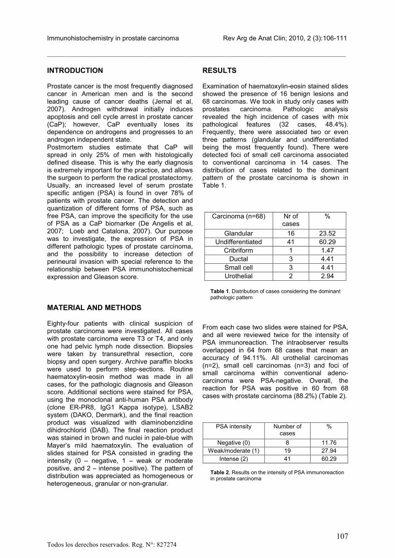
Examination of haematoxylin-eosin stained slides showed the presence of 16 benign lesions and 68 carcinomas. We took in study only cases with prostates carcinoma. Pathologic analysis revealed the high incidence of cases with mixpathological features (32 cases, 48.4%). Frequently, there were associated two or even three patterns (glandular and undifferentiated being the most frequently found). There were detected foci of small cell carcinoma associated to conventional carcinoma in 14 cases. The distribution of cases related to the dominant pattern of the prostate carcinoma is shown in Table 1.
Carcinoma (n=68) Nr of cases
%
Glandular 16 23.52Undifferentiated 41 60.29
Cribriform 1 1.47Ductal 3 4.41
Small cell 3 4.41Urothelial 2 2.94
Table 1. Distribution of cases considering the dominant pathologic pattern
From each case two slides were stained for PSA, and all were reviewed twice for the intensity of PSA immunoreaction. The intraobserver results overlapped in 64 from 68 cases that mean an accuracy of 94.11%. All urothelial carcinomas (n=2), small cell carcinomas (n=3) and foci of small carcinoma within conventional adeno-carcinoma were PSA-negative. Overall, the reaction for PSA was positive in 60 from 68 cases with prostate carcinoma (88.2%) (Table 2).
Table 2. Results on the intensity of PSA immunoreaction in prostate carcinoma
PSA intensity Number of cases
%
Negative (0) 8 11.76Weak/moderate (1) 19 27.94
Intense (2) 41 60.29

Immunohistochemistry in prostate carcinoma Rev Arg de Anat Clin; 2010, 2 (3):106-111
__________________________________________________________________________________________
Todos los derechos reservados. Reg. N°: 827274 108
In conventional adenocarcinoma the PSA reaction was positive as a fine granular final reaction product that occupies the cytoplasm. The reaction pattern was granular and rarely diffuse (Fig.1). A particular feature is related to the clear cell carcinoma (hypernephroid) and areas with signet cells, where the reaction product is always located at the periphery of the
cytoplasm, but all cells are intensely stained. In some cases the reaction was positive for all glandular cells (homogeneous pattern) and in others only isolated cells or clusters were intensely stained (heterogeneous pattern). The last aspect was noticed especially in cribiform carcinoma (Fig. 2).
Figure 1. Conventional adenocarcinoma, anti-PSA. Intense positive immunoreaction, granular and homogeneous ob. 20X
In well-differentiated adenocarcinoma (Gleason score 3 to 5, n=6) the reaction was positive in all but one case. The large majority of cases were characterized by weak reaction, and only isolated cases were appreciated as +2. Often, the intensity of the reaction was significantly weaker than in benign prostate hyperplasia. All cases with Gleason score from 6 to 8 (n=18) were positive. There were found 41 cases with undifferentiated carcinoma, with high Gleason score: 32 were intense positive (+2) withhomogeneous pattern (78%), 7 weak/moderate positive (17.07%), and 2 were negative (4.87%).
In cases with high Gleason score prevailed the heterogeneous pattern of PSA, and in cases with Gleason score less than 8 there were not found significant differences. Detection of the perineural invasion is extremely important for the management of the patient because it represents the main route for the progression of the tumor. There were investigated for this aspect only 63 cases because small cell and urothelial carcinomas were excluded. They are both negative, and therefore, the method cannot bring new information about the perineural invasion.

Immunohistochemistry in prostate carcinoma Rev Arg de Anat Clin; 2010, 2 (3):106-111
__________________________________________________________________________________________
Todos los derechos reservados. Reg. N°: 827274 109
Overall, we found perineural invasion on usual stained slides in only 15 cases (23.8%). Malignant cells were isolated, or arranged in small clusters, or even forming large glands that occupy the entire epineurium. On slides stained for PSA perineural invasion was found in 25
cases (39.68%) (Fig. 3). For both methods, perineural invasion was rarely noticed in specimens taken by core biopsy, but even in this case our results were better using anti-PSA immunohistochemistry.
Figure 2. Cribriform carcinoma with heterogeneous reaction for PSA. The presence of scattered and intensely stained malignant cells around the pseudo-lumens ob. 20X
DISCUSSION
Many problems that occur in the current pathologic diagnosis are related to undifferentiated tumors developed at the bladder neck, and lymph node metastasis of unknown primary. In the case of prostate cancer, identification of prostate specific antigen (PSA) is extremely helpful. PSA is highly but not completely specific. Monoclonal as well as polyclonal antibodies stain secretory cells of the benign tissue and malignant cells of glandular origin (excepting neuroendocrine cells). In prostate cancer the expression of PSA is found in
more than 95% of cases, but urothelial, squamous, and small cell carcinomas are always negative. Our data correspond with literature; in small cell carcinoma PSA was negative in all 3 cases but undifferentiated form that mimics urothelial carcinoma was positive for PSA (Table 1).PSA levels, Gleason score and TNM staging are established and considered essential to the prognostic of prostate cancer, when analyzed separately or jointly. Other factors such as the additional clinical morphologic ones and

Immunohistochemistry in prostate carcinoma Rev Arg de Anat Clin; 2010, 2 (3):106-111
__________________________________________________________________________________________
Todos los derechos reservados. Reg. N°: 827274 110
molecular markers can also contribute substantially (study by Hernandez and Thompson, 2004). Among the factors related to the biopsy, the percentage of positive fragments has presented a positive correlation with the potential risk of biochemical progression after treatment. In a study by Varma et al (2002) demonstrated that polyclonal anti-PSA has a superior sensitivity and they even recommend the high-grade prostate cancer as external control of PSA reaction. Our results are also supported by a recent study of Martinez-Pineiro et al (2003) that found a strong correlation
between PSA RT-PCR and Gleason score; we established that in cases with Gleason score less than 5 only isolated cases were estimated as +2 (intense reaction for PSA) but in cases with high Gleason score the reaction of PSA was intensely positive (Table 2) and was associated with high risk of progression of disease but was not supported by the study of Bostwick et al (1998) that found PSA immunoreactivity declined from benign epithelium to PIN and prostatic adenocarcinoma, suggesting that PSA is regulated differentially and decreased in expression with malignant transformation.
Figure 3. Perineural invasion in prostate carcinoma. PSA positive ob. 20X
PSA expression frequently is heterogeneous; therefore, immunostains may need to be performed on multiple blocks containing tumor when dealing with challenging cases (Shah et al, 2004). We found in cribriform carcinoma a heterogeneous pattern; only isolated cells or clusters (fig. 2) in accordance with literature but in other cases the reaction was positive for all glandular cells (homogeneous pattern); in cases with adenocarcinoma was found a strong
granular citoplasmatic reaction and the pattern was granular and rarely diffuse (Fig. 1).Perineural invasion is the hallmark of invasive prostate cancer, and it is found in more than 70% of cases on specimens of radical prostatectomy. Patients with perineural invasion were twice as likely to progress compared with patients without perineural invasion (Sebo et al, 2002). Before using PSA on routine examination we found perineural invasion only in 23.8% but after using immunohistochemistry we found that rate of

Immunohistochemistry in prostate carcinoma Rev Arg de Anat Clin; 2010, 2 (3):106-111
__________________________________________________________________________________________
Todos los derechos reservados. Reg. N°: 827274 111
perineural invasion significantly increased to 39.68% but even so is less than in the mentioned study. It is believed that every pathologist will immediately identify a large perineural invasion (as shown in Fig. 3). More hard is to diagnose small groups of malignant cells located close to thin nerve fibers. We may conclude that immunohistochemistry for PSA is extremely useful to detect perineural invasion in specimens taken by core biopsy and transurethral resection.In this study we investigated the carcinomas of the prostate that revealed the value of the PSA, for the differential diagnosis. The reaction was positive in 88.2% of cases, and it was negative in urothelial and small cell carcinomas. Our results showed intense expression in many cases with high Gleason score. Immunohistochemistry for PSA significantly increases the rate of detection of perineural invasion and confirms the prostatic origin of lymph node metastasis of apparently unknown primary site.
REFERENCES
Bostwick DG, Pacelli A, Blute M. 1998. Prostate specific membrane antigen expression in prostatic intraepithelial neoplasia and adenocarcinoma: a study of 184 cases. Cancer 82: 2256-61
De Angelis G, Rittenhouse HG, Mikolajczyk SD, Blair Shamel, Semjonow A. 2007. Twenty years of PSA: from prostate antigen to tumor marker. Rev Urol 9: 113-23
Hernandez J, Thompson IM. 2004. Prostate-specific antigen: a review of the validation of the most commonly used cancer biomarker. Cancer 101: 894-904.
Jemal A, Siegel R, Ward E, Murray T, Xu J, Thun MJ. 2007. Cancer statistics. CA Cancer J Clin 57: 43–66.
Loeb S, Catalona WJ. 2007. Prostate-specific antigen in clinical practice. Cancer Letters 249: 30-39
Martinez-Pineiro L, Rios E, Martinez-Gomariz. 2003. Molecular staging of prostatic cancer with RT-PCR assay for prostate specific antigen in peripheral blood and lymph nodes: comparison with standard histological staging and immunohistochemical assessment of occult regional lymph node metastases. Eur Urol 43: 342-50
Sebo TJ, Cheville JC, Riehle DL. 2002.Perineural invasion and MIB-1 positivity in addition to Gleason score are significant preoperative predictors of progression after radical retropubic prostatectomy for prostate cancer. Am J Surg Pathol 26: 431-39
Shah RB, Mehra R, Chinnaiyan AM. 2004. Androgen independent prostate cancer is a heterogeneous group of diseases: lessons from a rapid autopsy program. Cancer Res 64: 9209-16.
Varma M, Morgan M, Jasani B, Tamboli P, Amin MB. 2002. Polyclonal anti-PSA is more sensitive but less specific than monoclonal anti-PSA: implications for diagnostic prostatic pathology. Am J Clin Pathol 118: 202-07