
Immunohistochemical analysis of urokinase plasminogen activator and its prognostic value in canine...
6
Immunohistochemical analysis of urokinase plasminogen activator and its prognostic value in canine mammary tumours Andreia Santos a,b , Célia Lopes a , Raquel M. Marques a , Irina Amorim a , Jorge Ribeiro a , Carlos Frias a , Corália Vicente a , Fátima Gärtner a,c , Augusto de Matos a,b, * a Institute of Biomedical Sciences Abel Salazar (ICBAS), University of Porto, Largo Professor Abel Salazar, 2, 4099-003 Porto, Portugal b Multidisciplinary Unit for Biomedical Research (UMIB), University of Porto, Largo Professor Abel Salazar, 2, 4099-003 Porto, Portugal c The Institute of Molecular Pathology and Immunology of the University of Porto IPATIMUP, R. Dr. Roberto Frias, s/n, 4200-465 Porto, Portugal article info Article history: Accepted 22 May 2010 Keywords: Canine Mammary Neoplasia uPA Prognosis abstract Urokinase plasminogen activator (uPA) has been associated with aggressive behaviour and poor progno- sis in human breast cancer, but there is no information on its expression in canine mammary tumours (CMT). uPA immunohistochemical expression was studied in 119 CMT (24 benign, 95 malignant) to investigate its relationship with clinical and histopathological parameters. Dogs with malignant mam- mary tumours (MMT) underwent a 2-year follow-up evaluation. MMT expressed significantly more uPA than benign tumours. In MMT, high uPA stromal expression was significantly associated with larger tumour size, high Ki-67 expression, invasive growth, high histo- logical grade, regional lymph node metastases, development of distant metastases, and lower overall sur- vival (OS) and disease-free survival (DFS). On the basis of these results, uPA could be considered a useful prognostic factor in dogs with MMT. Ó 2010 Elsevier Ltd. All rights reserved. Introduction Tumour invasion and metastasis are the leading causes of mor- bidity and mortality in humans with breast cancer (Walker et al., 1997) and dogs with malignant mammary tumours (MMT) (Lana et al., 2007). These events involve multiple processes, such as deg- radation of extracellular matrix (ECM) and basement membrane (BM) (Ulisse et al., 2009). One of the major proteolytic systems in- volved in the ECM and BM degradation is the urokinase plasmino- gen activator (uPA) system, comprising uPA, its cell surface receptor (uPAR) and its inhibitors – plasminogen activator inhibi- tors 1 and 2 (PAI-1, PAI-2) (Han et al., 2006). uPA is a member of the serine protease family that is important to convert circulating plasminogen into the active serine protease, plasmin (Andreasen et al., 1997). Upon binding to its receptor, uPA induces direct plasmin-mediated proteolysis or indirect activation of other proteases, such as metalloproteinases (MMPs) (Han et al., 2006), contributing to the degradation of the ECM during tissue remodelling processes such as wound healing and post-lactational mammary gland involution. ECM degradation also allows tumour cells to access the systemic circulation and colonize different or- gans (Rabbani and Xing, 1998; Nielsen et al., 2001). Moreover uPA stimulates the release and activation of various growth factors, such as vascular endothelial growth factor (VEGF), fibroblast growth factor (FGF)-2 and transforming growth factor (TGF)-b (Guo et al., 2000), events that are considered determinant features of malignancy (Andreasen et al., 1997). The net result of this prote- olytic flux, combined with uPA-dependent intracellular signalling, is acceleration of tumour cell invasion and tumour-associated angiogenesis (Guo et al., 2000; Bajou et al., 2002). In addition, uPA is associated with cell proliferation, chemotaxis (Han et al., 2006) and apoptosis (Hildenbrand et al., 2009). The four major components of this system (uPA, uPAR, PAI-1 and PAI-2) have been established as prognostic factors in primary breast cancer (Dublin et al., 2000; Konecny et al., 2001; Harbeck et al., 2002; Han et al., 2006; Hildenbrand et al., 2009). Several studies assessing both mRNA and protein levels have found that elevated levels of uPA are associated with both aggressive tumour characteristics and poor prognosis (Ulisse et al., 2009). Canine mammary tumours (CMT), like human breast cancer, are a heterogeneous group of neoplasms showing great variability in behaviour. To the best of our knowledge, uPA has not been studied in CMT. Therefore, the main purpose of the present study was to investigate the immunohistochemical distribution pattern of uPA in CMT and its role in tumour behaviour through its association with clinicopathological parameters with recognized prognostic value. 1090-0233/$ - see front matter Ó 2010 Elsevier Ltd. All rights reserved. doi:10.1016/j.tvjl.2010.05.023 * Corresponding author at: Institute of Biomedical Sciences Abel Salazar (ICBAS), University of Porto, Largo Professor Abel Salazar, 2, 4099-003 Porto, Portugal. Tel./ fax: +351 222 062 260. E-mail address: [email protected] (A. de Matos). The Veterinary Journal 189 (2011) 43–48 Contents lists available at ScienceDirect The Veterinary Journal journal homepage: www.elsevier.com/locate/tvjl
-
Upload
andreia-santos -
Category
Documents
-
view
212 -
download
0
Transcript of Immunohistochemical analysis of urokinase plasminogen activator and its prognostic value in canine...
The Veterinary Journal 189 (2011) 43–48
Contents lists available at ScienceDirect
The Veterinary Journal
journal homepage: www.elsevier .com/ locate/ tv j l
Immunohistochemical analysis of urokinase plasminogen activatorand its prognostic value in canine mammary tumours
Andreia Santos a,b, Célia Lopes a, Raquel M. Marques a, Irina Amorim a, Jorge Ribeiro a, Carlos Frias a,Corália Vicente a, Fátima Gärtner a,c, Augusto de Matos a,b,*
a Institute of Biomedical Sciences Abel Salazar (ICBAS), University of Porto, Largo Professor Abel Salazar, 2, 4099-003 Porto, Portugalb Multidisciplinary Unit for Biomedical Research (UMIB), University of Porto, Largo Professor Abel Salazar, 2, 4099-003 Porto, Portugalc The Institute of Molecular Pathology and Immunology of the University of Porto IPATIMUP, R. Dr. Roberto Frias, s/n, 4200-465 Porto, Portugal
a r t i c l e i n f o a b s t r a c t
Article history:Accepted 22 May 2010
Keywords:CanineMammaryNeoplasiauPAPrognosis
1090-0233/$ - see front matter � 2010 Elsevier Ltd. Adoi:10.1016/j.tvjl.2010.05.023
* Corresponding author at: Institute of Biomedical SUniversity of Porto, Largo Professor Abel Salazar, 2, 4fax: +351 222 062 260.
E-mail address: [email protected] (A. de Matos)
Urokinase plasminogen activator (uPA) has been associated with aggressive behaviour and poor progno-sis in human breast cancer, but there is no information on its expression in canine mammary tumours(CMT). uPA immunohistochemical expression was studied in 119 CMT (24 benign, 95 malignant) toinvestigate its relationship with clinical and histopathological parameters. Dogs with malignant mam-mary tumours (MMT) underwent a 2-year follow-up evaluation.
MMT expressed significantly more uPA than benign tumours. In MMT, high uPA stromal expressionwas significantly associated with larger tumour size, high Ki-67 expression, invasive growth, high histo-logical grade, regional lymph node metastases, development of distant metastases, and lower overall sur-vival (OS) and disease-free survival (DFS). On the basis of these results, uPA could be considered a usefulprognostic factor in dogs with MMT.
� 2010 Elsevier Ltd. All rights reserved.
Introduction
Tumour invasion and metastasis are the leading causes of mor-bidity and mortality in humans with breast cancer (Walker et al.,1997) and dogs with malignant mammary tumours (MMT) (Lanaet al., 2007). These events involve multiple processes, such as deg-radation of extracellular matrix (ECM) and basement membrane(BM) (Ulisse et al., 2009). One of the major proteolytic systems in-volved in the ECM and BM degradation is the urokinase plasmino-gen activator (uPA) system, comprising uPA, its cell surfacereceptor (uPAR) and its inhibitors – plasminogen activator inhibi-tors 1 and 2 (PAI-1, PAI-2) (Han et al., 2006).
uPA is a member of the serine protease family that is importantto convert circulating plasminogen into the active serine protease,plasmin (Andreasen et al., 1997). Upon binding to its receptor, uPAinduces direct plasmin-mediated proteolysis or indirect activationof other proteases, such as metalloproteinases (MMPs) (Han et al.,2006), contributing to the degradation of the ECM during tissueremodelling processes such as wound healing and post-lactationalmammary gland involution. ECM degradation also allows tumourcells to access the systemic circulation and colonize different or-
ll rights reserved.
ciences Abel Salazar (ICBAS),099-003 Porto, Portugal. Tel./
.
gans (Rabbani and Xing, 1998; Nielsen et al., 2001). MoreoveruPA stimulates the release and activation of various growth factors,such as vascular endothelial growth factor (VEGF), fibroblastgrowth factor (FGF)-2 and transforming growth factor (TGF)-b(Guo et al., 2000), events that are considered determinant featuresof malignancy (Andreasen et al., 1997). The net result of this prote-olytic flux, combined with uPA-dependent intracellular signalling,is acceleration of tumour cell invasion and tumour-associatedangiogenesis (Guo et al., 2000; Bajou et al., 2002). In addition,uPA is associated with cell proliferation, chemotaxis (Han et al.,2006) and apoptosis (Hildenbrand et al., 2009).
The four major components of this system (uPA, uPAR, PAI-1and PAI-2) have been established as prognostic factors in primarybreast cancer (Dublin et al., 2000; Konecny et al., 2001; Harbecket al., 2002; Han et al., 2006; Hildenbrand et al., 2009). Severalstudies assessing both mRNA and protein levels have found thatelevated levels of uPA are associated with both aggressive tumourcharacteristics and poor prognosis (Ulisse et al., 2009).
Canine mammary tumours (CMT), like human breast cancer, area heterogeneous group of neoplasms showing great variability inbehaviour. To the best of our knowledge, uPA has not been studiedin CMT. Therefore, the main purpose of the present study was toinvestigate the immunohistochemical distribution pattern of uPAin CMT and its role in tumour behaviour through its associationwith clinicopathological parameters with recognized prognosticvalue.
44 A. Santos et al. / The Veterinary Journal 189 (2011) 43–48
Materials and methods
Specimens
Tumours (n = 119) were surgically removed from 119 female dogs, aged 5–16 years. Animals with MMT were enrolled in a 2-year post-operative follow-upstudy with no adjuvant therapy. The largest cross-sectional diameter of each tu-mour was recorded and categorized as either <3 cm or P3 cm. All specimens werefixed in 10% neutral buffered formalin for 48 h. Tumours 61 cm were paraffinembedded in one block, while larger tumours were cut sequentially at 5 mm inter-vals to provide a series of tissue blocks representative of the entire lesion.
After dehydration and embedding in paraffin wax, 3 lm sections were cut fromeach block. One section was stained with haematoxylin and eosin (HE) for diagnos-tic purposes and two representative blocks from tumours >1 cm were selected forimmunohistochemical studies. When available, local and regional lymph nodeswere processed and examined as previously described (Matos et al., 2006a).
Tumours and lymph nodes were evaluated independently by two observers(F.G. and I.A.) according to the criteria of the World Health Organization (WHO)for the histological classification of mammary tumours of domestic animals (Mis-dorp et al., 1999). Tumour histological grading was performed as previously de-scribed, according to the Nottingham method for human breast tumours (Matoset al., 2006a,b). The mode of growth of each tumour was assessed and classifiedas expansive (cohesive and well delimited growth of the tumour mass pushing nor-mal surrounding tissue) or invasive (when there was an infiltrative growth or whenlymphatic or blood vessels invasion was registered).
uPA immunohistochemistry (IHC)
Selected tumour sections adjacent to those used for HE staining were analysedby IHC using the modified avidin–biotin–peroxidase complex (ABC) method (Hsuet al., 1981). Sections were dewaxed in xylene and rehydrated in graded alcohols.Endogenous peroxidase activity was blocked by treating with 3% hydrogen peroxidein methanol for 10 min. Slides were then incubated with normal rabbit serum(Dako) diluted 1:5 in TBS containing 10% bovine serum albumin (BSA) for 20 minat room temperature. Excess serum was drained and slides were then incubatedovernight at 4 �C in a wet chamber with the anti-uPA (M-20) goat polyclonal anti-body (Santa Cruz Biotechnology; dilution 1:40 in TBS with 5% BSA). Slides werethen incubated with biotinylated rabbit anti-goat antibody (Santa Cruz Biotechnol-ogy; dilution 1:100 in TBS with 5% BSA) for 30 min and then with the ABC (Vector)for further 30 min. Colour was developed with a solution of 3,30-diaminobenzidineand the sections were then counterstained with haematoxylin, dehydrated, andmounted. To confirm the specificity of the IHC staining, the primary antibody wasreplaced with non-immune goat immunoglobulin. Positive controls consisted ofsections from human breast cancer tissue known to express uPA and canine normalrenal tissue (Bailey et al., 2006).
uPA expression evaluation was semi-quantitative and based on the percentageof neoplastic and stromal cells (fibroblasts) with cytoplasmic staining according tothe scoring method (cut-off 10%) used in human breast oncology (Hildenbrandet al., 2009). In carcinosarcomas, uPA expression was evaluated in both malignantcomponents (carcinomatous and sarcomatous) and in normal stromal fibroblasts.The slides were examined independently by two observers (A.S. and A.M.) and,when there was disagreement (<5% of the slides), a consensus was obtained usinga multi-head microscope.
MIB-1 immunohistochemistry
Selected tumour sections adjacent to those used for HE staining were immuno-stained and evaluated for Mindbomb homolog 1 (MIB-1) labelling index (Ki-67expression), as previously described (Matos et al., 2006b).
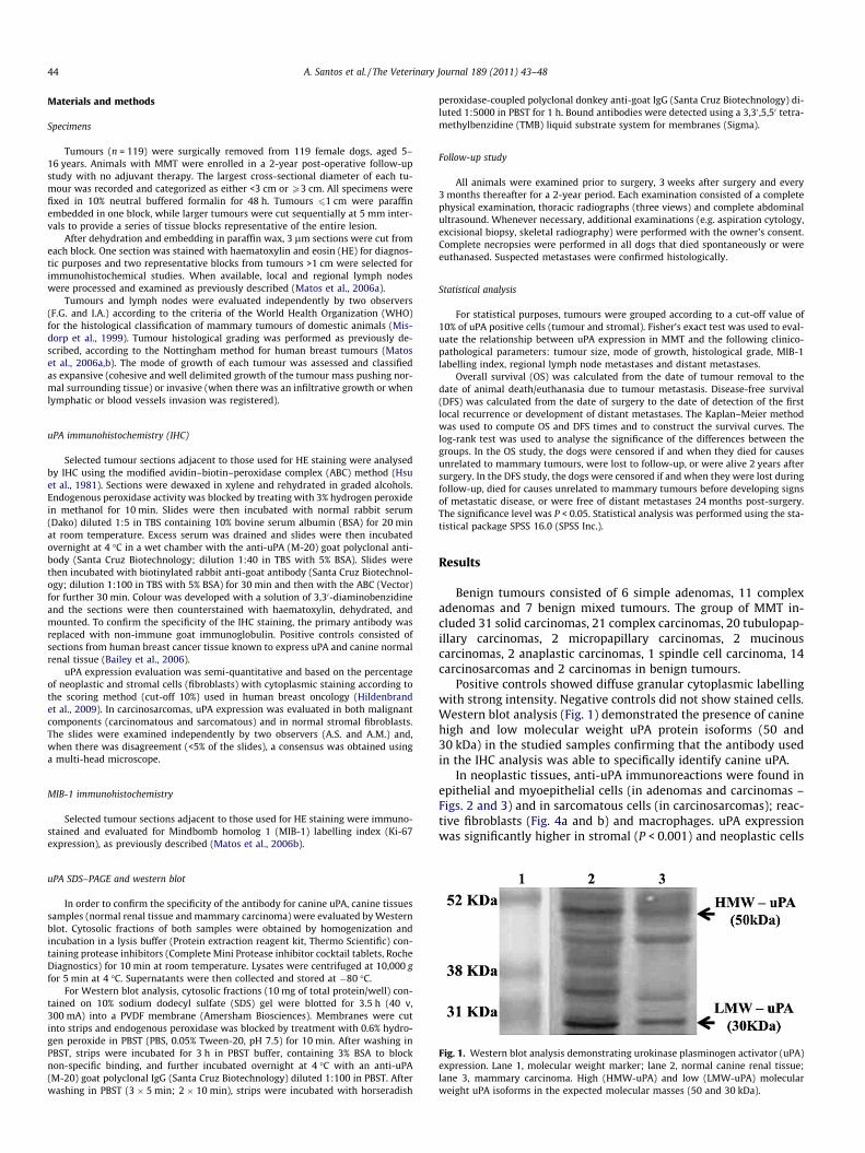
Fig. 1. Western blot analysis demonstrating urokinase plasminogen activator (uPA)expression. Lane 1, molecular weight marker; lane 2, normal canine renal tissue;lane 3, mammary carcinoma. High (HMW-uPA) and low (LMW-uPA) molecularweight uPA isoforms in the expected molecular masses (50 and 30 kDa).
uPA SDS–PAGE and western blot
In order to confirm the specificity of the antibody for canine uPA, canine tissuessamples (normal renal tissue and mammary carcinoma) were evaluated by Westernblot. Cytosolic fractions of both samples were obtained by homogenization andincubation in a lysis buffer (Protein extraction reagent kit, Thermo Scientific) con-taining protease inhibitors (Complete Mini Protease inhibitor cocktail tablets, RocheDiagnostics) for 10 min at room temperature. Lysates were centrifuged at 10,000 gfor 5 min at 4 �C. Supernatants were then collected and stored at �80 �C.
For Western blot analysis, cytosolic fractions (10 mg of total protein/well) con-tained on 10% sodium dodecyl sulfate (SDS) gel were blotted for 3.5 h (40 v,300 mA) into a PVDF membrane (Amersham Biosciences). Membranes were cutinto strips and endogenous peroxidase was blocked by treatment with 0.6% hydro-gen peroxide in PBST (PBS, 0.05% Tween-20, pH 7.5) for 10 min. After washing inPBST, strips were incubated for 3 h in PBST buffer, containing 3% BSA to blocknon-specific binding, and further incubated overnight at 4 �C with an anti-uPA(M-20) goat polyclonal IgG (Santa Cruz Biotechnology) diluted 1:100 in PBST. Afterwashing in PBST (3 � 5 min; 2 � 10 min), strips were incubated with horseradish
peroxidase-coupled polyclonal donkey anti-goat IgG (Santa Cruz Biotechnology) di-luted 1:5000 in PBST for 1 h. Bound antibodies were detected using a 3,30 ,5,50 tetra-methylbenzidine (TMB) liquid substrate system for membranes (Sigma).
Follow-up study
All animals were examined prior to surgery, 3 weeks after surgery and every3 months thereafter for a 2-year period. Each examination consisted of a completephysical examination, thoracic radiographs (three views) and complete abdominalultrasound. Whenever necessary, additional examinations (e.g. aspiration cytology,excisional biopsy, skeletal radiography) were performed with the owner’s consent.Complete necropsies were performed in all dogs that died spontaneously or wereeuthanased. Suspected metastases were confirmed histologically.
Statistical analysis
For statistical purposes, tumours were grouped according to a cut-off value of10% of uPA positive cells (tumour and stromal). Fisher’s exact test was used to eval-uate the relationship between uPA expression in MMT and the following clinico-pathological parameters: tumour size, mode of growth, histological grade, MIB-1labelling index, regional lymph node metastases and distant metastases.
Overall survival (OS) was calculated from the date of tumour removal to thedate of animal death/euthanasia due to tumour metastasis. Disease-free survival(DFS) was calculated from the date of surgery to the date of detection of the firstlocal recurrence or development of distant metastases. The Kaplan–Meier methodwas used to compute OS and DFS times and to construct the survival curves. Thelog-rank test was used to analyse the significance of the differences between thegroups. In the OS study, the dogs were censored if and when they died for causesunrelated to mammary tumours, were lost to follow-up, or were alive 2 years aftersurgery. In the DFS study, the dogs were censored if and when they were lost duringfollow-up, died for causes unrelated to mammary tumours before developing signsof metastatic disease, or were free of distant metastases 24 months post-surgery.The significance level was P < 0.05. Statistical analysis was performed using the sta-tistical package SPSS 16.0 (SPSS Inc.).
Results
Benign tumours consisted of 6 simple adenomas, 11 complexadenomas and 7 benign mixed tumours. The group of MMT in-cluded 31 solid carcinomas, 21 complex carcinomas, 20 tubulopap-illary carcinomas, 2 micropapillary carcinomas, 2 mucinouscarcinomas, 2 anaplastic carcinomas, 1 spindle cell carcinoma, 14carcinosarcomas and 2 carcinomas in benign tumours.
Positive controls showed diffuse granular cytoplasmic labellingwith strong intensity. Negative controls did not show stained cells.Western blot analysis (Fig. 1) demonstrated the presence of caninehigh and low molecular weight uPA protein isoforms (50 and30 kDa) in the studied samples confirming that the antibody usedin the IHC analysis was able to specifically identify canine uPA.
In neoplastic tissues, anti-uPA immunoreactions were found inepithelial and myoepithelial cells (in adenomas and carcinomas –Figs. 2 and 3) and in sarcomatous cells (in carcinosarcomas); reac-tive fibroblasts (Fig. 4a and b) and macrophages. uPA expressionwas significantly higher in stromal (P < 0.001) and neoplastic cells

Fig. 2. Immunohistochemistry (IHC) of canine mammary tumour. Simple adenoma.Strong uPA expression scattered in few neoplastic cells. No expression in stromalcells. Bar, 50 lm.
Fig. 3. IHC of canine mammary tumour. Tubulopapillary carcinoma. Neoplasticcells showing strong uPA immunoreactivity. Bar, 50 lm.
Fig. 4. IHC of canine mammary tumours. Complex carcinoma (a) and anaplasticcarcinoma (b). Reactive fibroblasts with strong immunoreactivity in contrast withneoplastic cells. Bars, 20 lm (a), 50 lm (b).
Table 1uPA expression in neoplastic and in stromal cells from benign and malignant caninemammary tumours.
Totalnumber
Number (%) of tumoursexhibiting neoplasticcells with uPAexpression
Number (%) of tumoursexhibiting stromal cellswith uPA expression
<10% >10% P <10% >10% P
Tumourtype
0.003 <0.001
Benign 24 10(41.7)
14(58.3)
18(75.0)
6 (25.0)
Malignant 95 12(12.6)
83(87.4)
26(27.4)
69(72.6)
A. Santos et al. / The Veterinary Journal 189 (2011) 43–48 45
(P = 0.003) from malignant tumours, when compared to benign tu-mours (Table 1).
In MMT, the elevated uPA expression in stromal cells was signif-icantly associated with larger tumour size (P = 0.011), invasivegrowth (P = 0.002), high histological grade (P = 0.005), regionallymph node (P = 0.002) and distant metastases (P = 0.014) and highMIB-1 labelling index (P < 0.001) (Table 2). Twelve of the 17 malig-nant tumours with lymphatic or vascular invasion exhibited morethan 50% stromal cells with strong immunoreactivity.
The OS study was performed in 66 patients and the DFS study in73 patients with MMT. uPA overexpression in stromal cells wassignificantly associated with lower OS (Table 3; Fig. 5) and lowerDFS (Table 4; Fig. 6).
Discussion
The uPA system is recognized for its major role in human cancerprogression, since its pleiotropic activity regulates the pathologicalbehaviour of epithelial and stromal cells within primary tumoursand metastases (Ulisse et al., 2009). In human medicine, high levelsof uPA are associated with poor prognosis for breast cancer (Dublinet al., 2000; Konecny et al., 2001; Harbeck et al., 2002; Hildenbrandet al., 2009). In the present study, we provide evidence that tumour
stromal cells (fibroblasts and macrophages) and neoplastic cells inCMT express uPA, as previously described in human breast cancer(Dublin et al., 2000; Nielsen et al., 2001, 2007; Hildenbrand et al.,2009; Hildenbrand and Schaaf, 2009).
uPA levels are significantly higher in malignant compared to be-nign human breast tumours (Ulisse et al., 2009). The higher expres-sion of uPA in malignant CMT suggests its possible involvement inmalignant behaviour. The uPA system can contribute to tumourgrowth and metastasis through multiple mechanisms, such asthe ECM and BM degradation, release of growth factors and stimu-lation of neoangiogenesis (Andreasen et al., 1997). Additionally,
Table 2Relationship between uPA expression in malignant mammary tumours and clinicopathological parameters.
Total number Number (%) of tumours exhibiting neoplastic cells withuPA expression
Number (%) of tumours exhibiting stromal cells withuPA expression
<10% >10% P <10% >10% P
Tumour sizea 0.043 0.011<3 cm 49 3 (6.1) 46 (93.9) 19 (38.8) 30 (61.2)P3 cm 45 9 (20.0) 36 (80.0) 7 (15.6) 38 (84.4)Mode of growth NS 0.002Expansive 25 2 (8.0) 23 (92.0) 11 (44.0) 14 (56.0)Invasive 70 10 (14.3) 60 (85.7) 15 (21.4) 55 (78.6)Histological grade NS 0.005I + II 64 8 (12.5) 56 (87.5) 23 (35.9) 41 (64.1)III 31 4 (12.9) 27 (87.1) 3 (9.7) 28 (90.3)Regional lymph node metastasesb NS 0.002Absent 50 4 (8.0) 46 (92.0) 19 (38.0) 31 (62.0)Present 23 4 (17.4) 18 (82.6) 1 (4.3) 22 (95.7)Distant metastasesc NS 0.014Absent 51 5 (9.8) 46 (90.2) 18 (35.3) 33 (64.7)Present 23 5 (21.7) 18 (78.3) 2 (8.7) 21 (91.3)MIB-1 labelling indexd NS <0.001<43% 55 6 (10.9) 49 (89.1) 22 (40.0) 33 (60.0)P43% 39 6 (15.4) 33 (84.6) 3 (7.7) 36 (92.3)
NS – not significant.a In one case, tumour size was not registered because it was multifocal throughout the mammary chain and incompletely excised, hampering the accurate measurement.b Lymph nodes were not submitted in 22 cases.C Fourteen dogs are still being followed. Seven dogs were lost to follow-up.d Due to technical reasons inherent to MIB-1 IHC analysis, the percentage of labelled cells was not generated in one tumour.
Table 3Association between uPA expression and overall survival in malignant mammarytumours.
uPAexpression
Numberof dogs
Dead/euthanased
Alive Mean OS time(±SE)
P
Tumour cells NS<10% 6 1 3 20.67 (3.04)>10% 60 17 32 19.72 (1.00)Stromal cells 0.002<10% 20 0 14 24>10% 46 18 21 17.98 (1.27)
NS – not significant.OS – overall survival.
Fig. 5. Kaplan–Meyer overall survival curves following surgical removal of malig-nant mammary tumours grouped on the basis of uPA expression in stromal cells.
46 A. Santos et al. / The Veterinary Journal 189 (2011) 43–48
this system may also affect tumour cell proliferation (via activationof ECM-associated mitogenic and angiogenic growth factors),adhesion and migration of tumour cells (through proteolysis ofsome ECM components, such as laminin and fibronectin), invasion(through BM breakdown), and growth at metastatic sites (Ulisseet al., 2009).
It is still unclear whether the high levels of uPA with prognosticvalue in breast cancer are of stromal or neoplastic origin (Castellóet al., 2007; Hildenbrand et al., 2009). Hildenbrand et al. (2009)found immunostaining in both stromal and neoplastic cells andsuggested that they have the same prognostic value; however,other authors (Dublin et al., 2000; Nielsen et al., 2001; Castellóet al., 2007) concluded that the strong uPA immunoreactivity dem-onstrated in stromal cells has a much greater implication in tu-mour behaviour and, therefore, stronger prognostic value. In ourstudy, the absence of a significant association between uPA expres-sion in neoplastic cells and histopathological parameters withprognostic value suggests that stromal rather than neoplastic cellexpression of uPA is directly related to malignant behaviour.
The predominant expression of uPA in tumour stroma impliesan interaction between the stromal and neoplastic components(Hildenbrand et al., 2009). During carcinogenesis, cancer cellsmay acquire the ability to control the expression of certain mole-
cules by neighbouring cells. uPA expression by different cell typesin a tumour is likely to depend on the complex paracrine signallingnetworks, involving growth factors and cytokines, through whichcancer cells and stromal cells communicate (Andreasen et al.,1997). Several molecules produced by cancer cells (e.g. TGF-band platelet-derived growth factor) induce the production of stro-mal proteases (MMPs and uPA) which in turn, mediate signallingevents essential for tumour cell migration (Rabbani and Gladu,2002; Têtu et al., 2006). Based on our observations, we hypothesize
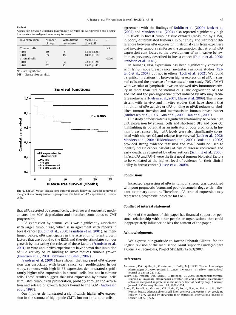
Table 4Association between urokinase plasminogen activator (uPA) expression and disease-free survival in malignant mammary tumours.
uPA expression Numberof dogs
With distantmetastases
Mean DFStime (±SE)
P
Tumour cells NS<10% 10 5 13.96 (3.26)>10% 63 19 18.07 (1.19)Stromal cells 0.009<10% 21 2 22.09 (1.28)>10% 52 22 15.65 (1.42)
NS – not significant.DSF – disease-free survival.
Fig. 6. Kaplan–Meyer disease-free survival curves following surgical removal ofmalignant mammary tumours grouped on the basis of uPA expression in stromalcells.
A. Santos et al. / The Veterinary Journal 189 (2011) 43–48 47
that uPA, secreted by stromal cells, drives several oncogenic mech-anisms, like ECM degradation and therefore contributes to CMTprogression.
uPA expression by stromal cells was significantly associatedwith larger tumour size, which is in agreement with reports inbreast cancer (Dublin et al., 2000; Frandsen et al., 2001). As men-tioned before, uPA participates in the activation of latent growthfactors that are bound to the ECM, and thereby stimulates tumourgrowth by increasing the release of these factors (Frandsen et al.,2001). In vitro and in vivo experiments have shown that inhibitionof uPA activity or its binding to uPAR reduces tumour growth(Frandsen et al., 2001; Rabbani and Gladu, 2002).
Frandsen et al. (2001) have shown that increased uPA expres-sion was associated with breast cancer cell proliferation. In ourstudy, tumours with high Ki-67 expression demonstrated signifi-cantly higher uPA expression in stromal cells, but not in tumourcells. These results suggest that uPA expression by stromal cellsstimulates tumour cell proliferation, probably through the activa-tion and release of growth factors bound to the ECM (Andreasenet al., 1997).
Our findings demonstrated a significantly higher uPA expres-sion in the stroma of high grade CMT’s but not in tumour cells in
agreement with the findings of Dublin et al. (2000). Look et al.(2002) and Manders et al. (2004) also reported significantly highuPA levels in breast tumour tissue extracts (measured by ELISA)in poorly differentiated tumours. In our study, the significant dif-ferences between uPA expression in stromal cells from expansiveand invasive tumours reinforces the assumption that stromal uPAexpression contributes to the development of an invasive behav-iour, as previously described in breast cancer (Dublin et al., 2000;Frandsen et al., 2001).
In humans, uPA expression has been significantly correlatedwith lymph node breast cancer metastasis in some studies (Cas-telló et al., 2007), but not in others (Look et al., 2002). We founda significant relationship between higher expression of uPA in stro-mal cells and the presence of metastases. In our study, 70% of MMTwith vascular or lymphatic invasion showed uPA immunoreactiv-ity in more than 50% of stromal cells. The degradation of ECMand BM and the pro-angiogenic effect induced by uPA may facili-tate metastasis (Nielsen et al., 2001; Ulisse et al., 2009). This is con-sistent with in vivo and in vitro studies that have shown thatinhibition of uPA activity or uPA-binding to uPAR reduces or abol-ishes tumour invasion and metastasis in human breast cancer(Andreasen et al., 1997; Guo et al., 2000; Han et al., 2006).
Our study demonstrated a significant relationship between highuPA expression by stromal cells and shortened DFS and poor OS,highlighting its potential as an indicator of poor prognosis. In hu-man breast cancer, high uPA levels were also significantly corre-lated with shorter OS and relapse-free survival (Look et al., 2002;Manders et al., 2004; Hildenbrand et al., 2009). Look et al. (2002)provided strong evidence that uPA and PAI-1 could be used toidentify breast cancer patients at risk of disease recurrence andearly death, as suggested by other authors (Schmitt et al., 2006).In fact, uPA and PAI-1 were the first novel tumour biological factorsto be validated at the highest level of evidence for their clinicalutility in breast cancer (Ulisse et al., 2009).
Conclusions
Increased expression of uPA in tumour stroma was associatedwith poor prognostic factors and poor outcome in dogs with malig-nant mammary tumours. Therefore, uPA stromal expression mayrepresent a prognostic indicator for CMT.
Conflict of interest statement
None of the authors of this paper has financial support or per-sonal relationship with other people or organisations that couldinappropriately influence or bias the content of the paper.
Acknowledgments
We express our gratitude to Doctor Deborah Gillette, for theEnglish revision of the manuscript. Grant support: Fundação paraa Ciência e Tecnologia, Project SFRH/BD/42363/2007.
References
Andreasen, P.A., Kjoller, L., Christense, L., Duffy, M.J., 1997. The urokinase-typeplasminogen activator system in cancer metastasis: a review. InternationalJournal of Cancer 72, 1–22.
Bailey, T.R., Paulsen, D.B., Sehgal, I., Hosgood, G., 2006. Immunohistochemicalstaining of urokinase plasminogen activator-like and urokinase plasminogenactivator receptor-like proteins in the urinary tract of healthy dogs. AmericanJournal of Veterinary Research 67, 1628–1634.
Bajou, K., Lewall, K., Martinez, C.R., Soria, C., Lu, H., Noél, A., Foidart, J.M., 2002.Human breast adenocarcinoma cell lines promote angiogenesis by providingcells with uPA-PAI and by enhancing their expression. International Journal ofCancer 100, 501–506.
48 A. Santos et al. / The Veterinary Journal 189 (2011) 43–48
Castelló, R., Landete, J.M., España, F., Vázquez, C., Fuster, C., Almenar, S.M., Ramón,L.A., Radtke, K., Estellés, A., 2007. Expression of plasminogen activator inhibitorstypes 1 and 3 and urokinase plasminogen activator protein and mRNA in breastcancer. Thrombosis Research 120, 753–762.
Dublin, E., Hanby, A., Patel, N.K., Liebman, R., Barnes, D., 2000.Immunohistochemical expression of uPA, uPAR, and PAI-1 in breastcarcinoma. American Journal of Pathology 157, 1219–1227.
Frandsen, T.L., Holst-Hansen, C., Nielsen, B.S., Christensen, I.J., Nyengaard, J.R.,Carmeliet, P., Brünner, N., 2001. Direct evidence of the importance of stromalurokinase plasminogen activator (uPA) in the growth of an experimentalhuman breast cancer using a combined uPA gene-disrupted andimmunodeficient xenograft model. Cancer Research 61, 532–537.
Guo, Y., Higazi, A.A.R., Arakelian, A., Sachais, B.S., Cines, D., Goldfarb, R.H., Jones, T.R.,Kwaan, H., Mazar, A.P., Rabbani, S.A., 2000. A peptide derived from thenonreceptor binding region of urokinase plasminogen activator (uPA) inhibitstumour progression and angiogenesis and induces tumour cell death in vivo.FASEB Journal 14, 1400–1410.
Han, B., Nakamura, M., Zhou, G., Ishii, A., Nakamura, A., Bai, Y., Mori, I., Kakudo, K.,2006. Calcitonin inhibits invasion of breast cancer cells: involvement ofurokinase-type plasminogen activator (uPA) and uPA receptor. InternationalJournal of Oncology 28, 807–814.
Harbeck, N., Kates, R.E., Schmitt, M., 2002. Clinical relevance of invasion factorsurokinase-type plasminogen activator and plasminogen activator inhibitor type1 for individualized therapy decisions in primary breast cancer is greatest whenused in combination. Journal of Clinical Oncology 20, 1000–1007.
Hildenbrand, R., Schaaf, A., 2009. The urokinase-system in tumour tissue stroma ofthe breast and breast cancer cell invasion. International Journal of Oncology 34,15–23.
Hildenbrand, R., Schaaf, A., Dorn-Beineke, A., Allgayer, H., Sütterlin, M., Marx, A.,Stroebel, P., 2009. Tumour stroma is the predominant uPA, uPAR, PAI-1expressing tissue in human breast cancer: prognostic impact. Histology andHistopathology 24, 869–877.
Hsu, S., Raine, L., Fanger, H., 1981. The use of anti avidin antibody and avidin–biotin–peroxidase complex in immunoperoxidase techniques. American Journalof Clinical Pathology 75, 816–821.
Konecny, G., Untch, M., Arboleda, J., Wilson, C., Kahlert, S., Boettcher, B., Felber, M.,Beryt, M., Lude, S., Hepp, H., Slamon, D., Pegram, M., 2001. HER-2/neu andurokinase-type plasminogen activator and its inhibitor in breast cancer. ClinicalCancer Research 7, 2448–2457.
Lana, S.E., Rutteman, G.R., Withrow, S.J., 2007. Tumors of the mammary gland. In:Withrow, S.J., MacEwen, E.G. (Eds.), Small Animal Clinical Oncology. SaundersElsevier, St. Louis, pp. 619–636.
Look, M.P., van Putten, W.L.J., Duffy, M.J., Harbeck, N., Christensen, I.J., Thomssen, C.,Kates R., Spyratos, F., Fernö, M., Eppenberger-Castori, S., Sweep, C.G., Ulm, K.,Peyrat, J.P., Martin, P.M., Magdelenat, H., Brünner, N., Duggan, C., Lisboa, B.W.,Bendahl, P.O., Quillien, V., Daver, A., Ricolleau, G., Meijer-van Gelder, M.E.,Manders, P., Fiets, W.E., Blankenstein, M.A., Broët, P., Romain, S., Daxenbichler,
G., Windbichler, G., Cufer, T., Borstnar, S., Kueng, W., Beex, L.V., Klijn, J.G.,O’Higgins, N., Eppenberger, U., Jänicke, F., Schmitt, M., Foekens, J.A., 2002.Pooled analysis of prognostic impact of urokinase-type plasminogen activatorand its inhibitor PAI-1 in 8377 breast cancer patients. Journal of National CancerInstitute 94, 116–128.
Manders, P., Tjan-Heijnen, V.C.G., Grebenchtchikov, N., Geurts-Moespot, A., vanTienoven, D.T.H., Beex, L.V.A.M., Sweep, F.C.G.J., 2004. Complex of urokinase-type plasminogen activator with its type 1 inhibitor predicts poor outcome in576 patients with lymph node-negative breast carcinoma. American CancerSociety 101, 3.
Matos, A.J.F., Faustino, A.M.R., Lopes, C., Rutteman, G.R., Gärtner, F., 2006a. Detectionof lymph node micrometastases in canine malignant mammary tumours withthe use of cytokeratin immunostaining. Veterinary Record 158, 626–629.
Matos, A.J.F., Lopes, C., Faustino, A.M.R., Carvalheira, J., Santos, M., Rutteman, G.R.,Gärtner, F., 2006b. MIB-1 indices according to clinico-pathological variables incanine mammary tumours: a multivariate study. Anticancer Research 26,1821–1826.
Misdorp, W., Else, R.W., Helmén, E., Lipscomb, T.P., 1999. Histological classificationof the mammary tumours of the dog and the cat. In: Shulman, Fl., (Ed.), WorldHealth Organization International Histological Classification of Tumours ofDomestic Animals, Second Series, vol. 7, Armed Forces Institute of Pathology,Washington, DC, pp. 16–29.
Nielsen, B.S., Sehested, M., Duun, S., Rank, F., Timshel, S., Rygaard, J., Johnsen, M.,Dano, K., 2001. Urokinase plasminogen activator is localized in stromal cells inductal breast cancer. Laboratory Investigation 81, 1485–1501.
Nielsen, B.S., Rank, F., Illeman, M., Lund, L.R., Dano, K., 2007. Stromal cells associatedwhit early invasive foci in human mammary ductal carcinoma in situ coexpressurokinase and urokinase receptor. International Journal of Cancer 120, 2086–2095.
Rabbani, S.A., Gladu, J., 2002. Urokinase receptor antibody can reduces tumourvolume and detect the presence of occult tumour metastases in vivo. CancerResearch 62, 2390–2397.
Rabbani, S.A., Xing, R.H., 1998. Role of urokinase (uPA) and its receptor (uPAR) ininvasion and metastasis of hormone-dependent malignancies. InternationalJournal of Oncology 12, 911–920.
Schmitt, M., Sturmheit, A.S., Welk, A., Schnelldorfer, C., Harbeck, N., 2006.Procedures for the quantitative protein determination of urokinase and itsinhibitor, PAI-1, in human breast cancer tissue extracts by ELISA. Methods inMolecular Medicine 120, 245–265.
Têtu, B., Trudel, D., Wang, C.S., 2006. Protéases des cellules stromales réactionnellesdu cancer: une cible thérapeutique attrayante. Bulletin du Cancer 93, 944–948.
Ulisse, S., Baldini, E., Sorrenti, S., D’Armiento, M., 2009. The urokinase plasminogenactivator system: a target for anti-cancer therapy. Current Cancer Drug Targets9, 32–71.
Walker, J.A., Jones, J.L., Chappell, S., Walsh, T., Shawn, J.A., 1997. Molecularpathology of breast cancer and its application to clinical management. CancerMetastasis Reviews 16, 5–27.


![Arecombinantchimeric plasminogenactivatorwithhighaffinity for … › content › pnas › 88 › 22 › 10337.full.pdf · urokinase-type plasminogen activator [scuPA(32kDa)], afi-brin-selective](https://static.fdocuments.us/doc/165x107/5f1cd2e4e4e08d6801761b19/arecombinantchimeric-plasminogenactivatorwithhighaffinity-for-a-content-a-pnas.jpg)

