Immune-Relevant and Antioxidant Activities of Vitellogenin ...
12
Review Immune-Relevant and Antioxidant Activities of Vitellogenin and Yolk Proteins in Fish Chen Sun and Shicui Zhang * Received: 15 July 2015 ; Accepted: 25 August 2015 ; Published: 22 October 2015 Laboratory for Evolution & Development, Institute of Evolution & Marine Biodiversity and Department of Marine Biology, Ocean University of China, Qingdao 266003, China; [email protected] * Correspondence: [email protected]; Tel./Fax: +86-532-82032787 Abstract: Vitellogenin (Vtg), the major egg yolk precursor protein, is traditionally thought to provide protein- and lipid-rich nutrients for developing embryos and larvae. However, the roles of Vtg as well as its derived yolk proteins lipovitellin (Lv) and phosvitin (Pv) extend beyond nutritional functions. Accumulating data have demonstrated that Vtg, Lv and Pv participate in host innate immune defense with multifaceted functions. They can all act as multivalent pattern recognition receptors capable of identifying invading microbes. Vtg and Pv can also act as immune effectors capable of killing bacteria and virus. Moreover, Vtg and Lv are shown to possess phagocytosis-promoting activity as opsonins. In addition to these immune-relevant functions, Vtg and Pv are found to have antioxidant activity, which is able to protect the host from oxidant stress. These non-nutritional functions clearly deepen our understanding of the physiological roles of the molecules, and at the same time, provide a sound basis for potential application of the molecules in human health. Keywords: vitellogenin; lipovitellin; phosvitin; immunity; antioxidant activity 1. Introduction Most fishes are oviparous, with their eggs being fertilized externally [1]. Eggs or haploid reproductive cells, which develop into viable embryos after fertilization, are the final product of oocyte growth and differentiation [2]. Generally, several steps are involved in oocyte development: formation of primordial germ-cells (PGCs), and transformation of PGCs into oogonia and then to oocytes. Subsequently, massive maternal information and molecules needed for early embryo development are deposited in growing oocytes during vitellogenesis, including RNAs, proteins, lipids, vitamins, and hormones [2,3]. One of the most important proteins deposited in oocytes is vitellogenin (Vtg), a member of the large lipid transfer protein (LLTP) superfamily [3–5]. Vtg is a high molecular mass glycolipophosphoprotein, usually circulating in the blood (vertebrates)/hemolymph (invertebrates) as a homodimer [4,6–8]. There are usually several isoforms of Vtg in a given species, which are encoded by a multigene family [9,10]. For instance, three vtg genes have been identified in chicken Gallus gallus [11,12], four in Africa frog Xenopus laevis [13,14], and six in nematode Caenorhabditis elegans [15]. Multiple vtg genes are also common in teleosts. It has been documented that there are seven vtg genes in zebrafish Danio rerio [16,17], two vtg genes in carp Cyprinus carpio [18], four vtg genes in medaka Oryzias latipes [10], three vtg genes in striped bass Morone saxatilis [19], and three vtg genes in white perch Morone americana [20]. All vitellogenins (Vtgs) encoded by multiple genes display a similar structure in vertebrates, such as fish, and invertebrates, particularly insects [21,22]. In most cases, Vtg contains three conserved domains, the LPD_N (also known as vitellogenin_N or LLT domain), which is identified at the N-terminus, the domain of unknown function (DUF) 1943, and the von Willebrand factor type D domain (vWD), which is located at the Nutrients 2015, 7, 8818–8829; doi:10.3390/nu7105432 www.mdpi.com/journal/nutrients
Transcript of Immune-Relevant and Antioxidant Activities of Vitellogenin ...

Review
Immune-Relevant and Antioxidant Activities ofVitellogenin and Yolk Proteins in Fish
Chen Sun and Shicui Zhang *
Received: 15 July 2015 ; Accepted: 25 August 2015 ; Published: 22 October 2015
Laboratory for Evolution & Development, Institute of Evolution & Marine Biodiversity and Department ofMarine Biology, Ocean University of China, Qingdao 266003, China; [email protected]* Correspondence: [email protected]; Tel./Fax: +86-532-82032787
Abstract: Vitellogenin (Vtg), the major egg yolk precursor protein, is traditionally thought toprovide protein- and lipid-rich nutrients for developing embryos and larvae. However, the rolesof Vtg as well as its derived yolk proteins lipovitellin (Lv) and phosvitin (Pv) extend beyondnutritional functions. Accumulating data have demonstrated that Vtg, Lv and Pv participate inhost innate immune defense with multifaceted functions. They can all act as multivalent patternrecognition receptors capable of identifying invading microbes. Vtg and Pv can also act as immuneeffectors capable of killing bacteria and virus. Moreover, Vtg and Lv are shown to possessphagocytosis-promoting activity as opsonins. In addition to these immune-relevant functions, Vtgand Pv are found to have antioxidant activity, which is able to protect the host from oxidant stress.These non-nutritional functions clearly deepen our understanding of the physiological roles of themolecules, and at the same time, provide a sound basis for potential application of the molecules inhuman health.
Keywords: vitellogenin; lipovitellin; phosvitin; immunity; antioxidant activity
1. Introduction
Most fishes are oviparous, with their eggs being fertilized externally [1]. Eggs or haploidreproductive cells, which develop into viable embryos after fertilization, are the final product ofoocyte growth and differentiation [2]. Generally, several steps are involved in oocyte development:formation of primordial germ-cells (PGCs), and transformation of PGCs into oogonia and thento oocytes. Subsequently, massive maternal information and molecules needed for early embryodevelopment are deposited in growing oocytes during vitellogenesis, including RNAs, proteins,lipids, vitamins, and hormones [2,3]. One of the most important proteins deposited in oocytes isvitellogenin (Vtg), a member of the large lipid transfer protein (LLTP) superfamily [3–5]. Vtg is a highmolecular mass glycolipophosphoprotein, usually circulating in the blood (vertebrates)/hemolymph(invertebrates) as a homodimer [4,6–8]. There are usually several isoforms of Vtg in a given species,which are encoded by a multigene family [9,10]. For instance, three vtg genes have been identifiedin chicken Gallus gallus [11,12], four in Africa frog Xenopus laevis [13,14], and six in nematodeCaenorhabditis elegans [15]. Multiple vtg genes are also common in teleosts. It has been documentedthat there are seven vtg genes in zebrafish Danio rerio [16,17], two vtg genes in carp Cyprinus carpio [18],four vtg genes in medaka Oryzias latipes [10], three vtg genes in striped bass Morone saxatilis [19],and three vtg genes in white perch Morone americana [20]. All vitellogenins (Vtgs) encoded bymultiple genes display a similar structure in vertebrates, such as fish, and invertebrates, particularlyinsects [21,22]. In most cases, Vtg contains three conserved domains, the LPD_N (also known asvitellogenin_N or LLT domain), which is identified at the N-terminus, the domain of unknownfunction (DUF) 1943, and the von Willebrand factor type D domain (vWD), which is located at the
Nutrients 2015, 7, 8818–8829; doi:10.3390/nu7105432 www.mdpi.com/journal/nutrients
Nutrients 2015, 7, 8818–8829
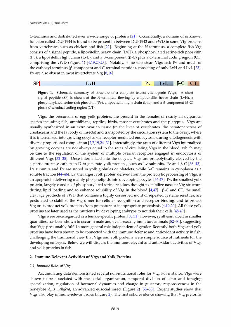
C-terminus and distributed over a wide range of proteins [21]. Occasionally, a domain of unknownfunction called DUF1944 is found to be present in between DUF1943 and vWD in some Vtg proteinsfrom vertebrates such as chicken and fish [22]. Beginning at the N-terminus, a complete fish Vtgconsists of a signal peptide, a lipovitellin heavy chain (LvH), a phosphorylated serine-rich phosvitin(Pv), a lipovitellin light chain (LvL), and a β-component (β-C) plus a C-terminal coding region (CT)comprising the vWD (Figure 1) [4,19,20,23]. Notably, some teleostean Vtgs lack Pv and much ofthe carboxyl-terminus (β-component and C-terminal peptide), consisting of only LvH and LvL [23].Pv are also absent in most invertebrate Vtg [8,16].
Nutrients 2015, 7 2 maternal information and molecules needed for early embryo development are deposited in growing oocytes during vitellogenesis, including RNAs, proteins, lipids, vitamins, and hormones [2,3]. One of the most important proteins deposited in oocytes is vitellogenin (Vtg), a member of the large lipid transfer protein (LLTP) superfamily [3–5]. Vtg is a high molecular mass glycolipophosphoprotein, usually circulating in the blood (vertebrates)/hemolymph (invertebrates) as a homodimer [4,6–8]. There are usually several isoforms of Vtg in a given species, which are encoded by a multigene family [9,10]. For instance, three vtg genes have been identified in chicken Gallus gallus [11,12], four in Africa frog Xenopus laevis [13,14], and six in nematode Caenorhabditis elegans [15]. Multiple vtg genes are also common in teleosts. It has been documented that there are seven vtg genes in zebrafish Danio rerio [16,17], two vtg genes in carp Cyprinus carpio [18], four vtg genes in medaka Oryzias latipes [10], three vtg genes in striped bass Morone saxatilis [19], and three vtg genes in white perch Morone americana [20]. All vitellogenins (Vtgs) encoded by multiple genes display a similar structure in vertebrates, such as fish, and invertebrates, particularly insects [21,22]. In most cases, Vtg contains three conserved domains, the LPD_N (also known as vitellogenin_N or LLT domain), which is identified at the N-terminus, the domain of unknown function (DUF) 1943, and the von Willebrand factor type D domain (vWD), which is located at the C-terminus and distributed over a wide range of proteins [21]. Occasionally, a domain of unknown function called DUF1944 is found to be present in between DUF1943 and vWD in some Vtg proteins from vertebrates such as chicken and fish [22]. Beginning at the N-terminus, a complete fish Vtg consists of a signal peptide, a lipovitellin heavy chain (LvH), a phosphorylated serine-rich phosvitin (Pv), a lipovitellin light chain (LvL), and a β-component (β-C) plus a C-terminal coding region (CT) comprising the vWD (Figure 1) [4,19,20,23]. Notably, some teleostean Vtgs lack Pv and much of the carboxyl-terminus (β-component and C-terminal peptide), consisting of only LvH and LvL [23]. Pv are also absent in most invertebrate Vtg [8,16].
Figure 1. Schematic summary of structure of a complete teleost vitellogenin (Vtg). A short signal peptide (SP) is shown at the N-terminus, flowing by a lipovitellin heave chain (LvH), a phosphorylated serine-rich phosvitin (Pv), a lipovitellin light chain (LvL), and a β-component (β-C) plus a C-terminal coding region (CT).
Vtgs, the precursors of egg yolk proteins, are present in the females of nearly all oviparous species including fish, amphibians, reptiles, birds, most invertebrates and the platypus. Vtgs are usually synthesized in an extra-ovarian tissue (in the liver of vertebrates, the hepatopancreas of crustaceans and the fat body of insects) and transported by the circulation system to the ovary, where it is internalized into growing oocytes via receptor-mediated endocytosis during vitellogenesis with diverse proportional composition [2,7,19,24–31]. Interestingly, the rates of different Vtgs internalized by growing oocytes are not always equal to the rates of circulating Vtgs in the blood, which may be due to the regulation of the system of multiple ovarian receptors engaged in endocytosis of different Vtgs [32–35]. Once internalized into the oocytes, Vtgs are proteolytically cleaved by the aspartic protease cathepsin D to generate yolk proteins, such as Lv subunits, Pv and β-C [36–43]. Lv subunits and Pv are stored in yolk globules or platelets, while β-C remains in cytoplasm as a soluble fraction [44–46]. Lv, the largest yolk
Figure 1. Schematic summary of structure of a complete teleost vitellogenin (Vtg). A shortsignal peptide (SP) is shown at the N-terminus, flowing by a lipovitellin heave chain (LvH), aphosphorylated serine-rich phosvitin (Pv), a lipovitellin light chain (LvL), and a β-component (β-C)plus a C-terminal coding region (CT).
Vtgs, the precursors of egg yolk proteins, are present in the females of nearly all oviparousspecies including fish, amphibians, reptiles, birds, most invertebrates and the platypus. Vtgs areusually synthesized in an extra-ovarian tissue (in the liver of vertebrates, the hepatopancreas ofcrustaceans and the fat body of insects) and transported by the circulation system to the ovary, whereit is internalized into growing oocytes via receptor-mediated endocytosis during vitellogenesis withdiverse proportional composition [2,7,19,24–31]. Interestingly, the rates of different Vtgs internalizedby growing oocytes are not always equal to the rates of circulating Vtgs in the blood, which maybe due to the regulation of the system of multiple ovarian receptors engaged in endocytosis ofdifferent Vtgs [32–35]. Once internalized into the oocytes, Vtgs are proteolytically cleaved by theaspartic protease cathepsin D to generate yolk proteins, such as Lv subunits, Pv and β-C [36–43].Lv subunits and Pv are stored in yolk globules or platelets, while β-C remains in cytoplasm as asoluble fraction [44–46]. Lv, the largest yolk protein derived from the proteolytic processing of Vtgs, isan apoprotein delivering mainly phospholipids into developing oocytes [36,47]. Pv, the smallest yolkprotein, largely consists of phosphorylated serine residues thought to stabilize nascent Vtg structureduring lipid loading and to enhance solubility of Vtg in the blood [4,47]. β-C and CT, the smallcleavage products of vWD that contains a highly conserved motif of repeated cysteine residues, arepostulated to stabilize the Vtg dimer for cellular recognition and receptor binding, and to protectVtg or its product yolk proteins from premature or inappropriate proteolysis [4,19,20]. All these yolkproteins are later used as the nutrients by developing embryos to nourish their cells [48,49].
Vtgs were once regarded as a female-specific protein [50,51]; however, synthesis, albeit in smallerquantities, has been shown to occur in male and even sexually immature animals [52–54], suggestingthat Vtgs presumably fulfill a more general role independent of gender. Recently, both Vtgs and yolkproteins have been shown to be connected with the immune defense and antioxidant activity in fish,challenging the traditional view that Vtgs and yolk proteins were simple source of nutrients for thedeveloping embryos. Below we will discuss the immune-relevant and antioxidant activities of Vtgsand yolk proteins in fish.
2. Immune-Relevant Activities of Vtgs and Yolk Proteins
2.1. Immune Roles of Vtgs
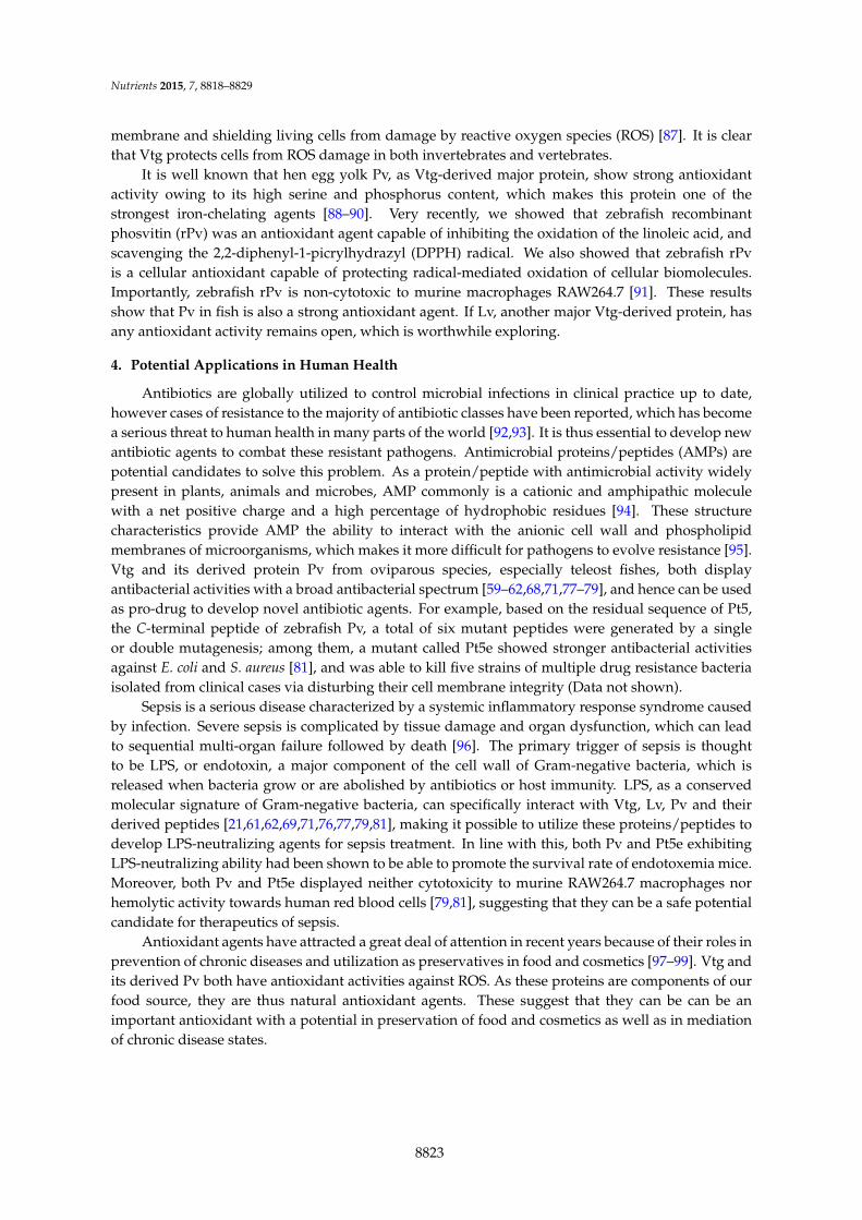
Accumulating data demonstrated several non-nutritional roles for Vtg. For instance, Vtgs wereshown to be associated with the social organization, temporal division of labor and foragingspecialization, regulation of hormonal dynamics and change in gustatory responsiveness in thehoneybee Apis mellifera, an advanced eusocial insect (Figure 2) [55–58]. Recent studies show thatVtgs also play immune-relevant roles (Figure 2). The first solid evidence showing that Vtg preforms
8819

Nutrients 2015, 7, 8818–8829
an immune-relevant role was the observation by Zhang et al., that Vtg purified from the ovaries ofthe protochordate amphioxus (Branchiostoma japonicum) exhibited hemagglutinating activity againstchick, toad and grass carp erythrocytes as well as antibacterial activity against the Gram-negativebacterium E. coli [59]. Soon after that, Vtg purified from the rosy barb Puntius conchonius was found tobe capable of inhibiting the growth of the Gram-negative bacteria E. coli, E. aerogenes and Pseudomonasputida as well as the Gram-positive bacteria Staphylococcus aureus, Bacillus subtilis and Streptococcuspyogenes [60], and Vtg from the carp capable of suppressing the growth of E. coli and S. aureus in adose dependent-manner [61]. Interestingly, Vtgs from protostomes also appear to have antibacterialactivity. Vtg from the scallop (Patinopecten yessoensis) was recently shown to have antibacterialactivity against Gram-positive and Gram-negative bacteria [62]. In addition, Vtg in the nematodeC. elegans seems also involved in its antibacterial defense. A reduced survival was observed in thevtg-knockdown C. elegans after pathogen infection [63]. Another evidence for a role of invertebrateVtg associated with resistance against bacteria was provided by the enhancement of resistance ofnematode against the pathogen Photorhabdus luminescens, when the production of Vtg was stimulatedby estrogen 17β-estradiol and phytoestrogen daidzein. However, reduction of Vtg caused by soyisoflavone genistein diminished the host resistance to P. luminescens [64]. Taken together, it appearsthat the antibacterial activity is a universal property of Vtgs from both vertebrates and invertebrates.Nutrients 2015, 7 4
Figure 2. Multiple roles of vitellogenin (Vtg). Vtg is traditionally thought to provide protein- and lipid-rich nutrients for developing embryos and larvae. However, accumulating data demonstrate that its roles extend beyond the nutritional function. In the advanced eusocial insect honeybee, Vtgs were shown to be associated with the social organization, temporal division of labor and foraging specialization, regulation of hormonal dynamics and change in gustatory responsiveness. Recent studies show that Vtgs also play immune-relevant roles. Vtg is able to recognize the invading microbes as a multivalent pattern recognition receptor, kill bacteria or neutralize virus as an effector molecule as well as enhance phagocytosis as an opsonin. Besides, Vtg also exhibits activities to hemagglutinate erythrocytes and aggregate pathogens. In addition to immune roles, Vtg plays another novel role as an antioxidant.
Shi et al. showed that intraperitoneal injection of E. coli was able to enhance the level of serum Vtg in male P. conchonius [60]. This has recently been confirmed by Lu et al., who showed that expression of vtg genes in the skin of zebrafish was induced following the challenge with Gram-negative bacterium Citrobacter freundii [65,66]. Moreover, an increased expression of vtg was also detectable in the insect Bactericera cockerelli after infected by “Candidatus Liberibacter solanacearum” via transcriptome analyses [67]. These data together suggest that Vtg may play an active role in the anti-infection of the host in vivo. Actually, Tong et al. showed that Vtg produced in male zebrafish as a consequence of
Figure 2. Multiple roles of vitellogenin (Vtg). Vtg is traditionally thought to provide protein- andlipid-rich nutrients for developing embryos and larvae. However, accumulating data demonstratethat its roles extend beyond the nutritional function. In the advanced eusocial insect honeybee,Vtgs were shown to be associated with the social organization, temporal division of labor andforaging specialization, regulation of hormonal dynamics and change in gustatory responsiveness.Recent studies show that Vtgs also play immune-relevant roles. Vtg is able to recognize the invadingmicrobes as a multivalent pattern recognition receptor, kill bacteria or neutralize virus as an effectormolecule as well as enhance phagocytosis as an opsonin. Besides, Vtg also exhibits activities tohemagglutinate erythrocytes and aggregate pathogens. In addition to immune roles, Vtg playsanother novel role as an antioxidant.
8820

Nutrients 2015, 7, 8818–8829
Shi et al. showed that intraperitoneal injection of E. coli was able to enhance the level of serum Vtgin male P. conchonius [60]. This has recently been confirmed by Lu et al., who showed that expression ofvtg genes in the skin of zebrafish was induced following the challenge with Gram-negative bacteriumCitrobacter freundii [65,66]. Moreover, an increased expression of vtg was also detectable in theinsect Bactericera cockerelli after infected by “Candidatus Liberibacter solanacearum” via transcriptomeanalyses [67]. These data together suggest that Vtg may play an active role in the anti-infection ofthe host in vivo. Actually, Tong et al. showed that Vtg produced in male zebrafish as a consequenceof induction by lipopolysaccharide (LPS) and lipoteichoic acid (LTA) is an acute phase reactant, withantibacterial activity against E. coli and S. aureus [68].
Vtg appears to play a multifaceted immune-relevant functions. Li et al. demonstrated thatVtg of the marine fish Hexagrammos otakii was able to bind to Gram-negative bacterium E. coli andGram-positive bacterium S. aureus as well as fungus Pichia pastoris [69]. The binding to E. coli andS. aureus was also detected for carp and zebrafish Vtgs [61,68]. The binding of Vtgs to bacteriaprovides them ability to aggregate pathogens as well as to recognize the invading microbes [61].Further examination via ELISA assay showed that Vtgs exhibited specific affinities to the componentsconserved within a class of microbes, called pathogen-associated molecular patterns (PAMPs),including LPS of Gram-negative bacteria, LTA of Gram-positive bacteria, peptidoglycan (PGN) ofGram-negative and positive bacteria, and glucan of fungi [61,69]. These observations indicate thatVtg first functions as a multivalent pattern recognition receptor capable of identifying invadingGram-negative and Gram-positive bacteria as well as fungi, and is involved in host immune defenseas a detector. In a recent study attempting to search PGN recognition proteins in giant tiger shrimp(Penaeus monodon), an 83 kDa protein was isolated by in vitro PGN pull-down binding assay andidentified as a Vtg-like protein via mass spectrometry as well as Western blots with monoclonalantibodies specific of Vtgs reported from P. monodon [70], implicating that invertebrate Vtg mayalso play a pattern recognition receptor role. Scanning electron microscopy as well as bacterial celland protoplast lysis assays showed that H. otakii Vtg was able to kill pathogenic bacteria by lysingthe whole cells (with cell walls) instead of protoplast (without cell walls) via interaction with LPSand LTA [71]. These show that Vtg functions as an effector molecule, capable of directly killingbacteria. Interestingly, Vtg was also shown to be able to enhance the phagocytosis of microbes bymacrophages. Li et al. first reported that H. otakii Vtg could facilitate engulfing of the microbesE. coli, S. aureus and P. pastoris by head-kidney-derived macrophages in vitro [69]. Later, Vtg ofcarp was shown to possess similar phagocytosis-promoting activity [61]. In an in-situ study ofimpacts of urban wastewater on freshwater mussel Elliptio complanata, it was observed that theproduction of Vtg-like proteins was strongly associated with phagocytosis [72], suggesting a relationbetween Vtg and phagocytosis in invertebrates. Besides, H. otakii Vtg was found to be capableof binding to the cell surface of macrophages but not that of red blood cells [61,69]. Collectively,these observations indicate that Vtg is an opsonin functioning as a bridging molecule between hostmacrophages and invading pathogens, thereby leading to enhanced phagocytosis. Notably, Liu et al.,established that the H. otakii Vtg was able to opsonize the fungus P. pastoris for phagocytosis bymacrophages isolated from sea bass Lateolabrax japonicas, implying that the opsonization of Vtg wasnot species-specific [73]. Further study revealed that Vtg-opsonized phagocytosis showed propertiestypical of type I phagocytosis, including pseudopod extension, tyrosine kinase dependence, andup-regulation of pro-inflammatory cytokine genes tnf-α and il-1β [73]. Therefore, Vtg is a patternrecognition receptor capable of identifying pathogens, a bactericidal molecule capable of damagingbacterial cell walls, and an opsonin capable of enhancing phagocytosis of pathogens by macrophages.The multifaceted immune-relevant activities of Vtg are in part endowed with its different domains.It was reported by Sun et al. that both DUF1943 and DUF1944 as well as vWD contribute to thefunction of Vtg as a pattern recognition receptor, and DUF1943 and DUF1944 (but not vWD) alsocontribute to the function of Vtg as an opsonin [21].
8821

Nutrients 2015, 7, 8818–8829
Recently, Garcia et al. showed that Atlantic salmon Vtg possessed neutralizing ability forinfectious pancreatic necrosis virus [74], suggesting that Vtg is also involved in host antiviralimmunity. This seems further supported by the observation that the mosquito (Anopheles gambiae) Vtgwas able to interfere with anti-plasmodium response [75]. These denote that in addition to antibacterialactivity, Vtg also has antiviral activity, which demands detailed study in the future.
2.2. Immune Roles of Yolk Proteins
Lv and Pv are the principal yolk proteins generated by the proteolytic cleavage of Vtg. As Vtg isan immune-competent molecule, it is thus reasonable to hypothesize that Lv and Pv also have similarimmune activities. This hypothesis was first tested by Zhang and Zhang [76]. They demonstratedthat the native Lv purified from ovulated eggs of the rosy barb P. conchonius was able to interactwith LPS, LTA and PGN, as well as E. coli and S. aureus, but not with self-molecules such as theegg extracts prepared, indicating that Lv is a molecule capable of recognizing non-self components.Moreover, the bacterial binding activity of Lv enabled it to enhance the phagocytosis of bacteria bymacrophages, suggesting that Lv is also an opsonin functional in developing embryos/larvae [76].Similarly, Pv was also shown to play a critical role in the immunity of zebrafish embryos via actingas a pattern recognition receptor and an antimicrobial effector molecule [77]. In line with this, henegg yolk Pv was also shown to be able to inhibit the growth of the Gram-negative bacterium E. coliand the Gram-positive bacterium S. aureus under thermal stress [78,79]. Of note, the affinity ofPv to LPS enabled the protein a capacity to neutralize endotoxin, promoting the survival rate ofendotoxemia mice [79]. It was recently shown that a truncated Pv (Pt5) consisting of the C-terminal55 residues of zebrafish Pv also displayed similar immune activities with Pv, including antimicrobialactivity against E. coli, Aeromonas hydrophila and S. aureus, and specific affinity to LPS, LTA, andPGN [77]. Intraperitoneal injection of this Pv-derived peptide was able to increase the survivalrate of zebrafish challenged with pathogenic A. hydrophila and to markedly decrease the numberof the pathogen in multiple tissues, suggesting that Pt5 could inhibit multiplication/disseminationof pathogen in host as an antimicrobial agent. In addition to direct antimicrobial activity, Pt5was also shown to be able to regulate the host immune responses via suppressing the expressionof pro-inflammatory cytokine genes (il-1β, il-6, tnf-α and ifn-γ) and simultaneously enhancingthe expression of anti-inflammatory cytokine genes (il-10 and il-4), suggesting a dual role of Pt5as both immune effector and modulator [80]. Recently, a mutant peptide of Pt5 (designated asPt5e), generated by site-directed mutagenesis, was shown to have stronger bactericidal activityand LPS-neutralizing activity [81]. Besides, Sun et al. demonstrated that recombinant zebrafishPv was capable of inhibiting the formation of the cytopathic effect in lymphocystis disease virus(LCDV)-infected cells and reducing the virus quantities in the infected cells as well as in the infectedzebrafish, suggesting that Pv possesses an antiviral activity and participates in immune defense ofhost against the infection by viruses like LCDV [82]. Taken together, these data show that like Vtg,Lv and Pv are both immune-competent molecules involved in immune response of the host againstinvading pathogenic microbes.
3. Antioxidant Activities of Vtgs and Yolk Proteins
In addition to immune roles, another novel role of Vtg is antioxidant activity (Figure 2).It was first shown by Ando and Yanagida that Vtg from the eel Anguilla japonica was able to resistthe copper-induced oxidation, and could protect the very low density lipoprotein (VLDL) againstcopper-induced oxidation [83]. This was the first observation reporting that Vtg has antioxidantactivity, and serves to suppress the free-radical reactions in fish oocytes. Similar antioxidant activitywas also suggested for the nematode (C. elegans) Vtg [84]. In the honeybee, Vtg was demonstratedto be able to reduce oxidative stress by scavenging free radicals, thereby increasing the lifespan inthe facultatively sterile worker castes and reproductive queen castes [85,86]. The honeybee Vtg wasalso demonstrated in a recent study to be capable of recognizing cell damage through its binding to
8822
Nutrients 2015, 7, 8818–8829
membrane and shielding living cells from damage by reactive oxygen species (ROS) [87]. It is clearthat Vtg protects cells from ROS damage in both invertebrates and vertebrates.
It is well known that hen egg yolk Pv, as Vtg-derived major protein, show strong antioxidantactivity owing to its high serine and phosphorus content, which makes this protein one of thestrongest iron-chelating agents [88–90]. Very recently, we showed that zebrafish recombinantphosvitin (rPv) was an antioxidant agent capable of inhibiting the oxidation of the linoleic acid, andscavenging the 2,2-diphenyl-1-picrylhydrazyl (DPPH) radical. We also showed that zebrafish rPvis a cellular antioxidant capable of protecting radical-mediated oxidation of cellular biomolecules.Importantly, zebrafish rPv is non-cytotoxic to murine macrophages RAW264.7 [91]. These resultsshow that Pv in fish is also a strong antioxidant agent. If Lv, another major Vtg-derived protein, hasany antioxidant activity remains open, which is worthwhile exploring.
4. Potential Applications in Human Health
Antibiotics are globally utilized to control microbial infections in clinical practice up to date,however cases of resistance to the majority of antibiotic classes have been reported, which has becomea serious threat to human health in many parts of the world [92,93]. It is thus essential to develop newantibiotic agents to combat these resistant pathogens. Antimicrobial proteins/peptides (AMPs) arepotential candidates to solve this problem. As a protein/peptide with antimicrobial activity widelypresent in plants, animals and microbes, AMP commonly is a cationic and amphipathic moleculewith a net positive charge and a high percentage of hydrophobic residues [94]. These structurecharacteristics provide AMP the ability to interact with the anionic cell wall and phospholipidmembranes of microorganisms, which makes it more difficult for pathogens to evolve resistance [95].Vtg and its derived protein Pv from oviparous species, especially teleost fishes, both displayantibacterial activities with a broad antibacterial spectrum [59–62,68,71,77–79], and hence can be usedas pro-drug to develop novel antibiotic agents. For example, based on the residual sequence of Pt5,the C-terminal peptide of zebrafish Pv, a total of six mutant peptides were generated by a singleor double mutagenesis; among them, a mutant called Pt5e showed stronger antibacterial activitiesagainst E. coli and S. aureus [81], and was able to kill five strains of multiple drug resistance bacteriaisolated from clinical cases via disturbing their cell membrane integrity (Data not shown).
Sepsis is a serious disease characterized by a systemic inflammatory response syndrome causedby infection. Severe sepsis is complicated by tissue damage and organ dysfunction, which can leadto sequential multi-organ failure followed by death [96]. The primary trigger of sepsis is thoughtto be LPS, or endotoxin, a major component of the cell wall of Gram-negative bacteria, which isreleased when bacteria grow or are abolished by antibiotics or host immunity. LPS, as a conservedmolecular signature of Gram-negative bacteria, can specifically interact with Vtg, Lv, Pv and theirderived peptides [21,61,62,69,71,76,77,79,81], making it possible to utilize these proteins/peptides todevelop LPS-neutralizing agents for sepsis treatment. In line with this, both Pv and Pt5e exhibitingLPS-neutralizing ability had been shown to be able to promote the survival rate of endotoxemia mice.Moreover, both Pv and Pt5e displayed neither cytotoxicity to murine RAW264.7 macrophages norhemolytic activity towards human red blood cells [79,81], suggesting that they can be a safe potentialcandidate for therapeutics of sepsis.
Antioxidant agents have attracted a great deal of attention in recent years because of their roles inprevention of chronic diseases and utilization as preservatives in food and cosmetics [97–99]. Vtg andits derived Pv both have antioxidant activities against ROS. As these proteins are components of ourfood source, they are thus natural antioxidant agents. These suggest that they can be can be animportant antioxidant with a potential in preservation of food and cosmetics as well as in mediationof chronic disease states.
8823

Nutrients 2015, 7, 8818–8829
5. Conclusions
Vtg, the precursor of major egg yolk proteins, is traditionally thought to provide protein- andlipid-rich nutrients for developing embryos and larvae. However, accumulating data indicate thatVtg as well as its derived yolk proteins Lv and Pv also play non-nutritional functions: they arenot only involved in immune defense but also antioxidant reaction. These non-nutritional functionsclearly better and deepen our understanding of the physiological roles of the molecules, and at thesame time, provide a sound basis for potential application of the molecules in human health.
Acknowledgments: This work was supported by Natural Science Foundation of China (No. 31402030;31372505), Fundamental Research Funds for the Central Universities (201462004), China Postdoctoral ScienceFoundation (2015T80745; 2014M560580) and Qingdao Municipal Science and Technology Commission(No. 13-1-4-206-jch).
Author Contributions: C. Sun wrote the manuscript. S. C. Zhang read and revised the manuscript.
Conflicts of Interest: The authors declare no conflict of interest.
References
1. Jalabert, B. Particularities of reproduction and oogenesis in teleost fish compared to mammals.Reprod. Nutr. Dev. 2005, 45, 261–279. [CrossRef] [PubMed]
2. Lubzens, E.; Young, G.; Bobe, J.; Cerda, J. Oogenesis in teleosts: How eggs are formed.Gen. Comp. Endocrinol. 2010, 165, 367–389. [CrossRef]
3. Patiño, R.; Sullivan, C. Ovarian follicle growth, maturation, and ovulation in teleost fish.Fish Physiol. Biochem. 2002, 26, 57–70. [CrossRef]
4. Finn, R.N. Vertebrate yolk complexes and the functional implications of phosvitins and other subdomainsin vitellogenins. Biol. Reprod. 2007, 76, 926–935. [CrossRef] [PubMed]
5. Smolenaars, M.M.; Madsen, O.; Rodenburg, K.W.; van der Horst, D.J. Molecular diversity and evolution ofthe large lipid transfer protein superfamily. J. Lipid Res. 2007, 48, 489–502. [CrossRef] [PubMed]
6. Avarre, J.C.; Lubzens, E.; Babin, P.J. Apolipocrustacein, formerly vitellogenin, is the major egg yolkprecursor protein in decapod crustaceans and is homologous to insect apolipophorin II/I and vertebrateapolipoprotein B. BMC Evol. Biol. 2007, 7. [CrossRef] [PubMed]
7. Tufail, M.; Takeda, M. Molecular characteristics of insect vitellogenins. J. Insect Physiol. 2008, 54, 1447–1458.[PubMed]
8. Matozzo, V.; Gagne, F.; Marin, M.G.; Ricciardi, F.; Blaise, C. Vitellogenin as a biomarker of exposure toestrogenic compounds in aquatic invertebrates: A review. Environ. Int. 2008, 34, 531–545. [PubMed]
9. Wu, L.T.; Hui, J.H.; Chu, K.H. Origin and evolution of yolk proteins: Expansion and functionaldiversification of large lipid transfer protein superfamily. Biol. Reprod. 2013, 88. [CrossRef] [PubMed]
10. Finn, R.N.; Kolarevic, J.; Kongshaug, H.; Nilsen, F. Evolution and differential expression of a vertebratevitellogenin gene cluster. BMC Evol. Biol. 2009, 9. [CrossRef] [PubMed]
11. Silva, R.; Fischer, A.H.; Burch, J.B. The major and minor chicken vitellogenin genes are each adjacent topartially deleted pseudogene copies of the other. Mol. Cell. Biol. 1989, 9, 3557–3562. [CrossRef] [PubMed]
12. Van het Schip, F.D.; Samallo, J.; Broos, J.; Ophuis, J.; Mojet, M.; Gruber, M.; Ab, G. Nucleotide sequenceof a chicken vitellogenin gene and derived amino acid sequence of the encoded yolk precursor protein.J. Mol. Biol. 1987, 196, 245–260. [CrossRef]
13. Germond, J.E.; Walker, P.; ten Heggeler, B.; Brown-Luedi, M.; de Bony, E.; Wahli, W. Evolution ofvitellogenin genes: Comparative analysis of the nucleotide sequences downstream of the transcriptioninitiation site of four Xenopus laevis and one chicken gene. Nucleic Acids Res. 1984, 12, 8595–8609. [CrossRef][PubMed]
14. Wahli, W.; Dawid, I.B.; Wyler, T.; Jaggi, R.B.; Weber, R.; Ryffel, G.U. Vitellogenin in Xenopus laevis is encodedin a small family of genes. Cell 1979, 16, 535–549. [CrossRef]
15. Blumenthal, T.; Squire, M.; Kirtland, S.; Cane, J.; Donegan, M.; Spieth, J.; Sharrock, W. Cloning of a yolkprotein gene family from Caenorhabditis elegans. J. Mol. Biol. 1984, 174, 1–18. [CrossRef]
8824

Nutrients 2015, 7, 8818–8829
16. Wang, H.; Yan, T.; Tan, J.T.; Gong, Z. A zebrafish vitellogenin gene (vg3) encodes a novel vitellogeninwithout a phosvitin domain and may represent a primitive vertebrate vitellogenin gene. Gene 2000, 256,303–310. [CrossRef]
17. Wang, H.; Tan, J.T.; Emelyanov, A.; Korzh, V.; Gong, Z. Hepatic and extrahepatic expression of vitellogeningenes in the zebrafish, Danio rerio. Gene 2005, 356, 91–100. [PubMed]
18. Kang, B.J.; Jung, J.H.; Lee, J.M.; Lim, S.G.; Saito, H.; Kim, M.H.; Kim, Y.J.; Saigusa, M.; Han, C.H. Structuraland expression analyses of two vitellogenin genes in the carp, Cyprinus carpio. Comp. Biochem. Physiol. Part BBiochem. Mol. Biol. 2007, 148, 445–453. [CrossRef] [PubMed]
19. Williams, V.N.; Reading, B.J.; Hiramatsu, N.; Amano, H.; Glassbrook, N.; Hara, A.; Sullivan, C.V. Multiplevitellogenins and product yolk proteins in striped bass, Morone saxatilis: Molecular characterization andprocessing during oocyte growth and maturation. Fish Physiol. Biochem. 2014, 40, 395–415. [CrossRef][PubMed]
20. Reading, B.J.; Hiramatsu, N.; Sawaguchi, S.; Matsubara, T.; Hara, A.; Lively, M.O.; Sullivan, C.V. Conservedand variant molecular and functional features of multiple egg yolk precursor proteins (vitellogenins) inwhite perch (Morone americana) and other teleosts. Mar. Biotechnol. 2009, 11, 169–187. [CrossRef] [PubMed]
21. Sun, C.; Hu, L.; Liu, S.; Gao, Z.; Zhang, S. Functional analysis of domain of unknown function(DUF) 1943, DUF1944 and von Willebrand factor type D domain (VWD) in vitellogenin 2 in zebrafish.Dev. Comp. Immunol. 2013, 41, 469–476. [CrossRef] [PubMed]
22. Hayward, A.; Takahashi, T.; Bendena, W.G.; Tobe, S.S.; Hui, J.H. Comparative genomic and phylogeneticanalysis of vitellogenin and other large lipid transfer proteins in metazoans. FEBS Lett. 2010, 584, 1273–1278.[CrossRef] [PubMed]
23. Finn, R.N.; Kristoffersen, B.A. Vertebrate vitellogenin gene duplication in relation to the “3R hypothesis”:Correlation to the pelagic egg and the oceanic radiation of teleosts. PLoS ONE 2007, 2, e169. [CrossRef][PubMed]
24. Meusy, J.J. Vitellogenin, the extraovarian precursor of the protein yolk in Crustacea: A review.Reprod. Nutr. Dev. 1980, 20, 1–21. [CrossRef] [PubMed]
25. Girish, B.P.; Swetha, C.; Reddy, P.S. Hepatopancreas but not ovary is the site of vitellogenin synthesis infemale fresh water crab, Oziothelphusa senex senex. Biochem. Biophys. Res. Commun. 2014, 447, 323–327.[CrossRef] [PubMed]
26. Mak, A.S.; Choi, C.L.; Tiu, S.H.; Hui, J.H.; He, J.G.; Tobe, S.S.; Chan, S.M. Vitellogenesis in the red crabCharybdis feriatus: Hepatopancreas-specific expression and farnesoic acid stimulation of vitellogenin geneexpression. Mol. Reprod. Dev. 2005, 70, 288–300. [CrossRef] [PubMed]
27. Kolarevic, J.; Nerland, A.; Nilsen, F.; Finn, R.N. Goldsinny wrasse (Ctenolabrus rupestris) is an extremevtgAa-type pelagophil teleost. Mol. Reprod. Dev. 2008, 75, 1011–1020. [CrossRef] [PubMed]
28. Sawaguchi, S.; Ohkubo, N.; Koya, Y.; Matsubara, T. Incorporation and utilization of multiple forms ofvitellogenin and their derivative yolk proteins during vitellogenesis and embryonic development in themosquitofish, Gambusia affinis. Zool. Sci. 2005, 22, 701–710. [CrossRef] [PubMed]
29. Amano, H.; Fujita, T.; Hiramatsu, N.; Kagawa, H.; Matsubara, T.; Sullivan, C.V.; Hara, A. Multiplevitellogenin-derived yolk proteins in gray mullet (Mugil cephalus): Disparate proteolytic patterns associatedwith ovarian follicle maturation. Mol. Reprod. Deve. 2008, 75, 1307–1317. [CrossRef] [PubMed]
30. Wallace, R.A.; Selman, K. Ultrastructural aspects of oogenesis and oocyte growth in fish and amphibians.J. Electron Microsc. Tech. 1990, 16, 175–201. [PubMed]
31. Conner, S.D.; Schmid, S.L. Regulated portals of entry into the cell. Nature 2003, 422, 37–44. [CrossRef][PubMed]
32. Hiramatsu, N.; Todo, T.; Sullivan, C.V.; Schilling, J.; Reading, B.J.; Matsubara, T.; Ryu, Y.W.; Mizuta, H.;Luo, W.; Nishimiya, O.; et al. Ovarian yolk formation in fishes: Molecular mechanisms underlyingformation of lipid droplets and vitellogenin-derived yolk proteins. Gen. Comp. Endocrinol. 2015. [CrossRef][PubMed]
33. Reading, B.J.; Hiramatsu, N.; Schilling, J.; Molloy, K.T.; Glassbrook, N.; Mizuta, H.; Luo, W.; Baltzegar, D.A.;Williams, V.N.; Todo, T.; et al. Lrp13 is a novel vertebrate lipoprotein receptor that binds vitellogenins inteleost fishes. J. Lipid Res. 2014, 55, 2287–2295. [CrossRef] [PubMed]
34. Reading, B.J.; Hiramatsu, N.; Sullivan, C.V. Disparate binding of three types of vitellogenin to multipleforms of vitellogenin receptor in white perch. Biol. Reprod. 2011, 84, 392–399. [CrossRef] [PubMed]
8825

Nutrients 2015, 7, 8818–8829
35. Williams, V.N.; Reading, B.J.; Amano, H.; Hiramatsu, N.; Schilling, J.; Salger, S.A.; Williams, T.I.; Gross, K.;Sullivan, C.V. Proportional accumulation of yolk proteins derived from multiple vitellogenins is preciselyregulated during vitellogenesis in striped bass (Morone saxatilis). J. Exp. Zool. A Ecol. Genet. Physiol. 2014,321, 301–315. [CrossRef] [PubMed]
36. Romano, M.; Rosanova, P.; Anteo, C.; Limatola, E. Vertebrate yolk proteins: A review. Mol. Reprod. Dev.2004, 69, 109–116. [CrossRef] [PubMed]
37. Carnevali, O.; Carletta, R.; Cambi, A.; Vita, A.; Bromage, N. Yolk formation and degradation during oocytematuration in seabream Sparus aurata: Involvement of two lysosomal proteinases. Biol. Reprod. 1999, 60,140–146. [CrossRef] [PubMed]
38. Carnevali, O.; Cionna, C.; Tosti, L.; Lubzens, E.; Maradonna, F. Role of cathepsins in ovarian follicle growthand maturation. Gen. Comp. Endocrinol. 2006, 146, 195–203. [CrossRef] [PubMed]
39. Hiramatsu, N.; Ichikawa, N.; Fukada, H.; Fujita, T.; Sullivan, C.V.; Hara, A. Identification andcharacterization of proteases involved in specific proteolysis of vitellogenin and yolk proteins in salmonids.J. Exp. Zool. 2002, 292, 11–25. [CrossRef] [PubMed]
40. Fabra, M.; Cerda, J. Ovarian cysteine proteinases in the teleost Fundulus heteroclitus: Molecular cloningand gene expression during vitellogenesis and oocyte maturation. Mol. Reprod. Dev. 2004, 67, 282–294.[CrossRef] [PubMed]
41. Opresko, L.K.; Karpf, R.A. Specific proteolysis regulates fusion between endocytic compartments inXenopus oocytes. Cell 1987, 51, 557–568. [CrossRef]
42. Sire, M.F.; Babin, P.J.; Vernier, J.M. Involvement of the lysosomal system in yolk protein deposit anddegradation during vitellogenesis and embryonic development in trout. J. Exp. Zool. 1994, 269, 69–83.[CrossRef]
43. Retzek, H.; Steyrer, E.; Sanders, E.J.; Nimpf, J.; Schneider, W.J. Molecular cloning and functionalcharacterization of chicken cathepsin D, a key enzyme for yolk formation. DNA Cell Biol. 1992, 11, 661–672.[CrossRef] [PubMed]
44. Babin, P.J. Apolipoproteins and the association of egg yolk proteins with plasma high density lipoproteinsafter ovulation and follicular atresia in the rainbow trout (Salmo gairdneri). J. Biol. Chem. 1987, 262,4290–4296. [PubMed]
45. Matsubara, T.; Ohkubo, N.; Andoh, T.; Sullivan, C.V.; Hara, A. Two forms of vitellogenin, yieldingtwo distinct lipovitellins, play different roles during oocyte maturation and early development of barfinflounder, Verasper moseri, a marine teleost that spawns pelagic eggs. Dev. Biol. 1999, 213, 18–32. [CrossRef][PubMed]
46. Tyler, C.R.; Sumpter, J.P.; Bromage, N.R. In vivo ovarian uptake and processing of vitellogenin in the rainbowtrout, Salmo gairdneri. J. Exp. Zool. 1988, 246, 171–179. [CrossRef]
47. Yilmaz, O.; Prat, F.; Ibanez, A.J.; Amano, H.; Koksoy, S.; Sullivan, C.V. Estrogen-induced yolk precursorsin European sea bass, Dicentrarchus labrax: Status and perspectives on multiplicity and functioning ofvitellogenins. Gen. Comp. Endocrinol. 2015. [CrossRef] [PubMed]
48. Finn, R.N.; Fyhn, H.J. Requirement for amino acids in ontogeny of fish. Aquac. Res. 2010, 41, 684–716.[CrossRef]
49. Arukwe, A.; Goksoyr, A. Eggshell and egg yolk proteins in fish: Hepatic proteins for the next generation:Oogenetic, population, and evolutionary implications of endocrine disruption. Comp. Hepatol. 2003, 2.[CrossRef]
50. Pan, M.L.; Bell, W.J.; Telfer, W.H. Vitellogenic blood protein synthesis by insect fat body. Science 1969, 165,393–394. [CrossRef] [PubMed]
51. Nath, P.; Sundararaj, B.I. Isolation and identification of female-specific serum lipophosphoprotein(vitellogenin) in the catfish, Heteropneustes fossilis (Bloch). Gen. Comp. Endocrinol. 1981, 43, 184–190.[CrossRef]
52. Shyu, A.B.; Raff, R.A.; Blumenthal, T. Expression of the vitellogenin gene in female and male sea urchin.Proc. Natl. Acad. Sci. USA 1986, 83, 3865–3869. [CrossRef] [PubMed]
53. Piulachs, M.D.; Guidugli, K.R.; Barchuk, A.R.; Cruz, J.; Simoes, Z.L.; Belles, X. The vitellogenin of the honeybee, Apis mellifera: Structural analysis of the cdna and expression studies. Insect Biochem. Mol. Biol. 2003, 33,459–465. [CrossRef]
8826
Nutrients 2015, 7, 8818–8829
54. Scharf, M.E.; Wu-Scharf, D.; Zhou, X.; Pittendrigh, B.R.; Bennett, G.W. Gene expression profiles amongimmature and adult reproductive castes of the termite Reticulitermes flavipes. Insect Mol. Biol. 2005, 14,31–44. [CrossRef] [PubMed]
55. Amdam, G.V.; Norberg, K.; Hagen, A.; Omholt, S.W. Social exploitation of vitellogenin. Proc. Natl. Acad.Sci. USA 2003, 100, 1799–1802. [CrossRef] [PubMed]
56. Amdam, G.V.; Norberg, K.; Page, R.E., Jr.; Erber, J.; Scheiner, R. Downregulation of vitellogenin gene activityincreases the gustatory responsiveness of honey bee workers (Apis mellifera). Behav. Brain Res. 2006, 169,201–205. [CrossRef] [PubMed]
57. Guidugli, K.R.; Nascimento, A.M.; Amdam, G.V.; Barchuk, A.R.; Omholt, S.; Simoes, Z.L.; Hartfelder, K.Vitellogenin regulates hormonal dynamics in the worker caste of a eusocial insect. FEBS Lett. 2005, 579,4961–4965. [CrossRef] [PubMed]
58. Nelson, C.M.; Ihle, K.E.; Fondrk, M.K.; Page, R.E.; Amdam, G.V. The gene vitellogenin has multiplecoordinating effects on social organization. PLoS Biol. 2007, 5, e62. [CrossRef] [PubMed]
59. Zhang, S.; Sun, Y.; Pang, Q.; Shi, X. Hemagglutinating and antibacterial activities of vitellogenin.Fish Shellfish Immunol. 2005, 19, 93–95. [CrossRef] [PubMed]
60. Shi, X.; Zhang, S.; Pang, Q. Vitellogenin is a novel player in defense reactions. Fish Shellfish Immunol. 2006,20, 769–772. [CrossRef] [PubMed]
61. Liu, Q.H.; Zhang, S.C.; Li, Z.J.; Gao, C.R. Characterization of a pattern recognition molecule vitellogeninfrom carp (Cyprinus carpio). Immunobiology 2009, 214, 257–267. [CrossRef] [PubMed]
62. Wu, B.; Liu, Z.; Zhou, L.; Ji, G.; Yang, A. Molecular cloning, expression, purification and characterizationof vitellogenin in scallop Patinopecten yessoensis with special emphasis on its antibacterial activity.Deve. Comp. Immunol. 2015, 49, 249–258. [CrossRef] [PubMed]
63. Fischer, M.; Regitz, C.; Kull, R.; Boll, M.; Wenzel, U. Vitellogenins increase stress resistance of Caenorhabditiselegans after Photorhabdus luminescens infection depending on the steroid-signaling pathway. Microbes Infect.2013, 15, 569–578. [CrossRef] [PubMed]
64. Fischer, M.; Regitz, C.; Kahl, M.; Werthebach, M.; Boll, M.; Wenzel, U. Phytoestrogens genistein anddaidzein affect immunity in the nematode Caenorhabditis elegans via alterations of vitellogenin expression.Mol. Nutr. Food Res. 2012, 56, 957–965. [CrossRef] [PubMed]
65. Lu, A.; Hu, X.; Wang, Y.; Shen, X.; Zhu, A.; Shen, L.; Ming, Q.; Feng, Z. Comparative analysis of the acuteresponse of zebrafish Danio rerio skin to two different bacterial infections. J. Aquat. Anim. Health 2013, 25,243–251. [CrossRef] [PubMed]
66. Lu, A.; Hu, X.; Xue, J.; Zhu, J.; Wang, Y.; Zhou, G. Gene expression profiling in the skin of zebrafish infectedwith Citrobacter freundii. Fish Shellfish Immunol. 2012, 32, 273–283. [CrossRef] [PubMed]
67. Nachappa, P.; Levy, J.; Tamborindeguy, C. Transcriptome analyses of Bactericera cockerelli adults in responseto “Candidatus Liberibacter solanacearum” infection. Mol. Genet. Genom. 2012, 287, 803–817. [CrossRef][PubMed]
68. Tong, Z.; Li, L.; Pawar, R.; Zhang, S. Vitellogenin is an acute phase protein with bacterial-binding andinhibiting activities. Immunobiology 2010, 215, 898–902. [CrossRef] [PubMed]
69. Li, Z.; Zhang, S.; Liu, Q. Vitellogenin functions as a multivalent pattern recognition receptor with an opsonicactivity. PLoS ONE 2008, 3, e1940. [CrossRef] [PubMed]
70. Udompetcharaporn, A.; Junkunlo, K.; Senapin, S.; Roytrakul, S.; Flegel, T.W.; Sritunyalucksana, K.Identification and characterization of a QM protein as a possible peptidoglycan recognition protein (PGRP)from the giant tiger shrimp Penaeus monodon. Dev. Comp. Immunol. 2014, 46, 146–154. [CrossRef] [PubMed]
71. Li, Z.; Zhang, S.; Zhang, J.; Liu, M.; Liu, Z. Vitellogenin is a cidal factor capable of killing bacteria viainteraction with lipopolysaccharide and lipoteichoic acid. Mol. Immunol. 2009, 46, 3232–3239. [CrossRef][PubMed]
72. Bouchard, B.; Gagne, F.; Fortier, M.; Fournier, M. An in-situ study of the impacts of urban wastewater onthe immune and reproductive systems of the freshwater mussel Elliptio complanata. Comp. Biochem. Physiol.C Toxicol. Pharm. 2009, 150, 132–140. [CrossRef] [PubMed]
73. Liu, M.; Pan, J.; Ji, H.; Zhao, B.; Zhang, S. Vitellogenin mediates phagocytosis through interaction withFcγR. Mol. Immunol. 2011, 49, 211–218. [CrossRef] [PubMed]
8827

Nutrients 2015, 7, 8818–8829
74. Garcia, J.; Munro, E.S.; Monte, M.M.; Fourrier, M.C.; Whitelaw, J.; Smail, D.A.; Ellis, A.E. Atlantic salmon(Salmo salar L.) serum vitellogenin neutralises infectivity of infectious pancreatic necrosis virus (IPNV).Fish Shellfish Immunol. 2010, 29, 293–297. [CrossRef] [PubMed]
75. Rono, M.K.; Whitten, M.M.; Oulad-Abdelghani, M.; Levashina, E.A.; Marois, E. The major yolk proteinvitellogenin interferes with the anti-plasmodium response in the malaria mosquito Anopheles gambiae.PLoS Biol. 2010, 8, e1000434. [CrossRef] [PubMed]
76. Zhang, J.; Zhang, S. Lipovitellin is a non-self recognition receptor with opsonic activity. Mar. Biotechnol.2011, 13, 441–450. [CrossRef] [PubMed]
77. Wang, S.; Wang, Y.; Ma, J.; Ding, Y.; Zhang, S. Phosvitin plays a critical role in the immunity of zebrafishembryos via acting as a pattern recognition receptor and an antimicrobial effector. J. Biol. Chem. 2011, 286,22653–22664. [CrossRef] [PubMed]
78. Sattar Khan, M.A.; Nakamura, S.; Ogawa, M.; Akita, E.; Azakami, H.; Kato, A. Bactericidal action of egg yolkphosvitin against Escherichia coli under thermal stress. J. Agric. Food Chem. 2000, 48, 1503–1506. [CrossRef][PubMed]
79. Ma, J.; Wang, H.; Wang, Y.; Zhang, S. Endotoxin-neutralizing activity of hen egg phosvitin. Mol. Immunol.2013, 53, 355–362. [CrossRef] [PubMed]
80. Ding, Y.; Liu, X.; Bu, L.; Li, H.; Zhang, S. Antimicrobial-immunomodulatory activities of zebrafishphosvitin-derived peptide Pt5. Peptides 2012, 37, 309–313. [CrossRef] [PubMed]
81. Hu, L.; Sun, C.; Wang, S.; Su, F.; Zhang, S. Lipopolysaccharide neutralization by a novel peptide derivedfrom phosvitin. Int. J. Biochem. Cell Biol. 2013, 45, 2622–2631. [CrossRef] [PubMed]
82. Sun, C.; Hu, L.; Liu, S.; Hu, G.; Zhang, S. Antiviral activity of phosvitin from zebrafish Danio rerio. Deve.Comp. Immunol. 2013, 40, 28–34. [CrossRef] [PubMed]
83. Ando, S.; Yanagida, K. Susceptibility to oxidation of copper-induced plasma lipoproteins from Japanese eel:Protective effect of vitellogenin on the oxidation of very low density lipoprotein. Comp. Biochem. Physiol.Part C Pharmacol. Toxicol. Endocrinol. 1999, 123, 1–7. [CrossRef]
84. Nakamura, A.; Yasuda, K.; Adachi, H.; Sakurai, Y.; Ishii, N.; Goto, S. Vitellogenin-6 is a major carbonylatedprotein in aged nematode, Caenorhabditis elegans. Biochem. Biophys. Res. Commun. 1999, 264, 580–583.[CrossRef] [PubMed]
85. Seehuus, S.C.; Norberg, K.; Gimsa, U.; Krekling, T.; Amdam, G.V. Reproductive protein protects functionallysterile honey bee workers from oxidative stress. Proc. Natl. Acad. Sci. USA 2006, 103, 962–967. [CrossRef][PubMed]
86. Corona, M.; Velarde, R.A.; Remolina, S.; Moran-Lauter, A.; Wang, Y.; Hughes, K.A.; Robinson, G.E.Vitellogenin, juvenile hormone, insulin signaling, and queen honey bee longevity. Proc. Natl. Acad. Sci. USA2007, 104, 7128–7133. [CrossRef] [PubMed]
87. Havukainen, H.; Munch, D.; Baumann, A.; Zhong, S.; Halskau, O.; Krogsgaard, M.; Amdam, G.V.Vitellogenin recognizes cell damage through membrane binding and shields living cells from reactiveoxygen species. J. Biol. Chem. 2013, 288, 28369–28381. [CrossRef] [PubMed]
88. Lu, C.L.; Baker, R.C. Characteristics of egg yolk phosvitin as an antioxidant for inhibiting metal-catalyzedphospholipid oxidations. Poult. Sci. 1986, 65, 2065–2070. [CrossRef] [PubMed]
89. Ishikawa, S.; Yano, Y.; Arihara, K.; Itoh, M. Egg yolk phosvitin inhibits hydroxyl radical formation from thefenton reaction. Biosci. Biotechnol. Biochem. 2004, 68, 1324–1331. [CrossRef] [PubMed]
90. Guérin-Dubiard, C.; Anton, M.; Dhene-Garcia, A.; Martinet, V.; Brulé, G. Hen egg and fish egg phosvitins:Composition and iron binding properties. Eur. Food Res. Technol. 2002, 214, 460–464. [CrossRef]
91. Hu, L.; Sun, C.; Luan, J.; Lu, L.; Zhang, S. Zebrafish phosvitin is an antioxidant with non-cytotoxic activity.Acta Biochim. Biophys. Sin. 2015, 47, 349–354. [CrossRef] [PubMed]
92. Maria-Neto, S.; de Almeida, K.C.; Macedo, M.L.; Franco, O.L. Understanding bacterial resistance toantimicrobial peptides: From the surface to deep inside. Biochim. Biophys. Acta 2015. [CrossRef] [PubMed]
93. Grundmann, H.; Klugman, K.P.; Walsh, T.; Ramon-Pardo, P.; Sigauque, B.; Khan, W.; Laxminarayan, R.;Heddini, A.; Stelling, J. A framework for global surveillance of antibiotic resistance. Drug Resist. Updates2011, 14, 79–87. [CrossRef] [PubMed]
94. Wiesner, J.; Vilcinskas, A. Antimicrobial peptides: The ancient arm of the human immune system. Virulence2010, 1, 440–464. [CrossRef] [PubMed]
8828
Nutrients 2015, 7, 8818–8829
95. Brown, K.L.; Hancock, R.E. Cationic host defense (antimicrobial) peptides. Curr. Opin. Immunol. 2006, 18,24–30. [CrossRef] [PubMed]
96. Uppu, D.S.; Ghosh, C.; Haldar, J. Surviving sepsis in the era of antibiotic resistance: Are there any alternativeapproaches to antibiotic therapy? Microb. Pathog. 2015, 80, 7–13. [CrossRef] [PubMed]
97. Knight, J.A. The biochemistry of aging. Adv. Clin. Chem. 2000, 35, 1–62. [PubMed]98. Lupo, M.P. Antioxidants and vitamins in cosmetics. Clin. Dermatol. 2001, 19, 467–473. [CrossRef]99. Bonilla, J.; Atares, L.; Chiralt, A.; Vargas, M. Recent patents on the use of antioxidant agents in food. Recent
Pat. Food Nutr. Agric. 2011, 3, 123–132. [CrossRef] [PubMed]
© 2015 by the authors; licensee MDPI, Basel, Switzerland. This article is an openaccess article distributed under the terms and conditions of the Creative Commons byAttribution (CC-BY) license (http://creativecommons.org/licenses/by/4.0/).
8829