Immune Inhibitory Molecules LAG-3 and PD-1 Synergistically ...
12
2012;72:917-927. Published OnlineFirst December 20, 2011. Cancer Res Seng-Ryong Woo, Meghan E. Turnis, Monica V. Goldberg, et al. Regulate T-cell Function to Promote Tumoral Immune Escape Immune Inhibitory Molecules LAG-3 and PD-1 Synergistically Updated Version 10.1158/0008-5472.CAN-11-1620 doi: Access the most recent version of this article at: Material Supplementary http://cancerres.aacrjournals.org/content/suppl/2011/12/19/0008-5472.CAN-11-1620.DC1.html Access the most recent supplemental material at: Cited Articles http://cancerres.aacrjournals.org/content/72/4/917.full.html#ref-list-1 This article cites 50 articles, 25 of which you can access for free at: Citing Articles http://cancerres.aacrjournals.org/content/72/4/917.full.html#related-urls This article has been cited by 1 HighWire-hosted articles. Access the articles at: E-mail alerts related to this article or journal. Sign up to receive free email-alerts Subscriptions Reprints and . [email protected] Department at To order reprints of this article or to subscribe to the journal, contact the AACR Publications Permissions . [email protected] To request permission to re-use all or part of this article, contact the AACR Publications Department at American Association for Cancer Research Copyright © 2012 on July 6, 2012 cancerres.aacrjournals.org Downloaded from Published OnlineFirst December 20, 2011; DOI:10.1158/0008-5472.CAN-11-1620
Transcript of Immune Inhibitory Molecules LAG-3 and PD-1 Synergistically ...

2012;72:917-927. Published OnlineFirst December 20, 2011.Cancer Res Seng-Ryong Woo, Meghan E. Turnis, Monica V. Goldberg, et al. Regulate T-cell Function to Promote Tumoral Immune EscapeImmune Inhibitory Molecules LAG-3 and PD-1 Synergistically
Updated Version 10.1158/0008-5472.CAN-11-1620doi:
Access the most recent version of this article at:
MaterialSupplementary
http://cancerres.aacrjournals.org/content/suppl/2011/12/19/0008-5472.CAN-11-1620.DC1.htmlAccess the most recent supplemental material at:
Cited Articles http://cancerres.aacrjournals.org/content/72/4/917.full.html#ref-list-1
This article cites 50 articles, 25 of which you can access for free at:
Citing Articles http://cancerres.aacrjournals.org/content/72/4/917.full.html#related-urls
This article has been cited by 1 HighWire-hosted articles. Access the articles at:
E-mail alerts related to this article or journal.Sign up to receive free email-alerts
SubscriptionsReprints and
[email protected] atTo order reprints of this article or to subscribe to the journal, contact the AACR Publications
To request permission to re-use all or part of this article, contact the AACR Publications Department at
American Association for Cancer Research Copyright © 2012 on July 6, 2012cancerres.aacrjournals.orgDownloaded from
Published OnlineFirst December 20, 2011; DOI:10.1158/0008-5472.CAN-11-1620
Microenvironment and Immunology
Immune InhibitoryMolecules LAG-3 and PD-1 SynergisticallyRegulate T-cell Function to Promote Tumoral ImmuneEscape
Seng-Ryong Woo1, Meghan E. Turnis1, Monica V. Goldberg4, Jaishree Bankoti1, Mark Selby8,Christopher J. Nirschl4, Matthew L. Bettini1, David M. Gravano1, Peter Vogel2, Chih Long Liu9,Stephanie Tangsombatvisit9, Joseph F. Grosso4, George Netto6, Matthew P. Smeltzer3, Alcides Chaux7,Paul J. Utz9, Creg J. Workman1, DrewM. Pardoll5, Alan J. Korman8, Charles G. Drake4, and Dario A.A. Vignali1
AbstractInhibitory receptors on immune cells are pivotal regulators of immune escape in cancer. Among these
inhibitory receptors, CTLA-4 (targeted clinically by ipilimumab) serves as a dominant off-switch while otherreceptors such as PD-1 and LAG-3 seem to serve more subtle rheostat functions. However, the extent of synergyand cooperative interactions between inhibitory pathways in cancer remain largely unexplored. Here, we revealextensive coexpression of PD-1 and LAG-3 on tumor-infiltrating CD4þ and CD8þ T cells in three distincttransplantable tumors. Dual anti–LAG-3/anti–PD-1 antibody treatment cured most mice of established tumorsthatwere largely resistant to single antibody treatment. Despiteminimal immunopathologic sequelae in PD-1 andLAG-3 single knockout mice, dual knockout mice abrogated self-tolerance with resultant autoimmune infiltratesin multiple organs, leading to eventual lethality. However, Lag3�/�Pdcd1�/� mice showed markedly increasedsurvival from and clearance of multiple transplantable tumors. Together, these results define a strong synergybetween the PD-1 and LAG-3 inhibitory pathways in tolerance to both self and tumor antigens. In addition, theyargue strongly that dual blockade of these molecules represents a promising combinatorial strategy for cancer.Cancer Res; 72(4); 917–27. �2011 AACR.
Introduction
T-cell–mediated antitumor immune responses are essentialfor effective deletion of primary tumor lesions and for protec-
tion against metastases (1). Although the immune system candetect and destroy malignant cells, tumors escape surveillanceby a variety of cell intrinsic and extrinsic mechanisms (1–3). Aswith chronic viral infection (4), tumor antigen-specific CD4þ
and CD8þ T cells display impaired effector function and anexhausted phenotype characterized by decreased productionof proinflammatory cytokines and hyporesponsiveness toantigenic restimulation (5). This is mediated by cell extrinsicmechanisms, such as regulatory T cells (Treg), and cell intrinsicmechanisms, such as inhibitorymolecules that are upregulatedon exhausted, tumor-infiltrating lymphocytes (TIL). In com-bination, these inhibitory mechanisms represent a formidablebarrier to effective antitumor immunity (6–10).
Inhibitory receptors such as cytotoxic T-lymphocyte–asso-ciated protein 4 (CTLA-4, CD152), lymphocyte-activation gene3 (LAG-3, CD223), and programmed cell death 1 (PD-1, CD279)function atmultiple levels to ensure appropriate T-cell homeo-stasis, activation, and differentiation (7, 11–17). Furthermore,all 3 inhibitory molecules also contribute to cell extrinsicregulation by controlling Treg homeostasis and function, medi-ating induced Treg development, and mitigating dendriticcell differentiation and function (13–16, 18, 19). Data fromgenetically manipulated mice indicate that CTLA-4 representsa basic and indispensible "off switch," whereas PD-1 and LAG-3 play more subtle roles in immune regulation. WhereasCtla4�/� mice develop a severe lymphoproliferative diseaseand are usually moribund by 3 to 4 weeks of age (20), Pdcd1�/�
Authors' Affiliations: 1Department of Immunology; 2Veterinary PathologyCore; 3Department of Biostatistics, St. Jude Children's Research Hospital,Memphis, Tennessee; 4Departments of Oncology, Immunology and Urol-ogy; 5Immunology and Hematopoiesis Division, Johns Hopkins SidneyKimmel Comprehensive Cancer Center; 6Departments of Oncology,Pathology and Urology; 7Department of Pathology, Johns Hopkins Uni-versity, Baltimore, Maryland; 8Biologics Discovery California, Bristol-Myers-Squibb, Milpitas; and 9Department of Medicine, Division ofImmunology and Rheumatology, Stanford University School of Medicine,Stanford, California
Note: Supplementary data for this article are available at Cancer ResearchOnline (http://cancerres.aacrjournals.org/).
Current address for JosephF.Grosso:Clinical Biomarkers/Immuno-Oncol-ogy, Bristol-Myers-Squibb, Princeton, NJ.
S.-R.Woo,M.E. Turnis, andM.V.Goldberg contributed equally to thiswork.
C.G. Drake and D.A.A. Vignali share senior authorship.
Corresponding Authors: Dario A.A. Vignali, Department of Immunology,St. Jude Children's Research Hospital, Memphis, TN. Phone: 901-595-2576; Fax: 901-595-3107; E-mail: [email protected]; and Charles G.Drake, Departments of Oncology, Immunology and Urology, Johns Hop-kinsSidneyKimmelComprehensiveCancer Center, Baltimore,MD. E-mail:[email protected]
doi: 10.1158/0008-5472.CAN-11-1620
�2011 American Association for Cancer Research.
CancerResearch
www.aacrjournals.org 917
American Association for Cancer Research Copyright © 2012 on July 6, 2012cancerres.aacrjournals.orgDownloaded from
Published OnlineFirst December 20, 2011; DOI:10.1158/0008-5472.CAN-11-1620

(which encodes PD-1)mice live beyond 1 year while developingsubtle and variable immune-based disease manifestationsdepending on genetic background; Pdcd1�/� BALB/c micedevelop dilated cardiomyopathy 5 to 30 weeks of age, whereasPdcd1�/� C57BL/6 mice develop a protracted lupus-like con-dition that takes over 6 months to develop (21, 22). Unmanip-ulated Lag3�/� C57BL/6 mice do not develop any diseasemanifestations within the first year of life (23).
Recent studies have revealed that LAG-3 and PD-1 arecoexpressed on tolerized TILs suggesting that they may con-tribute to tumor-mediated immune suppression (5, 24). Pre-clinical models using antibody treatment to block LAG-3 forcancer treatment showenhanced activation of antigen-specificT cells at the tumor site and disruption of tumor growth (25).Abrogation of PD1 signaling in mice leads to enhanced CTLkilling, cytokine production, and tumor-bearing animal sur-vival over several different tumor models (26). On the basis oftheir roles in T-cell inhibition and antitumor immune regula-tion, individual antibody blockade of both CTLA-4 and PD-1have been reported to show clinical utility (27, 28). Given thisinformation, LAG-3 and PD-1 represent a potentially beneficialpairing for dual pathway blockade in cancer therapy. However,little is known about the extent of cooperative interactionbetween these regulatory pathways, information critical to thedevelopment of combinatorial immunotherapy based onsimultaneous blockade of multiple receptors or ligands. Inthis study, we investigate whether there is synergy betweenLAG-3 and PD-1 by analysis of tumor growth and clearance inblocking antibody-treated mice and Lag3�/�Pdcd1�/� mice.
Materials and Methods
Mouse strains and cell linesC57BL/6micewerepurchased fromThe JacksonLaboratory.
Lag3�/� mice were provided by Y.H. Chien (StanfordUniversity) with permission from C. Benoist and D. Mathis(Joslin Diabetes Center; refs. 23, 29). Pdcd1�/� mice wereprovided by Lieping Chen (Johns Hopkins University) withpermission fromT. Honjo (Kyoto University; ref. 30). At St. JudeChildren's Research Hospital, the Lag3�/�, Pdcd1�/� andLag3�/�Pdcd1�/� mice were backcrossed onto a C57BL/6background an additional 5, 9, and 5 generations respectively,and a genome-wide single-nucleotide polymorphism analysisindicated that 100% of the markers were C57BL/6 for Lag3�/�
and Pdcd1�/�mice and 90% for the Lag3�/�Pdcd1�/�mice. AtJohns Hopkins, the Lag3�/�Pdcd1�/� were backcrossed 5generations onto a B10.D2 background and crossed with Clone4 (CL4) TCR transgenicmice. Animal experiments were carriedout in specific pathogen-free facilities accredited by the Amer-ican Association for the Accreditation of Laboratory AnimalCare (AAALAC) at St. Jude Children's Research Hospital andJohns Hopkins Kimmel Cancer Center and approved by therespective Animal Care and Use Committees. The mice at St.Jude Children's Research Hospital are also Helicobacter- andMNV free. B16 melanoma cells were obtained from MJ Turk(Dartmouth College, Hanover, NH). This line has been authen-ticated by the RADIL at the University of Missouri (September18, 2008) and maintained in continuous culture for no more
than 6months posttesting. It was also tested by IMPACT I PCRProfile at the RADIL at the University of Missouri (October 10,2008). MC38 cells were obtained from J.P. Allison (MemorialSloan-Kettering Cancer Center, NY), authenticated by theRADIL (March 10, 2003), and tested by IMPACT I at the RADILat the University of Missouri (March 18, 2010). Sa1N cells wereoriginally obtained from J.P. Allison (Memorial Sloan-KetteringCancer Center, NY). Although these cells have not been authen-ticated, they perform as described in the literature in syngeneicA/J mice (14) and tested by IMPACT I at the RADIL at theUniversity of Missouri (January 29, 2011).
Flow cytometry, intracellular cytokine staining, andcytokine analysis
Single-cell suspensions were prepared from spleens, ingui-nal, brachial, and axillary lymph nodes, and tumors. Cells werestained with fluorescent-labeled antibodies (BioLegend, BD-Bioscience Pharmingen, or eBiosciences) and analyzed byeither FACSCalibur or LSR II flow cytometer (BD). The fol-lowing clones were used: CD4 (GK1.5), CD8a (53–6.7), CD11c(N418), CD25 (PC61), CD44 (IM7), CD45R/B220 (RA3–6B2),CD69 (H1.2F3), PD-1 (RMP1–30), TCR-b (H57–597), Thy1.1(HIS51), IFN-g (XMG1.2), TNF-a (MP6-XT22), IL-17 (TC11–18H10), Foxp3 (150D), and LAG-3 (4–10-C9; ref. 31). Forintracellular cytokine staining, cells were activated with phor-bol 12-myristate 13-acetate (PMA; 100 ng/mL) plus ionomycin(500 ng/mL) for 4 hours in the presence of GolgiPlug (32),processed with a Cytofix/Cytoperm kit (32), and stained asindicated. Measurement of IFN-g , TNF-a, andMCP-1 in serumwas determined by IFN-g- or TNF-a–specific ELISA kits(eBioscience) and aMCP-1–specific bead based kit (Millipore).
Tumor growth experiments and TIL preparationB16 melanoma and MC38 colon adenocarcinoma models
were carried out as previously described with some modifica-tions (33, 34). Briefly, on day 0 mice were injected with 1.25 �105 to 5.0� 105 B16 cells i.d. in the back or 2.0� 106 to 5.0� 106
MC38 cells subcutaneously (s.c) in the right flank. Lag3�/�
Pdcd1�/� (and appropriate controls) were used at approxi-mately 5 weeks of age. Tumor diameter was measured every 2to 3 days with an electronic caliper and reported as volumeusing the formula m1
2 � m2 � p/6 (35). To isolate TILs, solidtumors were excised after 12 to 14 days, single-cell suspensionsprepared by mechanical dissociation, followed by densitygradient centrifugation on an 80%/40% Percoll (GE Health-care) gradient. For CD4þ and CD8þ T-cell depletion experi-ments, anti-mouse CD4 (GK1.5) and anti-mouse CD8 (2.43)ascites were administered i.p. on days 1, 2, 5, 8, and 11(pretitered for maximal deletion).
Dual antibody blocking experimentsSa1N fibrosarcoma cells or MC38 cells (2 � 106) were
implanted s.c. into A/J mice (Harlan) or C57Bl/6 mice (CharlesRiver), respectively. Tumor volumes were measured with anelectronic caliper (l � w � h/2) and randomized by size (10mice per group).Micewith palpable tumors (Sa1N�60mm3/2;MC38 �40 mm3/2) were injected i.p. at a dosage of 10 mg/kgfor chimeric mouse anti-PD-1 (4H2, IgG1; ref. 36) and/or rat
Woo et al.
Cancer Res; 72(4) February 15, 2012 Cancer Research918
American Association for Cancer Research Copyright © 2012 on July 6, 2012cancerres.aacrjournals.orgDownloaded from
Published OnlineFirst December 20, 2011; DOI:10.1158/0008-5472.CAN-11-1620

anti-mouse LAG-3 (C9B7W, IgG1; ref. 37). Control murine IgG1(MOPC 21; BioXCell) was dosed at 20 mg/kg or added toindividual anti–PD-1 or anti–LAG-3 antibody treatments at10mg/kg. Tumor growth inhibition (TGI) was calculated whenall mice within a groupwere available for tumormeasurement.
Adoptive transfer into Rag-1�/� miceSplenocytes and lymph node cells from female mice (5–7
weeks old) were pooled, and 107 cells injected i.v. into age-matched female Rag-1�/� (5–6weeks old)mice. CD4þ or CD8þ
cells were depleted from splenocytes and lymph node cellswith biotinylated anti-CD4 or anti-CD8 by macrophage sepa-ration using streptavidin-coupled beads (Milteny Biotec) toachieve purity above 95%.
HistopathologyFull necropsies were completed independently at St Jude
Children's Research Hospital and Johns Hopkins. Tissuesfrom knockout and control mice were fixed in 10% neutralbuffered formalin, except eyes, which were fixed for 24 hoursin Davidson's fixative, embedded in paraffin, sectioned at 4mm and stained with hematoxylin and eosin for histopath-ologic examination. Collagen deposition was detected by aMasson's Trichrome stain. Macrophages and T cells weredetected using rat anti-mouse MAC2 (CL8942AP; AccurateChemical) and goat anti-human CD3 (sc-1127; Santa CruzBiotechnology) antibodies, respectively. Heat-induced epi-tope retrieval was carried out by heating slides in ER2(AR9640; Leica) for 30 minutes and the Refine system(DS9800; Leica Microsystems) used for detection. Treg cellswere detected with a rat anti-mouse FoxP3 antibody (14–5773-82; eBioscience) with heat-induced retrieval, pH 6.0,target retrieval buffer (S699; Dako) followed by a horseradishperoxidase–labeled streptavidin detection system (TS-125-HR; Thermo Shandon). In all immunohistochemical assays,3,30-diaminobenzidine was used as the chromogenic sub-strate with a light hematoxylin counterstain.
Autoantibody analysisMouse sera were analyzed by indirect ELISA, alongside a
positive control (serum from a MRL/lpr mouse, a SLE diseasemodel), using 96-well Nunc MaxiSorp plates (Nalgene Nunc).Multiple antigen blot assay (MABA) was conducted asdescribed (38).
Clone 4 TCR transgenic T-cell experimentsCL4 adoptive transfer and in vivo CTL studies were carried
out as previously described (25, 39).
Statistical analysesSummary statistics are presented as mean � SEM. Group
means were compared with 2-sample t tests. Event-free sur-vival (moribund) estimates were calculated with the Kaplan–Meier method; mouse groups were compared by log-rank test.The proportions of tumor-free mice were evaluated with thebinomial distribution; synergy hypotheses were tested basedon the maximum likelihood method. Trends in weight overtime and tumor growth over time among differentmice groups
were analyzed usingmixedmodels. AllP values are 2-sided, andstatistical significance was assessed at the 0.05 level. Analysiswas conducted by SAS (version 9.2).
Results
Combinatorial anti–LAG-3/anti–PD-1 immunotherapyinhibits tumor growth
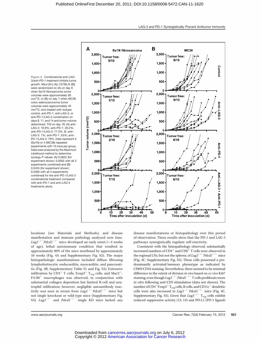
PD-1 monoclonal antibody treatment has shown clinicalefficacy against multiple malignancies including melanoma,prostate, renal cell, and lung cancer (27). LAG-3 has beensuggested to directly modulate the activity of PD-1þ cells (5);furthermore, coexpression of LAG-3 and PD-1 has beenshown in malignant mouse and human tissue (5, 24). Giventhese data, we hypothesized that LAG-3 and PD-1 actsynergistically to control immune homeostasis and mediatetumor-induced tolerance. Consistent with previous reports,a significant percentage of CD4þ and CD8þ TILs fromtransplanted B16 melanoma, MC38 colorectal adenocarci-noma, and Sa1N fibrosarcoma expressed high levels of LAG-3 and PD-1 (32, 34), whereas similar upregulation was notobserved on peripheral T-cell populations (Fig. 1). Next, weasked if antibody-mediated dual blockade of these pathwayswould reduce tumor growth by assessing the potentialefficacy of combined anti–LAG-3 and anti–PD-1 blockadein mice with established tumors. Reduced growth of Sa1Nfibrosarcoma and MC38 colorectal adenocarcinoma (32, 40–42) was observed in some but not all mice treated with theanti–LAG-3 or anti–PD-1 monotherapy (Fig. 2); only a fewmice were tumor free after 50 days (0%–40%). For anti–LAG-3, this is the first demonstration of TGI with anti–LAG-3 as amonotherapy. In striking contrast, 70% and 80% of the Sa1N-and MC38-inoculated mice, respectively, were tumor freeafter 50 days following combinatorial anti–LAG-3/anti–PD-1immunotherapy (Fig. 2). However, this regimen had no effectagainst established B16 tumors. Using the maximum likeli-hood method, there seemed to be a synergistic benefit ofanti–LAG-3/anti–PD-1 combinatorial immunotherapy thatis superior to either the additive effect of anti–LAG-3and anti–PD-1 or monotherapy. Dual treatment withanti–LAG-3/anti–PD-1 did not result in immunopathologicmanifestations such as lymphocytic infiltration in the Sa1Nfibrosarcoma model as determined by detailed histologicanalysis of multiple tissues. Despite efficient tumor clear-ance, no evidence of systemic or organ-specific autoimmu-nity was observed.
To investigate the mechanism underlying decreased tumorgrowth in antibody-treated mice, MC38 tumor-bearing micewere treated with the antibody combinations used above anddraining lymph node (DLN) T cells, non-DLN (NDLN) T cells,and TILs analyzed by flow cytometry for phenotype andeffector function. As expected, average tumor size of anti-PD1–treated or dual antibody–treated mice was significantlysmaller than isotype control or anti-LAG3–treated mice(Supplementary Fig. S1). A significantly higher percentage ofIFN-gþCD8þ T cells were found in the tumor-associated DLNsof dual antibody–treated mice compared with the monother-apy groups, or cells analyzed from NDLNs (Fig. 3A). Likewise, a
LAG-3 and PD-1 Synergistically Prevent Antitumor Immunity
www.aacrjournals.org Cancer Res; 72(4) February 15, 2012 919
American Association for Cancer Research Copyright © 2012 on July 6, 2012cancerres.aacrjournals.orgDownloaded from
Published OnlineFirst December 20, 2011; DOI:10.1158/0008-5472.CAN-11-1620

higher percentage of IFN-gþCD4þ and IFN-gþCD8þ TILs, andto a lesser extent TNF-aþCD4þ and CD8þ TILs, were observedin anti–LAG-3/anti–PD-1–treated mice than in control groups(Fig. 3B). Taken together, these data suggest that anti–LAG-3/anti–PD-1 combinatorial immunotherapy may act synergisti-cally to reduce tumor growth by increasing the proportion ofeffector T cells in the tumor and DLNs.
Lag3�/�Pdcd1�/� mice develop lethal systemicautoimmunity
To further investigate the synergy between these 2 inhibitorymolecules, we next assessed whether LAG-3 and PD-1 coop-erate to control immune homeostasis and mediate tumor-induced tolerance with a genetic approach. Lag3�/�Pdcd1�/�
C57BL/6 or B10.D2 mice were generated at 2 independent
Figure 1. TILs express LAG-3 andPD-1. TILs were isolated from B16,MC38, and Sa1N tumors resectedfrom wild-type mice 13 dayspostinoculation (average sizes:B16 ¼ 350 mm3, MC38 ¼ 1000mm3, and Sa1N ¼ 750 mm3) andstained for flow cytometricanalysis. Representative data(left; gated on live CD4þ or CD8þ
lymphocytes as indicated) orpooled data (right; n ¼ 8–10) ofLAG-3/PD-1 expression on CD4þ
or CD8þ TILs are shown.
Woo et al.
Cancer Res; 72(4) February 15, 2012 Cancer Research920
American Association for Cancer Research Copyright © 2012 on July 6, 2012cancerres.aacrjournals.orgDownloaded from
Published OnlineFirst December 20, 2011; DOI:10.1158/0008-5472.CAN-11-1620
locations (see Materials and Methods), and diseasemanifestation and immune pathology analyzed over time.Lag3�/�Pdcd1�/� mice developed an early onset (�4 weeksof age), lethal autoimmune condition that resulted inapproximately 80% of the mice moribund by approximately10 weeks (Fig. 4A and Supplementary Fig. S2). The majorhistopathologic manifestations included diffuse fibrosinglymphohistiocytic endocarditis, myocarditis, and pancreati-tis (Fig. 4B, Supplementary Table S1 and Fig. S3). Extensiveinfiltration by CD3þ T cells, Foxp3þ Treg cells, and Mac2þ-F4/80þ macrophages was observed, in conjunction withsubstantial collagen deposition but limited B-cell and neu-trophil infiltration; however, negligible autoantibody reac-tivity was seen in serum from Lag3�/�Pdcd1�/� mice butnot single knockout or wild-type mice (Supplementary Fig.S4). Lag3�/� and Pdcd1�/� single KO mice lacked any
disease manifestations or histopathology over this periodof observation. These results show that the PD-1 and LAG-3pathways synergistically regulate self-reactivity.
Consistent with the histopathology observed, substantiallyincreased numbers of CD4þ and CD8þ T cells were observed inthe regional LNs, but not the spleens, of Lag3�/�Pdcd1�/�mice(Fig. 4C, Supplementary Fig. S5). These cells possessed a pre-dominantly activated/memory phenotype as indicated byCD69/CD44 staining. Nevertheless, there seemed to beminimaldifference in the extent of division in vivo based on ex vivo Ki67staining, even thoughLag3�/�Pdcd1�/�T cells proliferatemorein vitro following anti-CD3 stimulation (data not shown). Thenumber ofCD4þFoxp3þTreg cells, B cells, andCD11c
þdendriticcells were also increased in Lag3�/�Pdcd1�/� mice (Fig. 4C,Supplementary Fig. S5). Given that Lag3�/� Treg cells exhibitreduced suppressive activity (13, 14) and PD-L1 (PD-1 ligand)
Figure 2. Combinatorial anti–LAG-3/anti–PD-1 treatment inhibits tumorgrowth. Mice [A/J (A); C57BL/6 (B)]were randomized on (A) on day 6when Sa1N fibrosarcoma tumorvolumes were approximately 60mm3/2, or (B) on day 7 when MC38colon adenocarcinoma tumorvolumes were approximately 40mm3/2, and treated with isotypecontrol, anti-PD-1, anti-LAG-3, oranti-PD-1/LAG-3 combination ondays 8, 11, and 14 and tumor volumedetermined. TGI on day 18: (A) anti–LAG-3: 18.9%; anti–PD-1: 26.2%;anti–PD-1/LAG-3: 77.3%. B, anti–LAG-3: 1%; anti–PD-1: 55%; anti–PD-1/LAG-3: 79%. Data represent 3(Sa1N) or 4 (MC38) repeatedexperiments with 10 mice per group.Data were analyzed by the MaximumLikelihood method to determinesynergy P values: (A) 0.0622 (forexperiment shown; 0.0002 with all 3experiments combined) and (B)0.0455 (for experiment shown;0.0366 with all 4 experimentscombined) for the anti–PD-1/LAG-3combinatorial treatment comparedwith anti–PD-1 and anti–LAG-3treatments alone.
LAG-3 and PD-1 Synergistically Prevent Antitumor Immunity
www.aacrjournals.org Cancer Res; 72(4) February 15, 2012 921
American Association for Cancer Research Copyright © 2012 on July 6, 2012cancerres.aacrjournals.orgDownloaded from
Published OnlineFirst December 20, 2011; DOI:10.1158/0008-5472.CAN-11-1620

contributes to iTreg development (15), it is possible that thecombined loss of LAG-3 and PD-1 alters Treg cell homeostasis.
To further probe the cellular defects in Lag3�/�Pdcd1�/�
mice, we adoptively transferred splenocytes into lymphopenicRag-1�/� mice. In contrast to healthy wild-type and singleknockout controls, Lag3�/�Pdcd1�/� splenocyte recipientsstarted to lose body weight approximately 6 days posttransferwith 100% morbidity by day 20 (Supplementary Fig. S6Aand S6B). Adoptive transfer experiments T-cell–depletedLag3�/�Pdcd1�/� splenocytes clearly showed that both CD4þ
or CD8þ T-cell populations contributed to the diseaseobserved, with a dominant role for the former (SupplementaryFig. S6C and S6D). Consistent with these survival andweight loss data, histologic analysis of CD4þ T-cell–depletedLag3�/�Pdcd1�/� splenocyte recipients revealed relativelynormal bone marrow cellularity and density, whereas
Lag3�/�Pdcd1�/� splenocyte recipients exhibited a near totalabsence of hematopoietic cell precursors in bone marrow andsevere lymphoid depletion in the spleen, LNs, and Peyer'spatches (Supplementary Table S2, Fig. S6E and S7). These dataindicate that CD4þ T cells are primarily responsible for thepathology observed. Cytokine analysis revealed high levels ofIFN-g , TNF-a, and MCP-1 in the serum of Lag3�/�Pdcd1�/�
recipients but not single knockout or wild-type control reci-pients (Supplementary Fig. S6F–S6H). Taken together, thesedata suggest that Lag3�/�Pdcd1�/� splenocyte recipients, incontrast with their single knockout and wild-type controls,develop an autoimmuneGvHD-like syndromewith evidence ofaplastic anemia and bone marrow failure as a cause of death.
The data thus far suggested that while a reasonable levelof tolerance is maintained in single knockout Lag3�/� orPdcd1�/� mice, dual loss of LAG-3 and PD-1 expression results
Figure 3. Combinatorial anti–LAG-3/anti–PD-1 treatment results inenhanced adaptive immuneresponses. Mice were inoculatedon day 0 with 2 � 106 MC38 cellss.c. in the right flank, euthanized atday 15, and tissues analyzed byflow cytometry. A, tumor draininginguinal (DLN) and nondrainingbrachial and axillary lymph nodes(NDLN)were isolated and activatedwith PMA þ ionomycin for 4 hoursin the presence of brefeldin A, thenanalyzed by intracellular stainingand flow cytometry (gated onlymphocytes). Nonparametric1-way ANOVA with Kruskal–Wallistest (P ¼ 0.0074) was used for partA. B, TILs were analyzed byintracellular staining and flowcytometry; plots represent 5 to 8animals per group. Numbers arepercentage cytokine-positiveinfiltrating lymphocytes.
Woo et al.
Cancer Res; 72(4) February 15, 2012 Cancer Research922
American Association for Cancer Research Copyright © 2012 on July 6, 2012cancerres.aacrjournals.orgDownloaded from
Published OnlineFirst December 20, 2011; DOI:10.1158/0008-5472.CAN-11-1620

in a loss of peripheral self-tolerance of CD4þ and CD8þ T cells.To test this in an antigen-specific system, we asked whetherhemagglutinin-specific tolerance induced in transgenic miceexpressing hemagglutinin as a self-antigen inmultiple epithelialtissues (C3-HAlo mice; refs. 13, 43), could also be broken ifadoptively transferred hemagglutinin-specific T cells [from
Clone 4 (CL4) TCR transgenics] lacked both inhibitory mole-cules. Compared with wild-type CL4 T cells, significant expan-sion of Lag3�/�Pdcd1�/� clonotypic CD8þ T cells was observed5 days posttransfer (Supplementary Fig. S6I). Although this wasnot substantially greater than that seen with Pdcd1�/� T cells,the Lag3�/�Pdcd1�/� T cells exhibited a significantly enhancedeffector phenotype, as determined by intracellular expression ofIFN-g and IL-17, compared with their single knockout and wild-type controls (Supplementary Fig. S6J and S6K). Similarly, whentolerance was broken in C3-HAlo transgenic mice (39), adoptivetransfer of antigen-specific Lag3�/�Pdcd1�/� CD8þ T cellsexpanded significantly more than their single knockout andwild-type controls, although enhanced in vivo CTL activity wascomparable in the Pdcd1�/� and Lag3�/�Pdcd1�/� CD8þ T cellrecipients (Supplementary Fig. S8). Collectively, these datasuggest that the loss of LAG-3 and PD-1 also results in loss oftolerance to a model self-antigen.
Reduced tumor growth and enhanced survival inLag3�/�Pdcd1�/� mice
To continue our analysis of Lag-3/PD-1 synergy in theregulation of antitumor immunity, we assessed tumor growthin Lag3�/�Pdcd1�/� mice and controls over time. Of the 3transplantable tumor models examined in this study, B16 isregarded as the least immunogenic and thus the hardest toeliminate by immunologic intervention (32, 34). A low doseof B16 cells (1.25 � 105) progressively grew in wild-typeand Lag3�/� mice inoculated intradermally at day 0,whereas limited growth was observed in Pdcd1�/� andLag3�/�Pdcd1�/� mice (Supplementary Fig. S9A). Althoughprevious studies suggested that PD-1 deletion did not affectsubcutaneously injected tumor growth (44), our experimentsrevealed reduced tumor growth in Pdcd1�/� mice comparedwith wild-type mice. Paradoxically, Lag3�/� mice developedslightly larger tumors. Whether this is due to reported defectsin natural killer cell cytolysis (23), or an unexpected role ofpDCs, which highly express LAG-3 (45), remains to be deter-mined. Statistical analysis with themaximum likelihoodmeth-od for synergy found that the lack of tumor growth in theLag3�/�Pdcd1�/�mice was greater than the additive effects oftumor growth in Lag3�/� mice and Pdcd1�/� mice at day 11(P< 0.05) andday 13 (P< 0.0005) suggesting that LAG-3 andPD-1 synergize to mediate tumor-induced tolerance. Depletion ofCD4þ and CD8þ T cells restored normal B16 tumor growth incompound-deficient mice, indicating the necessity of adaptiveimmunity to the antitumor response (Supplementary Fig. S9B).
As the difference in resistance to B16 growth betweenPdcd1�/� and Lag3�/�Pdcd1�/� mice seemed small, we eval-uated tumor B16 and MC38 growth at different doses. Atthe higher B16 dose (5 � 105 cells per mouse), wild-type andLag3�/� mice show uncontrolled tumor growth and lethalitywith an average survival time of less than 20 days (Fig. 5A andSupplementary Fig. S10). Lag3�/�Pdcd1�/� mice (80%) elim-inated tumors compared with only 40% of Pdcd1�/� mice;however, B16 survivors did not display autoimmune vitiligo asis often seen with this model (46). We also investigated growthof subcutaneously implanted MC38 adenocarcinoma cells at 2different doses. Whereas MC38 growth and survival were
Figure 4. Lag3�/�Pdcd1�/� mice develop lethal systemic autoimmunity.A, disease incidence for Lag3�/�Pdcd1�/� mice, plus single knockoutandWT littermate controls. Moribund curveswere analyzed for statisticalsignificance by log-rank test; ���, P < 0.001. B, representativehistopathology of the heart and pancreas of WT and Lag3�/�Pdcd1�/�
mice. C, number of various T-cell populations in the spleen and LNs(inguinal and brachial) is shown. Data represent 3 to 4 independentexperiments with 4 to 7mice total per group (5- to 7weeks old). Error barsrepresent SEM; �, P < 0.05; ��, P < 0.01 (unpaired t test).
LAG-3 and PD-1 Synergistically Prevent Antitumor Immunity
www.aacrjournals.org Cancer Res; 72(4) February 15, 2012 923
American Association for Cancer Research Copyright © 2012 on July 6, 2012cancerres.aacrjournals.orgDownloaded from
Published OnlineFirst December 20, 2011; DOI:10.1158/0008-5472.CAN-11-1620

comparable in Lag3�/�Pdcd1�/� and Pdcd1�/� mice when alow dose (2 � 105 cells per mouse) was used (70% vs.71%, respectively; Fig. 5B and Supplementary Fig. S10),Lag3�/�Pdcd1�/�mice were clearly more effective at prevent-ing high-dose (5 � 105 cells per mouse) MC38 tumor growthand ensuring survival (83% vs. 38%, respectively; Fig. 5C andSupplementary Fig. S10). Phenotypic analysis revealedenhanced IFN-g expression by Lag3�/�Pdcd1�/� CD4þ andCD8þ T cells in tumor DLNs compared with wild-typeand Lag3�/� mice, and NDLNs from all groups (Fig. 6). Thesedata suggest that combined loss of LAG-3 and PD-1 limitstumor-mediated tolerization and enhances tumor-specificimmunity and resistance to tumor growth.
To further investigate the killing efficacy of Lag3�/�
Pdcd1�/� T cells in vivo in the presence of an establishedtumor, clonotypic CL4 CD8þ T cells were transferred intoProTRAMP male mice, which develop prostate cancer drivenby the probasin promoter and express hemagglutinin. Aftervaccination with hemagglutinin-expressing Vaccinia virus,Lag3�/�Pdcd1�/� recipients showed significantly increasedkilling ability in comparison with the wild type, and slightlyincreased killing efficiency in comparison with Pdcd1�/�
single knockouts (Supplementary Fig. S11). These data sup-port the conclusion that Lag3�/�Pdcd1�/� CD8þ T cells areless susceptible to tumor-induced tolerance than wild-type cells. Taken together, these data clearly show thatLag3�/�Pdcd1�/� mice are more capable of resisting high-dose tumor growth than Pdcd1�/� and wild-type mice.
Discussion
The data presented here illustrate clear synergy between theinhibitory receptors LAG-3 and PD-1 in controlling immunehomeostasis, preventing autoimmunity, and enforcing tumor-induced tolerance. First, we show coexpression of LAG-3 andPD-1 on tumor-infiltrating lymphocytes. Second, we show thatdual blockade of these receptors leads to decreased tumorgrowth and enhanced antitumor immunity. Importantly, dualantibody–treated mice show more robust immune responsesthan either single-treated group. Third, analysis of mutantmice revealed a cooperative requirement for LAG-3 and PD-1 in maintaining immune homeostasis. Consistent with ourobservations following antibody-mediated blockade of LAG-3and PD-1, Lag3�/�Pdcd1�/� mice prevented growth of high-dose B16 and MC38 tumors and ensured survival while singleknockout controls and wild-type mice succumbed to disease.Taken together, these data reveal an unappreciated synergisticcooperation between LAG-3 and PD-1 in limiting tumorgrowth.
Although anti–LAG-3/anti–PD-1 combinatorial immuno-therapy effectively cleared established Sa1N andMC38 tumors,this therapy was not effective against established B16 tumors.In contrast, B16 tumors were more difficult to establish inLag3�/�Pdcd1�/� mice. B16 is a more difficult tumor toeradicate thanMC38 and Sa1N and thus there could be severalpossible explanations for this apparent discrepancy. First,expression of LAG-3/PD-1 on TILs from B16 is lower than for
Figure 5. Reduced tumor growth intumor-bearing Lag3�/�Pdcd1�/�
mice. Wild type, Lag3�/�,Pdcd1�/�, and Lag3�/�Pdcd1�/�
mice were inoculated on day 0 with5 � 105 B16 cells i.d. (A), 2 � 106
MC38 cells s.c. (B), or 5 � 106
MC38 cells s.c. (C). Tumors weremeasuredwith anelectronic caliperand reported as volume (seeMaterials and Methods). Data arecombined from 2 to 3 repeatedexperiments, 3 to 5 animals peranimals per group. Data wereanalyzed by the MaximumLikelihood method to determinesynergy p values: 0.0253 (A),0.94 (B), and 0.0273 (C) for theLag3�/�Pdcd1�/� mice comparedwith the additive effect of the 2single knockouts. Animals wereeuthanized when tumors becamelarge, ulcerated, and/or necrotic.
Woo et al.
Cancer Res; 72(4) February 15, 2012 Cancer Research924
American Association for Cancer Research Copyright © 2012 on July 6, 2012cancerres.aacrjournals.orgDownloaded from
Published OnlineFirst December 20, 2011; DOI:10.1158/0008-5472.CAN-11-1620

MC38 and Sa1N, and thismay be below a required threshold forefficacy upon antibody blockade in tumor-bearing mice.Second, as the knockout mice would lack LAG-3/PD-1 atthe initiation of tumor inoculation, the immune systemwould not have this impediment at the commencementof an antitumor response. In contrast, the initial stages ofLAG-3/PD-1 upregulation may already have occurred intumor-bearing mice at the time of antibody treatmentthereby establishing sufficient tolerance to prevent effectiveantitumor immunity. Third, there may be additional regu-latory mechanisms that contribute a greater role with B16that may be sufficient to prevent antitumor immunity whenLAG-3/PD-1 blockade is initiated in mice that already havetumors. Lastly, we cannot rule out the possibility that thegenetic deletion of LAG-3/PD-1 results in Tregs that have acell intrinsic defect in their regulatory capacity whichimpacts early tumor establishment. Of course, a combina-tion of these issues could also contribute to the difference inB16 clearance seen in LAG-3/PD-1 deficiency versus latestage antibody-mediated blockade. It is possible that alter-nate dosing regimens and therapeutic combinations couldresult in effective clearance of established B16 with anti–LAG-3/anti–PD-1 combinatorial immunotherapy.It is noteworthy that the percentage of IFN-gþ TILs is
higher in Lag3�/�Pdcd1�/� mice versus anti–LAG-3/anti–PD-1–treated mice (Fig. 6 vs. Fig. 3). These differences couldbe reflective of the differences in B16 elimination in LAG-3/PD-1–deficient versus mAb-treated mice. Alternatively, as
discussed above, these observations could be due to thedifferent tumors analyzed or temporal differences betweenthese experiments, as IFN-g expression was determined 1week after mAb treatment compared with 2 weeks aftertumor inoculation into Lag3�/�Pdcd1�/� mice. Finally, wecannot rule out the possibility that there is a phenotypicdifference in the immune cells in the Lag3�/�Pdcd1�/�mice.However, there does not seem to be an active systemic defectat the time of the experiment as high IFN-g expression is notobserved in the NDLN.
Although Lag3�/�Pdcd1�/� mice develop a lethal auto-immune condition, the disease is slower (�10 weeks vs. 3–4weeks) and less penetrant (80% vs. 100%) than the phenotypeobserved in Ctla4�/� mice (20). Recently, analogous obser-vations were reported by Honjo and colleagues in whichBALB/c mice harboring a loss-of-function mutation in Lag3combined with genetic deletion of Pdcd1 develop lethalmyocarditis (47). Heart-infiltrating T cells from these com-pound-deficient mice were shown to produce high amountsof IFNg compared with distal lymphoid organs such as thespleen. Our results are consistent with their data, as we alsoobserved enhanced production of proinflammatory cyto-kines by T cells infiltrating sites of inflammation, such asthe heart and pancreas in Lag3�/�Pdcd1�/� mice and intumors and DLNs in knockout and antibody-treated mice.Furthermore, the Honjo group also observed acceleratedautoimmune diabetes in NOD mice expressing a loss-of-function LAG-3 mutant, consistent with our recent observa-tions in Lag3�/� NOD mice (48). However, the mice used byHonjo and colleagues that lacked functional LAG-3/PD-1expression were on a BALB/c background, whereas our datawere derived frommice on a C57BL/6 or B10.D2 background.Strain-specific differences between these mice have beenwell documented (49, 50) and may have contributed tosubtle differences in phenotypic and mechanistic observa-tions reported. For instance, loss of LAG-3/PD-1 on a B10.D2background can lead to an increase in IL-17þ cells which wasnot seen in mice on a Balb/c background.
Although CTLA-4, PD-1, and LAG-3 are all negative regula-tors expressed during T-cell activation, high level, dual LAG-3/PD-1 expression is largely restricted to infiltrating TILs. Thus,LAG-3/PD-1 combinatorial immunotherapy may promotetumor-specific responses relative to nonspecific or self-anti-gen–specific immune responses and thus may be less toxicthan CTLA-4 blockade. Given the recent phase 3 results withanti–CTLA-4 treatment of patients withmetastaticmelanoma,showing a clear survival benefit (albeit with notable immunetoxicity; ref. 28), our results suggest that combined blockade ofPD-1 and LAG-3 is a highly promising combinatorial strategyfor the immune-based therapy of cancer.
Disclosure of Potential Conflict of Interest
The authors declare competing financial interests. D.M. Pardoll, D.A.A.Vignali, C.G. Drake, and C.J. Workman have submitted patents that arepending and are entitled to a share in net income generated from licensingof these patent rights for commercial development. A.J. Korman, M. Selby,and J.F. Grosso are employees of Bristol-Myers Squibb. C.G. Drake has anownership interest in Amplimmune and has served as a consultant toDendreon, Bristol-Myers Squibb, and Pfizer.
Figure 6. Tumor-draining (DLN; right) and nondraining (NDLN; left) LNT cells were isolated on day 14 post-B16 inoculation and were activatedwith PMA þ ionomycin for 4 hours in the presence of brefeldin A. Afterintracellular cytokine staining, CD8þIFN-gþ (A) and CD4þIFN-gþ (B) cellswere analyzed by flow cytometry. Data are representative of 10 to15 animals per group. Nonparametric 1-way ANOVA with Kruskal–Wallistest (P < 0.0001) was used; �, P < 0.05; ��, P < 0.01; and ���, P < 0.001 vs.Lag3�/�Pdcd1�/�.
LAG-3 and PD-1 Synergistically Prevent Antitumor Immunity
www.aacrjournals.org Cancer Res; 72(4) February 15, 2012 925
American Association for Cancer Research Copyright © 2012 on July 6, 2012cancerres.aacrjournals.orgDownloaded from
Published OnlineFirst December 20, 2011; DOI:10.1158/0008-5472.CAN-11-1620

Acknowledgments
The authors thank Many Jo Turk and Jim Allison for cell lines. At St. Jude, theauthors thank Karen Forbes, Amy Krause, and Ashley Castellaw formaintenanceand breeding of mouse colonies, Cliff Guy for help with cytokine analysis, PaulThomas for anti-CD4 and anti-CD8 depleting Abs, Richard Cross, Greig Lennonand StephanieMorgan for FACS, SongWu andHui Zhang for helpwith statisticalanalysis, the staff of the Shared Animal Resource Center at St Jude for the animalhusbandry, the Veterinary Pathology Core Laboratory at St. Jude for histologyand immunohistochemistry support, and the Hartwell Center for Biotechnologyand Bioinformatics at St Jude for real-time PCR primer/probe synthesis andMOG synthesis and purification. At Johns Hopkins, the authors thank Dih-DihHuang for the maintenance and breeding of the mouse colonies, and technicalsupport. At Bristol-Myers-Squibb, the authors thank David Klitzing and the staffof theMilpitas animal facility for carrying out the tumor experiments andRanganVanganipuram, Brian Lee, and Shilpa Mankikar for provision of antibodies.
Grant Support
This work was supported by the NIH (R01 AI39480 to D.A.A. Vignali and R01CA127153 and P50 CA58236–15 to C.G. Drake), a Hartwell Postdoctoral Fellow-ship (to M.L. Bettini), NCI Comprehensive Cancer Center Support CORE grant(CA21765, to D.A.A. Vignali), the American Lebanese Syrian Associated Charities(ALSAC, to D.A.A. Vignali), the Patrick C. Walsh Fund (to C.G. Drake), the KochFund (to C.G. Drake), NHLBI contract - HHSN-268201999934C and CIHR -20R92141 to P.J. Utz, and NIAID F32 AI080086 to C.L. Liu. C.G. Drake is a DamonRunyon-Lilly Clinical Investigator.
The costs of publication of this article were defrayed in part by the payment ofpage charges. This article must therefore be hereby marked advertisement inaccordance with 18 U.S.C. Section 1734 solely to indicate this fact.
ReceivedMay 11, 2011; revisedDecember 7, 2011; accepted December 10, 2011;published OnlineFirst December 20, 2011.
References1. Vesely MD, KershawMH, Schreiber RD, SmythMJ. Natural innate and
adaptive immunity to cancer. Annu Rev Immunol 2011;29:235–71.2. Dunn GP, Bruce AT, Ikeda H, Old LJ, Schreiber RD. Cancer immu-
noediting: from immunosurveillance to tumor escape. Nat Immunol2002;3:991–8.
3. Leen AM, Rooney CM, Foster AE. Improving T cell therapy for cancer.Annu Rev Immunol 2007;25:243–65.
4. Blackburn SD, Shin H,HainingWN, Zou T,WorkmanCJ, Polley A, et al.Coregulation ofCD8þT cell exhaustion bymultiple inhibitory receptorsduring chronic viral infection. Nat Immunol 2009;10:29–37.
5. Matsuzaki J,Gnjatic S,Mhawech-Fauceglia P,BeckA,Miller A, Tsuji T,et al. Tumor-infiltratingNY-ESO-1-specificCD8þ T cells are negativelyregulatedby LAG-3 andPD-1 in humanovarian cancer. ProcNatl AcadSci U S A 2010;107:7875–80.
6. Driessens G, Kline J, Gajewski TF. Costimulatory and coinhibitoryreceptors in anti-tumor immunity. Immunol Rev 2009;229:126–44.
7. Fife BT, Pauken KE, Eagar TN, Obu T, Wu J, Tang Q, et al. Interactionsbetween PD-1 and PD-L1 promote tolerance by blocking the TCR-induced stop signal. Nat Immunol 2009;10:1185–92.
8. Francisco LM, Sage PT, Sharpe AH. The PD-1 pathway in toleranceand autoimmunity. Immunol Rev 2010;236:219–42.
9. Goldberg MV, Drake CG. LAG-3 in cancer immunotherapy. Curr TopMicrobiol Immunol 2010;344:269–78.
10. Vignali DA,Collison LW,WorkmanCJ.How regulatory Tcellswork.NatRev Immunol 2008;8:523–32.
11. WorkmanCJ, Dugger KJ, Vignali DA. Cutting edge: molecular analysisof the negative regulatory function of lymphocyte activation gene-3.J Immunol 2002;169:5392–5.
12. Okazaki T, Honjo T. The PD-1-PD-L pathway in immunological toler-ance. Trends Immunol 2006;27:195–201.
13. HuangCT,WorkmanCJ, Flies D, Pan X,Marson AL, ZhouG, et al. Roleof LAG-3 in regulatory T cells. Immunity 2004;21:503–13.
14. Workman CJ, Vignali DA. Negative regulation of T cell homeostasis bylymphocyte activation gene-3 (CD223). J Immunol 2005;174:688–95.
15. Francisco LM, Salinas VH, Brown KE, Vanguri VK, Freeman GJ,Kuchroo VK, et al. PD-L1 regulates the development, maintenance,and function of induced regulatory T cells. J Exp Med 2009;206:3015–29.
16. Ise W, Kohyama M, Nutsch KM, Lee HM, Suri A, Unanue ER, et al.CTLA-4 suppresses the pathogenicity of self antigen-specific T cellsby cell-intrinsic and cell-extrinsic mechanisms. Nat Immunol 2010;11:129–35.
17. Chambers CA, Sullivan TJ, Allison JP. Lymphoproliferation in CTLA-4-deficient mice is mediated by costimulation-dependent activation ofCD4þ T cells. Immunity 1997;7:885–95.
18. WingK,Onishi Y, Prieto-Martin P, Yamaguchi T,MiyaraM, Fehervari Z,et al. CTLA-4 control over Foxp3þ regulatory T cell function. Science2008;322:271–5.
19. Liang B, Workman C, Lee J, Chew C, Dale BM, Colonna L, et al.RegulatoryTcells inhibit dendritic cells by lymphocyte activation gene-3 engagement of MHC class II. J Immunol 2008;180:5916–26.
20. Waterhouse P, Penninger JM, Timms E,WakehamA, Shahinian A, LeeKP, et al. Lymphoproliferative disorders with early lethality in micedeficient in Ctla-4. Science 1995;270:985–8.
21. Nishimura H, Nose M, Hiai H, Minato N, Honjo T. Development oflupus-like autoimmune diseases by disruption of the PD-1 geneencoding an ITIM motif-carrying immunoreceptor. Immunity 1999;11:141–51.
22. Nishimura H, Okazaki T, Tanaka Y, Nakatani K, Hara M, Matsumori A,et al. Autoimmune dilated cardiomyopathy in PD-1 receptor-deficientmice. Science 2001;291:319–22.
23. Miyazaki T, Dierich A, Benoist C, Mathis D. Independent modes ofnatural killing distinguished in mice lacking Lag3. Science 1996;272:405–8.
24. Grosso JF, Goldberg MV, Getnet D, Bruno TC, Yen HR, Pyle KJ, et al.Functionally distinct LAG-3 and PD-1 subsets on activated and chron-ically stimulated CD8 T cells. J Immunol 2009;182:6659–69.
25. Grosso JF, Kelleher CC, Harris TJ, Maris CH, Hipkiss EL, De Marzo A,et al. LAG-3 regulates CD8þ T cell accumulation and effector functionin murine self- and tumor-tolerance systems. J Clin Invest 2007;117:3383–92.
26. Keir ME, Butte MJ, Freeman GJ, Sharpe AH. PD-1 and its ligands intolerance and immunity. Annu Rev Immunol 2008;26:677–704.
27. Brahmer JR, Drake CG, Wollner I, Powderly JD, Picus J, SharfmanWH, et al. Phase I study of single-agent anti-programmed death-1(MDX-1106) in refractory solid tumors: safety, clinical activity, phar-macodynamics, and immunologic correlates. J Clin Oncol 2010;28:3167–75.
28. Hodi FS, O'Day SJ, McDermott DF, Weber RW, Sosman JA, HaanenJB, et al. Improved survival with ipilimumab in patients with metastaticmelanoma. N Engl J Med 2010;363:711–23.
29. Workman CJ, Vignali DA. The CD4-related molecule, LAG-3 (CD223),regulates the expansion of activated T cells. Eur J Immunol 2003;33:970–9.
30. Nishimura H, Minato N, Nakano T, Honjo T. Immunological studies onPD-1 deficient mice: implication of PD-1 as a negative regulator for Bcell responses. Int Immunol 1998;10:1563–72.
31. Woo SR, Li N, Bruno TC, Forbes K, Brown S, Workman C, et al.Differential subcellular localization of the regulatory T-cellprotein LAG-3 and the coreceptor CD4. Eur J Immunol 2010;40:1768–77.
32. Kocak E, Lute K, Chang X, May KF Jr, Exten KR, Zhang H, et al.Combination therapy with anti-CTL antigen-4 and anti-4–1BB anti-bodies enhances cancer immunity and reduces autoimmunity. CancerRes 2006;66:7276–84.
33. Collison LW, Chaturvedi V, Henderson AL, Giacomin PR, Guy C,Bankoti J, et al. IL-35-mediated induction of a potent regulatory T cellpopulation. Nat Immunol 2010;11:1093–101.
34. TurkMJ, Guevara-Patino JA, RizzutoGA, EngelhornME, Sakaguchi S,Houghton AN. Concomitant tumor immunity to a poorly immunogenicmelanoma is prevented by regulatory T cells. J Exp Med 2004;200:771–82.
Woo et al.
Cancer Res; 72(4) February 15, 2012 Cancer Research926
American Association for Cancer Research Copyright © 2012 on July 6, 2012cancerres.aacrjournals.orgDownloaded from
Published OnlineFirst December 20, 2011; DOI:10.1158/0008-5472.CAN-11-1620

35. Park D, Lapteva N, Seethammagari M, Slawin KM, Spencer DM. Anessential role for Akt1 in dendritic cell function and tumor immuno-therapy. Nat Biotechnol 2006;24:1581–90.
36. Li B, VanRoey M, Wang C, Chen TH, Korman A, Jooss K. Anti-programmed death-1 synergizes with granulocyte macrophage colo-ny-stimulating factor–secreting tumor cell immunotherapy providingtherapeutic benefit to mice with established tumors. Clin Cancer Res2009;15:1623–34.
37. Workman CJ, Rice DS, Dugger KJ, Kurschner C, Vignali DA. Pheno-typic analysis of themurine CD4-related glycoprotein, CD223 (LAG-3).Eur J Immunol 2002;32:2255–63.
38. Noya O, Alarcon de Noya B. The multiple antigen blot assay (MABA): asimple immunoenzymatic technique for simultaneous screening ofmultiple antigens. Immunol Lett 1998;63:53–6.
39. Goldberg MV, Maris CH, Hipkiss EL, Flies AS, Zhen L, Tuder RM, et al.Role of PD-1 and its ligand, B7-H1, in early fate decisions of CD8 Tcells. Blood 2007;110:186–92.
40. Paradis TJ, Floyd E, Burkwit J, Cole SH, Brunson B, Elliott E, et al.The anti-tumor activity of anti-CTLA-4 is mediated through itsinduction of IFN gamma. Cancer Immunol Immunother 2001;50:125–33.
41. Leach DR, Krummel MF, Allison JP. Enhancement of antitumor immu-nity by CTLA-4 blockade. Science 1996;271:1734–6.
42. RosenbergSA, Spiess P, Lafreniere R. A newapproach to the adoptiveimmunotherapyof cancerwith tumor-infiltrating lymphocytes. Science1986;233:1318–21.
43. Adler AJ, Marsh DW, Yochum GS, Guzzo JL, Nigam A, Nelson WG,et al. CD4þ T cell tolerance to parenchymal self-antigens requirespresentation by bone marrow-derived antigen-presenting cells. J ExpMed 1998;187:1555–64.
44. Iwai Y, Terawaki S, Honjo T. PD-1 blockade inhibits hematogenousspread of poorly immunogenic tumor cells by enhanced recruitment ofeffector T cells. Int Immunol 2005;17:133–44.
45. WorkmanCJ,WangY, El Kasmi KC, Pardoll DM,Murray PJ, DrakeCG,et al. LAG-3 regulates plasmacytoid dendritic cell homeostasis.J Immunol 2009;182:1885–91.
46. Zhang P, Cote AL, de Vries VC, Usherwood EJ, Turk MJ. Induction ofpostsurgical tumor immunity and T-cell memory by a poorly immuno-genic tumor. Cancer Res 2007;67:6468–76.
47. Okazaki T, Okazaki IM, Wang J, Sugiura D, Nakaki F, Yoshida T, et al.PD-1 and LAG-3 inhibitory co-receptors act synergistically to preventautoimmunity in mice. J Exp Med 2011;208:395–407.
48. Bettini M, Szymczak-Workman AL, Forbes K, Castellaw AH, Selby M,Pan X, et al. Cutting edge: accelerated autoimmune diabetes in theabsence of LAG-3. J Immunol 2011;187:3493–8.
49. Brenner GJ, Cohen N, Moynihan JA. Similar immune response tononlethal infection with herpes simplex virus-1 in sensitive (BALB/c)and resistant (C57BL/6) strains ofmice. Cell Immunol 1994;157:510–24.
50. Niewiesk S, Brinckmann U, Bankamp B, Sirak S, Liebert UG, terMeulen V. Susceptibility to measles virus-induced encephalitis inmicecorrelates with impaired antigen presentation to cytotoxic T lympho-cytes. J Virol 1993;67:75–81.
LAG-3 and PD-1 Synergistically Prevent Antitumor Immunity
www.aacrjournals.org Cancer Res; 72(4) February 15, 2012 927
American Association for Cancer Research Copyright © 2012 on July 6, 2012cancerres.aacrjournals.orgDownloaded from
Published OnlineFirst December 20, 2011; DOI:10.1158/0008-5472.CAN-11-1620

![Vibrio cholerae use pili and flagella synergistically to ...wonglab.seas.ucla.edu/pdf/2014 Nat Commun [Utada, Wong] Vibrio... · Vibrio cholerae use pili and flagella synergistically](https://static.fdocuments.us/doc/165x107/5afa9d177f8b9a32348e07cc/vibrio-cholerae-use-pili-and-flagella-synergistically-to-nat-commun-utada.jpg)