IMMEDIATE LOADING WITH MINI DENTAL IMPLANTS IN THE FULLY EDENTULOUS MANDIBLE
11
*Corresponding Author Address: Dr Abu-Hussein Muhamad Email: [email protected] International Journal of Dental and Health Sciences Volume 02,Issue 06 Original Article IMMEDIATE LOADING WITH MINI DENTAL IMPLANTS IN THE FULLY EDENTULOUS MANDIBLE Abdulgani Azzaldeen * , Abusalih Ahmet ,Hakki Ismail ,Chlorokostas Georges, Abu-Hussein Muhamad Al-Quds University, Faculty of Dentistry,Jerusalem,Palestine ABSTRACT: Mini dental implants (MDI) have become increasingly popular in the past decade and have been approved for many long-term uses in dentistry. There are many advantages of the use of mini dental implants from both a practitioner and patient perspective. For the general dentist starting out in implant dentistry, their placement can be more challenging than conventional implants. It requires a different skill set, but one which can be learned with proper guidance and practice.In the study are presented clinical cases with mini implants with spherical joints for retention of removable overimplant mandibular dentures. Key words: mini dental implants, immediate loading implants Prosthetics, overdenture INTRODUCTION: The key features and the prime requisites of an ideal prosthesis for the rehabilitation of the stomatognathic system include the restoration of normal contour, function, esthetics, comfort, speech, and health. Assimilation of these features in any prosthesis delivered to the patient is the ideal goal of modern dentistry. However, with the highly complicated and challenging clinical situations which are commonly encountered in the general practice, an ideal replacement of the lost tissues using the conventional techniques may not be always possible. Answer to such a clinical dilemma would probably be Implant therapy.[1,2] Implant dentistry is unique because of its ability to achieve an ideal replacement of the lost tissues, regardless of the atrophy, disease, or injury of the stomatognathic system[2,3]. This has significantly increased the acceptance of osseointegrated supported prosthesis by the patients. However, greater the destruction of the stomatognathic system, the more challenging is the task of rehabilitation. As a result of the current availability of the advanced diagnostic tools which aid in treatment planning, the improved implant designs, materials, and techniques as a result of continuous research, many challenging clinical situations can be successfully managed with predictable success.[1,2,4,5,6] Recently, mini-implant has been used as transitional implants to support dentures during the healing phase of implant denture restoration.It is also used as a permanent single implant crown in inadequate space for standard implants and limited bone availability situations.[4,5] The use of dental implants of smaller diameters in various forms has been present for almost 20 years. Those are
-
Upload
abu-hussein-muhamad -
Category
Health & Medicine
-
view
457 -
download
0
Transcript of IMMEDIATE LOADING WITH MINI DENTAL IMPLANTS IN THE FULLY EDENTULOUS MANDIBLE

*Corresponding Author Address: Dr Abu-Hussein Muhamad Email: [email protected]
International Journal of Dental and Health Sciences
Volume 02,Issue 06
Original Article
IMMEDIATE LOADING WITH MINI DENTAL IMPLANTS IN
THE FULLY EDENTULOUS MANDIBLE
Abdulgani Azzaldeen *, Abusalih Ahmet
,Hakki Ismail
,Chlorokostas Georges, Abu-Hussein Muhamad
Al-Quds University, Faculty of Dentistry,Jerusalem,Palestine
ABSTRACT:
Mini dental implants (MDI) have become increasingly popular in the past decade and have been approved for many long-term uses in dentistry. There are many advantages of the use of mini dental implants from both a practitioner and patient perspective. For the general dentist starting out in implant dentistry, their placement can be more challenging than conventional implants. It requires a different skill set, but one which can be learned with proper guidance and practice.In the study are presented clinical cases with mini implants with spherical joints for retention of removable overimplant mandibular dentures. Key words: mini dental implants, immediate loading implants Prosthetics, overdenture INTRODUCTION:
The key features and the prime
requisites of an ideal prosthesis for the
rehabilitation of the stomatognathic
system include the restoration of normal
contour, function, esthetics, comfort,
speech, and health. Assimilation of these
features in any prosthesis delivered to
the patient is the ideal goal of modern
dentistry. However, with the highly
complicated and challenging clinical
situations which are commonly
encountered in the general practice, an
ideal replacement of the lost tissues
using the conventional techniques may
not be always possible. Answer to such a
clinical dilemma would probably be
Implant therapy.[1,2]
Implant dentistry is unique because of its
ability to achieve an ideal replacement of
the lost tissues, regardless of the
atrophy, disease, or injury of the
stomatognathic system[2,3]. This has
significantly increased the acceptance of
osseointegrated supported prosthesis by
the patients. However, greater the
destruction of the stomatognathic
system, the more challenging is the task
of rehabilitation. As a result of the
current availability of the advanced
diagnostic tools which aid in treatment
planning, the improved implant designs,
materials, and techniques as a result of
continuous research, many challenging
clinical situations can be successfully
managed with predictable
success.[1,2,4,5,6]
Recently, mini-implant has been used as
transitional implants to support dentures
during the healing phase of implant
denture restoration.It is also used as a
permanent single implant crown in
inadequate space for standard implants
and limited bone availability
situations.[4,5]
The use of dental implants of smaller
diameters in various forms has been
present for almost 20 years. Those are
Azzaldeen A.et al, Int J Dent Health Sci 2015; 2(6):1071-1079
1072
generally 2.75 mm to 3.3 mm in
diameter, and they are frequently used
in cases of limited bone volume. Mini
dental implants (MDIs) are even smaller,
with diameters ranging from 1.8 mm to
2.4 mm [4,5,6].
In the last few years mini-implants
became widely used as an orthodontic
anchorage, single and multiple tooth
fixed replacement, bridge repair and
removable prosthesis retention, where
they became a key solution for many
challenging situations [7,8]. Further, the
evolution of the dental implantology
science generates technological break
throughs in the miniimplant design. This
development includes enhancement of
the implant shape, thread patterns and
its surface treatments, which have
considerably improve primary stability
and lead to faster osseointegration
[9,10]. Implant size influences the area
of possible retention in bones.
Additionally, factors such as occlusion,
masticatory forces, number of implants
and their position within the prosthesis
affect the forces acting on the bone
adjacent to the implants[11,12]
. Holmgren et al., added that load
direction in addition to implant diameter
and shape influence stress
distribution.[13]
They are simply placed into the jawbone.
and have several advantages over
standard-size implants:
a-Minimum trauma to the implant site,
b- immediate stability upon completion
of placement.
c- Mini-dental implants are surprisingly
affordable and are usually available at a
fraction of the cost of traditional
implants.
In the study are presented clinical cases
with mini implants with spherical joints
for retention of removable overimplant
mandibular dentures
MATERIALS AND METHODS:
Patient in the age of 54came for
examination in our clinical department.
He was not satisfied with the existing
removable dentures, especially the
lower one. He had been informed about
the possibilities of implant therapy and
fixed prosthodontic construction, but he
could not afford it. Figure 1,2
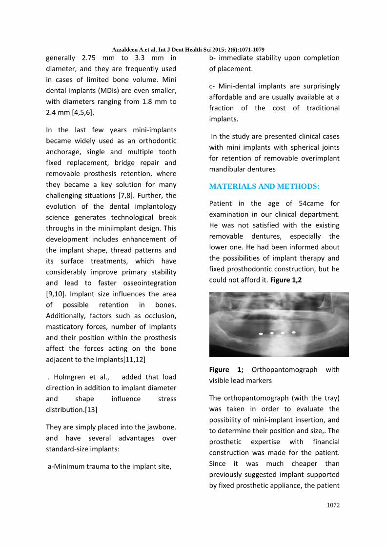
Figure 1; Orthopantomograph with
visible lead markers
The orthopantomograph (with the tray)
was taken in order to evaluate the
possibility of mini-implant insertion, and
to determine their position and size,. The
prosthetic expertise with financial
construction was made for the patient.
Since it was much cheaper than
previously suggested implant supported
by fixed prosthetic appliance, the patient

Azzaldeen A.et al, Int J Dent Health Sci 2015; 2(6):1071-1079
1073
decided to make lower removable
denture (overdenture) supported with
four MDIs Sendax type with ball
attachments.
Figure 2;Pre-operative mandibular arch
According to the orthopantomograph
findings, correction of future implant
sites was performed. The tray was
punctured on selected spots by grinding
bur and placed into the patient’s mouth.
The implant sites were marked through
the holes in acrylic baseplate with
surgical marker and transgingival
implantation was performed. The gingiva
was punctured on the marked spots, and
the bone was initially drilled with the
locator drill according to the marks
made with surgical marker Figure 3. The
bone drilling was performed by using
disposable surgical drill of 1.1 mm
diameter to the depth of 1 length of
implant as recommended by the
manufacturer. Parallelization of the
implants was achieved with the insertion
of sterile, previously used, surgical drills
into each drilled implant site. After
drilling, the MDIs Sendax Classic
Standard, O-Ball dimensions 1,8 mm
(diameter) x 15 mm (length) were
screwed firstly by using manual screwing
instrument , and afterwards
Figure 3 Implantation of mini dental
implants with ratchet (torque 35 N/cm ).
Since it was not possible to screw MDI to
the end of the length, it was unscrewed
and displaced. For that reason, the
primarily drilled holes were deepened to
the depth of 2/3 of the implants length,
and in repeated screwing, it was possible
to screw MDI to the end . Figure 4
Figure 4.; Placed mini dental implants
The laboratory implants were inserted
into the impression copings (Figure 6),
and the models were poured in hard
stone . Micro metal housings were
placed onto the laboratory implants ,
and the metal base of the lower
overdenture was produced . Further
clinical and laboratory procedures were
performed according to the routine
procedure for lower denture
production. Figure 5,6
Azzaldeen A.et al, Int J Dent Health Sci 2015; 2(6):1071-1079
1074
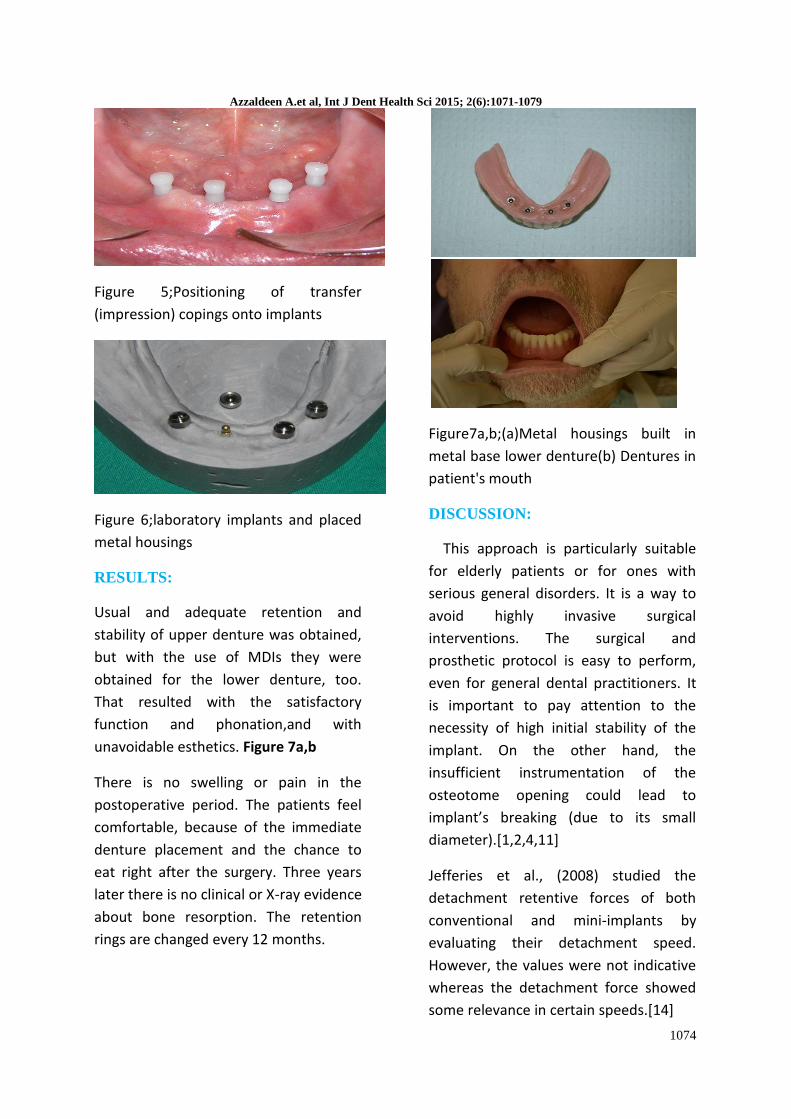
Figure 5;Positioning of transfer
(impression) copings onto implants
Figure 6;laboratory implants and placed
metal housings
RESULTS:
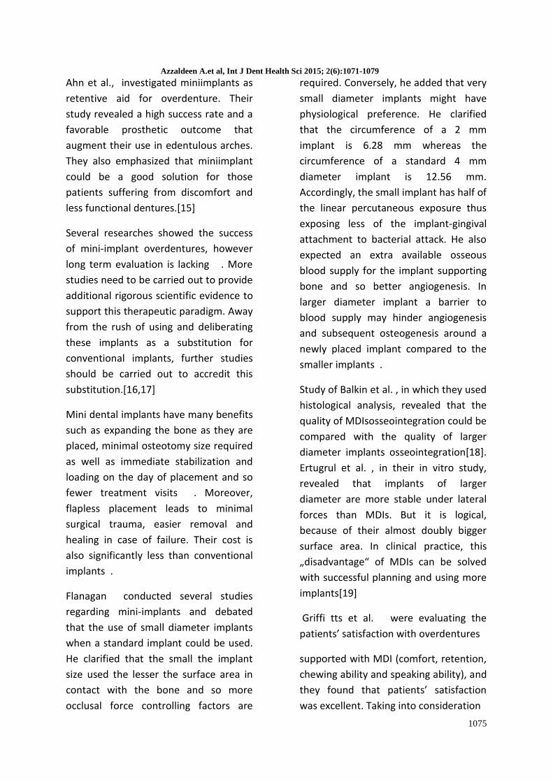
Usual and adequate retention and
stability of upper denture was obtained,
but with the use of MDIs they were
obtained for the lower denture, too.
That resulted with the satisfactory
function and phonation,and with
unavoidable esthetics. Figure 7a,b
There is no swelling or pain in the
postoperative period. The patients feel
comfortable, because of the immediate
denture placement and the chance to
eat right after the surgery. Three years
later there is no clinical or X-ray evidence
about bone resorption. The retention
rings are changed every 12 months.
Figure7a,b;(a)Metal housings built in
metal base lower denture(b) Dentures in
patient's mouth
DISCUSSION:
This approach is particularly suitable
for elderly patients or for ones with
serious general disorders. It is a way to
avoid highly invasive surgical
interventions. The surgical and
prosthetic protocol is easy to perform,
even for general dental practitioners. It
is important to pay attention to the
necessity of high initial stability of the
implant. On the other hand, the
insufficient instrumentation of the
osteotome opening could lead to
implant’s breaking (due to its small
diameter).[1,2,4,11]
Jefferies et al., (2008) studied the
detachment retentive forces of both
conventional and mini-implants by
evaluating their detachment speed.
However, the values were not indicative
whereas the detachment force showed
some relevance in certain speeds.[14]
Azzaldeen A.et al, Int J Dent Health Sci 2015; 2(6):1071-1079
1075
Ahn et al., investigated miniimplants as
retentive aid for overdenture. Their
study revealed a high success rate and a
favorable prosthetic outcome that
augment their use in edentulous arches.
They also emphasized that miniimplant
could be a good solution for those
patients suffering from discomfort and
less functional dentures.[15]
Several researches showed the success
of mini-implant overdentures, however
long term evaluation is lacking . More
studies need to be carried out to provide
additional rigorous scientific evidence to
support this therapeutic paradigm. Away
from the rush of using and deliberating
these implants as a substitution for
conventional implants, further studies
should be carried out to accredit this
substitution.[16,17]
Mini dental implants have many benefits
such as expanding the bone as they are
placed, minimal osteotomy size required
as well as immediate stabilization and
loading on the day of placement and so
fewer treatment visits . Moreover,
flapless placement leads to minimal
surgical trauma, easier removal and
healing in case of failure. Their cost is
also significantly less than conventional
implants .
Flanagan conducted several studies
regarding mini-implants and debated
that the use of small diameter implants
when a standard implant could be used.
He clarified that the small the implant
size used the lesser the surface area in
contact with the bone and so more
occlusal force controlling factors are
required. Conversely, he added that very
small diameter implants might have
physiological preference. He clarified
that the circumference of a 2 mm
implant is 6.28 mm whereas the
circumference of a standard 4 mm
diameter implant is 12.56 mm.
Accordingly, the small implant has half of
the linear percutaneous exposure thus
exposing less of the implant-gingival
attachment to bacterial attack. He also
expected an extra available osseous
blood supply for the implant supporting
bone and so better angiogenesis. In
larger diameter implant a barrier to
blood supply may hinder angiogenesis
and subsequent osteogenesis around a
newly placed implant compared to the
smaller implants .
Study of Balkin et al. , in which they used
histological analysis, revealed that the
quality of MDIsosseointegration could be
compared with the quality of larger
diameter implants osseointegration[18].
Ertugrul et al. , in their in vitro study,
revealed that implants of larger
diameter are more stable under lateral
forces than MDIs. But it is logical,
because of their almost doubly bigger
surface area. In clinical practice, this
„disadvantage“ of MDIs can be solved
with successful planning and using more
implants[19]
Griffi tts et al. were evaluating the
patients’ satisfaction with overdentures
supported with MDI (comfort, retention,
chewing ability and speaking ability), and
they found that patients’ satisfaction
was excellent. Taking into consideration
Azzaldeen A.et al, Int J Dent Health Sci 2015; 2(6):1071-1079
1076
all advantages of MDI (success rates,
surgical technique, fi nancial advantages,
possibilities of immediate loading), it can
be concluded that MDI are highly
successful implant option for edentulous
mandible.[20]
Shatkin et al , in their retrospective
analysis over five years of 2514 MDIs,
which equally supported fixed and
removable prostheses, found the overall
implant survival rate of 94.2%. Initial
stability is important for the successful
osseointegration and high implant
success rate. It is stipulated with bone
quality, implant design, and surgical
technique that is used.[21]
A recent study in which six mini-implants
were installed to stabilise full maxillary
dentures with or without palatal
coverage also reported high implant
failure rates; 21,6% and 46,2%,
respectively [22] . The authors attributed
the high failure rate to facial angulations
of maxillary implants, a thick masticatory
mucosa that necessitated longer implant
abutments, and disparallelism of the
unsplinted implants that may have
produced micromovements in
conjunction with multiple insertions and
removals of the prosthesis.[22]
The original implant dimension, as
described by Branemark, was 3.7 by
10mm. Branemark felt this dimension
fulfilled the need for all implant therapy.
In today’s practice, implants of varying
dimensions are now available. Implants
with a diameter of 3.75 mm have been
considered standard, below and above
which have been considered as narrow
and wide diameter, respectively.
Implants with a length of less than
10mm are considered short. A reduction
of implant diameter and length results in
a proportional decrease of implant
surface area. This infers a decreased
implant-to-bone contact area[23].
The biomechanical impact of smaller
dimensional implants was discussed
earlier in this paper with there being
higher crestal strain, lower pull out force
and lower structural integrity as the
implant dimension is reduced.
Theoretically, this could translate to
lower clinical success rates for implants
of lesser dimensions.[24]
The use of CBCT scans for treatment
planning of dental implants has become
largely recognized as a high quality, time
and cost effective, imaging method .
Several studies have confirmed that
linear measurements on CBCTs images
present the necessary accuracy for use in
dentistry ). In well controlled studies,
Mozzo et al. (1998) and Moreira et al.
assessed the CBCT’s geometric accuracy
and reported that differences between
simulated mandibular bone and dry
human skulls to images generated from
CBCT’s ranged from 0.15 to 2 %, for
linear measurements (in width and
height, respectively) and 0.33 % for
angular measurements ). Based on the
ability of the system to reconstruct
anatomic structures with dimensions
considered “close to real”, the data
obtained with CBCT scans were used in
the present investigation as reference
Azzaldeen A.et al, Int J Dent Health Sci 2015; 2(6):1071-1079
1077
for comparisons with the data assessed
with the Conventional method.[25,26]
Two categories of complications can
occur in implant therapy: biological and
technical or mechanical. Biological
complications refer to any disturbance in
the peri-implant tissue that results in a
decrease of function or eventual loss of
the implant fixture. This includes peri-
implantitis/progressive bone loss, peri-
implant mucositis, periapical implantitis,
or sensory disturbance. Technical or
mechanical complications refer to
mechanical damage of the implant
fixture and/or implant components and
its suprastructures. This can include
screw loosening, screw fracture, fixture
fracture, and prosthetic issues. Review of
the reported complications revealed
more reports on complications of
technical or mechanical nature than
complications biological in
nature.[27,28.29]
The short- and medium length mini-
implants (7– 10 mm) presented a higher
failure rate than the long mini-implants
(14 mm), 38% versus 3%. The fact that
the use of the long implants in the
replacement of the lost implants
resulted in maintenance of the implants
in proper function throughout the
observation period further indicates that
long implants should be selected for the
best prognosis of the treatment[1].
An insertion torque of 35 N/cm is
necessary for narrow implants to achieve
a degree of primary stability sufficient
for immediate loading. In the present
case, an insertion torque of 35 N/cm was
achieved without fracture of the
mandible or implants.[1,2]
The biomechanical aspects of the narrow
implant, such as the distribution and
control of the forces and movement of
the prosthesis, should also be
considered and evaluated. Masticatory
forces produce axial forces and bending
moments that could result in stress on
the implant as well as on the bone,
thereby compromising the longevity of
the implant. Narrow implants have a
smaller surface and,therefore, an overall
increase in the magnitude of stress and
strain experienced by the load-carrying
system compared to conventional
implants. However, this aspect does not
contraindicate the use of narrow
implants in older patients,because, in
most cases, occlusal forces are slightly
reduced owing to age-related
deterioration of the dentition. The
masticatory forces and the quality of
cancellous bone should always be
evaluated before narrow-implant–
retained overdentures are selected as a
treatment .[2,31,32]
It is necessary to reline the complete
dentures on a regular basis and to
perform occlusal adjustments for better
force and movement distribution in all
narrow implant– supported
overdentures in order to avoid implant
fracture and overloadinduced bone loss
around the implants. These steps were
followed in the present case and were
essential for the longevity of the
success.[1,32,33,34]
Azzaldeen A.et al, Int J Dent Health Sci 2015; 2(6):1071-1079
1078
Although mini-implant overdenture is a
successful alternative for conventional
two-implant overdenture, the
conventional overdenture treatment
option exhibited more favorable clinical
and radiographic outcome than mini-
implant overdenture.
CONCLUSION:
The use of implants in the edentulous
arch has changed the way in which
patients can be treated. Standard
diameter implants have been utilized
successfully for more than twenty years
for overdenture patients, and more
recently narrow-diameter implants
have been utilized. Both standard and
narrow-diameter implants have
demonstrated high success and survival
rates and are associated with
improvements in function and patient
comfort.
In conclusion, the placement of mini-
implants as retentive elements for full
dentures with poor functional stability
had a marked positive effect on the
patients’ perception of oral function and
comfort as well as security in social life.
However, the treatment approach may
be less predictable in the maxilla and
with the use of short implants .
REFERENCES:
1. Abu-Hussein M. , Abdulgani A.,
Bajali M., Chlorokostas G .; The
Mandibular Two-Implant
Overdenture.Journal of Dental
and Allied Sciences , 2014 , Vol
3,1; 58-62
2. Abu-Hussein M ., Abdulgani A .
;MANDIBULAR IMPLANT
OVERDENTURE RETAINED WITH
O-RING BALL, Int J Dent Health
Sci 2014; 1(6):984-991
3. Bressan E, Tomasi C, Stellini E,
Sivolella S, Favero G, Berglundh T.
Implant-supported mandibular
overdentures: a cross-sectional
study. Clin Oral Implants Res.
2012;23:814–819.
4. Christensen GJ, Swift EJ Jr. Mini
implants: good or bad for the
long-term service? J Esthet Restor
Dent. 2008;20:343– 348.
5. LaBarre EE, Ahlstrom RH, Noble
WH. Narrow diameter implants
for mandibular denture
retention. J Calif Dent Assoc.
2008;36:283–286.
6. Flanagan D. Implant-supported
fixed prosthetic treatment using
very small-diameter implants: a
case report. J Oral Implantol.
2006;32:34–37.
7. Bryant SR, MacDonald JD, Kim K.
Does the type of implant
prosthesis affect outcomes for
the completely edentulous arch?
Int J Oral Maxillofacial Implants.
2007; 22(Suppl):117-139.
8. Shawneen MG. Cortical bone
thickness of the maxilla and
mandible for mini-implant
placement. PhD thesis, The
University of Iowa August 2008

Azzaldeen A.et al, Int J Dent Health Sci 2015; 2(6):1071-1079
1072
9. Jones AA, Cochran DL.
Consequences of Implant Design.
Dent Clin of North Am 2006;
50(3):339-360.
10. Sakoh J, Wahlmann U, Stender E,
Nat R, Al- Nawas B, Wagner W.
Primary stability of a conical
implant and a hybrid, cylindric
screwtype implant in vitro.
International Journal of Oral &
Maxillofacial Implants. 2006;
21(4):560-566.
11. Christensen GJ. The mini-implant
has arrived. J Am Dent Assoc
2006;137(3):387-90.
12. Froum SJ, Simon H, Stuart J. Cho
SC, Elian N, Michael DR, Tarnow
DP. Histologic evaluation of bone
implant contact of immediately
loaded transitional implants after
six to 27 months. J Oral
Maxillofac Implants 2005;20:54-
60.
13. Holmgren EP, Seckinger RJ,
Kilgren LM, Mante F. Evaluating
parameters of osseointegrated
dental implants using finite
element analysis; a two
dimensional comparative study
examining the effects of implant
diameter, implant shape, and
load direction. J Oral Implantol
1998; 24:80-88
14. Jefferies SR, Boston DW, Damrow
MP, Galbraith CT. Comparison of
detachment forces of two
implant overdenture attachment
types: effect of detachment
speed. Am J Dent.
2008;21(4):244-250.
15. Ahn MR, An KM, Choi JH, Sohn
DS. Immediate Loading With Mini
Dental Implants in the Fully
Edentulous Mandible. Implant
Dent 2004; 13: 367-72.
16. Mazor Z, Steigmann M, Leshem R,
Peleg M. Mini-implants to
reconstruct missing teeth in a
severe ridge deficiency and small
inter dental space: a 5 year case
series. Implant Dent.
2004;13:336–341.
17. Vigolo P, Givani A. Clinical
evaluation of single mini-implant
restorations: a five year
retrospective study. J Prosthet
Dent. 2000;84:50–54.
18. Balkin BE, Stefl ik DE, Naval F.
Mini-dental implant insertion
with the auto-advance technique
for ongoing applications. J Oral
Implantol. 2001;27(1):32-7.
19. Ertugrul HZ, Pipko DJ. Measuring
mobility of 2 dental implant fi
xtures of different confi
gurations: an in vitro study.
Implant Dent. 2006;15(3):290-7.
20. Griffi tts TM, Collins CP, Collins
PC. Mini dental implants: an
adjunct for retention, stability,
and comfort for the edentulous
patient. Oral Surg Oral Med Oral
Pathol Oral Radiol Endod.
2005;100(5):e81-4.
21. Shatkin TE, Shatkin S,
Oppenheimer AJ. Mini dental
implants for the general dentists:
A novel technical approach for
small-diameter implant
placement. Compend Contin

Azzaldeen A.et al, Int J Dent Health Sci 2015; 2(6):1071-1079
1073
Educ Dent. 2003;24(Suppl 1):26-
34.
22. Elsyad MA, Ghoneem NE, El-
Sharkawy H. Marginal bone loss
aroundunsplinted mini-implants
supporting maxillary
overdentures: a preliminary
comparative study between
partial and full palatal coverage.
Quintessence Int. 2013;44:45–52.
23. Malo P, De Araujo Nobre M,
Rangert B.Short Implants placed
one stage in maxillae and
mandibles: a retrospective clinical
study with 1 To 9 Years of follow-
-‐up. Clin Implant Dent Relat Res
2007;9(1):15--‐21.
24. Allum SR, Tomlinson RA, Joshi R.
The Impact of loads on standard
diameter, small diameter and
mini implants: a comparative
laboratory study.Clin Oral
ImplantsRes,2008;19(6):553--‐9.
25. Mozzo P, Procacci C, Tacconi A,
Martini PT, Andreis IA. A new
volumetric CT machine for dental
imaging based on the cone-beam
technique: preliminary results.
Eur Radiol 1998;8(9):1558-64.
26. Moreira CR, Sales MA, Lopes PM,
Cavalcanti MG. Assessment of
linear and angular measurements
on three-dimensional cone-beam
computed tomographic images.
Oral Surg Oral Med Oral Pathol
Oral Radiol Endod 2009
Sep;108(3):430-6
27. Dilek OC, Tezulas E. Treatment of
a narrow, single tooth edentulous
area with mini-dental implants: a
clinical report. Oral Surg Oral
Med Oral Pathol Oral Radiol
Endod. 2007;103(2):e22-5.
28. Bulard RA, Vance JB. Multi clinic
evaluation using mini-dental
implants for long term denture
stabilization: a preliminary
biometric evaluation. Compend
Contin Educ Dent. 2005;26:892–
897.\
29. Linkevicius T, Apse P, Grybauskas
S, Puisys A. The influence of soft
tissue thickness on crestal bone
changes around implants: a 1-
year prospective controlled
clinical trial. Int J Oral Maxillofac
Implants. 2009; 24:712–719.
30. Scepanovic M, Calvo-Guirado JL,
Markovic A, Delgardo-Ruiz R,
Todorovic A, Millicic B, et al. A 1-
year prospective cohort study on
mandibular overdentures
retained by mini dental implants.
Eur J Oral Implantol 2012; 5: 367-
79.
31. Cehreli MC, Akca K. Narrow-
diameter implants as terminal
support for occlusal three-unit
FPDs: a biomechanical analysis.
Int J Periodontics Restorative
Dent.2004;24(6):513-519.
32. Morneburg TR, Proschel PA.
Success rates of microimplants in
edentulous patients with residual
ridge resorption. Int J Oral
Maxillofac Implants.
2008;23(2):270-276.
33. Singh RD, Ramashanker, Chand P.
Management of atrophic
mandibular ridge with mini

Azzaldeen A.et al, Int J Dent Health Sci 2015; 2(6):1071-1079
1074
dental implant system. Natl J
Maxillofac Surg. 2010;1(2):176-
178.
34. Bidra AS, Almas K. Mini implants
for definitive prosthodontic
treatment: a systematic review. J
Prosthet Dent. 2013;109(3):156-
164.