Immature and Mature Dengue Serotype 1 Virus Structures...
8
Immature and Mature Dengue Serotype 1 Virus Structures Provide Insight into the Maturation Process Victor A. Kostyuchenko, a,b Qian Zhang, a,b Joanne L. Tan, a,b Thiam-Seng Ng, a,b Shee-Mei Lok a,b Program in Emerging Infectious Diseases, Duke-NUS Graduate Medical School, Singapore a ; Center for Bioimaging Sciences, National University of Singapore, Singapore b Dengue virus is a major human pathogen that has four serotypes (DENV1 to -4). Here we report the cryoelectron microscopy (cryo-EM) structures of immature and mature DENV1 at 6- and 4.5-Å resolution, respectively. The subnanometer-resolution maps allow accurate placement of all of the surface proteins. Although the immature and mature viruses showed vastly different surface protein organizations, the envelope protein transmembrane (E-TM) regions remain in similar positions. The pivotal role of the E-TM regions leads to the identification of the start and end positions of all surface proteins during maturation. D engue virus (DENV), the cause of dengue fever, infects 100 million people worldwide every year. It is a member of the Flaviviridae family (1), with four serotypes: DENV1, -2, -3, and -4. Infection with DENV usually causes a self-limiting fever accom- panied by rashes and joint pain in patients but might lead to den- gue hemorrhagic fever and dengue shock syndrome, which may be fatal. DENV consists of an 500-Å-diameter protein shell embed- ded in a host-derived lipid membrane and encapsidates an 11-kb single-stranded positive-sense RNA genome. The dengue genome encodes three structural proteins, the core (or capsid), the pre- membrane (prM), and the envelope (E) protein, that form the virus particle, as well as seven nonstructural proteins (1) that are involved in replication of the virus genome. The newly synthe- sized immature DENV has a spiky appearance (2)(Fig. 1A) and is typically noninfectious unless it is complexed with certain anti- bodies (3). Virus maturation occurs during transportation of the virus particle through the trans-Golgi component network (TGN). The acidic environment of these compartments induces structural rearrangement of the virus surface proteins. During this initial maturation process, the furin protease cleaves prM mole- cules on the virus into pr and M. After leaving the cell, the cleaved pr dissociates from the virus surface, resulting in smooth, fully mature infectious virus particles (4)(Fig. 1B). The E protein is the major structural component of the viral surface. The ectodomain of E protein (5) contains three distinct domains, DI, DII, and DIII (also shown in Fig. 1C), which are connected by flexible links that allow rearrangement of domains during virus assembly, maturation, and infection (5–7). DIII is involved in attachment to host cell receptors, whereas DII is re- sponsible for fusion to the host endosomal membrane during in- fection (8). The ectodomain is connected to the stem made from amphipathic helices 1 and 2. The stem, in turn, is anchored to the virus lipid membrane by two transmembrane (TM) -helices, TM1 and TM2. The crystal structure of an E-prM complex (6) shows that pr has a -barrel fold and caps the fusion loop of the E protein, consistent with its function in preventing the newly synthesized virus from fusing back into the cell during maturation. The furin cleavage site lies between the pr molecule and the ectodomain of M protein, which exists as a linear polypeptide chain (6). The prM protein also has a stem region with a single amphipathic -helix followed by two TM -helices (9)(Fig. 2B). The core protein (10) associates with the viral RNA (11); however, it is not observed in cryoelectron microscopy (cryo-EM) reconstructions of flavivi- ruses (2, 9), indicating that the core proteins do not form icosa- hedral structures in the virus particle. Cryo-EM structures of immature (2) and mature (9, 12) DENV2 have been described previously. Both are icosahedral structures with three E and M (prM in immature virus) het- erodimers per asymmetric unit. The surface of the immature virus (Fig. 1A) contains 60 spikes, each made from three prM-E het- erodimers (2, 6). In contrast, the mature virus (Fig. 1B) has a smooth surface assembled from 90 E protein dimers organized in a characteristic “herringbone” pattern (13) with the M protein lying underneath (9). Until very recently, the cryo-EM recon- structions of homologous DENV2 have been calculated to a reso- lution of 12.5 Å for the immature virus (5) and 9.5 Å for mature virus (9). Although it is possible to define the domain organi- zation of the surface proteins at these resolutions, it is not possible to identify molecular interactions between the com- ponents that drive assembly, maturation, or infection. A 7-Å- resolution cryo-EM reconstruction of a complex of mature DENV1 with Fab molecules has also been published recently (14); however, this structure likely does not represent the infectious virus particle, as Fab binding may induce some structural changes (15). A very recent publication on a 3.5-Å cryo-EM reconstruction of mature DENV2 (12) allows a comparison and validation of our findings. Here we report 4.5- and 6-Å structures of the mature and immature dengue serotype 1 virus, respectively. At these resolu- tions, component proteins can be accurately placed, revealing es- sential molecular interactions between molecules that guide as- sembly, drive maturation, and confer stability to the infectious virus. Furthermore, a previously unobserved connection between the TM and stem regions in the immature virus is now clearly visible, allowing us to unambiguously deduce the start and end positions of the E and M proteins during virus maturation. Received 22 January 2013 Accepted 24 April 2013 Published ahead of print 1 May 2013 Address correspondence to Shee-Mei Lok, [email protected]. V.A.K., Q.Z., and J.L.T. contributed equally to this paper. Copyright © 2013, American Society for Microbiology. All Rights Reserved. doi:10.1128/JVI.00197-13 7700 jvi.asm.org Journal of Virology p. 7700 –7707 July 2013 Volume 87 Number 13 on July 10, 2018 by guest http://jvi.asm.org/ Downloaded from
Transcript of Immature and Mature Dengue Serotype 1 Virus Structures...

Immature and Mature Dengue Serotype 1 Virus Structures ProvideInsight into the Maturation Process
Victor A. Kostyuchenko,a,b Qian Zhang,a,b Joanne L. Tan,a,b Thiam-Seng Ng,a,b Shee-Mei Loka,b
Program in Emerging Infectious Diseases, Duke-NUS Graduate Medical School, Singaporea; Center for Bioimaging Sciences, National University of Singapore, Singaporeb
Dengue virus is a major human pathogen that has four serotypes (DENV1 to -4). Here we report the cryoelectron microscopy(cryo-EM) structures of immature and mature DENV1 at 6- and 4.5-Å resolution, respectively. The subnanometer-resolutionmaps allow accurate placement of all of the surface proteins. Although the immature and mature viruses showed vastly differentsurface protein organizations, the envelope protein transmembrane (E-TM) regions remain in similar positions. The pivotal roleof the E-TM regions leads to the identification of the start and end positions of all surface proteins during maturation.
Dengue virus (DENV), the cause of dengue fever, infects 100million people worldwide every year. It is a member of the
Flaviviridae family (1), with four serotypes: DENV1, -2, -3, and -4.Infection with DENV usually causes a self-limiting fever accom-panied by rashes and joint pain in patients but might lead to den-gue hemorrhagic fever and dengue shock syndrome, which maybe fatal.
DENV consists of an �500-Å-diameter protein shell embed-ded in a host-derived lipid membrane and encapsidates an 11-kbsingle-stranded positive-sense RNA genome. The dengue genomeencodes three structural proteins, the core (or capsid), the pre-membrane (prM), and the envelope (E) protein, that form thevirus particle, as well as seven nonstructural proteins (1) that areinvolved in replication of the virus genome. The newly synthe-sized immature DENV has a spiky appearance (2) (Fig. 1A) and istypically noninfectious unless it is complexed with certain anti-bodies (3). Virus maturation occurs during transportation of thevirus particle through the trans-Golgi component network(TGN). The acidic environment of these compartments inducesstructural rearrangement of the virus surface proteins. During thisinitial maturation process, the furin protease cleaves prM mole-cules on the virus into pr and M. After leaving the cell, the cleavedpr dissociates from the virus surface, resulting in smooth, fullymature infectious virus particles (4) (Fig. 1B).
The E protein is the major structural component of the viralsurface. The ectodomain of E protein (5) contains three distinctdomains, DI, DII, and DIII (also shown in Fig. 1C), which areconnected by flexible links that allow rearrangement of domainsduring virus assembly, maturation, and infection (5–7). DIII isinvolved in attachment to host cell receptors, whereas DII is re-sponsible for fusion to the host endosomal membrane during in-fection (8). The ectodomain is connected to the stem made fromamphipathic helices �1 and �2. The stem, in turn, is anchored tothe virus lipid membrane by two transmembrane (TM) �-helices,TM1 and TM2.
The crystal structure of an E-prM complex (6) shows that prhas a �-barrel fold and caps the fusion loop of the E protein,consistent with its function in preventing the newly synthesizedvirus from fusing back into the cell during maturation. The furincleavage site lies between the pr molecule and the ectodomain ofM protein, which exists as a linear polypeptide chain (6). The prMprotein also has a stem region with a single amphipathic �-helixfollowed by two TM �-helices (9) (Fig. 2B). The core protein (10)
associates with the viral RNA (11); however, it is not observed incryoelectron microscopy (cryo-EM) reconstructions of flavivi-ruses (2, 9), indicating that the core proteins do not form icosa-hedral structures in the virus particle.
Cryo-EM structures of immature (2) and mature (9, 12)DENV2 have been described previously. Both are icosahedralstructures with three E and M (prM in immature virus) het-erodimers per asymmetric unit. The surface of the immature virus(Fig. 1A) contains 60 spikes, each made from three prM-E het-erodimers (2, 6). In contrast, the mature virus (Fig. 1B) has asmooth surface assembled from 90 E protein dimers organized ina characteristic “herringbone” pattern (13) with the M proteinlying underneath (9). Until very recently, the cryo-EM recon-structions of homologous DENV2 have been calculated to a reso-lution of 12.5 Å for the immature virus (5) and 9.5 Å for maturevirus (9). Although it is possible to define the domain organi-zation of the surface proteins at these resolutions, it is notpossible to identify molecular interactions between the com-ponents that drive assembly, maturation, or infection. A 7-Å-resolution cryo-EM reconstruction of a complex of matureDENV1 with Fab molecules has also been published recently (14);however, this structure likely does not represent the infectiousvirus particle, as Fab binding may induce some structural changes(15). A very recent publication on a 3.5-Å cryo-EM reconstructionof mature DENV2 (12) allows a comparison and validation of ourfindings. Here we report 4.5- and 6-Å structures of the mature andimmature dengue serotype 1 virus, respectively. At these resolu-tions, component proteins can be accurately placed, revealing es-sential molecular interactions between molecules that guide as-sembly, drive maturation, and confer stability to the infectiousvirus. Furthermore, a previously unobserved connection betweenthe TM and stem regions in the immature virus is now clearlyvisible, allowing us to unambiguously deduce the start and endpositions of the E and M proteins during virus maturation.
Received 22 January 2013 Accepted 24 April 2013
Published ahead of print 1 May 2013
Address correspondence to Shee-Mei Lok, [email protected].
V.A.K., Q.Z., and J.L.T. contributed equally to this paper.
Copyright © 2013, American Society for Microbiology. All Rights Reserved.
doi:10.1128/JVI.00197-13
7700 jvi.asm.org Journal of Virology p. 7700–7707 July 2013 Volume 87 Number 13
on July 10, 2018 by guesthttp://jvi.asm
.org/D
ownloaded from

MATERIALS AND METHODSPurification of immature and mature DENV. Production and purifica-tion of immature (2) and mature (13) dengue virus had been describedpreviously. Briefly, immature DENV1 (DEN1/SG/07K3640DK1/2008)(16) was grown in C6/36 cells in a 10-cell stack containing minimumessential medium (MEM; Gibco) and 10% (vol/vol) fetal bovine serum(FBS) (Gibco) and infected at a multiplicity of infection (MOI) of 1 at29°C. At 2 h postinfection, the medium was replaced with MEM supple-mented with 2% (vol/vol) FBS and 40 mM NH4Cl and incubated for 2days. Mature virus was grown under similar conditions except using
RPMI 1640 (Gibco) medium containing 25 mM HEPES and 10% (vol/vol) FBS with no NH4Cl used. Cells were infected at an MOI of 0.1, and thevirus was harvested 96 h postinfection.
The purification steps were similar for both samples. Briefly, the har-vested medium was centrifuged at 6,000 rpm for 30 min, and the super-natant was precipitated with 8% (wt/vol) PEG 8000 (Sigma-Aldrich) byincubation at 4°C overnight. The suspension was centrifuged, and thepellet was resuspended in NTE buffer (12 mM Tris, pH 8.0, 120 mM NaCl,1 mM EDTA). The virus was purified through a 24% (wt/vol) sucrosecushion and a linear 10 to 30% (wt/vol) potassium tartrate-glycerol gra-
FIG 1 Cryo-EM structures of the DENV1. (A) Cryo-EM density of immature virus. The scale bar is 100 Å. The density is colored radially: radii between 0 and160 Å, between 161 and 210 Å, between 211 and 270 Å, and 271 Å and above are colored in yellow, green, light blue, and magenta, respectively. For clarity, thecryo-EM maps in panels A and B were low-pass filtered to an 8-Å resolution. (B) Cryo-EM density of mature DENV1. The density is colored radially: radiibetween 0 to 160 Å, between 161 and 210 Å, between 211 and 250 Å, and 251Å and above are colored in yellow, green, light blue, and magenta, respectively. (Cand D) Zoomed-in view of fitted surface proteins into the cryo-EM density maps (gray) of immature (C) and mature (D) virus. Black arrows in panel D pointto density corresponding to glycosylation sites. DI, DII, and DIII of the E ectodomain are colored in red, yellow, and blue, respectively. The pr part of the prMmolecule is colored in cyan, and the M protein is colored in purple. The E stem and transmembrane (TM) regions are colored in dark green. (E) E protein stemhelix �2 fitted into the 4.5-Å-resolution mature DENV1 virus map. Densities of some side chains are resolved (black arrows). The side chain models were notdetermined experimentally and are shown to indicate the protein sequence. (F) Close-up view of a part of the E protein DI main chain in the 4.5-Å-resolutionmap of the mature DENV1 virus. The densities for �-strands are separated. (G) Fourier shell correlation versus resolution plots for the individual lipid bilayer(red) and the E-prM protein shell (green) of the immature DENV1 map and the shell including the lipid bilayer and the outer surface protein of the matureDENV1 (blue).
Dengue Serotype 1 Virus Cryo-EM Structures
July 2013 Volume 87 Number 13 jvi.asm.org 7701
on July 10, 2018 by guesthttp://jvi.asm
.org/D
ownloaded from

dient. The virus band was collected, buffer exchanged to NTE buffer, andconcentrated in an Amicon Ultra centrifugal filter (100 kDa; Millipore).The purity and concentration of DENV were estimated using Coomassieblue-stained SDS-PAGE gel.
Cryo-electron microscopy and image processing. Copper grids cov-ered with lacey carbon and a layer of thin carbon were used for bothsamples. A 2.5-�l sample was pipetted onto the grid and then blotted withfilter paper for 2 s before being flash frozen in liquid ethane using FEIVitrobot Mark IV (The Netherlands). Image acquisitions were done byusing an FEI Titan Krios electron microscope operating at 300 kV at liquidnitrogen temperature with a nominal magnification of 75,000 and anelectron dose of 18 e�/Å2 at a defocus range of 0.7 to 3.7 �m. The imageswere collected with a Gatan 4K by 4K charge-coupled device (CCD) cam-era with a pixel size of 1.2 Å. Virus particles were selected manually using
the boxer tool from an EMAN (17) software package. The astigmaticdefocus parameters for each micrograph were estimated with CTFFIND(18). A total of 13,204 and 14,126 particles were selected for the immatureand mature virus reconstructions, respectively.
3D reconstruction, structure validation, and model building. Thethree-dimensional (3D) reconstructions for both data sets were per-formed in a similar way: the orientation assignment for all images wascarried out using the MultiPath Simulated Annealing protocol (19), fol-lowed by 3D reconstruction using the make3d program from EMAN (17).DENV2 immature (16-Å-resolution) (2) and mature (9.5-Å-resolution)(9) virus maps were used as starting models. For both structures, after�25 cycles of iterations, the reconstructions had converged. To producethe immature and mature virus maps, 10,588 and 9,447 images were se-lected from the data sets, respectively. Resolution was determined by plot-
FIG 2 Organization of the E and prM ectodomains in the immature DENV1 and their interactions. (A) Organization of the E and prM protein trimeric spikeson the surface of the virus. Three molecules in the asymmetric unit are shown as ribbons and are labeled A, B, and C. Symmetry-related molecules are shown assurfaces, and E protein surfaces are colored in gray whereas the pr surfaces are colored in cyan. The symmetry-related molecules are labeled a, b, and c. The blacktriangle represents one asymmetric unit. (B) Structure of an E protein complexed with prM protein. Color schemes are defined in Fig. 1. (C) Interactions betweenpr molecules (cyan) at the tip of the spike. Residues are identified as interacting by having a distance of less than 8 Å between C� chains. (C to F) The interactingresidues are colored in orange, and the interacting interface is indicated by an asterisk. (D) Stereodiagram showing the interactions between DII (yellow) of themolecules (labeled A, B, and C) in the asymmetric unit and DIII (blue) from neighboring symmetry-related molecules (labeled a, b, and c). (E) Interactionsbetween M molecule (pink) and the DII (yellow) of the E ectodomain. The furin cleavage site on the prM protein is colored in green and marked with an “X.” (F)Interactions of the E stem region (dark green) with DI of the E protein (red). (G) Fit of the E protein stem �-helices into the 6-Å cryo-EM map. The density thatconnects the DIII of the E protein to the stem region (indicated by “*”) and the stem to the transmembrane region (indicated by “#”) can be observed.
Kostyuchenko et al.
7702 jvi.asm.org Journal of Virology
on July 10, 2018 by guesthttp://jvi.asm
.org/D
ownloaded from
ting the Fourier shell correlation coefficient between reconstructions gen-erated from two half-data sets with a cutoff value of 0.5 (Fig. 1G).
The crystal structures of the DENV2 soluble fragment of E protein(Protein Data Bank [PDB] code 1TG8) (5) for mature virus and theprM-E ectodomain complex (6) (PDB code 3C6E) for immature viruswere used to fit into the cryo-EM maps by using Chimera (20). The fittedstructures were then modified to contain a DENV1 amino acid sequence.The missing stem and transmembrane helices were added using Coot(21). In the immature virus map, only part of the M ectodomain density iscontinuous: the density that connects to the pr molecule and the densitynear domain II of the E protein. The visible part of the M ectodomain wasthen connected to the nearest M stem region. Molecular dynamic flexiblefitting (MDFF) (22), with symmetry restraints (23) calculated usingNAMD (24), was used to produce the molecules in the asymmetric unit ofthe final virus structures. It involved 3,000 steps of energy minimization,followed by 5,000,000 steps of molecular dynamics run and another 3,000steps of minimization. The structures obtained have correct polypeptidechain geometry and were observed to fit the electron density well.
Protein structure accession numbers. The cryo-EM maps of the im-mature (EMD-2141) and mature (EMD-2142) DENV1 were deposited inthe Electron Microscopy Databank. The coordinates of the modeled com-ponent proteins for immature (PDB code 4B03) and mature (PDB code4AZX) virus were deposited in the Protein Data Bank.
RESULTS
The immature DENV1 is approximately 600 Å in diameter andhas a highly contoured spiky surface (Fig. 1A). The overall resolu-tion of the cryo-EM map is 6 Å; however, the TM layer density haspoorer resolution (8 Å) (Fig. 1G), probably due to slight differ-ences in the symmetry-related positions of the TM regions. Dis-tinct shapes of three pairs of structurally independent E and prproteins are clearly visible in each spike in the asymmetric unit(Fig. 1C and 2A). The crystal structure of a complex of solublefragments of DENV2 E and prM (6) (70% protein sequence iden-tity to DENV1) was fitted into the cryo-EM map as a rigid bodyand served as a base from which the complete structures of theDENV1 E and prM proteins, including the stem and TM regions,were built (Fig. 1C and 2B, F, and G).
The contacts between proteins were identified by measuringthe distances between C� positions, with pairs less than 8 Å apartconsidered to be interacting. In each spike, the arrangement of theprM-E complexes is stabilized at two sites. The first interactionbetween pr molecules occurs at the tip of the spike (Fig. 2A and C)and is likely responsible for holding the spike structure together.The second site of interaction is between DII of one E protein andDIII of another E protein from a neighboring spike (Fig. 2D),linking the spikes to make a shell. The small number of contactsbetween molecules suggests that the virus structure is labile andcan be easily disrupted during virus maturation. The N terminusof the M protein (Fig. 2B) contains a highly conserved hydropho-bic patch that interacts with two �-helices (residues 209 to 215 and258 to 265) of the E protein DII (Fig. 2E). The underside of the Eprotein DI (residues 19 to 25, a conserved hydrophobic sequence)contacts the highly conserved hydrophobic loop between helices�1 and �2 of its stem region (Fig. 2F). There was no obviousdensity in the cryo-EM map that would correspond to the knowncapsid protein structure, consistent with the previous results, in-dicating that the capsid protein is either disordered or differentlyordered in different virus particles (5).
At a 4.5-Å resolution, the map of the mature DENV1 (Fig. 1B)shows well-resolved densities for �-helical structures in the stemand TM region (Fig. 1D). Densities of several side chains were
readily visible (Fig. 1E), as was the separation of �-strands withinthe E protein (Fig. 1F). However, similar to 4.5-Å-resolution crys-tal structures, most side chains are not resolved; hence, protein-protein interactions were identified as having a distance of less
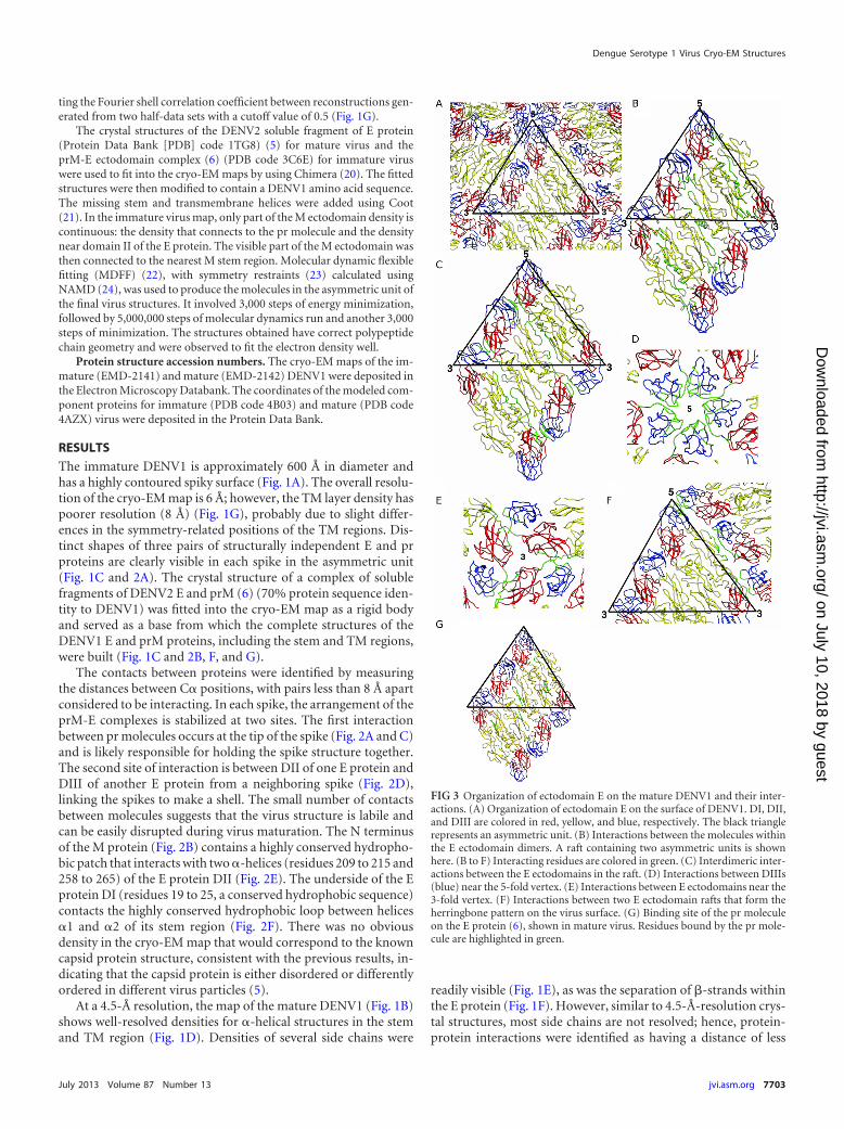
FIG 3 Organization of ectodomain E on the mature DENV1 and their inter-actions. (A) Organization of ectodomain E on the surface of DENV1. DI, DII,and DIII are colored in red, yellow, and blue, respectively. The black trianglerepresents an asymmetric unit. (B) Interactions between the molecules withinthe E ectodomain dimers. A raft containing two asymmetric units is shownhere. (B to F) Interacting residues are colored in green. (C) Interdimeric inter-actions between the E ectodomains in the raft. (D) Interactions between DIIIs(blue) near the 5-fold vertex. (E) Interactions between E ectodomains near the3-fold vertex. (F) Interactions between two E ectodomain rafts that form theherringbone pattern on the virus surface. (G) Binding site of the pr moleculeon the E protein (6), shown in mature virus. Residues bound by the pr mole-cule are highlighted in green.
Dengue Serotype 1 Virus Cryo-EM Structures
July 2013 Volume 87 Number 13 jvi.asm.org 7703
on July 10, 2018 by guesthttp://jvi.asm
.org/D
ownloaded from

than 8 Å between C� positions. A superposition of C� chains ofthe E and M proteins with those from the 3.5-Å DENV2 cryo-EMstructure (12) did not show any significant deviations.
The surface of the mature virus contains 180 copies of E pro-tein organized into 30 “rafts” of three dimers lying parallel to eachother (Fig. 3A). Each raft is stabilized by intra- and interdimericcontacts between ectodomains of the E protein (Fig. 3B and C,respectively), with DII playing a dominant role. In contrast, the
interaction between rafts is highly dependent on DIII (Fig. 3D toF). At the 5-fold vertex, DIIIs from different rafts have extensivecontacts with each other (Fig. 3D). Near the 3-fold vertex, thehinge between DIII and DI interacts with DI of a neighboring Eprotein from another raft (Fig. 3E). The DI-DIII hinge of an Eprotein from the middle dimer in the raft also contacts DII fromthe E protein in an adjacent raft (Fig. 3F).
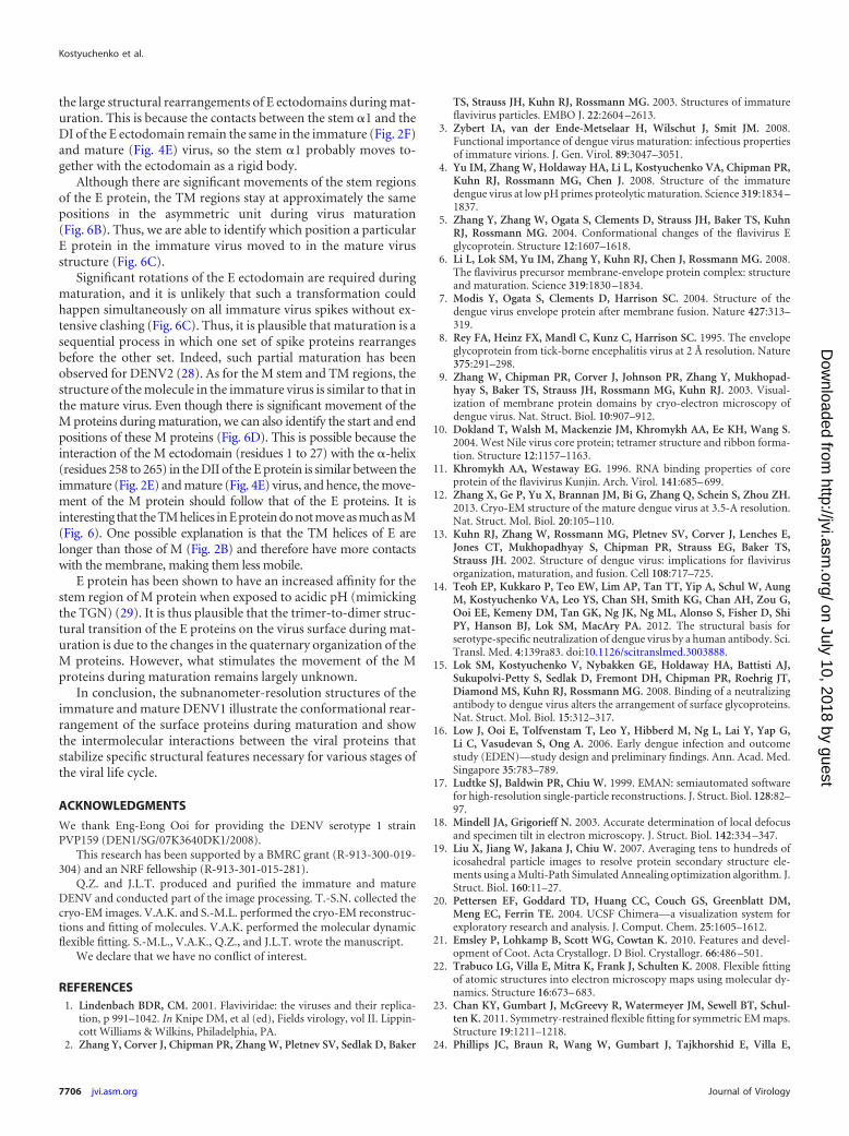
Unlike the immature virus, the TM and stem regions of E pro-teins of the mature virus interact with the M protein dimer (Fig.4A). The ectodomain of M (the polypeptide chain preceding thestem helix) was thought to be a linear polypeptide lying along theectodomain of E based on the 12.5-Å-resolution immatureDENV2 map (6). However, the M ectodomain was not observedin the 9.5-Å-resolution mature DENV2 map (9). In our map, theresolution is such that the N terminus of M (residues 1 to 27) (Fig.4B) is observed and can be fully traced, similarly to the 3.5-Å-resolution DENV2 structure (12). It interacts with the stem helix(residues 27 to 39) from the other M molecule in the dimer (Fig.4C). The C-terminal end of the TM regions also interacts with thesame region of the opposite M protein (Fig. 4C). The N-terminalend of the M protein makes contacts with two E protein loops, theloop that connects E stem �2 to the TM region and another loopthat connects E �1 and �2 (Fig. 4D). Examination of the interac-tions between E ectodomain and its stem region and M proteinshows that the underside (facing the virus membrane) from DI ofE protein interacts with the entire E stem �1, the highly conservedloop, and �2. The underside of E DII has extensive contacts with
FIG 4 Organization of membrane-associated proteins in the mature DENV1virus. (A) Organization of the stem and TM regions of E proteins (dark green)and M proteins (purple) in the virus. The black triangle represents an asym-metric unit. The M protein forms homodimeric interactions. (B) M ectodo-main (N-terminal residues 1 to 26) and stem region (N-terminal residues 27 to39) (black rectangle box) in the mature DENV. Domains DI and DII of theinteracting E protein are shown in red and yellow, respectively. (C) Stereodia-gram of the interactions of the M protein (purple) with the other molecule(pink) in the homodimeric structure. (C to E) Interacting residues are coloredin light green. (D) Stereodiagram of the interactions of the M protein with thestem and TM regions of the E protein. (E) Open-book view of the interactionsbetween the E ectodomain and the M protein as well as the stem and TMregions of E protein. View of the E ectodomain from inside the virion while theTM regions are viewed from the outside.
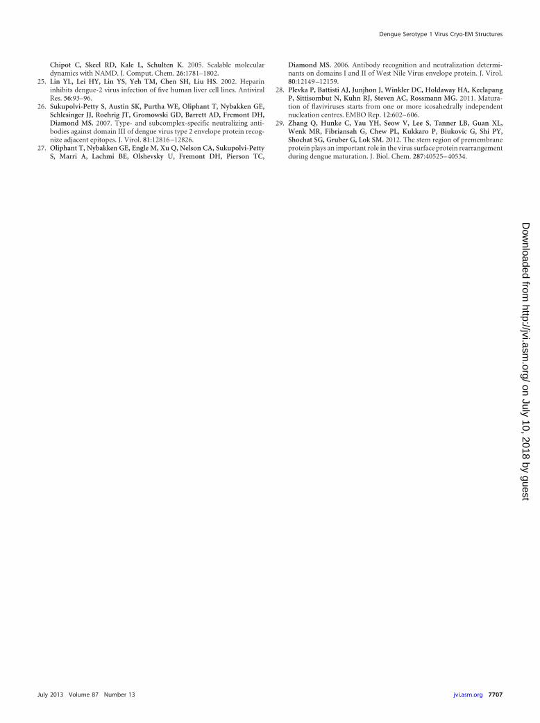
FIG 5 Comparing the surface E proteins between the mature DENV1 andDENV2. (A) Electrostatic charges on the surfaces of DENV1 and DENV2.Surfaces of a raft consisting of two asymmetric units of the E ectodomain areshown. Positive and negative charges are colored in blue and red, respectively.The DENV2 surface contains larger numbers of positively charged residues.(B) Location of nonconserved residues on an E ectodomain raft. The noncon-served residues (spheres) are present on all domains of the E ectodomainprotein except at the hinge between DI and DII, the hinge between DI and DIII,and the fusion loop at the tip of DII. (C) Location of nonconserved residues onan E ectodomain, side view. The epitope recognized by highly potent serotype-specific antibodies is located at the lateral side of DIII (purple circle). Theepitopes bound by weakly neutralizing antibodies, which are generally cross-reactive to all serotypes, are circled in green.
Kostyuchenko et al.
7704 jvi.asm.org Journal of Virology
on July 10, 2018 by guesthttp://jvi.asm
.org/D
ownloaded from

the M ectodomain (Fig. 4E), similar to those observed in the im-mature structure (Fig. 2E and F).
DISCUSSION
Comparison of the amino acid sequences of the E proteins ofDENV1 and DENV2 shows that 8% of the residues are noncon-served. Heparin is an attachment factor on cells that likely binds tostretches of positive charges on the DENV surface. Infection ofcells with DENV2 was shown to be highly dependent on bindingto heparan sulfate, while the other serotypes were less dependent(25). Comparison of the surface charges of the E proteins on theDENV1 and DENV2 (12) cryo-EM structures showed thatDENV2 has more positively charged residues (Fig. 5A).
Most of the nonconserved residues between DENV1 andDENV2 congregate on the surface-exposed regions on the virus(Fig. 5B), suggesting that the variations of residues defining thedifferent serotypes may have been evolved as a means of evadingthe host immune system. Nonconserved residues are present onall surface-exposed regions of the E protein domains with theexception of the hinge between DII and DIII, the hinge betweenDI and DII, and the fusion loop at the tip of DII (Fig. 5B). Thehighly potent DIII antibodies are serotype specific and bind to thelateral ridge of DIII (26), consistent with the location of the non-conserved regions in DIII (Fig. 5C). Weaker antibodies that aregenerally flavivirus cross-reactive bind to either the conserved re-gions next to the lateral ridge on DIII (26) or the fusion loop at thetip of DII (27) (Fig. 5C).
When exposed to the low-pH environment of the TGN duringmaturation, the surface proteins of immature virus undergo ex-tensive rearrangement to adopt a structure similar to that of themature virus except with regard to the pr molecule attached toeach E protein (4). Analysis of the pr binding site on the E proteins(6) in the cryo-EM mature virus structure (Fig. 3G) showed thatbound pr molecules would prevent dimeric interactions betweenE protein monomers (Fig. 3B). This suggests that the packing ofsurface E protein in the immature virus structure at low pH islikely loose. This loose structure may allow the reversibility of thesurface structural change of immature virus structure observedwhen pH is reversed from a neutral- to a low-pH environment (4).Hence, after the prM molecule is cleaved by furin, the dimericstructure of the E protein becomes stabilized when the virus isreleased into the neutral-pH environment outside the cell, thuspreventing the previously reversible structural change and lockingthe structure into a stable lattice.
In addition to the rearrangements of the ectodomain of the Eand prM proteins, there are also significant movements of thestem and TM regions during DENV maturation (Fig. 6). By align-ing the orientation of the TM regions of E protein from the im-mature and mature virus structures (Fig. 6A), movements of �1and �2 of the E stem region can be visualized. Helix �1 is rotatedby about 180 degrees relative to �2, and �2 is rotated approxi-mately 45 degrees counterclockwise relative to TM1. The rotationof the E stem helices likely plays an important role in facilitating
FIG 6 Movement of surface proteins during DENV maturation. (A) Move-ment of the E stem region during maturation. The positions of TM1 and TM2of the E proteins of the immature and mature virus are aligned so as to com-pare the positions of the stem �1 and �2. Helix �1 is rotated by about 180degrees relative to �2, and �2 is rotated approximately 45 degrees counter-clockwise relative to TM1. (B) Positions of the stem and TM regions of the Eprotein in immature (left) and mature (right) structures. There are some lo-calized rotations occurring in the stem region and also the TM region of the Eproteins during maturation. However, the position of the E transmembrane inthe asymmetric unit of the virus did not change significantly between theimmature and mature virus. (C) Rearrangement of the E ectodomain mole-cules in the immature and mature structures. The three independent E proteinmolecules in the asymmetric unit are colored red, green, and blue. The posi-tions of their TM regions allow us to identify which position the E molecule in
the immature virus structure moves into in the mature virus structure—redmoves to red, green moves to green, and blue moves to blue. (D) Movementsof the M protein. The three independent M protein molecules in the asymmet-ric unit are colored in red, green, and blue. There is significant movement ofthe M proteins in the virus membrane.
Dengue Serotype 1 Virus Cryo-EM Structures
July 2013 Volume 87 Number 13 jvi.asm.org 7705
on July 10, 2018 by guesthttp://jvi.asm
.org/D
ownloaded from
the large structural rearrangements of E ectodomains during mat-uration. This is because the contacts between the stem �1 and theDI of the E ectodomain remain the same in the immature (Fig. 2F)and mature (Fig. 4E) virus, so the stem �1 probably moves to-gether with the ectodomain as a rigid body.
Although there are significant movements of the stem regionsof the E protein, the TM regions stay at approximately the samepositions in the asymmetric unit during virus maturation(Fig. 6B). Thus, we are able to identify which position a particularE protein in the immature virus moved to in the mature virusstructure (Fig. 6C).
Significant rotations of the E ectodomain are required duringmaturation, and it is unlikely that such a transformation couldhappen simultaneously on all immature virus spikes without ex-tensive clashing (Fig. 6C). Thus, it is plausible that maturation is asequential process in which one set of spike proteins rearrangesbefore the other set. Indeed, such partial maturation has beenobserved for DENV2 (28). As for the M stem and TM regions, thestructure of the molecule in the immature virus is similar to that inthe mature virus. Even though there is significant movement of theM proteins during maturation, we can also identify the start and endpositions of these M proteins (Fig. 6D). This is possible because theinteraction of the M ectodomain (residues 1 to 27) with the �-helix(residues 258 to 265) in the DII of the E protein is similar between theimmature (Fig. 2E) and mature (Fig. 4E) virus, and hence, the move-ment of the M protein should follow that of the E proteins. It isinteresting that the TM helices in E protein do not move as much as M(Fig. 6). One possible explanation is that the TM helices of E arelonger than those of M (Fig. 2B) and therefore have more contactswith the membrane, making them less mobile.
E protein has been shown to have an increased affinity for thestem region of M protein when exposed to acidic pH (mimickingthe TGN) (29). It is thus plausible that the trimer-to-dimer struc-tural transition of the E proteins on the virus surface during mat-uration is due to the changes in the quaternary organization of theM proteins. However, what stimulates the movement of the Mproteins during maturation remains largely unknown.
In conclusion, the subnanometer-resolution structures of theimmature and mature DENV1 illustrate the conformational rear-rangement of the surface proteins during maturation and showthe intermolecular interactions between the viral proteins thatstabilize specific structural features necessary for various stages ofthe viral life cycle.
ACKNOWLEDGMENTS
We thank Eng-Eong Ooi for providing the DENV serotype 1 strainPVP159 (DEN1/SG/07K3640DK1/2008).
This research has been supported by a BMRC grant (R-913-300-019-304) and an NRF fellowship (R-913-301-015-281).
Q.Z. and J.L.T. produced and purified the immature and matureDENV and conducted part of the image processing. T.-S.N. collected thecryo-EM images. V.A.K. and S.-M.L. performed the cryo-EM reconstruc-tions and fitting of molecules. V.A.K. performed the molecular dynamicflexible fitting. S.-M.L., V.A.K., Q.Z., and J.L.T. wrote the manuscript.
We declare that we have no conflict of interest.
REFERENCES1. Lindenbach BDR, CM. 2001. Flaviviridae: the viruses and their replica-
tion, p 991–1042. In Knipe DM, et al (ed), Fields virology, vol II. Lippin-cott Williams & Wilkins, Philadelphia, PA.
2. Zhang Y, Corver J, Chipman PR, Zhang W, Pletnev SV, Sedlak D, Baker
TS, Strauss JH, Kuhn RJ, Rossmann MG. 2003. Structures of immatureflavivirus particles. EMBO J. 22:2604 –2613.
3. Zybert IA, van der Ende-Metselaar H, Wilschut J, Smit JM. 2008.Functional importance of dengue virus maturation: infectious propertiesof immature virions. J. Gen. Virol. 89:3047–3051.
4. Yu IM, Zhang W, Holdaway HA, Li L, Kostyuchenko VA, Chipman PR,Kuhn RJ, Rossmann MG, Chen J. 2008. Structure of the immaturedengue virus at low pH primes proteolytic maturation. Science 319:1834 –1837.
5. Zhang Y, Zhang W, Ogata S, Clements D, Strauss JH, Baker TS, KuhnRJ, Rossmann MG. 2004. Conformational changes of the flavivirus Eglycoprotein. Structure 12:1607–1618.
6. Li L, Lok SM, Yu IM, Zhang Y, Kuhn RJ, Chen J, Rossmann MG. 2008.The flavivirus precursor membrane-envelope protein complex: structureand maturation. Science 319:1830 –1834.
7. Modis Y, Ogata S, Clements D, Harrison SC. 2004. Structure of thedengue virus envelope protein after membrane fusion. Nature 427:313–319.
8. Rey FA, Heinz FX, Mandl C, Kunz C, Harrison SC. 1995. The envelopeglycoprotein from tick-borne encephalitis virus at 2 Å resolution. Nature375:291–298.
9. Zhang W, Chipman PR, Corver J, Johnson PR, Zhang Y, Mukhopad-hyay S, Baker TS, Strauss JH, Rossmann MG, Kuhn RJ. 2003. Visual-ization of membrane protein domains by cryo-electron microscopy ofdengue virus. Nat. Struct. Biol. 10:907–912.
10. Dokland T, Walsh M, Mackenzie JM, Khromykh AA, Ee KH, Wang S.2004. West Nile virus core protein; tetramer structure and ribbon forma-tion. Structure 12:1157–1163.
11. Khromykh AA, Westaway EG. 1996. RNA binding properties of coreprotein of the flavivirus Kunjin. Arch. Virol. 141:685– 699.
12. Zhang X, Ge P, Yu X, Brannan JM, Bi G, Zhang Q, Schein S, Zhou ZH.2013. Cryo-EM structure of the mature dengue virus at 3.5-A resolution.Nat. Struct. Mol. Biol. 20:105–110.
13. Kuhn RJ, Zhang W, Rossmann MG, Pletnev SV, Corver J, Lenches E,Jones CT, Mukhopadhyay S, Chipman PR, Strauss EG, Baker TS,Strauss JH. 2002. Structure of dengue virus: implications for flavivirusorganization, maturation, and fusion. Cell 108:717–725.
14. Teoh EP, Kukkaro P, Teo EW, Lim AP, Tan TT, Yip A, Schul W, AungM, Kostyuchenko VA, Leo YS, Chan SH, Smith KG, Chan AH, Zou G,Ooi EE, Kemeny DM, Tan GK, Ng JK, Ng ML, Alonso S, Fisher D, ShiPY, Hanson BJ, Lok SM, MacAry PA. 2012. The structural basis forserotype-specific neutralization of dengue virus by a human antibody. Sci.Transl. Med. 4:139ra83. doi:10.1126/scitranslmed.3003888.
15. Lok SM, Kostyuchenko V, Nybakken GE, Holdaway HA, Battisti AJ,Sukupolvi-Petty S, Sedlak D, Fremont DH, Chipman PR, Roehrig JT,Diamond MS, Kuhn RJ, Rossmann MG. 2008. Binding of a neutralizingantibody to dengue virus alters the arrangement of surface glycoproteins.Nat. Struct. Mol. Biol. 15:312–317.
16. Low J, Ooi E, Tolfvenstam T, Leo Y, Hibberd M, Ng L, Lai Y, Yap G,Li C, Vasudevan S, Ong A. 2006. Early dengue infection and outcomestudy (EDEN)—study design and preliminary findings. Ann. Acad. Med.Singapore 35:783–789.
17. Ludtke SJ, Baldwin PR, Chiu W. 1999. EMAN: semiautomated softwarefor high-resolution single-particle reconstructions. J. Struct. Biol. 128:82–97.
18. Mindell JA, Grigorieff N. 2003. Accurate determination of local defocusand specimen tilt in electron microscopy. J. Struct. Biol. 142:334 –347.
19. Liu X, Jiang W, Jakana J, Chiu W. 2007. Averaging tens to hundreds oficosahedral particle images to resolve protein secondary structure ele-ments using a Multi-Path Simulated Annealing optimization algorithm. J.Struct. Biol. 160:11–27.
20. Pettersen EF, Goddard TD, Huang CC, Couch GS, Greenblatt DM,Meng EC, Ferrin TE. 2004. UCSF Chimera—a visualization system forexploratory research and analysis. J. Comput. Chem. 25:1605–1612.
21. Emsley P, Lohkamp B, Scott WG, Cowtan K. 2010. Features and devel-opment of Coot. Acta Crystallogr. D Biol. Crystallogr. 66:486 –501.
22. Trabuco LG, Villa E, Mitra K, Frank J, Schulten K. 2008. Flexible fittingof atomic structures into electron microscopy maps using molecular dy-namics. Structure 16:673– 683.
23. Chan KY, Gumbart J, McGreevy R, Watermeyer JM, Sewell BT, Schul-ten K. 2011. Symmetry-restrained flexible fitting for symmetric EM maps.Structure 19:1211–1218.
24. Phillips JC, Braun R, Wang W, Gumbart J, Tajkhorshid E, Villa E,
Kostyuchenko et al.
7706 jvi.asm.org Journal of Virology
on July 10, 2018 by guesthttp://jvi.asm
.org/D
ownloaded from
Chipot C, Skeel RD, Kale L, Schulten K. 2005. Scalable moleculardynamics with NAMD. J. Comput. Chem. 26:1781–1802.
25. Lin YL, Lei HY, Lin YS, Yeh TM, Chen SH, Liu HS. 2002. Heparininhibits dengue-2 virus infection of five human liver cell lines. AntiviralRes. 56:93–96.
26. Sukupolvi-Petty S, Austin SK, Purtha WE, Oliphant T, Nybakken GE,Schlesinger JJ, Roehrig JT, Gromowski GD, Barrett AD, Fremont DH,Diamond MS. 2007. Type- and subcomplex-specific neutralizing anti-bodies against domain III of dengue virus type 2 envelope protein recog-nize adjacent epitopes. J. Virol. 81:12816 –12826.
27. Oliphant T, Nybakken GE, Engle M, Xu Q, Nelson CA, Sukupolvi-PettyS, Marri A, Lachmi BE, Olshevsky U, Fremont DH, Pierson TC,
Diamond MS. 2006. Antibody recognition and neutralization determi-nants on domains I and II of West Nile Virus envelope protein. J. Virol.80:12149 –12159.
28. Plevka P, Battisti AJ, Junjhon J, Winkler DC, Holdaway HA, KeelapangP, Sittisombut N, Kuhn RJ, Steven AC, Rossmann MG. 2011. Matura-tion of flaviviruses starts from one or more icosahedrally independentnucleation centres. EMBO Rep. 12:602– 606.
29. Zhang Q, Hunke C, Yau YH, Seow V, Lee S, Tanner LB, Guan XL,Wenk MR, Fibriansah G, Chew PL, Kukkaro P, Biukovic G, Shi PY,Shochat SG, Gruber G, Lok SM. 2012. The stem region of premembraneprotein plays an important role in the virus surface protein rearrangementduring dengue maturation. J. Biol. Chem. 287:40525– 40534.
Dengue Serotype 1 Virus Cryo-EM Structures
July 2013 Volume 87 Number 13 jvi.asm.org 7707
on July 10, 2018 by guesthttp://jvi.asm
.org/D
ownloaded from