Imaging Primary CNS Lymphoma - Lieberman's...
48
Primary CNS Lymphoma in Immunocompetent and Immunocompromised Patients Ruth Foreman, PhD, HMSIII Gillian Lieberman, MD December 2009
Transcript of Imaging Primary CNS Lymphoma - Lieberman's...

Primary CNS Lymphoma in Immunocompetent and Immunocompromised
Patients
Ruth Foreman, PhD, HMSIIIGillian Lieberman, MD
December 2009
Objectives
Learn the differential diagnosis of brain masses Understand the common imaging features of primary CNS lymphoma on both CT and MRILearn the differences in imaging features of primary CNS lymphoma between immunocompetent and immunocompromised patientsUnderstand the role of imaging in primary CNS lymphoma
We will be doing all of this with the aid of patient cases
Our patient, AG
81 year old female who presented to BIDMC ED with a one-month history of word-finding difficulty and a one-week history of seizure activity
Neuroradiographic Imaging
Used for detection of lesions, localization, determining extent of disease and characterization
CTMRI
Contrast enhancement increases the sensitivity of both modalities for the detection of brain tumors
Neuro CTUtilized for evaluating acute brain hemorrhage
speedhigh sensitivity in detectionwide-spread availability
Better than MRI at detecting calcifications -evaluating for cerebrovascular diseaseIndications:
sudden-onset neurologic symptomsif patient has contraindications for MRI (ferromagnetic implants, unstable)often performed if patient presents to ED
Neuro MRI
Most sensitive for neurologic lesion detectionSuperior tissue resolution
Multiplanar capability - determination of tumor site of origin
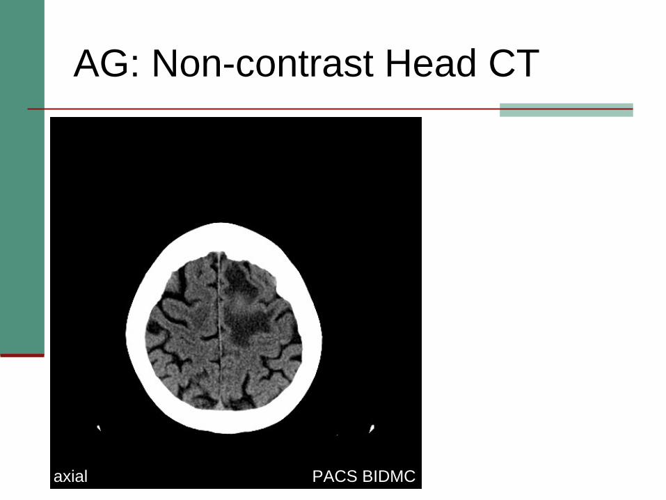
AG: Non-contrast Head CT
PACS BIDMCaxial
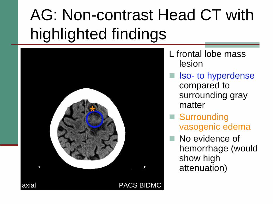
AG: Non-contrast Head CT with highlighted findings
PACS BIDMC
L frontal lobe mass lesionIso- to hyperdensecompared to surrounding gray matterSurrounding vasogenic edemaNo evidence of hemorrhage (would show high attenuation)
*
axial
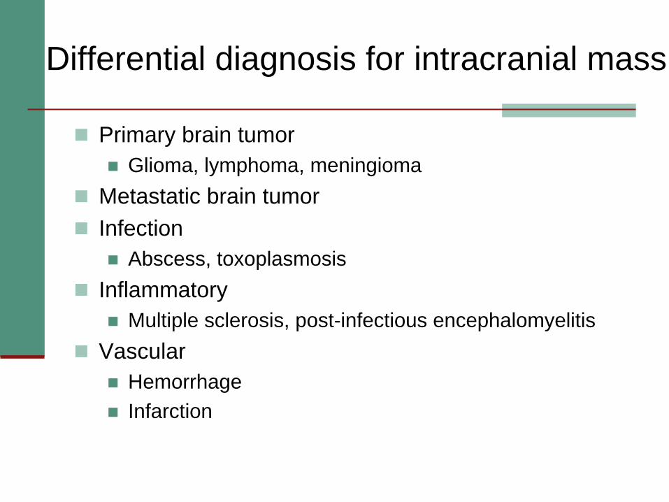
Differential diagnosis for intracranial mass
Primary brain tumorGlioma, lymphoma, meningioma
Metastatic brain tumorInfection
Abscess, toxoplasmosisInflammatory
Multiple sclerosis, post-infectious encephalomyelitisVascular
HemorrhageInfarction
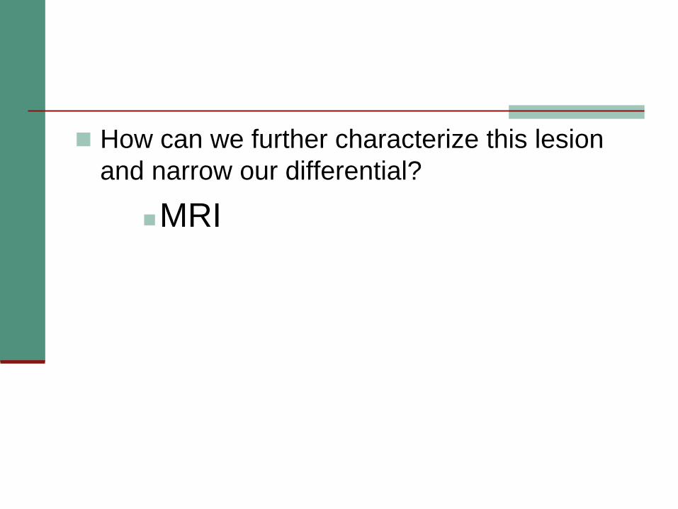
How can we further characterize this lesion and narrow our differential?
MRI
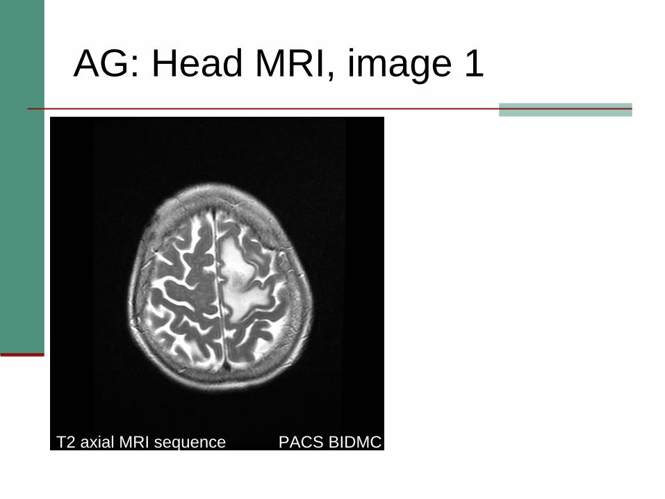
AG: Head MRI, image 1
PACS BIDMCT2 axial MRI sequence
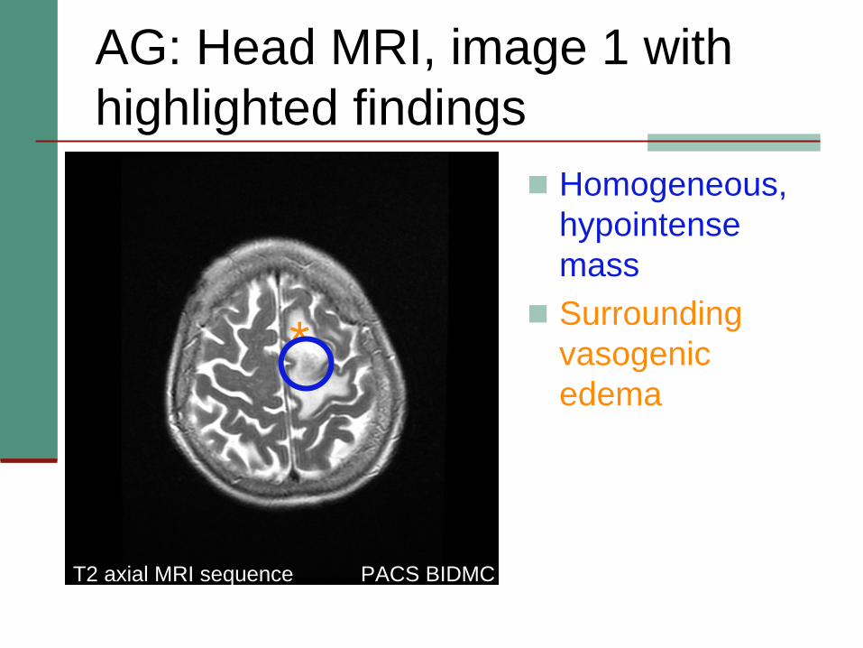
AG: Head MRI, image 1 with highlighted findings
Homogeneous, hypointensemassSurrounding vasogenicedema
PACS BIDMCT2 axial MRI sequence
*
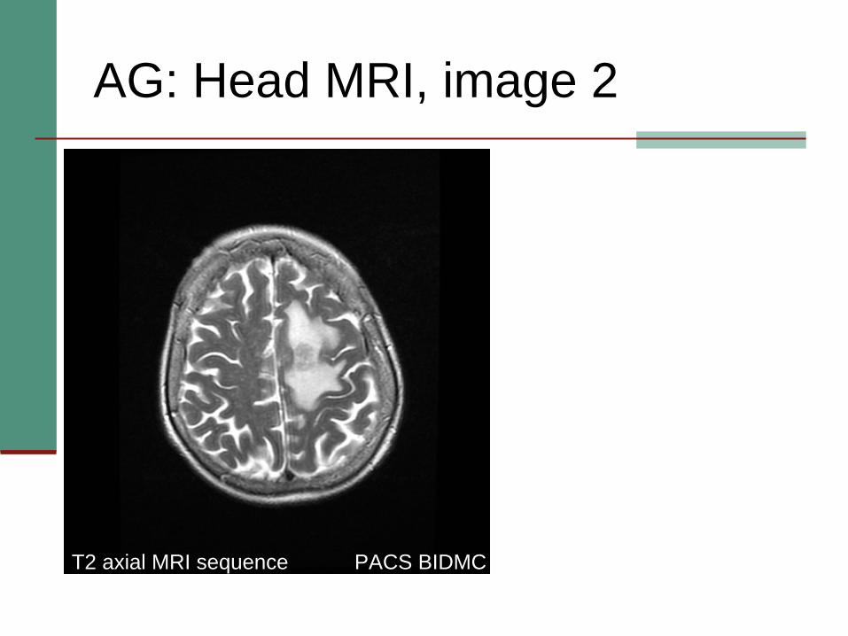
AG: Head MRI, image 2
PACS BIDMCT2 axial MRI sequence
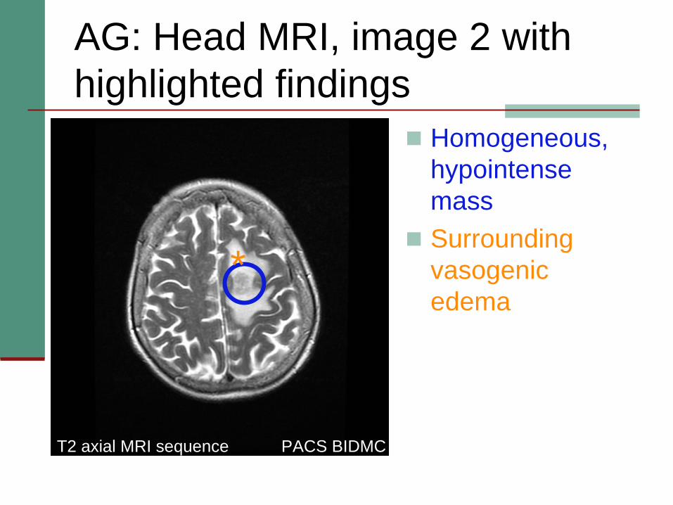
AG: Head MRI, image 2 with highlighted findings
PACS BIDMCT2 axial MRI sequence
Homogeneous, hypointensemassSurrounding vasogenicedema
*
AG: Head MRI findings from T2 sequences
Homogeneous, hypointense mass involving left cerebral hemisphere (frontal lobe)Surrounding vasogenic edema
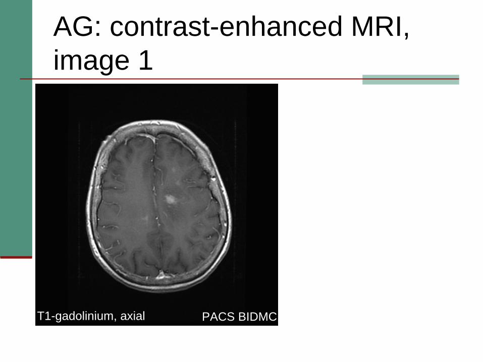
AG: contrast-enhanced MRI, image 1
PACS BIDMCT1-gadolinium, axial
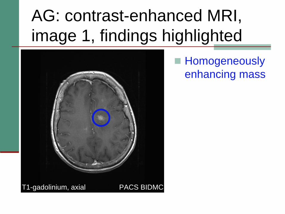
AG: contrast-enhanced MRI, image 1, findings highlighted
PACS BIDMCT1-gadolinium, axial
Homogeneously enhancing mass
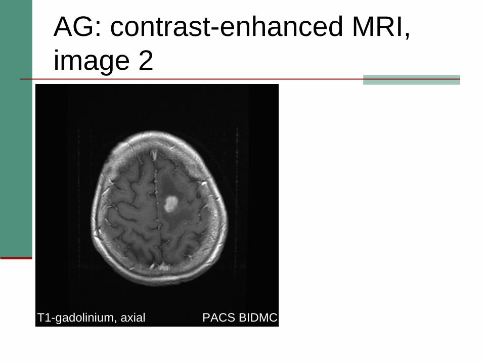
AG: contrast-enhanced MRI, image 2
PACS BIDMC
PACS BIDMCT1-gadolinium, axial
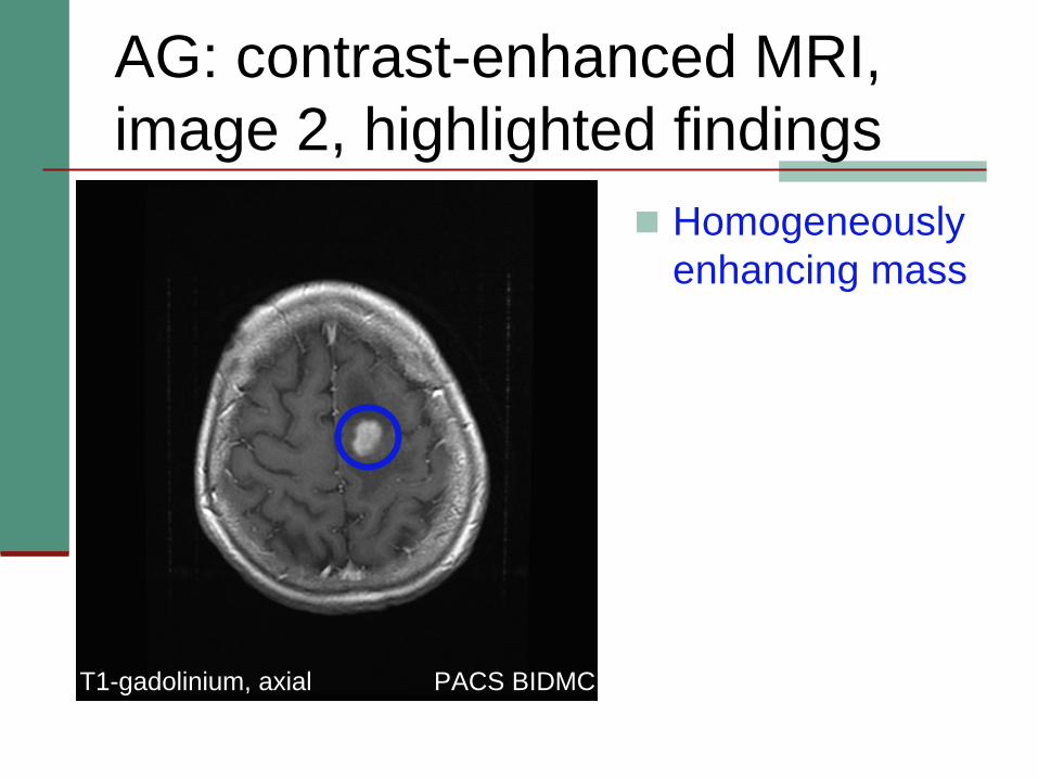
AG: contrast-enhanced MRI, image 2, highlighted findings
PACS BIDMC
Homogeneously enhancing mass
PACS BIDMCT1-gadolinium, axial

AG: contrast-enhanced MRI, image 3
PACS BIDMC
PACS BIDMCT1-gadolinium, axial
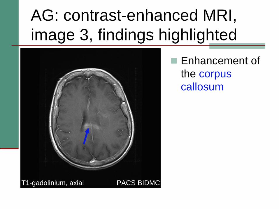
AG: contrast-enhanced MRI, image 3, findings highlighted
Enhancement of the corpus callosum
PACS BIDMC
PACS BIDMCT1-gadolinium, axial
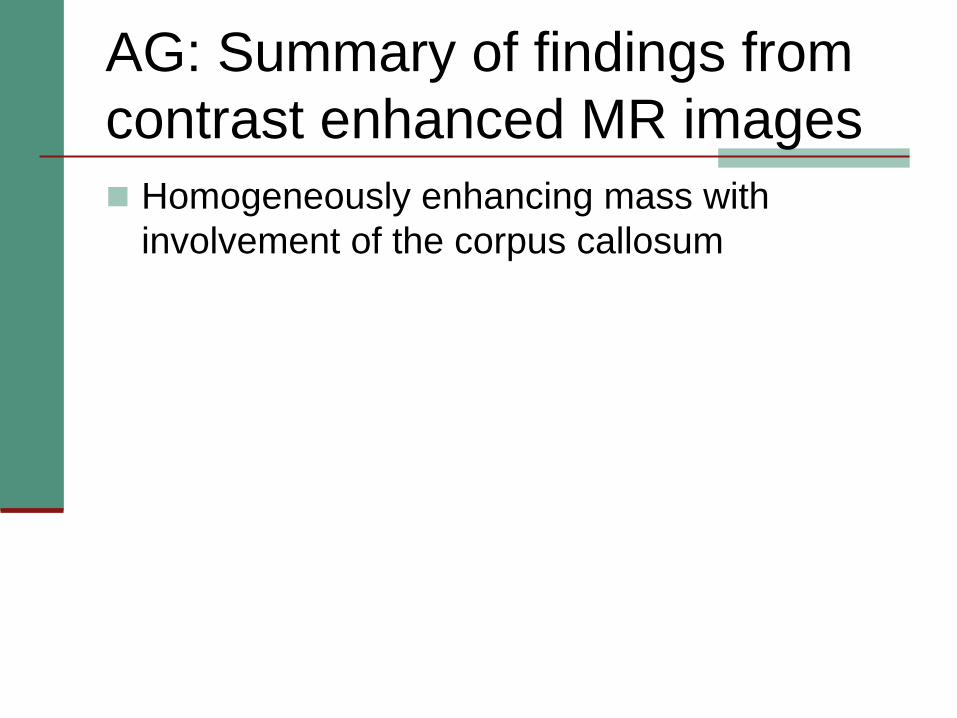
AG: Summary of findings from contrast enhanced MR images
Homogeneously enhancing mass with involvement of the corpus callosum
PACS BIDMC
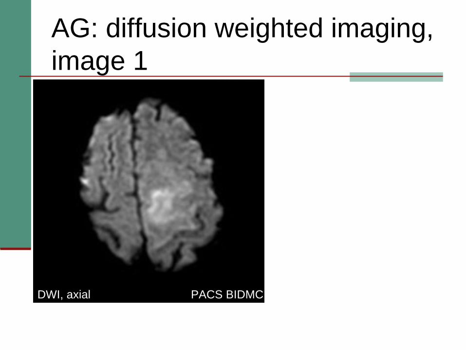
AG: diffusion weighted imaging, image 1
PACS BIDMC
PACS BIDMCDWI, axial
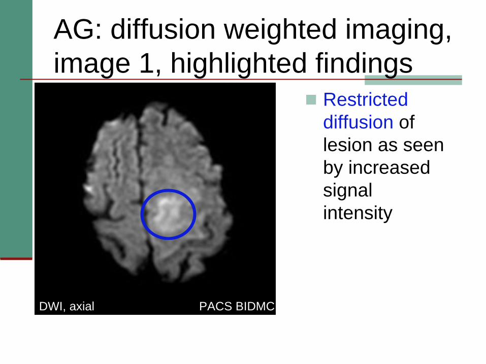
AG: diffusion weighted imaging, image 1, highlighted findings
Restricted diffusion of lesion as seen by increased signal intensity
PACS BIDMC
PACS BIDMCDWI, axial
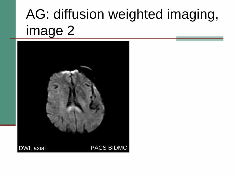
AG: diffusion weighted imaging, image 2
PACS BIDMC
PACS BIDMCDWI, axial
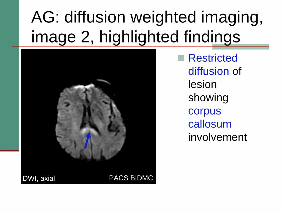
AG: diffusion weighted imaging, image 2, highlighted findings
Restricted diffusion of lesion showing corpus callosuminvolvement
PACS BIDMC
PACS BIDMCDWI, axial

Summary of AG’s radiographic findings
CT: isodense to hyperdense mass lesionMRI:
T2: homogeneous, hypointense signal with vasogenicedemaContrast enhanced: homogeneously enhancing lesion with involvement of corpus callosumDWI: restricted diffusion of lesion with corpus callosuminvolvement
Does this now help us narrow our original differential?
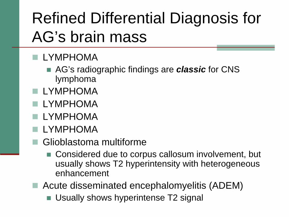
Refined Differential Diagnosis for AG’s brain mass
LYMPHOMA AG’s radiographic findings are classic for CNS lymphoma
LYMPHOMALYMPHOMALYMPHOMALYMPHOMAGlioblastoma multiforme
Considered due to corpus callosum involvement, but usually shows T2 hyperintensity with heterogeneous enhancement
Acute disseminated encephalomyelitis (ADEM)Usually shows hyperintense T2 signal
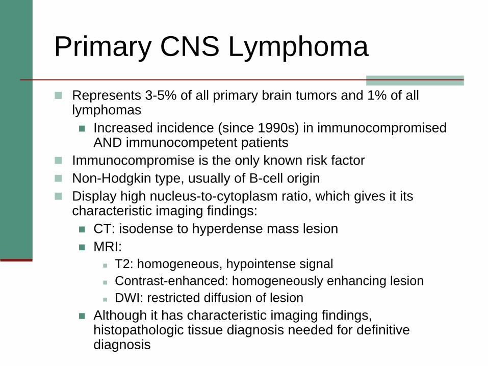
Primary CNS LymphomaRepresents 3-5% of all primary brain tumors and 1% of all lymphomas
Increased incidence (since 1990s) in immunocompromisedAND immunocompetent patients
Immunocompromise is the only known risk factorNon-Hodgkin type, usually of B-cell originDisplay high nucleus-to-cytoplasm ratio, which gives it its characteristic imaging findings:
CT: isodense to hyperdense mass lesionMRI:
T2: homogeneous, hypointense signalContrast-enhanced: homogeneously enhancing lesionDWI: restricted diffusion of lesion
Although it has characteristic imaging findings, histopathologic tissue diagnosis needed for definitive diagnosis
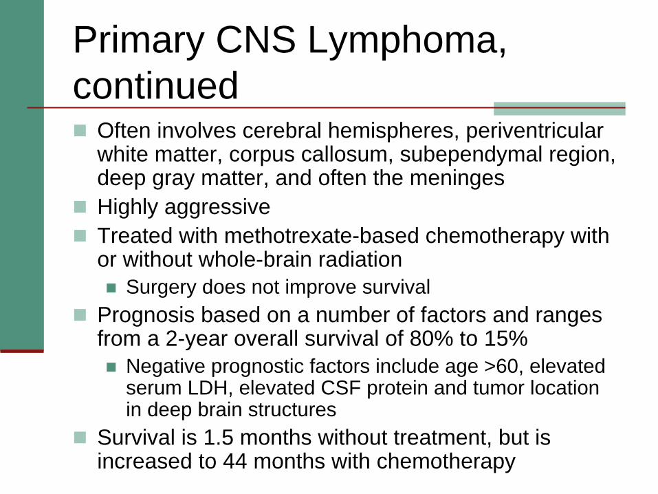
Primary CNS Lymphoma, continued
Often involves cerebral hemispheres, periventricularwhite matter, corpus callosum, subependymal region, deep gray matter, and often the meningesHighly aggressiveTreated with methotrexate-based chemotherapy with or without whole-brain radiation
Surgery does not improve survival Prognosis based on a number of factors and ranges from a 2-year overall survival of 80% to 15%
Negative prognostic factors include age >60, elevated serum LDH, elevated CSF protein and tumor location in deep brain structures
Survival is 1.5 months without treatment, but is increased to 44 months with chemotherapy
Role of imaging in further diagnosis of CNS lymphoma
Stereotactic needle biopsyOverlay MRI with intra-operative CT using software to determine the coordinates for precise needle biopsyThis is done because it is frequently difficult accurately image the extent of lesion on CT
QuickTime™ and a decompressor
are needed to see this picture.
http://www.elekta.com/assets/stereotactic_neurosurgery/images/Biopsy application + Arc with counter scale HIGH RES.jpg
Back to AG: tissue diagnosis
Primary CNS lymphoma was confirmed from tissue analysis taken by stereotactic biopsyShe was referred to a neuro-oncologist for further care management
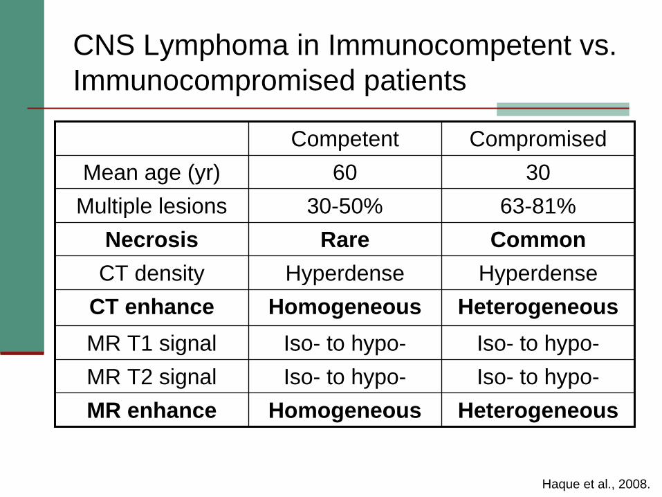
CNS Lymphoma in Immunocompetent vs. Immunocompromised patients
Competent CompromisedMean age (yr) 60 30
Multiple lesions 30-50% 63-81%Necrosis Rare Common
CT density Hyperdense HyperdenseCT enhance Homogeneous HeterogeneousMR T1 signal Iso- to hypo- Iso- to hypo-MR T2 signal Iso- to hypo- Iso- to hypo-MR enhance Homogeneous Heterogeneous
Haque et al., 2008.
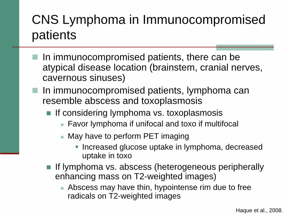
CNS Lymphoma in Immunocompromised patients
In immunocompromised patients, there can be atypical disease location (brainstem, cranial nerves, cavernous sinuses)In immunocompromised patients, lymphoma can resemble abscess and toxoplasmosis
If considering lymphoma vs. toxoplasmosisFavor lymphoma if unifocal and toxo if multifocalMay have to perform PET imaging
Increased glucose uptake in lymphoma, decreased uptake in toxo
If lymphoma vs. abscess (heterogeneous peripherally enhancing mass on T2-weighted images)
Abscess may have thin, hypointense rim due to free radicals on T2-weighted images
Haque et al., 2008.
Let’s review the common findings of primary CNS lymphoma again, using a companion patient, LM
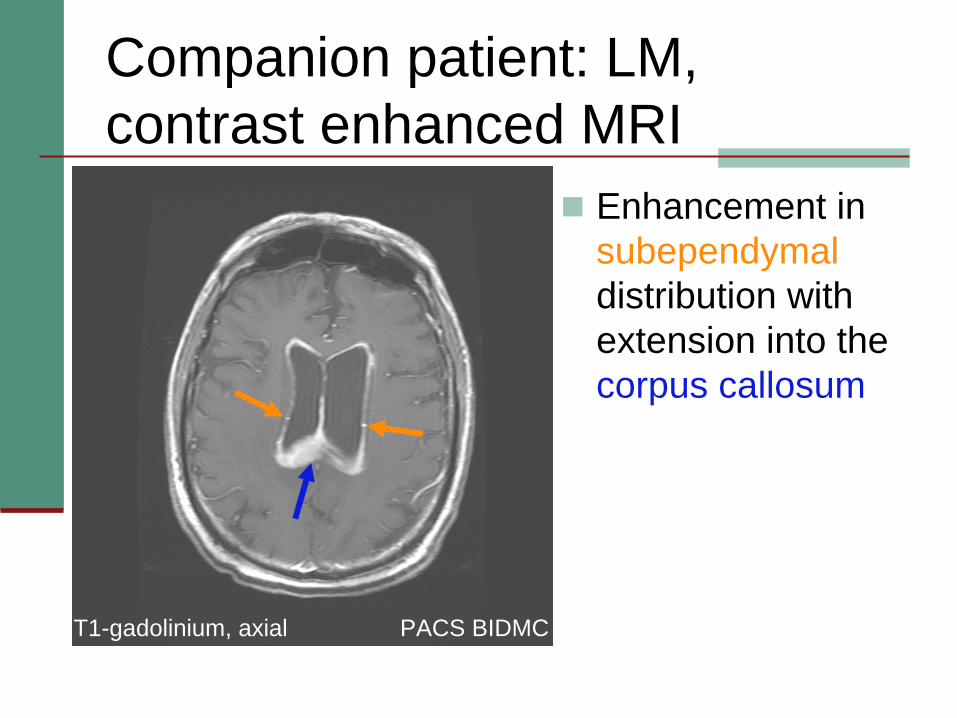
Companion patient: LM, contrast enhanced MRI
Enhancement in subependymaldistribution with extension into the corpus callosum
PACS BIDMC PACS BIDMCT1-gadolinium, axial
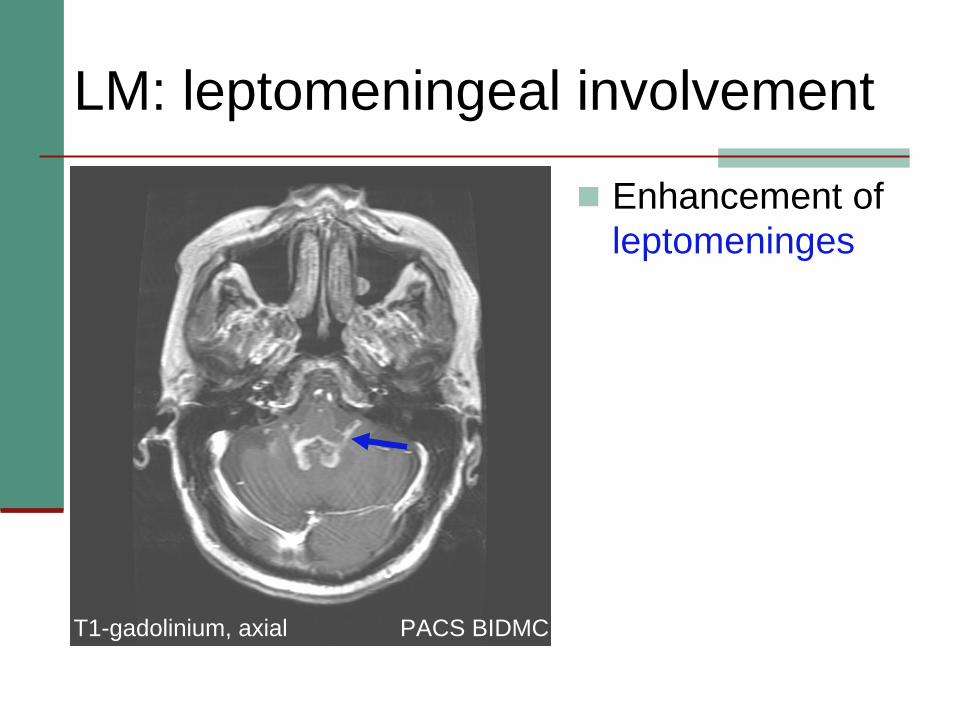
LM: leptomeningeal involvement
Enhancement of leptomeninges
PACS BIDMC
PACS BIDMCT1-gadolinium, axial
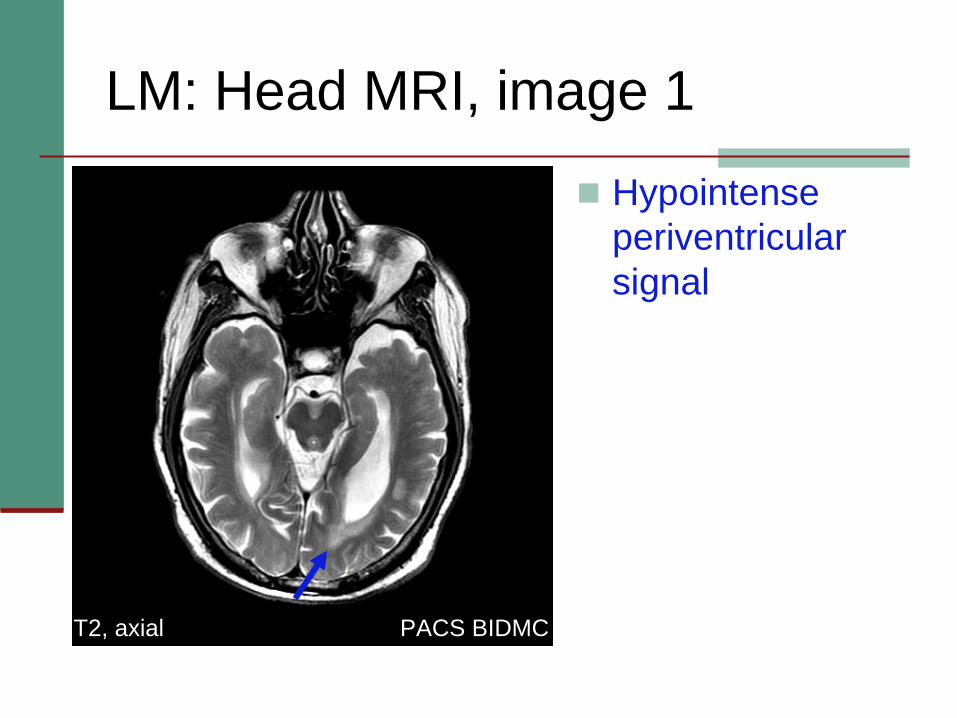
LM: Head MRI, image 1
Hypointenseperiventricularsignal
PACS BIDMC
PACS BIDMCT2, axial
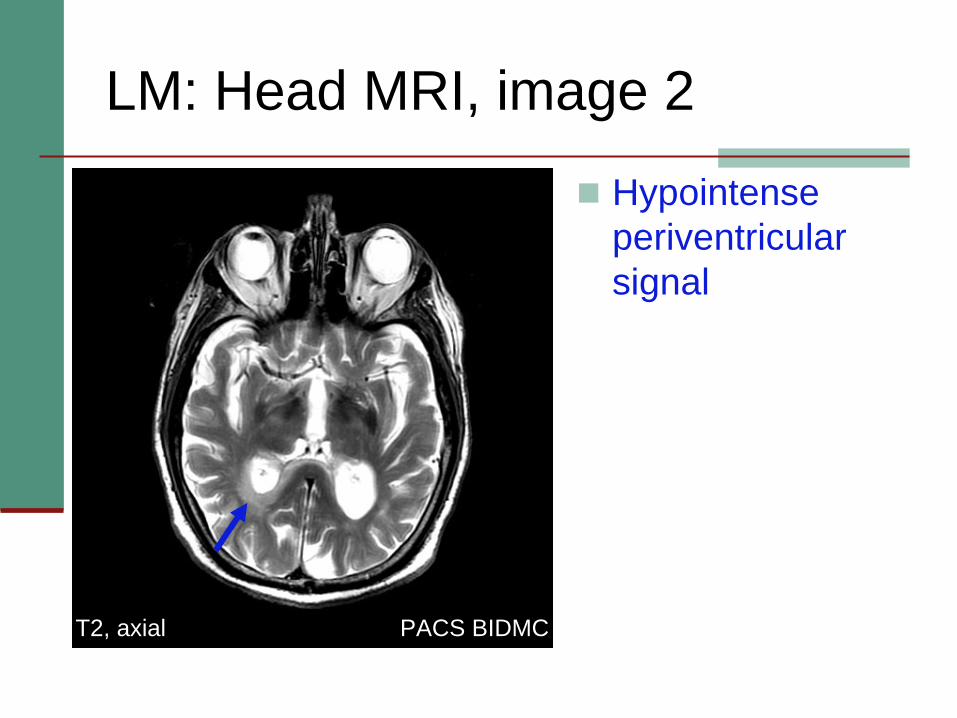
LM: Head MRI, image 2
Hypointenseperiventricularsignal
PACS BIDMC
PACS BIDMCT2, axial
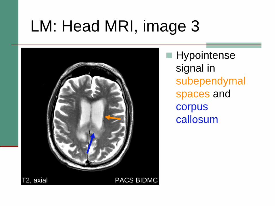
LM: Head MRI, image 3
Hypointensesignal in subependymalspaces andcorpus callosum
PACS BIDMC
PACS BIDMCT2, axial
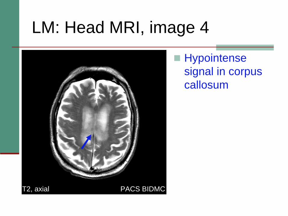
LM: Head MRI, image 4
Hypointensesignal in corpus callosum
PACS BIDMCT2, axial
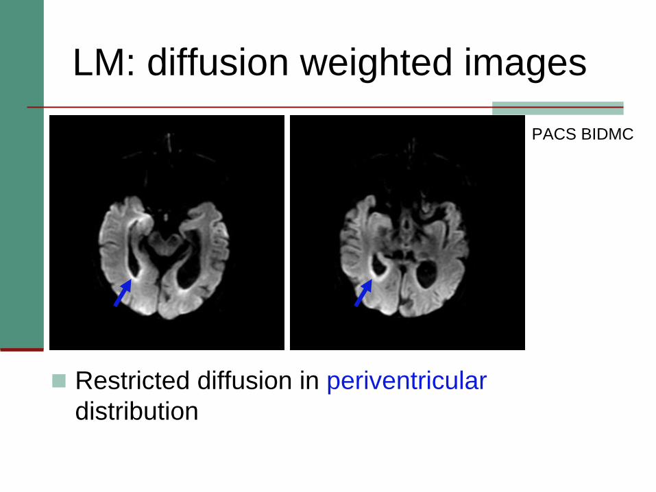
LM: diffusion weighted images
Restricted diffusion in periventriculardistribution
PACS BIDMC
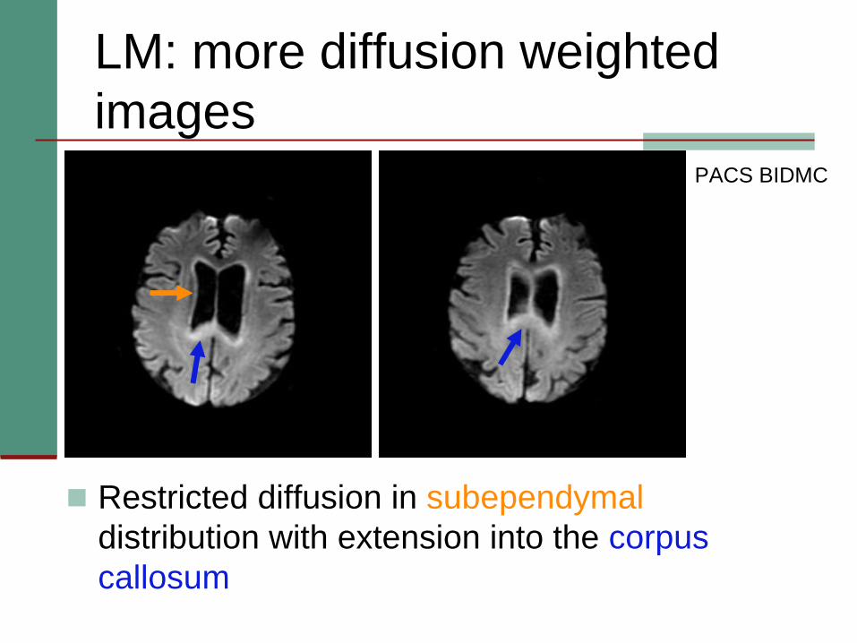
LM: more diffusion weighted images
Restricted diffusion in subependymaldistribution with extension into the corpus callosum
PACS BIDMC
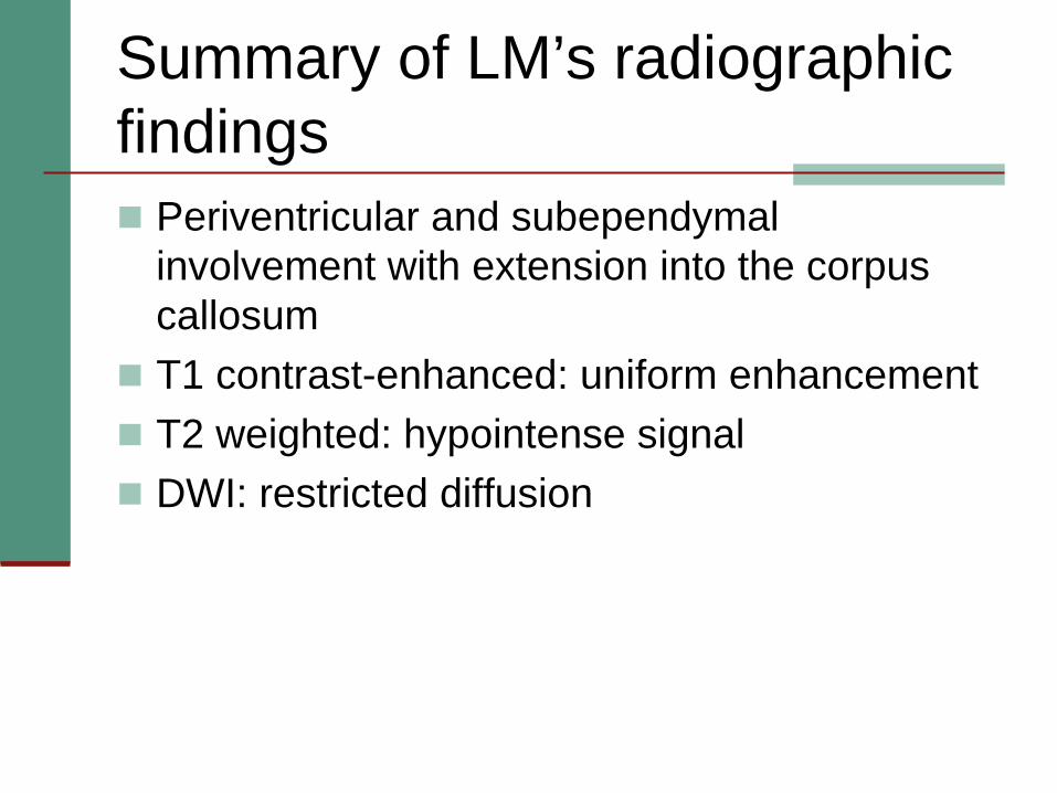
Summary of LM’s radiographic findings
Periventricular and subependymalinvolvement with extension into the corpus callosumT1 contrast-enhanced: uniform enhancementT2 weighted: hypointense signalDWI: restricted diffusion

Review of the radiologic features of primary CNS lymphoma
CT: isodense to hyperdense lesionMRI:
T2 weighted: homogeneous, hypointense signalCan be heterogeneous if patient is immunocompromised
Contrast enhanced T1: homogeneously enhancing lesionDWI: restricted diffusion of lesion
Typically involves cerebral hemispheres, corpus callosum, periventricular white matter, subependymalregion
Atypical locations in immunocompromised patients
Summary
Discussed the differential diagnosis of brain masses Familiarized with the common imaging features of primary CNS lymphoma on both CT and MRIDiscussed differences in imaging features of primary CNS lymphoma between immunocompetent and immunocompromisedpatientsGained an understanding for the role of imaging in primary CNS lymphoma

Many Thanks!
Dr. Gillian LiebermanDr. Gul MoonisDr. Iva PetkovskaMaria Levantakis
ReferencesBatchelor T and Loeffler J. (2006). Primary CNS lymphoma. Jour
Clin Onc 24, 1281-1288.Haque S, Law M, Abrey LE, and Young RJ. (2008). Imaging of
lymphoma of the central nervous system, spine and orbit. Radiol Clin North Am 46, 339-361, ix.
Patel MR, and Tse V. (2004). Diagnosis and staging of brain tumors. Semin Roentgenol 39, 347-360.
Hochberg FH, Batchelor T, and Loeffler JS. “Clinical presentation, pathologic features and diagnosis of primary central nervous system lymphoma.” Up To Date Online. 2008. 13 Dec 2009. http://utdol.com/online/content/topic.do?topicKey=lymphoma/12 708&selectedTitle=1%7E39&source=search_result.
Wippold FJ, et al. “ACR appropriateness criteria: Focal neurologic deficit.” ACR.org. 2008. 13 Dec 2009. http://www.acr.org/SecondaryMainMenuCategories/quality_safet y/app_criteria/pdf/ExpertPanelonNeurologicImaging/FocalNeurol ogicDeficitDoc4.aspx