Imaging features in pulmonary and extra-pulmonary · PDF fileImaging features in pulmonary and...
33
Page 1 of 33 Imaging features in pulmonary and extra-pulmonary tuberculosis Poster No.: C-0985 Congress: ECR 2011 Type: Educational Exhibit Authors: P. A. Bonaffini , D. Ippolito, A. C. Cadonici, C. Capraro, L. Monguzzi, S. Sironi; Monza (MB)/IT Keywords: Infection, Education, MR, CT, Neuroradiology brain, Lung, Abdomen, Cavitation, Abscess DOI: 10.1594/ecr2011/C-0985 Any information contained in this pdf file is automatically generated from digital material submitted to EPOS by third parties in the form of scientific presentations. References to any names, marks, products, or services of third parties or hypertext links to third- party sites or information are provided solely as a convenience to you and do not in any way constitute or imply ECR's endorsement, sponsorship or recommendation of the third party, information, product or service. ECR is not responsible for the content of these pages and does not make any representations regarding the content or accuracy of material in this file. As per copyright regulations, any unauthorised use of the material or parts thereof as well as commercial reproduction or multiple distribution by any traditional or electronically based reproduction/publication method ist strictly prohibited. You agree to defend, indemnify, and hold ECR harmless from and against any and all claims, damages, costs, and expenses, including attorneys' fees, arising from or related to your use of these pages. Please note: Links to movies, ppt slideshows and any other multimedia files are not available in the pdf version of presentations. www.myESR.org
Transcript of Imaging features in pulmonary and extra-pulmonary · PDF fileImaging features in pulmonary and...

Page 1 of 33
Imaging features in pulmonary and extra-pulmonarytuberculosis
Poster No.: C-0985
Congress: ECR 2011
Type: Educational Exhibit
Authors: P. A. Bonaffini, D. Ippolito, A. C. Cadonici, C. Capraro, L.Monguzzi, S. Sironi; Monza (MB)/IT
Keywords: Infection, Education, MR, CT, Neuroradiology brain, Lung,Abdomen, Cavitation, Abscess
DOI: 10.1594/ecr2011/C-0985
Any information contained in this pdf file is automatically generated from digital materialsubmitted to EPOS by third parties in the form of scientific presentations. Referencesto any names, marks, products, or services of third parties or hypertext links to third-party sites or information are provided solely as a convenience to you and do not inany way constitute or imply ECR's endorsement, sponsorship or recommendation of thethird party, information, product or service. ECR is not responsible for the content ofthese pages and does not make any representations regarding the content or accuracyof material in this file.As per copyright regulations, any unauthorised use of the material or parts thereof aswell as commercial reproduction or multiple distribution by any traditional or electronicallybased reproduction/publication method ist strictly prohibited.You agree to defend, indemnify, and hold ECR harmless from and against any and allclaims, damages, costs, and expenses, including attorneys' fees, arising from or relatedto your use of these pages.Please note: Links to movies, ppt slideshows and any other multimedia files are notavailable in the pdf version of presentations.www.myESR.org

Page 2 of 33
Learning objectives
To illustrate the CT and MRI imaging findings in pulmonary and extra-pulmonarytuberculosis, with emphasis to nodal, genitourinary, adrenal, skeletal and nervouslocalizations.
Background
INTRODUCTION
Tuberculosis (TBC) is an airborne communicable disease caused by Mycobacteriumtuberculosis, transmitted by patients with pulmonary TBC.
Infection occurs more commonly in adults (25-39 and 60-75 years old patients).
Population groups with increased risk: immunocompromised individuals (AIDS,lymphoma, leukemia), diabetics patients, children and the aged, alcoholics, the poor,immigrants from third-world countries, prisoners, nursing home residents, health careworkers and the homeless.
Up until the mid 1980s, there was a steady decline in the prevalence of tuberculosis.Since that time, however, there has been a resurgence of tuberculosis. His increasehas been seen not only in Africa and Asia, but also in Europe and so TBC remains animportant cause of morbidity and mortality worldwide.
This resurgence is due to the acquired immunodeficiency syndrome (AIDS) epidemicand the increasing number of drug-resistant strains of Mycobacterium tuberculosis(Multidrug-resistant Tuberculosis or MDR).
- MDR
MDR tuberculosis is no more infective than normal tuberculosis. However, it is amore serious infection, requiring prolonged administration of more toxic second-line drugs, associated with higher morbidity and mortality rates. Patients also remaininfectious for a longer period once treatment has been started.
- IMMUNOCOMPROMISED PATIENTS

Page 3 of 33
They have a significantly higher prevalence of tuberculosis than does the generalpopulation and are also more likely to be infected with MDR tuberculosis. Thepattern of disease is different in immunocompromised patients, who have a higherprevalence of extra-pulmonary involvement.
SUBTYPE OF TUBERCULOSIS
Tuberculosis is usually confined clinically to the respiratory system (pulmonary TBC,80% of cases). However, it can affect any organ system (extra-pulmonary TBC),particularly in immunocompromised individuals.
Pulmonary tuberculosis is classically divided into primary and postprimary(reactivation) tuberculosis.
In extra-pulmonary TBC the more common sites involved are: bones, genitourinarysystem and central nervous system. Other sites involved are, in order of frequency:abdomen, heart (cardiac TBC), eye (ocular TBC)
RADIOLOGIST'S ROLE
Tuberculosis demonstrates a variety of clinical and radiologic features dependingon the organ site involved and has a known propensity for dissemination from itsprimary site. Thus, tuberculosis can mimic a number of other disease entities, and itis important to be familiar with the various radiologic features of tuberculosis to ensureearly, accurate diagnosis.
Furthermore, there is a considerable overlap in the radiologic manifestations of bothprimary and postprimary pulmonary TBC. However, confirming the diagnosis is moreimportant than identifying the subtype, because this allows initiation of a correctclinical management.
Imaging findings OR Procedure details
PART I - PULMONARY TBC
PRIMARY PULMONARY TBC

Page 4 of 33
It's seen in patients not previously exposed to M. tuberculosis and it's most commonin infants and children. It has the highest prevalence in children under 5 years of age.
Although primary tuberculosis is the most common form of pulmonary TBC in infants andchildren, it has been increasingly encountered in adult patients and now accounts for23%-34% of all adult cases of tuberculosis.
Chest radiography (FIG. 1) remains the mainstay of diagnosis; however, normalradiographic findings may be seen in up to 15% of patients with proved tuberculosis.High-resolution CT (FIG. 2) is more sensitive.
Primary tuberculosis typically manifests radiologically with these main entities:
• parenchymal disease• lymphadenopathy• pleural effusion (one-fourth of patients, usually unilateral; possible
calcification), PNX (FIG. 1)• miliary disease (1-7%, overall in elderly, infants and immunocompromised)• atelectasis (either lobar or segmental, frequently seen in children under 2
years of age)
Typical presentation # "primary complex":
a) parenchymal disease (FIG. 1 and 2): dense, homogeneous parenchymalconsolidation that affects the areas of greatest ventilation # the most common sitesare the middle lobe, the lower lobes, and the anterior segment of the upper lobes; itsappearance is often indistinguishable from that of bacterial pneumonia.
b) lymphadenopathy (up to 96% of children and 43% of adults): typically unilateral andright sided, involving the hilum and right paratracheal region, although it is bilateral inabout one-third of cases.
Evolution of primary complex:
• in approximately two-thirds of cases, the parenchymal focus resolveswithout sequelae (it can take up to 2 years);
• radiologic scar that can calcify in up to 15% of cases, entity that is known asa Ghon focus;
• persistent masslike opacities called tuberculomas, that can cavitate(approximately 9% of cases) (FIG. 2)
LYMPHADENOPATHY (FIG. 3)

Page 5 of 33
Although lymphadenopathy is usually associated with other manifestations oftuberculosis, it can be the sole radiographic feature, a finding that is more common ininfants and decreases in frequency with age.
Nodes involved often calcify (6 month/>). The combination of calcified hilar nodes anda Ghon focus is called a Ranke complex and is suggestive of previous tuberculosis,although it can also result from histoplasmosis.
CT is more sensitive than chest radiography for assessing lymphadenopathy.
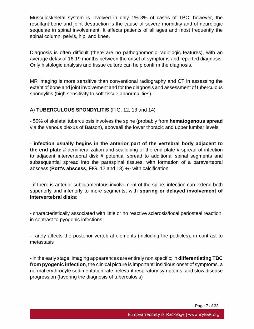
Any nodes greater than 2 cm in diameter generally have a low-attenuation centersecondary to necrosis at CT (FIG. 3). This finding is highly suggestive of active disease,in the appropriate clinical setting.
The radiologic differential diagnosis for tuberculous lymphadenopathy includesmetastases (FIG. 4) and histoplasmosis in endemic areas.
POST-PRIMARY TBC
The term post-primary tuberculosis is generally used to refer to both re-infectionwith (30-60%) and reactivation of tuberculosis (40-70%) in patients withimmunodepression.
It remains primarily a disease of adolescence and adulthood (40-60 years old) and itoccurs in patients previously sensitized to M. tuberculosis.
Primary tuberculosis is usually self-limiting, whereas post-primary tuberculosis isprogressive, with cavitation as its hallmark, resulting in hematogenous disseminationof the disease as well as disease spread throughout the lungs.
The features of primary and post-primary tuberculosis may overlap.However, the distinguishing features of post-primary tuberculosis include:
• predilection for the upper lobes• absence of lymphadenopathy• presence of cavitation
CT FINDINGS

Page 6 of 33
Parenchymal disease (the earliest finding, FIG. 7, 8 and 9): patchy, poorly definedconsolidation, particularly in the apical and posterior segments of the upper lobes,bilateral in one-third of cases.
Cavitation (FIG. 9 and 10): the hallmark of post-primary tuberculosis, that affects about50% of patients and indicates a high likelihood of activity. The cavities typically have thick,irregular walls, which become smooth and thin with successful treatment. Cavities areusually multiple and occur within areas of consolidation.
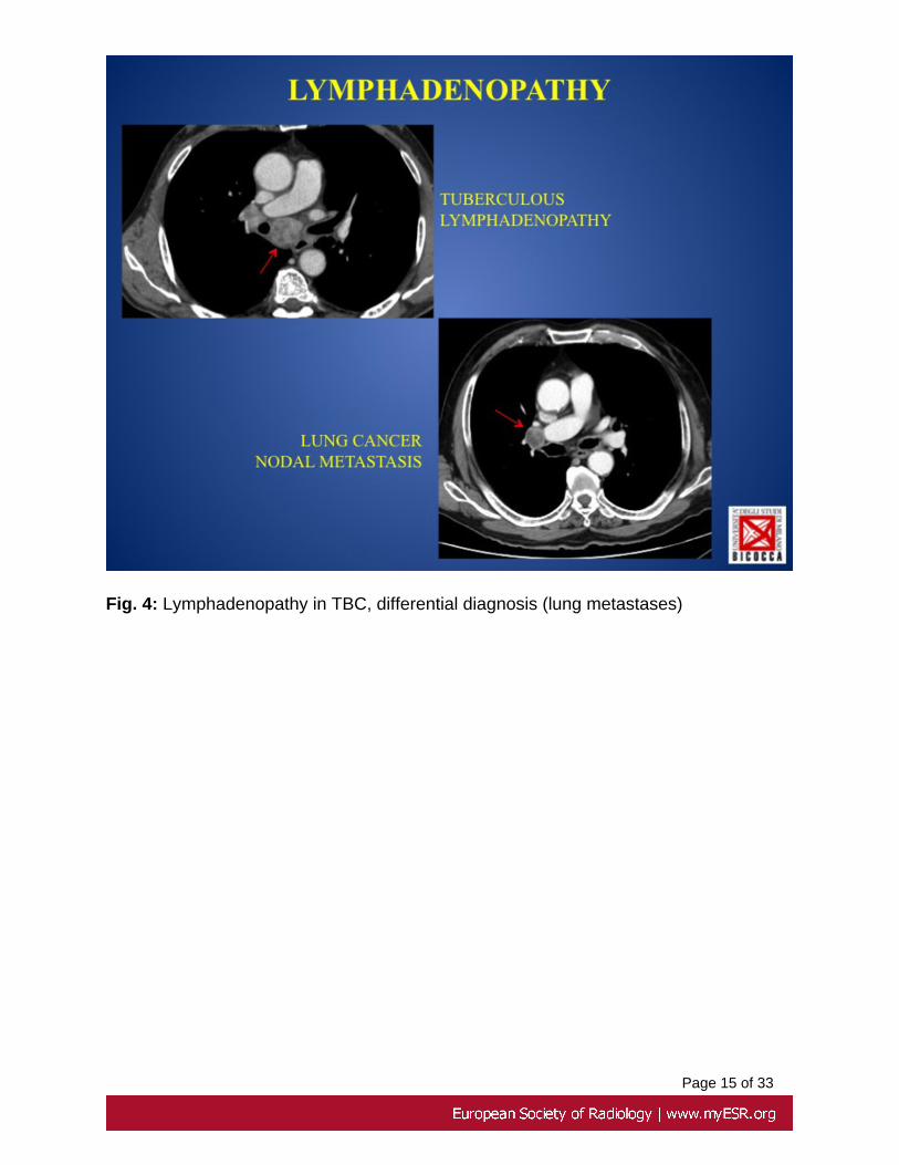
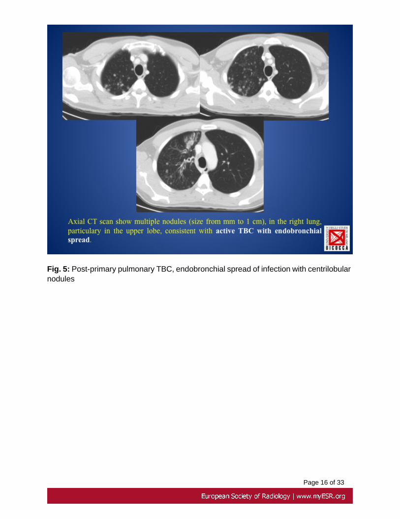
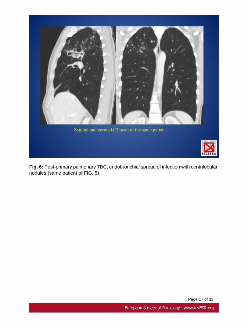
Centrilobular nodules and branching centrilobular areas of increased opacity (FIG5, 6 and 7): if there is airway disease and, in particular, endobronchial spread ofinfection (most common complication of TBC cavitation), "tree-in-bud" opacities maydevelop; this is characteristic but non pathognomonic for active TBC (can also beseen with viral, fungal, and parasitic infections and also in allergic bronchopulmonaryaspergillosis).
Airway involvement: characterized by bronchial stenosis, leading to lobar collapse andby traction bronchiectasis.
Pleural effusion (18%, usually with parenchymal disease; possible calcification).
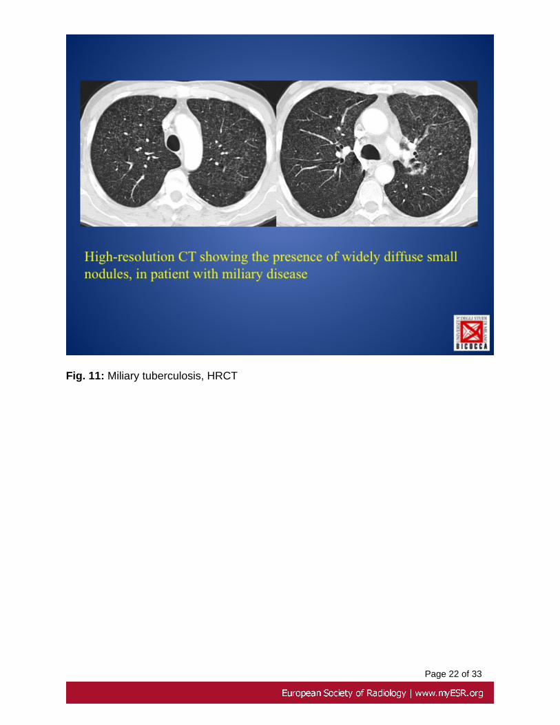
MILIARY DISEASE
Miliary tuberculosis occurs in 2% to 6% of primary TBC and also occur in post-primaryTBC, when the host's defence mechanisms are overwhelmed.
High-resolution CT (FIG. 11) is more sensitive than conventional radiography and showin 85% of cases a mixture of both sharply and poorly defined nodules of 1 to 2 mm,widely disseminated throughout the lungs (haematogenous spread of infection),with a slight lower lobe predominance.
The nodules usually resolve within 2-6 months with treatment, without scarring orcalcification.
PART II - EXTRA-PULMONARY TBC
PART IIA - MUSCULOSKELETAL TUBERCULOSIS
Page 7 of 33
Musculoskeletal system is involved in only 1%-3% of cases of TBC; however, theresultant bone and joint destruction is the cause of severe morbidity and of neurologicsequelae in spinal involvement. It affects patients of all ages and most frequently thespinal column, pelvis, hip, and knee.
Diagnosis is often difficult (there are no pathognomonic radiologic features), with anaverage delay of 16-19 months between the onset of symptoms and reported diagnosis.Only histologic analysis and tissue culture can help confirm the diagnosis.
MR imaging is more sensitive than conventional radiography and CT in assessing theextent of bone and joint involvement and for the diagnosis and assessment of tuberculousspondylitis (high sensitivity to soft-tissue abnormalities).
A) TUBERCULOUS SPONDYLITIS (FIG. 12, 13 and 14)
- 50% of skeletal tuberculosis involves the spine (probably from hematogenous spreadvia the venous plexus of Batson), aboveall the lower thoracic and upper lumbar levels.
- infection usually begins in the anterior part of the vertebral body adjacent tothe end plate # demineralization and scalloping of the end plate # spread of infectionto adjacent intervertebral disk # potential spread to additional spinal segments andsubsequential spread into the paraspinal tissues, with formation of a paravertebralabscess (Pott's abscess, FIG. 12 and 13) +/- with calcification;
- if there is anterior subligamentous involvement of the spine, infection can extend bothsuperiorly and inferiorly to more segments, with sparing or delayed involvement ofintervertebral disks;
- characteristically associated with little or no reactive sclerosis/local periosteal reaction,in contrast to pyogenic infections;
- rarely affects the posterior vertebral elements (including the pedicles), in contrast tometastasis
- in the early stage, imaging appearances are entirely non specific; in differentiating TBCfrom pyogenic infection, the clinical picture is important: insidious onset of symptoms, anormal erythrocyte sedimentation rate, relevant respiratory symptoms, and slow diseaseprogression (favoring the diagnosis of tuberculosis)

Page 8 of 33
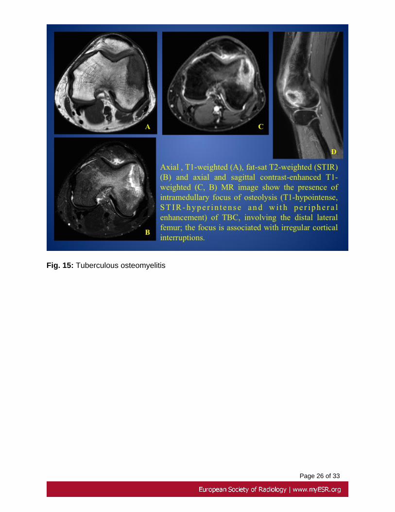
B) TUBERCULOUS OSTEMYELITIS (EXTRASPINAL TBC OSTEOMYELITIS) (FIG.15)
- tuberculous osteomyelitis is usually hematogenous in origin and the isolated form inthe absence of associated tuberculous arthritis is relatively rare
- is most commonly seen in bones of the extremities (femur, tibia), including the smallbones of the hands and feet
- in long, tubular bones, often involves the epiphyses; in children metaphyseal foci caninvolve the growth plate (this feature differentiates tuberculosis from pyogenic infection)
- initial radiologic appearance is similar to that of other types of osteomyelitis and includesfoci of osteolysis with varying degrees of eburnation and periostitis.
- the diagnosis is usually made after considerable delay, and radiographic changes areseen at clinical presentation; in contrast, in pyogenic infection, radiographic changesoccur 2-3 weeks after presentation.
C) TUBERCULOUS ARTHRITIS (FIG. 16)
- may be secondary to direct invasion from an adjacent focus of tuberculousosteomyelitis or may result from hematogenous dissemination
- is typically monoarticular and primarily involves large weight-bearing joints (hip andknee)
- the imaging findings are similar to those of other infectious and inflammatory arthritidesand are, therefore, non specific; these findings include osteopenia, synovitis andother soft-tissue swellings, marginal erosions, and varying degrees of cartilagedestruction
- the end result is usually fibrous ankylosis of the joint
- the differential diagnosis includes pyogenic and fungal infections factors # favoringa diagnosis of TBC include insidious onset, minimal sclerosis, the relative absence ofperiosteal reaction and bone proliferation

Page 9 of 33
PART IIB - TUBERCULOSIS INVOLVING THE CNS
Most tuberculous infections of the central nervous system (neurotuberculosis, 5% ofpatients) are usually a result of hematogenous spread. It results in two relatedpathologic processes: tuberculous meningitis (the most common manifestation, acrossall age groups) and intracranial tuberculomas and abscesses. Its prevalence is greaterin immunocompromised patients.
A) MENINGEAL INVOLVEMENT (TUBERCULOUS MENINGITIS)
- typical radiographic finding, better seen at gadolinium-enhanced MR imaging than atCT # abnormal meningeal enhancement (FIG. 16)
- appearance non specific and with wide differential diagnosis: meningitis fromother infective agents, inflammatory diseases (rheumatoid arthritis and sarcoidosis) andneoplastic causes (both primary and secondary)
- most common complication # communicating hydrocephalus
B) PARENCHYMAL INVOLVEMENT (can exist in conjunction with tuberculousmeningitis)
- most common parenchymal lesion # tuberculoma (non-caseating granuloma),solitary, multiple, or miliary, anywhere within the brain parenchyma (above all withinfrontal and parietal lobes)
- it demonstrate homogeneous enhancement, with irregular wall of varying thicknessand in one-third with central calcification
- caseating tuberculoma with necrotic center demonstrate a ring enhancement (FIG.17) and usually have a variable amount of surrounding edema; abscesses can be similarin appearance
PART IIC - GENITOURINARY TUBERCULOSIS
TBC may involve the genitourinary tract as a secondary site following hematogenousdissemination from the lung. Genitourinary TBC is the most common clinicalmanifestation of extrapulmonary tuberculosis.
A) KIDNEYS (75% unilateral) (FIG. 18)
- the most common CT finding is renal calcification (50%)

Page 10 of 33
- renal parenchymal cavitation may be detected as irregular pools of contrast material
- "moth-eaten" abnormality of calix at urography, due to erosions
B) BLADDER (tuberculous cystitis)
It manifests as a shrunken bladder with wall thickening and reduced bladder capacity.
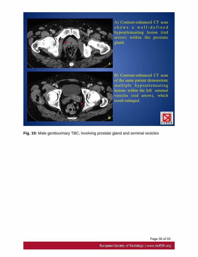
C) GENITALIA
- female: obstruction of the fallopian tubes (salpingitis, 94% cases) with multiple areasof constriction, and calcified lymph nodes in the adnexal region, endometrial adhesionswith deformity and obliteration of endometrial cavity
- male: involvement of the prostate gland or seminal vesicles (FIG. 19) may lead tonecrosis, calcification, caseation (hypoattenuating lesions at CT), and cavitation
PART IIIC - ABDOMINAL TUBERCULOSIS
The abdomen is the most common focus of extrapulmonary tuberculosis, with the solidviscera being affected more often than the gastrointestinal tract.Although abdominal tuberculosis is usually secondary to pulmonary tuberculosis,radiologic evaluation often shows no evidence of lung disease.CT is the mainstay for investigating possible abdominal tuberculosis.
A) LYMPHADENOPATHY
The most common manifestation (FIG. 20) of abdominal TBC (55-60%), with enlargednodes with hypoattenuating centers and hyperattenuating enhancing rims at CT(40-60% of cases), findings that are characteristic of, but not pathognomonic for, caseousnecrosis.
The mesenteric, omental, and peripancreatic lymph nodes are most commonly involved.
B) GASTROINTESTINAL TBC (rare)
It can occur elsewhere in the small bowel, but almost always involves ileocecal region(90% of cases), usually both the terminal ileum and the cecum. Rare localitations areesophagus, stomach and proximal small bowel. The most common CT finding (FIG. 21)is concentric mural thickening, with associated luminal narrowing with or withoutproximal dilatation.

Page 11 of 33
C) HEPATOSPLENIC TBC
- common in patients with disseminated disease, either miliary or macronodular
- micronodular/miliary involvement: seen in patients with miliary pulmonary TBC,characterized by innumerable 0.5-2.0-mm hypoattenuating nodules. The liver appearshyperechoic at US.
- macronodular involvement: uncommon, with liver or splenic enlargement andhypoattenuating lesions at CT, with irregular ill-defined margins and minimal butdefinite peripheral contrast enhancement (imaging appearances non specific andsimilar to those of multiple metastases and abscesses). At MR imaging, these lesionsare hypointense with T1-weighted sequences and hyperintense with T2-weightedsequences.
- hepatic tuberculomas eventually tend to calcify; the presence of calcified granulomasat CT in patients with known risk factors and in the absence of a known primary tumorshould raise suspicion for TBC
D) ADRENAL TBC
- it's seen in up to 6% of patients with active tuberculosis.
- bilateral adrenal involvement and an Addisonian type clinical picture
- CT signs: enlarged glands associated with large, hypoattenuating necrotic areas,with or without dotlike calcification
Images for this section:

Page 12 of 33
Fig. 1: Primary pulmonary TBC, chest X-Ray

Page 13 of 33
Fig. 2: Primary pulmonary TBC at CT

Page 14 of 33
Fig. 3: Lymphadenopathy in TBC
Page 15 of 33
Fig. 4: Lymphadenopathy in TBC, differential diagnosis (lung metastases)
Page 16 of 33
Fig. 5: Post-primary pulmonary TBC, endobronchial spread of infection with centrilobularnodules
Page 17 of 33
Fig. 6: Post-primary pulmonary TBC, endobronchial spread of infection with centrilobularnodules (same patient of FIG. 5)

Page 18 of 33
Fig. 7: Post-primary pulmonary TBC, parenchymal disease

Page 19 of 33
Fig. 8: Post-primary pulmonary TBC, parenchymal disease and endobronchial spread ofinfection with centrilobular nodules

Page 20 of 33
Fig. 9: Post-primary pulmonary TBC, parenchymal disease and cavitation

Page 21 of 33
Fig. 10: Post-primary pulmonary TBC, cavitation
Page 22 of 33
Fig. 11: Miliary tuberculosis, HRCT

Page 23 of 33
Fig. 12: Tuberculous spondylitis with Pott's abscess

Page 24 of 33
Fig. 13: Tuberculous spondylitis with Pott's abscess

Page 25 of 33
Fig. 14: Tuberculous spondylitis, MRI
Page 26 of 33
Fig. 15: Tuberculous osteomyelitis

Page 27 of 33
Fig. 16: CT and MRI features of tuberculous arthritis of left sacroiliac joint with abscess

Page 28 of 33
Fig. 17: MRI features of tuberculous infection of the central nervous system: tuberculousmeningitis and multiple abscesses

Page 29 of 33
Fig. 18: Genitourinary TBC: kidney's involvement
Page 30 of 33
Fig. 19: Male genitourinary TBC, involving prostate gland and seminal vesicles

Page 31 of 33
Fig. 20: Abdominal TBC with lymphadenopathy

Page 32 of 33
Fig. 21: Gastrointestinal TBC

Page 33 of 33
Conclusion
The infection of Mycobacterium tuberculosis had in the last years an evidentrecrudescence, due to HIV epidemic, to constant migrant population and to the spreadof drug resistant strains (MDR).
The two main pathologic form include pulmonary and extra-pulmonary infection. Bothhave a variety of clinical and radiologic features depending on the organ siteinvolved, also in the patients with decreased immunity.
The radiologist has an important role in the evaluation of the several manifestationsand sites of infection, keeping in mind that tuberculosis can mimic a number of otherdisease entities.
Personal Information
References
• Burrill J, Williams CJ , Bain G, Conder G, Hine AL, Misra RR.Tuberculosis: A Radiologic Review RadioGraphics 2007 Sep-Oct;27(5):1255-73
• Harisinghani MG, McLoud TC, Shepard JA, Ko JP, Shroff MM, MuellerPR. Tuberculosis from head to toe RadioGraphics 2000 Mar-Apr;20(2):449-7
• Lee JY, Lee KS, Jung KJ, Han J, Kwon OJ, Kim J, Kim TS. Pulmonarytuberculosis: CT and pathologic correlation J Comput Assist Tomogr. 2000Sep-Oct;24(5):691-8