
III Taller de Resonancia Magnética - unl.edu.ar · Santa Fe, Argentina, 31 de marzo - 1 de abril...
49
Transcript of III Taller de Resonancia Magnética - unl.edu.ar · Santa Fe, Argentina, 31 de marzo - 1 de abril...

Santa Fe, Argentina, 31 de marzo - 1 de abril de 2016
Page1
III Taller de Resonancia Magnética
NMR and EPR at the Forefront of Research
Comité Organizador
Carlos Brondino (FBCB, UNL/CONICET)
Ana M. Gennaro (FBCB, UNL e IFIS Litoral, UNL/CONICET)
Roberto R. Gil (Carnegie Mellon University, USA)
Pablo González (FBCB, UNL/ CONICET)
Nicolás Neuman (FBCB, UNL/ CONICET)
Alberto Rizzi (FBCB, UNL)
Comité Científico
Rafael Calvo (FBCB, UNL e IFIS Litoral, UNL/CONICET)
Sergio Dalosto (IFIS Litoral, UNL/CONICET)
Claudio Fernández (IBR, UNR/CONICET)
Alejandro Granados (INFIQC, UNC/CONICET)
María A. Grela (UNMdP/CONICET)
Rosana Misico (UMYMFOR (CONICET-UBA)
Gustavo Monti (FAMAF e Instituto de Física E. Gaviola, UNC/CONICET)
Susana Puntarulo (IBIMOL, UBA/CONICET)
Carlos Ramos (CAB, CNEA y UNCo)
Rodolfo Rasia (IBR, UNR/CONICET)
Sandra Signorella (IQUIR, UNR/CONICET)
Manuel Tovar (Instituto Balseiro, UNCu/CONICET)
Colaboradores
María Cecilia Gómez (FBCB, UNL/CONICET)
Ana Laura Perez (FBCB, UNL/CONICET)
Sede
Hotel UNL-ATE

Santa Fe, Argentina, 31 de marzo - 1 de abril de 2016
Page2
III Taller de Resonancia Magnética
NMR and EPR at the Forefront of Research
Sponsors

Santa Fe, Argentina, 31 de marzo - 1 de abril de 2016
Page3
Programa del Taller y Cursos Satélites
Curso Aplicaciones de Resonancias Magnéticas III Taller de Resonancia Magnética
Hora LUN 28/3 MAR 29/3 MIE 30/3 Hora JUE 31/3 VIE 1/4
9-13
Conceptos Generales de Resonancia Magnética (R. Gil).
RMN de Moléculas Orgánicas en Solución (R. Misico, M. García, R. Gil)
RMN de alta resolución en sólidos para espines ½ (G. Monti)
RMN en la caracterización de sistemas porosos (R. Acosta)
EPR en Metaloproteínas (C. Brondino)
Site Directed Spin Labeling en proteínas y péptidos (A. Costa Filho)
EPR en estudio de Radicales libres. Stress oxidativo. (S. Puntarulo)
Resonancia Ferromagnética (A. Butera)
9:00-9:30 inscripción CP5: A.Vila
9:30-9:40 apertura
9:40-10:20
CP1: C. Griesinger
CP6: S. Puntarulo
10:20-10:40
CO1 M. Garcia
CO7 E. Winkler
10:40-11:10
Café
11:10-11:50
CP2: A. Costa Filho Orales cortas
de estudiantes (4 de 10’ c/u)
11:50-12.10
CO2: P. Gonzalez
12:10-12:30
CO3: S. Pellegrinet
CO8: M. Riveira
12:30-14.00
Almuerzo
13-15 Almuerzo 15-18
RMN de Biomoléculas en Solucion (R.Rasia)
Clase introductoria EPR. (A. Gennaro)
EPR: anisotropías y efectos dinámicos (A. Rizzi)
Sesiones en paralelo: EPR: TP en laboratorio (N. Neuman, P. González) RMN: - RMN en medios anisotrópicos: análisis estructural de Biomoléculas y Moléculas Orgánicas en solución (R. Rasia, R. Gil)
14:00-14:40
CP3: B. Luy CP7: R. Kopke Salinas
14:40-15:00
CO4: S. Signorella
CO9: R. Misico
15:00-15:30
Café
15:30-16:10
CP4: E. Ribeiro de Azevedo
CP8: A. Binolfi
16:10-16:30
CO5: F. Vaca Chávez
CO10: G. Moyna
16:30-16:50
CO6 N. Neuman
Conferencia Bruker:
M. Chaykovsky
16:50-17:10
café + Posters
café
17:10-17:40
Charla SNRM: G. Alberto – G.
Mayada F 17:40-18:00
Mesa Redonda
18:00- Clausura 19:00 Visita
Cervecería Santa Fe /
cena

Santa Fe, Argentina, 31 de marzo - 1 de abril de 2016
Page4
Curso Operadores de equipos de RMN III Taller de Resonancia Magnética
Hora LUN 28/3 MAR 29/3 MIE 30/3 Hora JUE 31/3 VIE 1/4
9-13
Conceptos Generales de Resonancia Magnética (R. Gil).
RMN de Moléculas Orgánicas en Solución (R. Misico, M. García, R. Gil)
RMN de alta resolución en sólidos para espines ½ (G. Monti)
RMN en la caracterización de sistemas porosos (R. Acosta)
Introducción básica para adquisición y procesamiento de espectros de RMN: Descripción del menú principal del Topspin. Comandos y tablas importantes. Adquisición y procesamiento de los datos, etc. (R.Gil, S. Tindiglia, P. Duché)
9:00-9:30 inscripción CP5: A.Vila
9:30-9:40 apertura
9:40-10:20
CP1: C. Griesinger
CP6: S. Puntarulo
10:20-10:40
CO1: M. Garcia
CO7 E. Winkler
10:40-11:10
Café
11:10-11:50
CP2: A. Costa Filho Orales cortas
de estudiantes (4 de 10’+5’)
11:50-12.10
CO2: P. Gonzalez
12:10-12:30
CO3: S. Pellegrinet
CO8: M. Riveira
12:30-14.00
Almuerzo
13-15 Almuerzo 15-18
RMN de Biomoléculas en Solucion (R. Rasia)
Descripción del equipo de RMN: Descripción del sistema, mantenimiento y calibración del espectrómetro, etc. (R.Gil, S. Tindiglia, P. Duché)
Guía básica para utilizar Topspin, NMR guide, Plot Editor y ICON. (R.Gil, S. Tindiglia, P. Duché)
14:00-14:40
CP3: B. Luy CP7: R. Kopke Salinas
14:40-15:00
CO4: S. Signorella
CO9: R. Misico
15:00-15:30
Café
15:30-16:10
CP4: E. Ribeiro de Azevedo
CP8: A. Binolfi
16:10-16:30
CO5: F. Vaca Chávez
CO10: G. Moyna
16:30-16:50
CO6: N. Neuman
Conferencia Bruker:
M. Chaykovsky16:50-17:10
café + Posters
cafe
17:10-17:40
Charla SNRM: G. Alberto – G.
Mayada F 17:40-18:00
Mesa Redonda
18:00- Clausura 19:00 Visita
Cerveceria Santa Fe /
cena

Santa Fe, Argentina, 31 de marzo - 1 de abril de 2016
Page5
Titulos de charlas
Plenarias (35’ +5’)
CP1- C. Griesinger: NMR to solve questions of structural chemistry and biology as well as medicine CP2- A. Costa Filho: “Better alone than in bad company” – An unpaired spin dance in a party of biomolecules CP3- B. Luy: Fast experiments and configurational analysis: novel developments in small molecule NMR spectroscopy CP4- E. Ribeiro de Azevedo: Solid-State NMR on Plant Cell Walls: developments and applications to second generation biofuels technologies CP5- A. Vila: Alternative Ground States in Electron Transfer Copper Proteins Probed by EPR and NMR Spectroscopies CP6- S. Puntarulo: Free Radicals in Biological Systems: Detection by Electron Paramagnetic Resonance (EPR) CP7- R. Kopke Salinas: NMR studies of protein subunits of a bacterial killing type IV secretion system CP8- A. Binolfi: High-resolution in-cell magnetic resonance studies of the protein alpha-synuclein inside mammalian cells
Comunicaciones orales (15+5’):
CO1- M. García: Structural Analysis of small organic molecules by residual dipolar couplings (RDC) using "pure-shift" experiments CO2- P. Gonzalez: CW- and pulsed-EPR studies on the chlorite dismutase from Magnetospirillum sp CO3- S. Pellegrinet: Use of derivatives of mandelic acid to determine the optical purity and absolute configuration of cyclohexenols by NMR CO4- S. Signorella: Unraveling the catalytic cycle of redox reactions mediated by metal-based biomimetic complexes through EPR and NMR spectroscopy CO5- F. Vaca Chávez: NMR as a tool in the study of ionic liquids CO6- N. Neuman: Anisotropic magnetic interactions in transition metal clusters CO7- E. Winkler: Magnetic nanoparticles: internal structure from Electron Magnetic Resonance CO8- M. Riveira: NMR in anisotropic media in the identification of new domino reactions CO9- R. Misico: Impact of NMR from academia to industry CO10- G. Moyna: From metabolomics to isotope effects on nuclear shielding: NMR spectroscopy meets the Uruguayan countryside
Comunicaciones orales cortas (10+5’):
COC1- G. Bottini: Study of interionic hydrogen bonds in ionic liquids through determination of H/D isotope effects on nuclear shielding COC2- F. Campise: Macromolecular structure: average molecular weight between crosslinks of dry polymer networks by NMR COC3- M. S. Islas: Preparation of aliskiren copper(II) complex and solution studies by EPR and UVvis spectroscopies COC4- M. C. Miotto: NMR structural characterization of the complexation of Cu(I) by acetylated alpha-Synuclein Conferencia Bruker - M. Chaykovsky: Driving Innovation of Magnetic Resonance Charla SNRM - Gustavo Alberto - Gastón Mayada F.: Sistema de Gestión de Turnos SNRM: Implementación y avances

III Taller de Resonancia Magnética NMR and EPR at the Forefront of Research
Santa Fe, Argentina, 31 de marzo - 1 de abril de 2016
Page6
Conferencias Plenarias

III Taller de Resonancia Magnética NMR and EPR at the Forefront of Research
Santa Fe, Argentina, 31 de marzo - 1 de abril de 2016
Page7
CP1: NMR TO SOLVE QUESTIONS OF STRUCTURAL CHEMISTRY AND BIOLOGY AS WELL AS MEDICINE
N. Nath1, M. Schmidt1, C. Smith1,2, D. Ban1, D. Lee1, T.M. Sabo1, S. Villinger1, M. Bayrhuber1, R. Schneider1, S. Ryzanov1,3, L. Antonschmidt1,3, A. Martinez Hernandez4, H.Y. Agbemenyah5, S. Shi6, H. Grubmuller2, B. de Groot2, X. Salvatella7, R.B. Fenwick7, A. Navarro-Vazquez8, R. Gil9, A. Fischer5, G. Eichele4, S. Becker1, A. Leonov1,4, R. Benz10, M. Zweckstetter1,4, A. Giese6, and C. Griesinger1,4 1Dept. for NMR-based Struct. Biology & 2Dept. for Theoretical Biophysics, Max-Planck Institute for Biophysical Chemistry, Göttingen; 3DFG-Center for the Molecular Physiology of the Brain, Göttingen; 4Genes and Behavior Dept., Max-Planck Institute for Biophysical Chemistry, Göttingen; 5European Neuroscience Institute Göttingen; 6 Center for Neuropathology and prion research, LMU, Munich, Germany; 7Institute for Research in Biomedicine Barcelona, Spain; 8University de Pernambuco, Recife, Brazil; 9Carnegie Mellon University, Pittsburgh, USA; 10Jacobs University of Bremen, Germany
Keywords: NMR, dynamics, membrane proteins, neuroprotection.
The dynamics of bio-molecules is essential for their function. On the example of small molecules whose structure is described by an ensemble, the configuration can be determined with residual chemical shift anisotropy, which can be measured very accurately. NMR spectroscopy supported by other biophysical techniques is not only able to explore the structural heterogeneity of biomolecular ensembles but also the kinetics of interconversion between these different structures. On the example of ubiquitin (1) as well as the membrane proteins VDAC (2) and CitA we give examples how protein dynamics and its kinetics correlates with function. Finally, a more applied project is discussed in the field of neurodegeneration and -protection. Neurodegenerative diseases as well as diabetes II feature aggregates of proteins. In Alzheimer’s disease they are formed by A and tau protein leading to amyloids and neurofibrillary tangles which are connected to neuronal dysfunction and death. In diabetes II, IAPP aggregates and is connected to pancreatic islet cell dysfunction and death. The structural biology of some of the proteins is shown indicating that oligomers due to their toxicity are a promising target (3). The “aggregation landscape” can then be modified by small molecules to exclude the most toxic species (4). Biophysically these small molecules prevent pore formation by Ab, tau, aS and IAPP oligomers in membranes, an activity of aggregates that is discussed since long as a reason for dysfunction and toxicity. Oral application of these highly bioavailable compounds to various mouse models of PD, AD and T2DM reduces the aggregates and leads to an improvement of the behavioural phenotype with respect to locomotion in PD, learning and memory in AD, survival in CJD as well as PD and AD as well as normalization of the glucose level in T2DM.
REFERENCES 1. Lange, O., Lakomek, N. A. et al. Recognition dynamics up to microseconds revealed from an RDC-derived ubiquitin ensemble in solution. Science, 2008, 320, 1471-1475 2. Bayrhuber, M. et al. (2008) Structure of the human voltage-dependent anion channel, PNAS, 105 (40): 15370-15375; 3. P. Karpinar, et al. “Structure based pre-fibrillar -synuclein variants increase toxicity in Parkinson`s Disease models”, EMBO J 28, 3256-3268 (2009) 4. J. Wagner et al. Anle138b: a novel oligomer modulator for disease-modifying therapy of neurodegenerative diseases such as prion and Parkinson´s disease. Acta Neuropath. 125, 795-813 (2013):
ACKNOWLEDGEMENTS
MPG, DFG, MJFF, Humboldt Foundation, EU, BMBF

III Taller de Resonancia Magnética NMR and EPR at the Forefront of Research
Santa Fe, Argentina, 31 de marzo - 1 de abril de 2016
Page8
CP2: “BETTER ALONE THAN IN BAD COMPANY” – AN UNPAIRED SPIN DANCE IN A PARTY OF BIOMOLECULES
A. J. Costa-Filho
Departamento de Física, Faculdade de Filosofia, Ciências e Letras de Ribeirão Preto, Universidade de São Paulo, Ribeirão Preto, SP, Brazil
[email protected] Keywords: electron spin resonance, protein interactions
Understanding the way biological macromolecules, which can span space and time scales of several orders of magnitude, behave is a central problem at the interface of Biology/Physics/Chemistry and one of the major goals of many research teams in the world. Several experimental and theoretical approaches are available to tackle that problem. In this talk, we will focus on a minimalist method in which we consider that the understanding of complex macroscopic systems can, in principle, be described by the behavior of its molecular components. Our group is mostly interested in detecting changes in the tridimensional structure of proteins and membranes during their functional cycle. To do so we make use of probes, which contain a single (unpaired) electron, that are attached to the molecules under investigation. Such probes thus work as reporters of dynamical and structural alterations when, for instance, a protein binds ions that are relevant for its function. In this context, we will discuss a series of problems studied in our group: (1) a calcium binding protein of the S100 family, (2) a fatty acid binding protein from human brain, (3) a dehydrogenase involved in nucleotide biosynthesis and used as target for drug development, and (4) fusion peptides. In all cases, electron spin resonance (ESR) was used in several of its possibilities, including site-directed spin labeling and pulsed methods, such as double electron-electron resonance. Coexistence of protein states, conformational changes upon interaction and distance measurements are among the main results obtained and enabled us to build molecular models for protein function and/or interaction. Financial support: FAPESP, CNPq, CAPES.

III Taller de Resonancia Magnética NMR and EPR at the Forefront of Research
Santa Fe, Argentina, 31 de marzo - 1 de abril de 2016
Page9
CP3: FAST EXPERIMENTS AND CONFIGURATIONAL ANALYSIS: NOVEL DEVELOPMENTS IN SMALL MOLECULE NMR SPECTROSCOPY
Burkhard Luy
Institute for Biological Interfaces 4 and Institute of Organic Chemistry, Karlsruhe Institute of Technology (KIT), Fritz-Haber-Weg 6, 76131 Karlsruhe, Germany
Several concepts for the improved acquisition of routine experiments and structure determination of small molecules with (limited) inherent flexibility using RDCs and molecular dynamics with orientation constraints (MDOC) will be introduced.
Experiments include the CLIP-COSY as a fast alternative to conventional COSY experiments with inphase multiplets and the possibility for homonuclear decoupling. In addition, specifically tilted HMQC/HMBC-type experiments are introduced that allow homonuclear decoupling based on projection reconstruction methods and fast experiments based on the ASAP-HSQC approach are presented.
III Taller de Resonancia Magnética NMR and EPR at the Forefront of Research
Santa Fe, Argentina, 31 de marzo - 1 de abril de 2016
Page10
CP4: SOLID STATE NMR ON PLANT CELL WALLS: DEVELOPMENTS AND APPLICATIONS TO SECOND GENERATION BIOFUELS TECHNOLOGIES
Eduardo Ribeiro de Azevedo
Universidade de São Paulo- Instituto de Física de São Carlos, São Paulo, Brasil
Aiming for advanced biofuel production from the renewable feedstocks (such as sugarcane bagasse, for example), it is essential to convert the biomass carbohydrates into simple sugars. The efficiency of such process depends mostly on the development of effective pretreatment processes and on less expensive and more efficient enzymatic cocktails. The former are necessary to decrease the recalcitrance of the lignocellulosic materials thus facilitating the enzymatic hydrolysis to fermentable sugars, and to minimize the stress-tolerant microbial biocatalysts. The development of specific biomass depolymerization strategies requires a global understanding of the physical, chemical and morphological characteristics of the plant cell walls, as well as of the effects of the pretreatments and of the enzyme action on these biomass properties. In this presentation we will show some of our recent applications of solid-state nuclear magnetic resonance (SSNMR) to evaluate chemical composition and structure of plant cell wall components of biomasses aiming the development of depolymerization strategies. SSNMR was used to together with other characterization methods to provide an in-depth understanding of the effect of chemical and physical pre-treatments and enzymatic action on the composition of biomasses. The effect of distinct chemical and physical pre-treatments on the biomass of different types and origin allowed to identify the advantage and disadvantages of each type of pre-treatments in terms of providing a enhanced enzymatic hydrolysis yield. Complementary, a quantitative SSNMR excitation method based on the 1H-13C cross-polarization performed in multiple blocks (Multi-CP) was combined with a spectral subtraction strategy to isolate the NMR signals of the native cellulose and to evaluate the crystallinity index (CI) of cellulose inside sugarcane biomasses without chemical extraction. This allowed further evaluating the effect of pre-treatments and enzymatic action on the cellulose crystallinity.
III Taller de Resonancia Magnética NMR and EPR at the Forefront of Research
Santa Fe, Argentina, 31 de marzo - 1 de abril de 2016
Page11
CP5: ALTERNATIVE GROUND STATES IN ELECTRON TRANSFER COPPER PROTEINS PROBED BY EPR AND NMR SPECTROSCOPIES
Andrés Espinoza-Cara, Alcides Leguto, M. Eugenia Llases, Marcos Morgada,
Alejandro J. Vila
Instituto de Biología Molecular y Celular de Rosario (IBR), Facultad de Ciencias Bioquímicas y Farmacéuticas, Universidad Nacional de Rosario-Conicet, Rosario, Argentina [email protected]
Keywords: cytochrome c oxidase, CuA, paramagnetic resonance
The dinuclear copper center CuA is the electron entry point of cytochrome c oxidase (COX). CuA funnels the electrons from reduced cytochrome c to the subunit I of COX where O2 is then reduced to water molecules. These electron transfer (ET) reactions are highly efficient and this efficiency is given by the characteristic coordination chemistry of this center.
Paramagnetic spectroscopies of the CuA center in its oxidized state (Cu+1.5-Cu+1.5) revealed the presence of two electronic levels, with different orbital symmetries, σu* and πu. EPR spectroscopy studies of COX II from Thermus thermophilus (Tt-COX II) was compatible with a ΔEσ/π of ca. 5000 cm-1,1 thus the σu* has been considered as the only redox active. NMR spectroscopy later revealed the presence of signals with Non-Curie temperature dependence behavior, indicating that these levels are very close in energy, ca. 600 cm-1, and both can be populated at room temperature.2
Studies made with mutants of the first coordination sphere of the CuA site in Tt-COX II, showed that the energy gap between these two electronic levels can be fine tuned and mutations that cause an increase in the population of the πu state are not detrimental in the ET process, demonstrating that πu state is also redox active in the electron transfer process.3
Recently we also found that mutations in the second coordination sphere also tune the energy gap, incrementing the population in the πu states, by replacing the loops surrounding the metal site in Tt-COX II by the ones present in an eukaryotic homologue, COX II*, maintaining the same ET capabilities.4
Here we will show that a first coordination sphere mutations on COX II* tune the electronic structure of the CuA site in the same direction as in Tt-COX II. In fact, combining both axial ligand mutations and second sphere perturbations we generated a soluble and ET functional mutant with completely degenerated molecular orbitals. This is the first time a COX II variant with equal populations in both alternative ground states is reported.
REFERENCES (1) Gorelsky, S. I. et al. J Am Chem Soc 2006, 128, 16452
(2) Abriata, L. A. et al. J Am Chem Soc 2009, 131, 1939.
(3) Abriata, L. A. et al. Proc Natl Acad Sci U S A 2012, 109, 17348.
(4) Morgada, M. N. et al. Angew Chem Int Ed Engl 2014, 53, 6188.
III Taller de Resonancia Magnética NMR and EPR at the Forefront of Research
Santa Fe, Argentina, 31 de marzo - 1 de abril de 2016
Page12
CP6: FREE RADICALS IN BIOLOGICAL SYSTEMS: DETECTION BY ELECTRON PARAMAGNETIC RESONANCE (EPR)
Susana Puntarulo
Physical Chemistry- Institute of Biochemistry and Molecular Medicine (IBIMOL). UBA-CONICET, Buenos Aires, Argentina. e-mail: [email protected]
Keywords: radicals, biological systems, cellular redox balance.
EPR is the only analytical approach that permits the direct detection of free radicals. This technique reports on the magnetic properties of unpaired electrons and their molecular environment [1]. Biologically important paramagnetic species include free radicals and many transition elements. There is a substantial and rapidly growing use of synthetic stable free radicals (spin labels) to obtain information on a wide variety of complex biochemical environments (macromolecules and membranes). EPR techniques have become increasingly used in biochemical and biophysical investigations, since biologically oriented scientists have became aware of the capabilities of these techniques. Further correlations are needed to substantiate the relevance of an EPR signal. Even more conclusive, kinetic analysis of a sample system or reaction wherein EPR signal intensities obey the relationships expected can provide especially firm support for the role imputed to the radical. Because free radicals characteristically are highly reactive entities whose lifetimes in chemical or biochemical reactions tend to be very short, a common problem in making EPR measurements with such systems is to find experimental techniques that will allow time-consuming EPR spectra to be recorded from detectable levels of short–lived paramagnetic intermediates. Free radical detection by EPR requires concentrations on the order of 10-8 M [2]. Either one of the following approaches serve to retain intrinsically short-lived intermediates at constant concentrations during the relatively long periods for EPR measurements: i) trapping, in which normally reactive species are sequestered in an unreactive matrix, ii) regenerative procedures, which produce dynamic steady states whose stationary concentrations can be maintained over suitable prolonged intervals, and iii) fast perturbation methods that can be cycled repeatedly when only kinetic EPR data are sought. Stable secondary radical species, such as ascorbyl radical can be detected directly by EPR performed at room temperature, meanwhile the detection of some other radicals, such as protein radicals and Fe, requires the use of low temperature (77K) to stabilize the radicals. More stable radical species are also formed by adding exogenous “spin traps”-molecules that react with primary radical species to give more enduring radicals adducts, with characteristic EPR “signatures”. These spin traps, can also be used to label biomolecules and probe basal and oxidative-induced molecular events in protein and lipid environments [3]. At room temperature, spin traps had been successfully used to detect hydroxyl and lipid radicals. In some other cases, such as nitric oxide detection, both strategies, spin traping and low temperature are employed. Although probe instability, tissue metabolism, lack of spin specificity are drawback factors for employing EPR for the in vivo determination of free radicals, the dependability of this technique, mostly by combining it with other biochemical strategies, drastically enhance the value of these procedures. REFERENCES 1. Tarpey, M. M.; Wink, D. A.; Grisham, M. B.. Am. J. Physiol. Regul. Integr. Comp. Physiol. 286, 2004, R431-R444. 2. Atkins, P.; de Paula, J. In: Atkins, P.; de Paula, J. eds. Atkins´s Physical Chemistry. Oxford, University Press; 2006, 549-555. 3. Borbat, P. P.; Costa-Filho, A. J.; Earle, K. A.; Moscicki, J. K.; Freed, J. H. Science 291, 2001, 266-269. ACKNOWLEDGEMENTS: CONICET, ANPCYT, UBA.

III Taller de Resonancia Magnética NMR and EPR at the Forefront of Research
Santa Fe, Argentina, 31 de marzo - 1 de abril de 2016
Page13
CP7: NMR STUDIES OF PROTEIN SUBUNITS OF A BACTERIAL KILLING TYPE IV SECRETION SYSTEM
Luciana C. de Oliveira1, Gabriel U. Oka1, Diorge P. Souza1, Denize C. Favaro1,3, Hans Wienk2, Rolf Boelens2, Chuck S. Farah1, Roberto K. Salinas1
1Departamento de Bioquímica, Instituto de Química, Universidade de São Paulo, São Paulo SP, 05508-000, Brasil 2NMR Spectroscopy, Bijvoet Center for Biomolecular Research, Utrecht University, Utrecht, 3584 CH, The Netherlands 3Departamento de Química, Instituto de Ciências Exatas, Universidade Federal de Minas Gerais, Belo Horizonte, Brazil The Type IV Secretion System (T4SS) from the phytopathogen Xanthomonas citri (Xac) is a bacterial killing nanomachine. The T4SS core complex is a symmetric ring composed of multiple copies of VirB7-VirB9-VirB10 heterotrimers. We have previously determined the structure of the non-canonical Xac VirB7 subunit and shown that it interacts with VirB9 and with itself. Here, the NMR structure of the N-terminal domain of VirB7 (VirB7NT) bound to the C-terminal domain of VirB9 (VirB9CT) from the Xac T4SS is described. VirB7NT folds up into a short β-strand and stabilizes the VirB9CT β-sandwich. A tight interaction between them is shown to be essential for T4SS assembly and antibacterial activity. Disruption of VirB7-VirB7 interactions or deletion of VirB7’s C-terminal globular domain impairs the antibacterial activity without disturbing T4SS assembly. These findings reveal protein interactions within the core complex that are critical for a functional T4SS.

III Taller de Resonancia Magnética NMR and EPR at the Forefront of Research
Santa Fe, Argentina, 31 de marzo - 1 de abril de 2016
Page14
CP8: HIGH-RESOLUTION IN-CELL MAGNETIC RESONANCE STUDIES OF THE PROTEIN ALPHA-SYNUCLEIN INSIDE MAMMALIAN CELLS
Andrés Binolfi1,2, François X. Theillet2, Antonio Limatola2,3, Beata Bekei2, Andrea Martorana4, Honor M. Rose2, Silvia Verzini2, Daniella Goldfarb4 and Philipp Selenko2
1Max Planck Laboratory for Structural Biology, Chemistry and Molecular Biophysics of Rosario (MPLbioR-UNR) and Instituto de Investigaciones para el Descubrimiento de Fármacos de Rosario (IIDEFAR-CONICET), Rosario, Argentina; 2In-Cell NMR Laboratory, Department of NMR-supported Structural Biology, Leibniz Institute of Molecular Pharmacology (FMP Berlin), Berlin, Germany; 3Department of Pharmacy, University of Naples 'Federico II', Naples, Italy; 4Department of Chemical Physics, Weizmann Institute of Science, Rehovot, Israel. [email protected]
Keywords: in-cell NMR, alpha-synuclein, oxidative stress, Parkinson´s disease
Intracellular aggregates of the intrinsically disordered human protein alpha-synuclein (AS) are pathological hallmarks of Parkinson’s disease (PD)1. Most structural studies on AS have been performed on isolated protein samples, under conditions that differ substantially from the crowded in vivo environments of intact cells and the question remains whether the features observed for AS in vitro correlate with its cellular behaviour. Until recently, there were no means of looking into live cells with high enough resolution to answer these questions. The development of in-cell NMR spectroscopy techniques changed this notion2. Here, we present high-resolution in-cell NMR and EPR data on the structural and dynamic properties of AS inside five different mammalian cell lines. By using a novel approach to efficiently deliver isotopically enriched protein samples into the cytosol of cultured mammalian cells3 we recorded in-cell heteronuclear NMR spectra and obtained residue specific structural and dynamic information. The analysis of such NMR results and the comparison with data from different in vitro environments mimicking intracellular viscosity and macromolecular crowding allowed us to delineate physical and biological contributions to AS’s in vivo behaviours3. In addition, by using oxidatively modified AS molecules, as observed in PD, we were able to study enzymatic redox repair pathways and to identify permanently modified sites that might accumulate and exert neuronal toxicity under stress conditions4. Results emerging from this work contribute to the understanding of the native conformations of AS and lays the ground to further perform high-resolution in situ investigations of intracellular aggregation and PD related neurodegeneration.
REFERENCES Lashuel, H. A. et al (2012) Nat. Rev. Neurosci., 14, 38-48 Freedberg, D. I. and Selenko, P. (2014) Annu. Rev. Biophys., 43, 171-192 Theillet, F-X. et al (2016) Nature, 530, 45-50. Binolfi et al (2016) Nat. Comm. In press, DOI: 10.1038/ncomms10251

III Taller de Resonancia Magnética NMR and EPR at the Forefront of Research
Santa Fe, Argentina, 31 de marzo - 1 de abril de 2016
Page15
Comunicaciones Orales

III Taller de Resonancia Magnética NMR and EPR at the Forefront of Research
Santa Fe, Argentina, 31 de marzo - 1 de abril de 2016
Page16
CO1: STRUCTURAL ANALYSIS OF SMALL ORGANIC MOLECULES BY RESIDUAL DIPOLAR COUPLINGS (RDC) USING "PURE-SHIFT"
EXPERIMENTS.
M. García 1INFIQC-CONICET, Dpto. de Química Orgánica, Facultad de Ciencias Químicas, Universidad Nacional de Córdoba, Córdoba, Argentina [email protected]
Keywords: RDC, Pure-Shift, Small Molecules
With the relatively recent access to aligning media compatible with organic solvents,1 Residual Dipolar Couplings (RDCs) became an extremely powerful NMR tool for the constitutional, configurational, and conformational analysis of small organic molecules.2,3 The great power of RDCs is that they provide information about the relative orientation between the internuclear vectors (e.g., H-H, H-C, H-N, C-C, etc.), regardless of the distance between them, that enables determination of the relative configuration of remotely located centers. Particularly in challenging structural problems wherein conventional NMR experiments such as NOE and 3J coupling constant analysis fail to provide a unique solution.4
Lately, there has been significant progress in the use of the RDC methodology, mainly as regards to the development of alignment media compatible with organic solvents, pulse programs, affordable and easy to use experimental methodology.5
One of the most common problems when measuring RDCs is the significant signal broadening in F2 of HSQC experiments due to proton-proton dipolar couplings. In order to circumvent this problem we implemented proton homonuclear broadband decoupling capabilities (pure-shift, PS) to the J Scaled-BIRD-HSQC to boost resolution in F2 and to extract F1 1DCH more sensitively. Pure-shift is achieved during the acquisition of a single FID, without any special data processing, using trains of BIRD-based homonuclear decoupling.6
It is important to note, that signal multiplicities collapse efficiently in F2 and allow easy extraction of data in anisotropic media without multiplets overlapping. This would be particularly useful in complex molecules with many proton-carbon correlation peaks.
Indeed, the addition of PS has introduced better resolution in F2 and better signal to noise (S/N) ratio in most of the cross-peaks. We particularly appreciate the increase in S/N since the amount of sample that can be loaded in the gel is limited.
The experiments are demonstrated with differents small organic molecules, such as indoloquinolinones, selenoalkyl-phtalimides, and structurally complex natural products.
REFERENCES Gil, R.R.; Gayathri, C.; Tsarevsky, N.; Matyjaszewski, K. J. Org. Chem. 2008, 73, 840. Gil, R.R. Angew. Chem. Int. Ed. 2011, 50, 7222. Kummerlöwe, G.; Luy, B.; Graham, A. W. Ann. Rep. NMR Spectrosc. 2009, 68, 193. Gil, R.R.; Griesinger, C.; Navarro-Vázquez, A.; Sun, H. In Structure Elucidation in Organic
Chemistry, Wiley-VCH Verlag GmbH & Co. KGaA, 2015, 279. Becker, J.; Luy, B. Magn Reson Chem. 2015, 53, 878. Castañar, L.; Parella, T. Magn. Reson. Chem, 2015, 53, 399.
ACKNOWLEDGEMENTS
FONCyT, SECyT, NSF.

III Taller de Resonancia Magnética NMR and EPR at the Forefront of Research
Santa Fe, Argentina, 31 de marzo - 1 de abril de 2016
Page17
CO2: CW- AND PULSED-EPR STUDIES ON THE CHLORITE DISMUTASE FROM MAGNETOSPIRILLUM SP.
P.J. Gonzalez1, A. De Schutter,2 H.D. Correia,3 D.M. Freire,3 M.G. Rivas,1 A. Rizzi,1
T. Santos-Silva,3 S. Van Doorslaer2 1 Departamento de Física, FBCB-UNL, Santa Fe, Argentina. 2 BIMEF Laboratory, Department of Physics, University of Antwerp, Wilrijk, Belgium. 3 UCIBIO@REQUIMTE, Departamento de Química, FCT-UNL, Caparica, Portugal e-mail: [email protected] Keywords: chlorite dismutase, EPR spectroscopy, X-ray crystallography, ESEEM, HYSCORE, Magnetospirillum.
Chlorite dismutase (Cld) is a b-type heme-containing enzyme that catalyzes the reduction of chlorite into chloride plus dioxygen. For this reason, it has gained attention for the development of bioremediation processes, biosensors, and controlled dioxygen production. Biochemical, UV-Vis spectrophotometry, EPR spectroscopy and X-ray crystallographic methods were used in this study. We worked with the Cld from Magnetospirillum sp., a ~140 kDa homopentamer that decomposes chlorite at high turnover rates (40k s-1) being 6.0 the optimal pH. The ligand binding investigated through EPR reveals a large heterogeneity in the structure of wild-type Cld, showing a variety of low- and high-spin ferric heme forms. Addition of an axial ligand, such as azide or imidazole, removes this heterogeneity almost entirely. This is in line with the two high resolution crystal structures of Cld obtained in the presence of azide and thiocyanate, that show the coordination of the ligands to the heme iron. The crystal structure of the Cld−azide complex reveals a single well-defined orientation of the azide molecule in the heme pocket. However, EPR shows a pH-dependent heme structure, probably due to acid−base transitions of the surrounding amino-acid residues stabilizing azide. For the azide and imidazole complex of Cld, the hyperfine and nuclear quadrupole interactions with the close-by 14N and 1H nuclei were determined using pulsed-EPR. These values are compared to the corresponding data for the low-spin forms observed in the ferric wild-type Cld and to existing EPR data on azide and imidazole complexes of other heme proteins.
REFERENCES 1. D.M. Freire et al. J. Inorg. Biochem. 2015, 151, 1-9. 2. A. De Schutter et al. J. Phys. Chem. B. 2015, 119, 13859−13869.
ACKNOWLEDGEMENTS
CONICET (Argentina), Fundação para a Ciência e a Tecnologia - Portugal (Programa Ciência 2008, POCTI/QUI/55435/2004, grant Pest-C/EQB/LA0006/2011 and grant UID/Multi/04378/2013), Fundação Calouste Gulbenkian - Portugal, European Community’s Seventh Framework Programme (FP7/2007-2013) under BioStruct-X (grant agreement N°283570), and FWO/MINCyT (International exchange project VS.005.14N), Agency for Innovation by Science and Technology in Flanders, and beamline scientists of ID23-1 from the ESRF (France) and of PXIII from SLS (Switzerland) for their assistance in X-ray data collection.

III Taller de Resonancia Magnética NMR and EPR at the Forefront of Research
Santa Fe, Argentina, 31 de marzo - 1 de abril de 2016
Page18
CO3: USE OF DERIVATIVES OF MANDELIC ACID TO DETERMINE THE OPTICAL PURITY AND ABSOLUTE CONFIGURATION OF
CYCLOHEXENOLS BY NMR
Pablo L. Pisano, Ariel M. Sarotti and Silvina C. Pellegrinet
Instituto de Química Rosario (CONICET), Facultad de Ciencias Bioquímicas y Farmacéuticas, Universidad Nacional de Rosario, Suipacha 531, Rosario (2000), Argentina. [email protected] Keywords: chiral derivatizing agents, optical purity, absolute configuration.
NMR spectroscopy is one of the most useful methods to determine the optical purity and the absolute configuration of chiral compounds.1 The methodology relies on the derivatization of the studied compound with a chiral derivatizing agent (CDA) and comparison of the chemical shifts of the resulting diastereoisomers. Our interest in the development of asymmetric Diels-Alder reactions of boron-activated dienophiles led us to examine the use of the readily available O-substituted mandelic acids2 as CDAs for a set of secondary cyclohexenols, obtained by oxidation of the boron cycloadducts. Cyclohexenols are versatile building blocks for the synthesis of natural and pharmaceutical products. Initially, we found that 1H NMR spectra of the O-acetylmandelate and mandelate derivatives could be efficiently used to determine the optical purity and the absolute configuration of endo- and exo-norborn-5-en-2-ol (1), generated with cyclopentadiene as diene.3 The signals of the bridgehead protons attached to C1 of the four diastereomeric O-acetylmandelates 2 were nicely resolved (Figure 1). The spectra of the mandelates 3, prepared by selective hydrolysis of the acetate group, showed Δδs (Δδ = δR - δS) even higher than those for the O-acetylmandelates. The conformational properties of these derivatives were analyzed using theoretical methods, showing that all major conformers were synperiplanar, as proposed by the empirical model for secondary O-methylmandelates. The methodology was finally validated by computing GIAO 1H NMR Boltzmann weighted average chemical shifts, which were in good agreement with the experimental δ and Δδ values. This experimental/theoretical protocol was also applied to the bicyclic and monocyclic secondary cyclohexenols derived from 1,3-cyclohexadiene, trans,trans-1,4-diphenyl-1,3-butadiene and isoprene.4
(ppm)2.62.83.03.2
H-1NR
H-1XR
H-1NS
H-1XS
H-4
Figure 1 REFERENCES
1. Seco, J. M.; Quiñoá, E.; Riguera, R. Chem. Rev. 2004, 104, 17. 2. (a) Dale, J. A.; Mosher, H. S. J. Am. Chem. Soc. 1973, 95, 512. (b) Chataigner, I.;
Lebreton, J.; Durand, D.; Guingant, A.; Villiéras, J. Tetrahedron Lett. 1998, 39, 1759. 3. Pisano, P. L.; Sarotti, A. M.; Pellegrinet, S. C. Tetrahedron Lett. 2009, 50, 6121. 4. Sarotti, A. M.; Pisano, P. L.; Pellegrinet, S. C. Arkivoc, 2011, vii, 343.
ACKNOWLEDGEMENTS CONICET, UNR, ANPCYT

III Taller de Resonancia Magnética NMR and EPR at the Forefront of Research
Santa Fe, Argentina, 31 de marzo - 1 de abril de 2016
Page19
CO4: UNRAVELING THE CATALYTIC CYCLE OF REDOX REACTIONS MEDIATED BY METAL-BASED BIOMIMETIC COMPLEXES THROUGH EPR
AND NMR SPECTROSCOPY
S. Signorella
IQUIR (Instituto de Química Rosario), CONICET, Facultad de Ciencias Bioquímicas y Farmacéuticas, Universidad Nacional de Rosario, Rosario, Argentina [email protected]
Keywords: Mn catalase, Mn superoxide dismutase, mimics, reaction intermediates.
The special role played by manganese in the redox buffering of living cells results of its unrivaled repertoire of redox chemistry. This redox function of Mn is reflected in the active site of specialized enzyme systems that allow the cell to create an effective defense against oxidative challenges. Manganese catalases (MnCAT) and superoxide dismutases (MnSOD) are among these antioxidant enzymes. Evidence on the effect of the metal environment on the mechanism and oxidation states employed during catalysis has been obtained from studies of low molecular weight MnCAT and MnSOD mimics.1
EPR and 1H NMR spectroscopies have proven useful for identifying oxidation states of the metal and the structure of Mn complexes in solution.2 Particularly, in dinuclear systems, EPR is a powerful tool for characterizing the paramagnetic Mn oxidations states that originate from the exchange coupling between the two Mn ions, while 1H NMR is a probe for the nature of the bridging ligand. We have used EPR and 1H NMR spectroscopies to monitor Mn complexes under catalytic conditions in the presence of H2O2 (CAT activity) or O2
∙- (SOD activity) to characterize the Mn species involved in the catalytic cycle. Two examples of paramagnetic diMn species detected during the catalytic reaction of EPR silent Mn(III)2 CAT mimics and H2O2 are shown in Figure 1.
Figure 1: X-band EPR spectra taken during the reaction of a MnCAT mimic with H2O2. (a) Mn(II)2 intermediate. (b) Mn(II)Mn(III) intermediate.
Mechanistic insights into the catalytic cycle of synthetic models obtained through EPR and 1H NMR studies and their implication in the development of more efficient catalysts will be discussed.
REFERENCES 1. Grau M.; Rigodanza F.; White A. J. P.; Soraru A.; Carraro M.; Bonchio M. and Britovsek
G. J. P. Chem. Commun., 2014, 50, 4607. 2. Angelone D.; Abdolahzadeh S.; de Boer J. W. and Browne W. R. Eur. J. Inorg. Chem.
2015, 3532.
ACKNOWLEDGEMENTS
CONICET, SPU, UNR, SECTeI, CNRS, UNIVERSITÉ SORBONNE PARIS CITÉ

III Taller de Resonancia Magnética NMR and EPR at the Forefront of Research
Santa Fe, Argentina, 31 de marzo - 1 de abril de 2016
Page20
CO5: NMR AS A TOOL IN THE STUDY OF IONIC LIQUIDS
F. Vaca Chávez
IFEG - Facultad de Matemática, Astronomía y Física. Universidad Nacional de Córdoba, Córdoba, Argentina e-mail: [email protected]
Keywords: Ionic liquids, dynamics, molecular order, relaxation.
Ionic liquids (ILs) are compounds composed solely by anions and cations (salts) which are liquid below 100 oC [1]. These compounds have gained a huge interest in the last twenty years by the scientific community and industry due to their wide range of applications on engineering, biotechnology and material science. ILs can be used, for instance, as solvents for catalytic reactions or chemical synthesis, as electrolytes in batteries, capacitors, fuel cells, CO2 capture, extraction of oil and tar from sand. These inumerous applications are due to their unique properties such as being liquid over a wide range of temperature, nonvolatile, nonflammable, relatively thermally and electrically stable and because they possess an excellent dissolving power for a large number of organic and inorganic materials.
Here we present the use of NMR, namely relaxometry, spectrocospy and diffusometry in the characterization of ILs to obtain information about the molecular dynamcs and molecular organization. The results include solvation of carbon dioxide in a ionic liquid, the effect on the dynamics due to nanoconfiments and the solvation mechanisms of IL+ aromatic compounds [2,3,4].
REFERENCES 1. Wasserscheid P. and Welton T., eds., Ionic liquids in synthesis, Wiley-VCH Verlag
GmbH & Co. KGaA, 2002. 2. Dias N.; Shimizu K.; Morgado P.; Filipe E.; Canongia Lopes J. N. and Vaca Chávez F.
J. Phys. Chem. B, 2014, 118, 5772. 3. Martins C. F.; Neves L.; Coelhoso I. M.; Vaca Chávez F.; Crespo J. G. and Sebastião P.
J. Fuel Cells, 2013, 13 (6), 1166. 4. Daniel C.; Vaca Chávez F.; Portugal C.; Crespo J. G. and Sebastião P. J. J. Phys.
Chem B, 2015, 119 (35), 11740.
ACKNOWLEDGEMENTS
CONICET

III Taller de Resonancia Magnética NMR and EPR at the Forefront of Research
Santa Fe, Argentina, 31 de marzo - 1 de abril de 2016
Page21
CO6: ANISOTROPIC MAGNETIC INTERACTIONS IN TRANSITION METAL CLUSTERS
Nicolás I. Neuman, Ana Laura Pérez, Alberto C. Rizzi and Carlos D. Brondino
Departamento de Física, FBCB, UNL - CONICET, Santa Fe, Argentina [email protected] Keywords: Magnetic Interactions, EPR, Cobalt
Magnetic interactions between transition metal ions are relevant in coordination chemistry, materials science and biology. (ref) For interacting spin-1/2 centers such as Cu(II) extensive theoretical and experimental work has been performed; but for high spin ions such as Co(II) both aspects are less developed. In this work simple formulas are derived for magnetic interactions involving high spin ions that behave as effective S´=1/2 centers under experimental conditions. These formulae reduce the number of unknowns in a pair spin Hamiltonian, thus allowing to more reliably extract structural information, for example from magnetic dipolar interactions. Figure 1 shows a simulation of the energy levels of a S = 3/2 spin pair using a complete spin-Hamiltonian including Zeeman, individual zero-field splitting (ZFS) and spin-spin exchange (J) and dipolar (D) interactions (black lines) and the same system using effective S´= 1/2 spins, effective g´-tensors and effective spin-spin interactions (J´ and D´), but no ZFS.
Figure 1: Energy levels as a function of the magnetic field for a pair of high spin Co(II) ions treated as real S = 3/2 (black lines) or effective S´ = 1/2 centers (red lines).
The obtained formulae are valid for any dimer or cluster containing half-integer high-spin ions where the individual ZFS is much larger (> 5 times) than the interspin magnetic interactions, and therefore applicable to most metalloprotein systems containing natural or substituted high-spin ions.
REFERENCES 1. Kahn, O. Molecular Magnetism; VCH Publishers: New York, 1993.
2. Bencini, A.; Gatteschi, D. Electron paramagnetic resonance of exchange coupled systems; Springer: Berlin, 1990.
3. Neuman, N. I.; Winkler, E.; Peña, O.; Passeggi, M. C.; Rizzi, A. C.; Brondino, C. D. Inorganic chemistry 2014, 53, 2535.
ACKNOWLEDGEMENTS
CONICET, ANPCYT

III Taller de Resonancia Magnética NMR and EPR at the Forefront of Research
Santa Fe, Argentina, 31 de marzo - 1 de abril de 2016
Page22
CO7: MAGNETIC NANOPARTICLES: INTERNAL STRUCTURE FROM ELECTRON MAGNETIC RESONANCE
Elin L. Winkler
Div. Resonancias Magnéticas, Centro Atómico Bariloche, 8400 S. C. de Bariloche, RN, Argentina [email protected] Keywords: magnetic nanoparticles, internal structure of nanoparticles, ESR, FMR.
The study and development of new magnetic nanoparticle systems have gained increasing interest due to the novel properties manifested at the nanoscale and also due to its wide range of applications: that ranging from biomedical use as for example in drug delivery and hypertermia, to technological devices, as high density data storage and permanent magnets. In the last years new fabrication techniques, with high control of the parameters that govern the nanoparticles growth, allowed the fabrication of designed systems according to the selected academic study or technological development. In particular the fabrication of core/shell nanoparticles combining materials of different phases increases the range of possibilities to design materials with suited properties. Because the large surface to volume ratio, the quality of the interfaces, the surface reactivity and the surface disorder are very sensitive at the nanoscale; as a consequence, an exhaustive characterization of the system is mandatory. The Electron Spin Resonance (ESR) is a highly sensitive technique which allow to characterize both the local composition and the magnetic structure of the core/shell nanoparticles. In this talk the size dependence of the magnetic properties of two nanoparticles systems, formed by an antiferromagnetic core encapsulated with a ferrimagnetic shell, study by ESR will be presented. The former system consists of MnO core with passivated Mn3O4 or -Mn2O3 shell of 3 nm to 25 nm of diameter [1], while the second system is constituted by FexO-core/Fe3O4-shell nanoparticles of 9 nm and 45 nm of diameter [2]. We found that the physicochemical properties of both systems are very sensitive to the nanoparticle size. In the MnO nanoparticles, the shell composition of the smallest nanoparticles is mainly -Mn2O3 while the oxide shell changes in composition towards Mn3O4 for increasing particle size. On the other hand the core of the FexO/Fe3O4 in larger nanoparticles is antiferromagnetic below 240 K, the small particle is highly non-stoichimetric and do not evidence magnetic order. The size dependence of the effective magnetic anisotropy is also analized. These results can explain the broad dispersion of magnetic properties reported for these core/shell system and show the great potential of the ESR spectroscopy to provide qualitative and quantitative information.
REFERENCES 1. López-Ortega, D. Tobia, E. Winkler, I. V. Golosovsky, G. Salazar-Alvarez, S. Estradé,
M. Estrader, J. Sort, M. A. González, S. Suriñach, J. Arbiol, F. Peiró, R. D. Zysler, M. D. Baró, and J. Nogués. Journal of the American Chemical Society 132, 2010, 9398.
2. M. Estrader, A. López-Ortega, I. V. Golosovsky, S. Estradé, A. G. Roca, G. Salazar-Alvarez, L. López-Conesa, D. Tobia, E. Winkler, J. D. Ardisson, W. A.A. Macedo, A. Morphis, M. Vasilakaki, K. N. Trohidou, A. Gukasov, I. Mirebeau, O. L. Makarova, R. D. Zysler, F. Peiró, M. D. Baró, L. Bergström, and J. Nogués, Nanoscale 7, 2015, 3002.
ACKNOWLEDGEMENTS
CONICET, ANPCYT, UNCuyo.
III Taller de Resonancia Magnética NMR and EPR at the Forefront of Research
Santa Fe, Argentina, 31 de marzo - 1 de abril de 2016
Page23
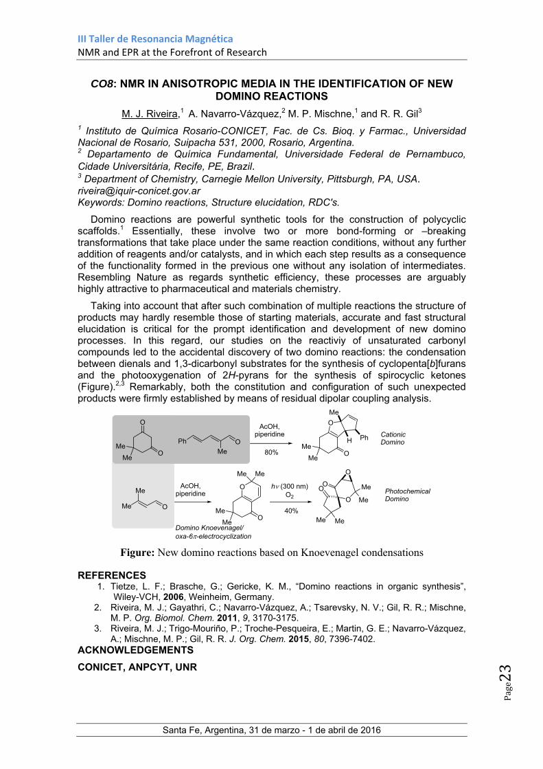
CO8: NMR IN ANISOTROPIC MEDIA IN THE IDENTIFICATION OF NEW DOMINO REACTIONS
M. J. Riveira,1 A. Navarro-Vázquez,2 M. P. Mischne,1 and R. R. Gil3 1 Instituto de Química Rosario-CONICET, Fac. de Cs. Bioq. y Farmac., Universidad Nacional de Rosario, Suipacha 531, 2000, Rosario, Argentina. 2 Departamento de Química Fundamental, Universidade Federal de Pernambuco, Cidade Universitária, Recife, PE, Brazil. 3 Department of Chemistry, Carnegie Mellon University, Pittsburgh, PA, USA. [email protected] Keywords: Domino reactions, Structure elucidation, RDC's.
Domino reactions are powerful synthetic tools for the construction of polycyclic scaffolds.1 Essentially, these involve two or more bond-forming or –breaking transformations that take place under the same reaction conditions, without any further addition of reagents and/or catalysts, and in which each step results as a consequence of the functionality formed in the previous one without any isolation of intermediates. Resembling Nature as regards synthetic efficiency, these processes are arguably highly attractive to pharmaceutical and materials chemistry.
Taking into account that after such combination of multiple reactions the structure of products may hardly resemble those of starting materials, accurate and fast structural elucidation is critical for the prompt identification and development of new domino processes. In this regard, our studies on the reactiviy of unsaturated carbonyl compounds led to the accidental discovery of two domino reactions: the condensation between dienals and 1,3-dicarbonyl substrates for the synthesis of cyclopenta[b]furans and the photooxygenation of 2H-pyrans for the synthesis of spirocyclic ketones (Figure).2,3 Remarkably, both the constitution and configuration of such unexpected products were firmly established by means of residual dipolar coupling analysis.
O
O
Me
O
Domino Knoevenagel/oxa-6-electrocyclization
O
O
Me
PhH
Me
Ph
O
OMe
Me
O
Me
Me
Me
Me
Me
Me
Me
O
Me Me
OO Me
Me
O
80%
40%
Cationic Domino
Photochemical Domino
h (300 nm)O2
AcOH, piperidine
AcOH, piperidine
Figure: New domino reactions based on Knoevenagel condensations
REFERENCES 1. Tietze, L. F.; Brasche, G.; Gericke, K. M., “Domino reactions in organic synthesis”,
Wiley-VCH, 2006, Weinheim, Germany. 2. Riveira, M. J.; Gayathri, C.; Navarro-Vázquez, A.; Tsarevsky, N. V.; Gil, R. R.; Mischne,
M. P. Org. Biomol. Chem. 2011, 9, 3170-3175. 3. Riveira, M. J.; Trigo-Mouriño, P.; Troche-Pesqueira, E.; Martin, G. E.; Navarro-Vázquez,
A.; Mischne, M. P.; Gil, R. R. J. Org. Chem. 2015, 80, 7396-7402. ACKNOWLEDGEMENTS
CONICET, ANPCYT, UNR

III Taller de Resonancia Magnética NMR and EPR at the Forefront of Research
Santa Fe, Argentina, 31 de marzo - 1 de abril de 2016
Page24
CO9: IMPACT OF NMR FROM ACADEMIA TO INDUSTRY
Rosana I. Misico
Departamento de Química Orgánica y UMYMFOR (CONICET-UBA), Facultad de Ciencias Exactas y Naturales, Universidad de Buenos Aires, Buenos Aires, Argentina. e-mail: [email protected] Keywords: structural elucidation, pharmaceutical impurities
Nuclear magnetic resonance (NMR) has been used successfully in the area of organic chemistry to the structural elucidation of organic compounds. Moreover, in the pharmaceutical industry standards and regulations require the identification of the impurities that accompany the drugs in their production process. At this point, NMR has become a very important method for evaluating the quality of the drug, as it not only allows the structural characterization of pharmaceutical active but also the determination of impurities and degradation products of drugs and their pharmaceutical formulations.
Through NMR it is possible to know the title of pharmaceutical ingredients for which there is no standards as well also the presence of active or excipients formulated and the impurity content of which do not have standards.
In this opportunity, the studies of a variety of pharmaceutical impurities made in our department in connection with different pharmaceutical industries will be described.
ACKNOWLEDGEMENTS
CONICET, UBA.

III Taller de Resonancia Magnética NMR and EPR at the Forefront of Research
Santa Fe, Argentina, 31 de marzo - 1 de abril de 2016
Page25
CO10: FROM METABOLOMICS TO ISOTOPE EFFECTS ON NUCLEAR SHIELDING: NMR SPECTROSCOPY MEETS THE URUGUAYAN
COUNTRYSIDE
Guillermo Moyna
Departamento de Química del Litoral, CENUR Litoral Norte, Universidad de la República, Ruta 3 Km 363, Paysandú 60000, Uruguay.
[email protected] Keywords: Ionic liquids; Lipidomics; Metabolomics; carbohydrate structure, conformation, and dynamics.
The research lines of the laboratories at the Departamento de Química del Litoral in Paysandú, Uruguay, a research and teaching facility which has been recently established and is part of the decentralization efforts of the Universidad de la República throughout the country, will be presented. Some of the projects that will be briefly described include the determination of metabolic and lipid profiles of agricultural products through the use of NMR-based metabolomic methods,1 the analysis of interionic hydrogen-bonding in ionic liquids by measurement of isotope effects on nuclear shielding (IENS),2 the study of charbohydrate conformation and dynamics using on- and off-resonance R1 relaxation techniques that have been previously employed with small RNA molecules,3 and the structure elucidation of oligosaccharides using solely NMR data and genetic information available in databases.4 The more important goal of the presentation is, however, to identify common insterests and potential areas of collaboration with other research groups from the region.
REFERENCES 1. Larroque, G.; Fernandez, P.; Grille, L.; Lopez, A.; Moyna, G. Cangüe 2015, 36, 3.
2. Khrizman, A.; Cheng, H. Y.; Bottini, G.; Moyna, G. Chem. Commun. 2015, 51, 3193. 3. Dethoff, E. A.; Petzold, K.; Chugh, J.; Casiano-Negroni, A.; Al-Hashimi, H. M. Nature
2012, 491, 724. 4. Fontana, C.; Lundborg, M.; Weintraub, A.; Widmalm, G. Glycobiology 2014, 24, 450.
ACKNOWLEDGEMENTS
ANII (Uruguay), CSIC (Uruguay), PEDECIBA (Uruguay), RSC (UK)

III Taller de Resonancia Magnética NMR and EPR at the Forefront of Research
Santa Fe, Argentina, 31 de marzo - 1 de abril de 2016
Page26
CHARLA BRUKER - DRIVING INNOVATION OF MAGNETIC RESONANCE
M. Chaykovsky
Executive Vice President at Bruker Biospin Corp, Billerica, Massachusetts
III Taller de Resonancia Magnética NMR and EPR at the Forefront of Research
Santa Fe, Argentina, 31 de marzo - 1 de abril de 2016
Page27
SISTEMA DE GESTIÓN DE TURNOS SNRM: IMPLEMENTACIÓN Y AVANCES
Gastón Mayada y Gustavo Alberto
Sistema Nacional de Resonancia Magnética
El Sistema Nacional de Resonancia Magnética tiene como propósito optimizar el funcionamiento de los grandes equipamientos de resonancia magnética, en todos sus tipos, adquiridos con fondos públicos, y mejorar en forma continua la calidad de las prestaciones. En ese marco lanza un sistema para la gestión del equipamiento que busca mejorar las condiciones de visibilidad, transparencia y acceso a los equipos y brindar información sobre su uso para la toma de decisiones estratégicas a nivel local y nacional. Durante la charla se presentará el sistema ya implementado en los Sistemas Nacionales de Microscopía, Espectrometría de Masas y Citometría de Flujos, se dará cuenta de los resultados obtenidos y se brindarán detalles sobre la pronta implementación del mismo en la red de centros adheridos al SNRM.

III Taller de Resonancia Magnética NMR and EPR at the Forefront of Research
Santa Fe, Argentina, 31 de marzo - 1 de abril de 2016
Page28
Posters

III Taller de Resonancia Magnética NMR and EPR at the Forefront of Research
Santa Fe, Argentina, 31 de marzo - 1 de abril de 2016
Page29
P1: STUDY OF INTERIONIC HYDROGEN BONDS IN IONIC LIQUIDS THROUGH DETERMINATION OF H/D ISOTOPE EFFECTS ON NUCLEAR
SHIELDING
Gualberto Bottini and Guillermo Moyna*
Departamento de Química del Litoral, CENUR Litoral Norte, Universidad de la República, Ruta 3 Km 363, Paysandú 60000, Uruguay. [email protected] Keywords: 19F NMR; Deuterium; Ionic liquids; Isotope effects on nuclear shielding.
Ionic liquids (ILs) interact with a variety of solutes and solvents, and have countless applications. Deciphering how interionic interactions in ILs affects their solvation properties and dynamics is therefore critical in the design of new processes based on these materials. In this work we present a study of interionic hydrogen bonds (H-bonds) in ILs through measurement of H/D isotope effects on nuclear shielding.
As we have previously shown, the formation of interionic H-bond in ILs can be detected through the measurement of H/D isotope effects on the shielding of reporter nuclei in the anion.1 Interactions between protons in either aromatic or aliphatic C-H groups can be investigated.1,2 In order to compare the relative strength of these two, we decided to prepare a series of [C4mim]BF4 and [C4mim]PF6 deuterium isotopologues labeled sequentially along the alkyl chains and the imidazole ring. Measurement of the H/D isotope effects on the 19F signal (Figure 1), or Δ19F(H,D), of the BF4
- and PF6- anions in the different [C4mim]+ isotopologues reveals that they are
roughly proportional to the polarization of the C-H group, and therefore the strength of the H-bond. Indeed, and as expected, the Δ19F(H,D) data show that the strongest H-bond is between the anion and the proton in the C-2 position. The results also reveal that the H-bonds to aliphatic protons on positions C-1’ and C-1” are comparable (PF6
-) or stronger (BF4
-) than to aromatic protons on C-4 and C-5. In summary, our findings show that intraionic H-bonds in ILs can be studied semi-quantitatively by means of isotope effects on nuclear shielding, providing a valuable tool to investigate these important interactions in these novel materials.
REFERENCES. 1. Remsing, R. C.; Rapp, A. L.; Wildin, J. L.; Moyna, G. J. Phys. Chem. B 2007, 111, 11619. 2. Khrizman, A.; Cheng, H. Y.; Bottini, G.; Moyna, G. Chem. Commun. 2015, 51, 3193.
ACKNOWLEDGEMENTS CSIC (Uruguay), PEDECIBA (Uruguay), NSF (USA)
Figure 1. Superposition of 19F spectra of the protiated (left) and deuterated (right) ILs.

III Taller de Resonancia Magnética NMR and EPR at the Forefront of Research
Santa Fe, Argentina, 31 de marzo - 1 de abril de 2016
Page30
P2: NMR-BASED METABOLIC PROFILING FOR THE DISCOVERY OF BIOMARKERS IN PATIENTS WITH PULMONARY TUBERCULOSIS (TB).
Burdisso Paula1;2, D'Attilio Luciano3, Díaz Ariana3, Bottasso Oscar3, Bay María L3, Vila Alejandro1;2, Rasia Rodolfo1;2
1Instituto de Biología Molecular y Celular de Rosario; Área Biofísica, Facultad de Ciencias, Bioquímicas y Farmacéuticas, Universidad Nacional de Rosario, 2000, Rosario, Argentina.
2 Plataforma de Biología Estructural y Metabolómica (PLABEM), 2000, Rosario, Argentina. 3 Instituto de Inmunología Clínica y Experimental de Rosario. CONICET-UNR, 2000, Rosario, Argentina. [email protected] Keywords: metabolomics, NMR, TB, biomarkers.
Omics sciences have allowed the use of global data analysis to explain biological processes. Among them, metabolomics deals with the study of metabolites, i.e small molecules of less than 1000 Da that represent the last product of celular processes reflecting the identity and the influence of the environment on an organism. In recent years advances in NMR allowed the acquisition of data in an effective, reproducible, and high-throughput manner. The challenge remains to develop highly reproducible methods and standardized protocols that minimize experimental or technical bias, allowing real interlaboratory comparisons of biomarker information. With the aim of developping the field in our lab, we investigated the metabolic profile of plasma samples of TB patients with pulmonary TB and healty controls (HCo). TB is a major health problem worldwide, with 1.5 millions of death per year. The workflow, including sample preparation, NMR calibration, data acquisition and processing was set up based on the literature1;2 and on bruker metabolomics specifications. In the present work 74 samples and 8 quality control samples were analyzed, giving hight quality 1H-NMR spectra. The data was digitized and and imported into MATLAB (2015, Mathworks Inc., USA), and automatically phase- and baseline-corrected. Each spectrum was normalized to the median spectrum, using glucose signals as reference. Plasma profiling analysis in combination with Principal Component Analysis (PCA) and Partial Least Squares Discriminant Analysis was performed to identify candidate biomarkers of TB. Several discriminant features are reported and the identification is made combining data bases, spike in and 2D spectra. The technique was succesfully implemented providing hight quality spectra and good models for human samples representing the startup for future projects in clinical research.
REFERENCES 1. Beckonert O, Keun HC, Ebbels TM, Bundy J, Holmes E, Lindon JC, Nicholson JK. Nat.
Protoc. 2007. 2, 2692–703. 2. Dona AC, Jiménez B, Schäfer H, Humpfer E, Spraul M, Lewis MR, Pearce JT, Holmes
E, Lindon JC, Nicholson JK. Anal Chem. 2014. 7;86(19):9887-94. ACKNOWLEDGEMENTS
CONICET, ANPCYT, GOBIERNO DE LA PROVINCIA DE SANTA FE.

III Taller de Resonancia Magnética NMR and EPR at the Forefront of Research
Santa Fe, Argentina, 31 de marzo - 1 de abril de 2016
Page31
P3: MACROMOLECULAR STRUCTURE: AVERAGE MOLECULAR WEIGHT BETWEEN CROSSLINKS OF DRY POLYMER NETWORKS BY NMR
F. Campise1, R.H. Acosta1, M.A. Villar2, E.M. Vallés2, G.A. Monti1, D.A. Vega3 1 FAMAF, Universidad Nacional de Córdoba, IFEG-CONICET, Córdoba, Argentina 2 Departamento de Ingenría Química, Planta Piloto de Ingeniería Química, CONICET, Bahía Blanca, Argentina 3 Departamento de Física, Instituto de Física del Sur (IFISUR), Universidad Nacional del Sur, CONICET, Bahía Blanca, Argentina [email protected] Keywords: Molecular weight between crosslinks; PDMS; NMR Double Quantum Coherence. The purpose of this work is to contribute to the structural characterization of polymer networks by Nuclear Magnetic Resonance (NMR) techniques. In particular, we pretend to study the possibility of determining the average molecular weight between crosslinks from the residual dipolar coupling constant obtained through NMR Double Quantum Coherence experiments. The residual dipolar coupling constant (Dres) is proportional to the dynamics chain order parameter S and the crosslink density [1], and can be related to the molecular weight between crosslinks (Mc). The relation between Dres and Mc depends on the theoretical (rubber elasticity) basis considered. If the phantom model is used as theoretical basis [2], instead of the ‘affine model’ [3], then a great dependence on the average functionality of the crosslinks arises. We evaluate both theories on a set of model polydimethylsiloxane (PDMS) networks prepared with varying functionality. Since transiently trapped entanglements contribute to the residual dipolar coupling, [4-5], we also made focus on how defects present in the networks can affect the determination on Mc through Dres. We compared NMR results with Miller-Macosko Mean Field Calculations for Mc. Although proportionality between Dres and Mc has already been investigated and several theoretical approaches have been considered [6-8], quantitative discrepancies were obtained, and contribution of defects to the determination of Dres is still a matter of interest. REFERENCES
1. Chassé W.; Lang M.; Jens-Uwe Sommer and Saalwächter K. Macromolecules, 2012, 899. 2. James H. M. and Guth E. J. Chem. Phys., 1943, 455. 3. Wall F. T. and Flory P. J. Chem.Phys., 1951, 1435. 4. Acosta R. H.; Monti G. A.; Villar M. A.; Vallés E. M. and Vega D. A. Macromolecules, 2009, 4674. 5. Campise F; Roth L. E.; Acosta R. H.; Monti G. A.; Villar M. A.; Vallés E. M. and Vega D. A. Macromolecules, 2016, 387. 6. Flory, P.; Rehner, J. J. Chem. Phys., 1943, 521−527 7. Flory, P. J. Chem. Phys., 1941, 660−661. 8. Huggins, M. J. Chem. Phys., 1941, 440.
ACKNOWLEDGEMENTS CONICET (PIP 11220130100746CO), ANPCyT (PICT 2014-1295), SECyT-UNC
III Taller de Resonancia Magnética NMR and EPR at the Forefront of Research
Santa Fe, Argentina, 31 de marzo - 1 de abril de 2016
Page32
P4: SITE DIRECTED MUTAGENESIS, KINETIC, UV-VIS, EPR, AND COMPUTATIONAL STUDIES OF MUTANTS PROVIDE INSIGHTS INTO CATALYSIS AND ELECTRON TRANSFER PROCESSES IN COPPER
NITRITE REDUCTASE
Julio C. Cristaldi,a María Cecilia Gomez,a Sergio D. Dalosto,b Alberto C. Rizzi,a Maria Gabriela Rivas,a Carlos D. Brondinob
a. Departamento de Física, FBCB, UNL Santa Fe, Argentina. b. Instituto de Física del Litoral (CONICET-UNL
Keywords: nitrite reductase, copper, denitrification, EPR
Denitrification is a key process in the nitrogen cycle that implicates dissimilatory transformation of nitrate or nitrite ions into gaseous compounds through a series of reactions which are catalysed by specific enzymes. Nitrite reductase (Nir) catalyzes the reduction of nitrite (NO2
-) to nitric oxide (NO).1,2 The present work studies the Nir from Sinorhizobium meliloti 2011 (SmNir), a rhizobia bacterium that lives in root nodules of legumes and is widely used in agriculture as fertilizers. SmNir is a homotrimeric protein that contains two Cu centers (T1 and T2) and can be heterologously produced in E. coli cells.3 T1 and T2 are the electron transfer and the catalytic centers, respectively, and are ~12 Å apart and bridged by a histidine-cysteine pathway. The proposed reaction mechanism in Nirs implies that nitrite binds to the T2 center and is converted to NO by one reducing equivalents delivered by an external physiological electron donor through the T1-T2 chemical pathway. We present kinetic, UV-vis, and EPR studies together with computational methods using first-principles combined with molecular mechanics (QM/MM) of SmNir mutants . of the Cys-His bridge through which electron transfer T1-T2 occurs. These data provide insight into how activity can be altered through mutational manipulation. Computational methods are used to evaluate the coordination around both copper centers in the mutant forms. The alteration of Cys to Asp results in enzyme inactivation with pseudoazurin as electron donor, which suggests that no direct transfer of electrons occurs from pseudoazurin to the catalytic T2 center. The mutation of His to Asp also yields inactive forms of Nirs, despite the mutant shows T1 and T2 centers, as indicated by UV-vis and EPR spectroscopies, and that the reduction potentials of both T1 and T2 centers present similar order of magnitude relative to the wild type SmMir.
REFERENCES Zumft W. G., Microbiol. Mol. Biol. R. (1997),61:533-616 Averill B. A., Chem. Rev. (1996),96:2951-2964 Ferroni F. M. et al., J. Inorg. Biochem. (2012),114:8-14
ACKNOWLEDGEMENTS
We thanks to CONICET, ANPCYT and UNL-CAI+D for financial support.
III Taller de Resonancia Magnética NMR and EPR at the Forefront of Research
Santa Fe, Argentina, 31 de marzo - 1 de abril de 2016
Page33
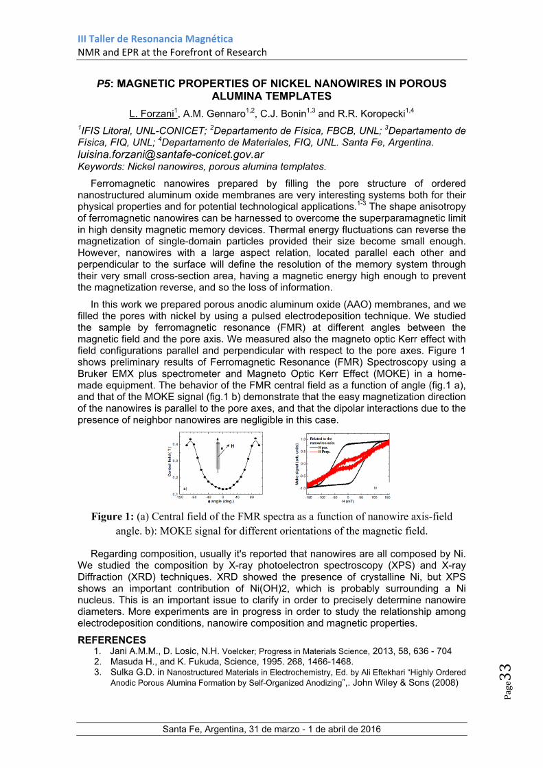
P5: MAGNETIC PROPERTIES OF NICKEL NANOWIRES IN POROUS ALUMINA TEMPLATES
L. Forzani1, A.M. Gennaro1,2, C.J. Bonin1,3 and R.R. Koropecki1,4 1IFIS Litoral, UNL-CONICET; 2Departamento de Física, FBCB, UNL; 3Departamento de Física, FIQ, UNL; 4Departamento de Materiales, FIQ, UNL. Santa Fe, Argentina. [email protected] Keywords: Nickel nanowires, porous alumina templates.
Ferromagnetic nanowires prepared by filling the pore structure of ordered nanostructured aluminum oxide membranes are very interesting systems both for their physical properties and for potential technological applications.1-3 The shape anisotropy of ferromagnetic nanowires can be harnessed to overcome the superparamagnetic limit in high density magnetic memory devices. Thermal energy fluctuations can reverse the magnetization of single-domain particles provided their size become small enough. However, nanowires with a large aspect relation, located parallel each other and perpendicular to the surface will define the resolution of the memory system through their very small cross-section area, having a magnetic energy high enough to prevent the magnetization reverse, and so the loss of information.
In this work we prepared porous anodic aluminum oxide (AAO) membranes, and we filled the pores with nickel by using a pulsed electrodeposition technique. We studied the sample by ferromagnetic resonance (FMR) at different angles between the magnetic field and the pore axis. We measured also the magneto optic Kerr effect with field configurations parallel and perpendicular with respect to the pore axes. Figure 1 shows preliminary results of Ferromagnetic Resonance (FMR) Spectroscopy using a Bruker EMX plus spectrometer and Magneto Optic Kerr Effect (MOKE) in a home-made equipment. The behavior of the FMR central field as a function of angle (fig.1 a), and that of the MOKE signal (fig.1 b) demonstrate that the easy magnetization direction of the nanowires is parallel to the pore axes, and that the dipolar interactions due to the presence of neighbor nanowires are negligible in this case.
Figure 1: (a) Central field of the FMR spectra as a function of nanowire axis-field angle. b): MOKE signal for different orientations of the magnetic field.
Regarding composition, usually it's reported that nanowires are all composed by Ni. We studied the composition by X-ray photoelectron spectroscopy (XPS) and X-ray Diffraction (XRD) techniques. XRD showed the presence of crystalline Ni, but XPS shows an important contribution of Ni(OH)2, which is probably surrounding a Ni nucleus. This is an important issue to clarify in order to precisely determine nanowire diameters. More experiments are in progress in order to study the relationship among electrodeposition conditions, nanowire composition and magnetic properties.
REFERENCES 1. Jani A.M.M., D. Losic, N.H. Voelcker; Progress in Materials Science, 2013, 58, 636 - 704 2. Masuda H., and K. Fukuda, Science, 1995. 268, 1466-1468. 3. Sulka G.D. in Nanostructured Materials in Electrochemistry, Ed. by Ali Eftekhari “Highly Ordered
Anodic Porous Alumina Formation by Self-Organized Anodizing”,. John Wiley & Sons (2008)

III Taller de Resonancia Magnética NMR and EPR at the Forefront of Research
Santa Fe, Argentina, 31 de marzo - 1 de abril de 2016
Page34
P6: PREPARATION OF ALISKIREN COPPER(II) COMPLEX AND SOLUTION STUDIES BY EPR AND UV-VIS SPECTROSCOPIES
M. S. Islas1, M. A. Grela2, E. G. Ferrer1, P. A. M. Williams1 1 Centro de Química Inorgánica (CEQUINOR), FCE, UNLP, 1900 La Plata, Argentina 2 Departamento de Química, FCEyN, UNMdP, 7600 Mar del Plata, Argentina. [email protected] Keywords: Aliskiren, copper(II), coordination complex, antihypertensive.
Aliskiren (Alk) represents the first in a novel class of renin inhibitors with the potential for treatment of hypertension and related cardiovascular diseases1. With the aim of improving the pharmacological effects of different compounds by inducing favorable conformational changes or by improving their bioavailability2, our group has been working with metal complexation. Herein, we have prepared a solid complex at pH 12 and studied the interaction of the ligand and the metal at different pH values using Electron Paramagnetic Resonance (EPR) and UV-visible spectroscopies.
Considering that Alk is commercially available as aliskiren hemifumarate, in a first step we have isolated aliskiren base. A solid CuAlk complex was synthesized by the addition of an aqueous solution of copper(II) nitrate to an ethanolic solution of Alk base. A violet complex, at pH 12, has been obtained and characterized by means of FTIR, EPR and electronic spectroscopies and elemental analysis as [Cu(Alk)2].2H2O.
Ethanolic solutions of CuAlk were prepared at different pH values and the EPR and UV-vis spectra were registered after the variation of one unit of pH in the range between 2 and 13 (Fig. 1).
500 600 700 8000,0
0,2
0,4
0,6
0,8
1,0
Ab
sorb
ance
(nm)
pH 2-4 5 6 7 8 9 10 11 12 13
a
2500 3000 3500 4000 4500
b
Field (G)
pH 2-13
Figure 1: Variation with pH of CuAlk in ethanolic solution a) visible spectra and b)EPR
It can be observed that at a pH value lower than 4, both EPR and UV-vis corresponded to the presence of free copper(II), while two different species were observed at higher pH values in agreement with the behavior in solid state. From the EPR spectra, the parameters of the violet complex can be determined. The complex shows an elongated octahedral geometry, g// > g, dX
2-y2 ground state (g// = 2.20; g = 2.04 and A// = 196x10-4 cm-1 ; A =2.5x10⁻³ cm-1 and an environment of nitrogen atoms3.
REFERENCES 1. Wood J. M.; Maibaum J.; et.al. Biochem. Biophys. Res. Commun. 2003, 308, 698–705.
2. Baran E. J. Química bioinorgánica, 1st ed.; McGraw-Hill Interamericana, 1994 3. Tabbì G.; Giuffrida A.; Bonomo R. P. J. Inorg. Biochem. 2013, 128, 137–145:
ACKNOWLEDGEMENTS
CONICET, (PICT 2013-0569) ANPCYT, UNLP, CICPBA.

III Taller de Resonancia Magnética NMR and EPR at the Forefront of Research
Santa Fe, Argentina, 31 de marzo - 1 de abril de 2016
Page35
P7: BIOGENESIS OF A COPPER CENTER IN PLANT CYTOCHROME OXIDASES
María Eugenia Llases1, Marcos N Morgada1, Alejandro J Vila1. 1 Instituto de Biología Molecular y Celular de Rosario (IBR), Facultad de Ciencias Bioquímicas y Farmacéuticas, Universidad Nacional de Rosario-Conicet, Rosario, Argentina [email protected]
Keywords: cytochrome oxidase, metal site assembly, metallochaperones, sco proteins
Copper is an essential transition metal in cells, where it binds to biomolecules with high affinity and plays a myriad of catalytic and signaling roles, all of them stemming from its redox capabilities. At the same time, these features make free copper ions toxic by outcompeting other metal ions in binding the target metalloproteins and as a source of reactive oxygen species. Copper metallochaperones assist copper in reaching vital destinations without inflicting damage or becoming trapped in adventitious binding sites. Copper ions are specifically released from copper metallochaperones upon contact with their cognate cuproproteins.1 Copper is an essential cofactor of cytochrome c oxidase (COX), the terminal oxidase of the respiratory chain in most organisms. COX I and COX II are the two copper-containing subunits harboring the CuB and CuA sites, respectively, conserved in heme-copper oxidases. Assembly of the oxidase is a complex process that requires several chaperones that assist the folding of the individual subunits and the incorporation of the metal cofactors. CuA is a binuclear copper center that provides the electron entry port of the oxidase in subunit II (COXII), located in the intermembrane space (IMS) of mitochondria or in the bacterial periplasm. A conserved family of proteins that has been related with copper insertion into COX, particularly into the COXII subunit, is the SCO family. SCO proteins are of prokaryotic origin and usually contain a transmembrane domain and a soluble domain that contains redox-active cysteines and a histidine presumably involved in copper binding.2 We have proposed distinct mechanisms for the assembly of CuA sites in Bacterial and Human oxidases.3,4 Based on recent reports on putative COXII assembly factors in plants, here we report the first steps towards the elucidation of the structural details of the biogenesis of CuA site in A. thaliana.5,6
REFERENCES 1.Robinson, N. J. & Winge, D. R. Annu. Rev. Biochem. 79, 537–62 2010 2.Banci, L., Bertini, I., Cavallaro, G. & Ciofi-Baffoni, S. FEBS J. 278, 2244–62 2011. 3.Abriata, L. A., Banci, L., Bertini, I., Ciofi-Baffoni, S., Gkazonis, P., Spyroulias, G., Vila, A. J. and Wang S. Nature Chem Bio. 4, 599–601 2009 4.Morgada, M. N., Abriata, L. A., Cefaro, C., Gaijda, K, Banci, L and Vila A. J., PNAS 112, 38, 11771-76, 2015 5.Steinebrunner, I., Gey, U., Andres, M., Garcia, L. & Gonzalez, D. H.. Front. Plant Sci. 5, 87, 2014 6.Attallah, C., Welchen, E., Martin, A. P., Spinelli, S. V., Bonnard G., Palatnik J. F., and Gonzalez D. H. J. Exp. Bot. 62, 4281–94, 2011 7.Horng, Y. C., Leary, S., Cobine, P. A.,Young, F. C., George G. N., Shoubridge E. A., and Winge D. R. J. Biol. Chem. 280, 34113–22, 2005 ACKNOWLEDGEMENTS CONICET, ANPCYT, PLABEM

III Taller de Resonancia Magnética NMR and EPR at the Forefront of Research
Santa Fe, Argentina, 31 de marzo - 1 de abril de 2016
Page36
P8: NMR STRUCTURAL CHARACTERIZATION OF THE COMPLEXATION OF CU(I) BY ACETYLATED ALPHA-SYNUCLEIN.
Marco C. Miotto1, Ariel A. Valiente-Gabioud1, Christian Griesinger2, Andrés Binolfi1, Claudio O. Fernández1 1 Max Planck Laboratory for Structural Biology, Chemistry and Molecular Biophysics of Rosario and Instituto de Investigaciones para el Descubrimiento de Fármacos de Rosario (IIDEFAR/CONICET-UNR), Universidad Nacional de Rosario, 27 de Febrero 210 bis, S2002LRK Rosario, Argentina 2 Department of NMR-based Structural Biology, Max Planck Institute for Biophysical Chemistry, Am Fassberg 11, D-37077 Göttingen, Germany
Keywords: Alpha-Synuclein, copper, Cu(I), acetylation.
Alpha-Synuclein (AS) amyloid aggregation is related to neurodegeneration in Parkinson´s disease (PD). Even though it remains uncertain how AS initiates neuronal death, there is growing evidence that supports a link among brain copper homeostasis, AS-copper complexes, and development of PD. Copper ions might represent the link between neuronal death and the pathological processes of protein aggregation and oxidative damage.1,2 Recently reported, it was described that AS experiences N-terminal acetylation in vivo (acAS), which abolishes Cu(II) binding to the N-terminal in vitro. However, since copper ions are predominantly found in their Cu(I) state in the reducing environment of living cells, it is the characterization of the AS−Cu(I) complexes that becomes fundamental to understand the impact of this interaction on the bioinorganic chemistry of PD. In our group, we have extensively studied and characterize the binding of Cu(I) to non-acetylated AS.3 Accordingly, we report here the detailed structural characterization of Cu(I) complexes with the physiologically relevant acetylated AS protein, assisted by several NMR approaches such as standard HSQC/HMQC and 13C direct detection experiments, and heteronuclear chemical shift, relaxation rates and NOE analysis. We found that the coordination chemistry of the acAS−Cu(I) complex is similar to that of non-acetylated AS, however, the tridimensional rearrangement is significantly different, since Cu(I) favor local conformations with α-helical secondary structure and restricted motility at the N-terminal region of acAS.
REFERENCES
1. Binolfi, A.; Quintanar, L.; Bertoncini, C. W.; Griesinger, C.; Fernandez, C. O. Coord. Chem. Rev. 2012, 256, 2188.
2. Miotto, M. C.; Rodriguez, E. E.; Valiente-Gabioud, A. A.; Torres-Monserrat, V.; Binolfi, A.; Quintanar, L.; Zweckstetter, M.; Griesinger, C.; Fernandez, C. O. Inorg. Chem. 2014, 53, 4350.
3. Miotto, M. C.; Binolfi, A.; Zweckstetter, M.; Griesinger, C.; Fernandez, C. O. J. Inorg. Biochem. 2014, 141, 208.
ACKNOWLEDGEMENTS
ANPCyT, FONCyT, CONICET, the Ministry of Education of Argentina, the Ministry of Health of Argentina, Fundación Medife, Fundación Bunge y Born, the Max Planck Society, and the Alexander von Humboldt Foundation.

III Taller de Resonancia Magnética NMR and EPR at the Forefront of Research
Santa Fe, Argentina, 31 de marzo - 1 de abril de 2016
Page37
P9: QUANTIFICATION OF SILANOL GROUPS ON MESOPOROUS MATERIALS USING SOLID STATE MAGNETIC RESONANCE
J. Villarroel-Rocha1, M.C. Valentinuzzi2, G.A. Monti2, K. Sapag1 1 Instituto de Física Aplicada, Universidad Nacional de San Luis, San Luis, Argentina 2 IFEG-CONICE y FaMAF-UNC y Córdoba, Argentina [email protected] Keywords: mesoporous materials, SiOH
Porous materials have properties which make them suitable for catalytic applications. They are classified according to pore size in: microporous (diameter less than 2 nm), mesoporous (diameter between 2 and 50 nm) and macroporous (greater than 50 nm diameter). In this work, we study mesoporous materials by Solid State Nuclear Magnetic Resonance. Such materials have three-dimensional arrangements of SiO4 and are characterized by a narrow and uniform pore size distribution. Different structures can be obtained according to the synthesis process; in this case we study the M41S and SBA families. From the M41S family we studied the MCM-41 and MCM-48 materials. MCM-41 is the most common and stable, with hexagonal phase. MCM-48 is the most complex phase, with channel systems that interlock to form a network of cubic symmetry. From the SBA Family we studied the SBA-15 materials; they are thermally more stable than the M41S family and have micropores connecting channels together randomly generating a dual porosity [1,2].
These mesoporous materials have characteristic SiOH arrangements that are identified by a chemical shift in the 29Si-NMR spectrum, with peaks approximately at -90, -100 and -110 ppm which are characteristic of the signals Q2 [(SiO)2(OH)2], Q3 [(SiO)3Si (OH)] and Q4 [(SiO)4] respectively. These signals represent different degrees of coordination of silicon present in these materials. We identified the groups present and calculated their relative proportion in each of the samples to determine the amount of silanol groups.
In all the spectra, Q4 is the most intense signal, indicating that among the species of silicon, the tetra coordinated one has the highest proportion. With respect to the total amount of silanol groups (2Q2 + Q3) corresponding to structures SBA-15 synthesized by Soxhlet extraction no significant differences were observed; instead a reduction of approximately 40% was found in calcinated samples.
REFERENCES 1. F. Rouquerol, J. Rouquerol, K.S.W. Sing, P. Llewellyn, G. Maurin, Adsorption by
powders and porous solids, Academic Press, San Diego, 2014. 2. M. Ide et al Phys Chem Chem Phys 15 2013 642.
ACKNOWLEDGEMENTS
CONICET, ANPCYT, SECyT-UNC

III Taller de Resonancia Magnética NMR and EPR at the Forefront of Research
Santa Fe, Argentina, 31 de marzo - 1 de abril de 2016
Page38
P10: SINGLE CRYSTAL EPR FOR THE STUDY OF ELECTRONIC AND MAGNETIC PROPERTIES IN TRANSITION METAL SYSTEMS
Ana Laura Pérez, Nicolás I. Neuman, Alberto C. Rizzi and Carlos D. Brondino
Departamento de Física, FBCB, UNL Santa Fe, Argentina [email protected]
Keywords: Single-crystal EPR, transition metals, magnetic interactions.
Magnetic interactions between paramagnetic transition metal ions or non-metal centered radicals are relevant to the fields of solid-state magnetism (including nano- and molecular magnetism), bioinorganic chemistry (metalloproteins) and polymer and macromolecular science (through the use of spin-labels). Most magnetic interactions in paramagnetic systems, which can be studied by electron paramagnetic resonance (EPR) are anisotropic and defined by matrices which contain information on orbital occupations of the unpaired electrons, delocalization or covalency, interspin distances, identity of bonding atoms, amongst other properties. In fluid samples some of these interactions are partially or totally averaged, while in polycrystalline (powder) or frozen solutin samples, the spectra are summed over all possible molecular orientations, obscuring part of the information. Therefore to fully understand the magnetic properties of some systems and to obtain correlations between magnetic interactions and subtle electronic structure factors it becomes necessary to perform EPR experiments on oriented single crystal samples. The angular variations of the resonance positions allow determination of many interactions (g-matrix, hyperfine matrix, zero-field splitting, and exchange interactions), sometimes providing information that cannot be extracted by any other technique. Our group has applied this technique to studies of copper(II) (S = 1/2) and cobalt(II) (S = 3/2) compounds presenting isolated, dimeric and chain topologies, developing in many cases new physical models to interpret the data. Some of these results are discussed here.
REFERENCES
1) Kahn O., Molecular Magnetism (1993). 2) Weil, J. A. y Bolton, J. R. (2007) Electron Paramagnetic Resonance: Elementary Theory and Practical Applications. John Wiley & Sons, New Jersey. 3) Neuman N.I. et al., J. Phys. Chem. A (2010), 114(59):13069-13075. 4) Neuman N.I. et al., Inorg. Chem. (2014), 53(5): 2535–2544. 5) Neuman N.I. et al., Inorg. Chem. Front. (2015), 2: 837-8456. 6) Rizzi A.C. et al., Eur. J. of Inorg. Chem. (2016), 2: 192-207.
ACKNOWLEDGEMENTS
CONICET, ANPCYT, UNL-CAI+D

III Taller de Resonancia Magnética NMR and EPR at the Forefront of Research
Santa Fe, Argentina, 31 de marzo - 1 de abril de 2016
Page39
P11: ROLE OF CONFORMATIONAL FLEXIBILITY IN THE FITNESS OF β-LACTAMASES ENZIMES
María Agustina Rossi1, Mariano González1, Alejandro J. Vila1.
1Instituto de Biología Molecular y Celular de Rosario (IBR), Facultad de Ciencias Bioquímicas y Farmacéuticas, Universidad Nacional de Rosario-Conicet, Rosario, Argentina [email protected]
Keywords: β-lactamases, conformational flexibility, CTX-M.
Alternate structural conformers can mediate alternate folds and functions, this diversity is the foundation of “protein evolvability”, the ability of proteins to rapidly adopt new functions within existing folds or even adopt entirely new folds.1,2. Antibiotic resistance mediated by β-lactamases represents an evolutionary paradigm in which organismal fitness depends on the catalytic efficiency of a single enzyme, which can be divided in two large families: serin-β-lactamases (SBLs) and metallo-β-lactamases (MBLs).3,4 CTX-M enzymes belong to the group of ESBLs (Extended spectrum β-lactamases). In the classic ESBLs, the increased hydrolytic spectrum came with enlargement of the active site.5 Instead, CTX -M expanded activity appears to be related, not with an increase in the active site volume, but with an increased flexibility. This increased flexibility is correlated with higher ceftazidimase activity and lower stability. Structurally, this can be reconciled with the substitutions D240G and V231A, which occur at either end of the B3 strand. 6,7 To test this hypothesis, we will study the dynamics of the polypeptide backbone of natural variants of CTX-M-14: CTX-M-9 (V231A), CTX-M-27 (D240G) y CTX-M-16 (double mutant) by NMR spectrosopy. It has been reported that the D240G substitution leads to reduced catalytic efficiency against cefotaxime, the natural substrate, but improves activity against bulky substrates. The V231A mutation does not affect enzyme activity but, in conjunction with D240G substitution, results in an increase activity against the natural substrate and the ceftazidime, the bulkier one. We cloned CTX-M-14 in a high expression plasmid and we generated, by site-directed mutagenesis, its natural variants: CTX-M-9, CTX-M-27 and CTX-M-16. CTX-M-14 was purified upon homogeneity in minimal medium M9, with a yield of 200 mg/L of protein. The activity of this enzyme was tested against different antibiotics: cefotaxime, cephalothin and ceftazidime, condition in which no hydrolysis was observed. Finally, the HSQC of CTX-M-14 was obtained, according with the signal dispersion of the spectrum the protein was properly folded, also it was stable during all time of the assignation experiments.
REFERENCES 1. Tokuriki, N.; Tawfik, D.S. Science. 2009, 324 (5924), 203‐207. 2. Boehr, D.D.; Nussinov, R.; Wright, P.E. Nat. Chem. Biol. 2009, 5 (11), 789‐796. 3. Tomatis, P.E, et al. Proc. Natl. Acad. Sci. U. S. A. 2008, 105 (52), 20605-20610. 4. Meini, M.R.; Tomatis,P.E.; Weinreich, D.M.; Vila, A.J. Mol. Biol. Evol. 2015, 32 (7), 1774-1787. 5. Perez, F.; Endimiani, A.; Hujer, K.M.; Bonomo, R.A. Curr Opin Pharmacol. 2007, 7(5), 459-469. 6. Chen, Y.; Delmas, J.; Sirot, J.; Shoichet, B.; Bonnet, R. J. Mol. Biol. 2005, 348 (2), 349‐362. 7. Delmas, J., et al. Mol Biol. 2008, 375 (1), 192-201
ACKNOWLEDGEMENTS CONICET, NIH
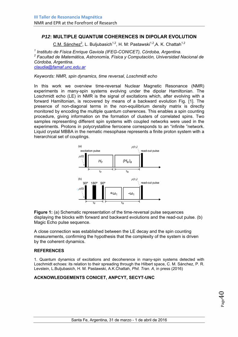
III Taller de Resonancia Magnética NMR and EPR at the Forefront of Research
Santa Fe, Argentina, 31 de marzo - 1 de abril de 2016
Page40
P12: MULTIPLE QUANTUM COHERENCES IN DIPOLAR EVOLUTION
C.M. Sánchez2, L. Buljubasich1,2, H. M: Pastawski1,2,A. K. Chattah1,2 1 Instituto de Física Enrique Gaviola (IFEG-CONICET), Córdoba, Argentina. 2 Facultad de Matemática, Astronomía, Física y Computación, Universidad Nacional de Córdoba, Argentina. [email protected]
Keywords: NMR, spin dynamics, time reversal, Loschmidt echo
In this work we overview time-reversal Nuclear Magnetic Resonance (NMR) experiments in many-spin systems evolving under the dipolar Hamiltonian. The Loschmidt echo (LE) in NMR is the signal of excitations which, after evolving with a forward Hamiltonian, is recovered by means of a backward evolution Fig. [1]. The presence of non-diagonal terms in the non-equilibrium density matrix is directly monitored by encoding the multiple quantum coherences. This enables a spin counting procedure, giving information on the formation of clusters of correlated spins. Two samples representing different spin systems with coupled networks were used in the experiments. Protons in polycrystalline ferrocene corresponds to an “infinite ”network. Liquid crystal MBBA in the nematic mesophase represents a finite proton system with a hierarchical set of couplings.
Figure 1: (a) Schematic representation of the time-reversal pulse sequences displaying the blocks with forward and backward evolutions and the read-out pulse. (b) Magic Echo pulse sequence.
A close connection was established between the LE decay and the spin counting measurements, confirming the hypothesis that the complexity of the system is driven by the coherent dynamics.
REFERENCES
1. Quantum dynamics of excitations and decoherence in many-spin systems detected with Loschmidt echoes: its relation to their spreading through the Hilbert space, C. M. Sánchez, P. R. Levstein, L.Buljubasich, H. M. Pastawski, A.K.Chattah, Phil. Tran. A, in press (2016)
ACKNOWLEDGEMENTS CONICET, ANPCYT, SECYT-UNC

III Taller de Resonancia Magnética NMR and EPR at the Forefront of Research
Santa Fe, Argentina, 31 de marzo - 1 de abril de 2016
Page41
P13: ACCESSING TO HELIX/COIL TRANSITION EVENTS INVOLVED IN THE CATALYTIC CYCLE OF THE E. COLI THIOL PEROXIDASE
Diego S. Vazquez1, William A. Agudelo1, Gerardo Ferrer-Sueta2 and Javier Santos1
1IQUIFIB (UBA-CONICET), Universidad de Buenos Aires, CABA, Argentina. 2Laboratorio de Fisicoquímica Biológica, UdelaR, Montevideo, Uruguay. e-mail: [email protected]
Keywords: peroxiredoxin, transition state, essential modes, protein dynamics.
Peroxiredoxins (PRDX) are ubiquitous enzymes that reduce different kind of peroxides via two catalytic cysteines [1]. All PRDX shows conformational changes during the catalytic cycle restricted to the region that contain the cysteines. The active fully folded (FF) conformation of the E. coli thiol peroxidase (EcTPX) requires a key peroxidatic cysteine (CP, located in the αH2). First, CP is oxidized to sulfenic acid and then, after a critically local unfolding step (LU), CP forms a disulfide bond with the resolving cysteine (CR, located in the αH3, see Figure 1AB) [2]. It is clear that the energetic cost to unfold both helix, has a direct impact to the catalytic ratio of substrate reduction, but the contribution has not been clarified yet. In this study, we performed a microsecond time-scale common molecular dynamic simulations of the WT and 3 point-mutants of αH3 to glycine in order to destabilize the helix and a conformational searching profile though several potential-biased accelerated molecular dynamic simulations (aMD) followed by essential mode analysis, in an attempt to determine the energetic cost of the FF→LU event; and the experimental determination of the ΔΔGN→U of unfolding. Preliminary results suggest that: I) the destabilization of the αH3 promote the local unfolding of αH3 in two of the three mutants but not in the wild-type EcTPX, II) The aMD simulations capture the FF→LU transition in the WT protein characterized by an intermediate with rotameric and secondary structure changes that involve αH2 and αH3 (Figure 1C).
Figure 1: (A) The loss of helical content detected in the far-UV circular dichroism
spectrum of reduced (black line) and oxidized (red line) state of EcTPX. (B) Superimposition of the FF (dark blue) and LU (gray) conformations (C) 3D heatmap of
the essential modes of the FF→LU transition obtained by AMD.
REFERENCES
1. S.G. Rhee, Overview on Peroxiredoxin, Mol. Cells, 2016, 39
2. L. Flohé et al., A Comparison of Thiol Peroxidase Mechanisms, Antiox Redox Signal, 2011, 15
ACKNOWLEDGEMENT: ANPCYT, UBACYT, CABBIO and CONICET.

III Taller de Resonancia Magnética NMR and EPR at the Forefront of Research
Santa Fe, Argentina, 31 de marzo - 1 de abril de 2016
Page42
P14: NMR CALCULATION AS A HELPFUL TOOL IN STRUCTURE ELUCIDATION
Zanardi, María Marta1,2; Grimblat, Nicolás1; Sarotti, Ariel M.1 1Instituto de Química Rosario (CONICET), Facultad de Ciencias Bioquímicas y Farmacéuticas, Universidad Nacional de Rosario, Suipacha 531, Rosario (2000), Argentina. [email protected] 2Facultad de Química e Ingeniería del Rosario, Pontificia Universidad Católica Argentina, Av. Pellegrini 3314, Rosario (2000), Argentina. [email protected] Keywords: NMR Calculations, Neural Networks, DP4.
The total synthesis of natural products is one of the most beautiful and exciting chemistry areas, lying somewhere between a fine art and a hard science. The enterprises are often hard and fraught with difficulties and detours. Eventually, after a considerable investment of time, money, and man-power the synthetic target is accomplished. However, surprisingly often such an exciting moment quickly moves to frustration once a mismatch between the NMR data of the synthesized compound and the natural product is detected.
Incorrectly assigned natural products are not uncommon, even in the golden age of NMR. High molecular complexity, human errors, signal ambiguity and sample impurities can be pointed to as the most common sources of misassignments. Hundreds of structural revisions have been published in the last decades, ranging from profound connectivity to subtle (but not least) stereochemical errors. Considering that the discrepancies are often detected after total synthesis of the originally proposed (wrong) structure, it is not unreasonable to assume that the real molecular architecture of many reported natural products remains unknown.
Modern computational chemistry has significantly contributed to prevent these mistakes. We have developed to tools wich allow to interpret the correlation between experimental and calculated NMR data contributing to the structural assignment.
The DP4 probability is one of the most sophisticated and popular approaches for the stereochemical assignment of organic molecules using GIAO NMR chemical shift calculations when only one set of experimental data is available. In order to improve the performance of the method, we have developed a modified probability (DP4+), whose main differences from the original DP4 are the inclusion of unscaled data and the use of higher levels of theory for the NMR calculation procedure. With these modifications, a significant improvement in the overall performance was achieved, providing accurate and confident results in establishing the stereochemistry of 48 challenging isomeric compounds.1
The structural validation problem using quantum chemistry approaches (confirm or reject a candidate structure) has been tackled with artificial neural network (ANN) mediated multidimensional pattern recognition from experimental and calculated 2D C−H COSY. In order to identify subtle errors (such as regio- or stereochemical), more
than 400 ANNs have been built and trained, and the most efficient in terms of classification ability were successfully validated in challenging real examples of natural product misassignments.2
Figure 1 REFERENCES
1. Grimblat, Nicolás; Zanardi, María M.; Sarotti, Ariel M. J. Org. Chem. 2015, 80, 12526. 2. Zanardi, María M.; Sarotti, Ariel M. J. Org. Chem. 2015, 80, 9371.
ACKNOWLEDGEMENTS: CONICET, ANPCYT, IQUIR, UNR, FBIOyF, UCA.

III Taller de Resonancia Magnética NMR and EPR at the Forefront of Research
Santa Fe, Argentina, 31 de marzo - 1 de abril de 2016
Page43
P15: SS-NMR APPLIED TO THE STRUCTURAL CHARACTERIZATION OF POLY(ETHYLENIMINE) (PEI) AND RELATED COMPOUNDS.
Ayelén F. Crespi1, Emilse Leal1, Graciela Y. Buldain1, Juan M. Lázaro Martínez1,2 1Universidad de Buenos Aires, Facultad de Farmacia y Bioquímica, Departamento de Química Orgánica – CONICET, CABA, Argentina. 2IQUIFIB-CONICET, Facultad de Farmacia y Bioquímica, CABA, Argentina. e-mail: [email protected] Keywords: PEI, Polymers, Cristallinity, Dynamics.
Linear Poly(ethyleneimine hydrochloride) (PEI.HCl) polymer is a cationic polymer which binds DNA through electrostatic interactions, being a non-viral gene delivery vehicles widely used in transfection processes. Many factors, such as the molecular weight, surface charge, charge density, and hydrophilicity, affect the transfection efficiency of these polymers. Therefore, it is necessary to optimize and develop new agents or formulations to achieve high transfection efficiency in the process.1
In the present study, we analyzed the impact of the lyophilization and deprotonation processes in PEI.HCl samples in the solid state. The main aim was to study the structural and dynamic homogeneity of three synthetic variants of linear PEI.HCl and PEIfree-base with different molecular weights (22, 87 and 217 KDa) in the crystallized or lyophilized states through 1D and 2D solid-state Nuclear Magnetic Resonance (ss-NMR) spectroscopy, together with X-ray photoelectron spectroscopy (XPS) and X-Ray Powder Diffraction (XRPD) measurements.1 In addition, changes after the uptake of Cu(II) ion by these polymeric matrices were explored, given the importance in the removal of heavy metal ions and the activation of hydrogen peroxide by the heterogeneous catalysts of Cu(II) for the degradation of pollutants with high environmental impact.2 Different 13C{1H} cross-polarization and magic angle spinning experiments using delayed contact time, 1H dipolar filter and 1H spin-lock filter were crucial to determine the different mobility domains in PEI samples. 13C direct polarization experiments allowed the quantification of the crystalline/amorphous segments in each sample. The lyophilization process produced an increase in the amorphous fraction, even when some ordered domains were present, whereas the coordination of copper/sulfate ions produced a significant decrease in the mobility of polymer chains, leading to homogeneous materials even when Cu was desorbed.
Figure 1: Preparation of the different PEI samples.
REFERENCES 1. Lázaro-Martínez et al. Macromolecules 2015, 48, 1115. 2. Lázaro Martínez et al. Appl. Catal.,B 2008, 82, 273; J. Mol. Catal. A: Chem. 2011,
339, 43; Langmuir 2014, 2903, 342; Appl. Catal. A General 2013, 467, 342.
ACKNOWLEDGEMENTS
ANCYPT (2012-0151) & UBACyT (2013-2016/043BA).

III Taller de Resonancia Magnética NMR and EPR at the Forefront of Research
Santa Fe, Argentina, 31 de marzo - 1 de abril de 2016
Page44
P16: STRUCTURAL ANALYSIS OF BACTERIAL POLYSACCHARIDES USING NMR DATA, THE PROGRAM CASPER AND PREDICTED
FUNCTIONS OF GLYCOSYLTRANSFERASES
C. Fontana1,2 and G. Widmalm2
1 Department of Organic Chemistry, Stockholm University, Arrhenius Laboratory, Stockholm University, S-106 91 Stockholm, Sweden 2 Current address: Departamento de Química del Litoral, Polo Agroalimentario y Agroindustrial de Paysandú, Facultad de Química, UdelaR, Paysandú, Uruguay. [email protected] Keywords: lipopolysaccharide, glycosyltransferases, absolute configuration, CASPER.
The lipopolisaccharide (LPS) is a critical virulence factor that confer protection to gram-negative bacteria, and the knowledage its O-antigen polysaccharide (PS) structure is of particular relevance in the development of new diagnostic and therapeutic strategies.1 The complexity of carbohydrates is evidenced by the large number of structures that can be generated with just a few monomers, and it become even more complex in the case of bacterial PS, for which more than a hundred different monosaccharide building blocks have been reported (with just ten of them constituting the “mammalian” glycome).2,3 NMR spectroscopy is one of the most powerful tools to address all the questions required for the characterization of PS, but the limited spectral dispersion of their 1H and 13C NMR resonances can make the NMR chemical shift assignment process tedious and time consuming. The program CASPER is a promising tool to help to overcome this problem, since the whole analysis of the NMR data can be carried out in an automated or semi-automated manner.4 The software uses liquid state NMR data to elucidate the structure of glycans based on their 1H and 13C chemical shifts, as well as 1H-1H or 1H-13C correlations from 2D experiments. We have recently demonstrated that CASPER can elucidate the structure of a regular PS in just a few seconds, using solely unassigned NMR data as input information.5 This was only possible after the development of a new methodology for determination of the absolute configuration of monosaccharides using NMR spectroscopy.6 Thus, what is considered the most tedious and time-consuming part of the structural elucidation process of carbohydrates (the assignment of resonances), can be considerably reduced from several hours of manual interpretation (or even days) to a few minutes of automated analysis. Furthermore, we have recently proposed a methodology that utilizes genetic information of glycosyltransferases to speed up the process carried out by CASPER, and we successfully validate this methodology in the structural elucidation of a PS of a previously unknown structure.7 The advantage of this methodology is that the sugar analysis and absolute configuration determination of the components is not required. Several features supported by the program CASPER, and its limitations, will be discussed using results from different projects we have been working on. REFERENCES
1. Aich, U.; Yarema, K. J. Carbohydrate-Based Vaccines and Immunotherapies, 2009, 1. 2. Wang, L.; Wang, Q.; Reeves, P. R. Endotoxins: Structure, Function and Recognition,
2010, 123. 3. Knirel, Y. A. Bacterial Lipopolysaccarides, 2011, 41. 4. Lundborg, M.; Widmalm, G. Anal. Chem. 2011, 83, 10. 5. Fontana, C.; Weintraub, A.; Widmalm, G. ChemPlusChem 2013, 78, 1327. 6. Lundborg, M.; Fontana, C.; Widmalm, G. Biomacromolecules 2011, 12, 3851. 7. Fontana, C.; Lundborg, M.; Weintraub, A.; Widmalm, G. Glycobiology 2014, 24, 450.
ACKNOWLEDGEMENTS
EUROPEAN COMMISSION (FP7/2007-2013, grant agreement No. 215536)
III Taller de Resonancia Magnética NMR and EPR at the Forefront of Research
Santa Fe, Argentina, 31 de marzo - 1 de abril de 2016
Page45
P17: NMR APLICATION IN RESOLVING MIXTURES OF ESBO PLASTICIZERS
Gustavo Zitto, Gabriela Foray.
CEPROCOR, Santa Maria de Punilla,Argentina. e-mail [email protected] Keywords: Plasticizer, Epoxidized Soybean Oil, NMR, identification.
Plasticizer are chemicals used in the industry in the formulation of plastics products to confer elastomeric properties to the final product. These chemical compounds are esters of aliphatic alcohols with aliphatic or aromatic mono-, di- or tricarboxilic acids, and esters of phosphoric acid. They are usually employed in differents mixtures. The search for replacement of those plasticizers considered harmful to health and the environment has led to the use of derivatives from renewable resources like epoxidized vegetables oils (EVO). In additon they show properties such as low volatility, miscibility and oxidative and thermal stabilizers, in polyvinyl chloride manufacture.
Epoxidized soybean oil (ESBO) is the most widely used additive. Although epoxidized products from lineseed, sunflower,olive and corn oils are also used. These oils are for nature a mix complex of glyceryl esters of the fatty acids epoxidized. The standard analitycal techniques used in the industrial quality control laboratory, are near and medium infra-red spectroscopy and gas chromatography (GC). EVO chromatographic analysis requires derivatizations to determine the epoxidized fatty acids composition, composition that could led to chemical information lost in complex sample.
The aim of this work was to attend the plastics industry providing approaches to resolve problems in the manufacture of final products from: a) qualification of the different batches of epoxidized oil; b) analysis of the additive composition in defective subproducts ; c) identification of plastizicers complex mixtures.
In this work we used 1H-NMR to qualify different batches of ESBO and detect differences in batches leading to defective products. 1H- y 13C-NMR and 2D-heteronuclear correlations experiments were used to analyze ESBO mixtures with another families of plasticizers in a 200 MHz spectrometer. These assays allowed to identify components of complex mixtures without having to use the respective reference materials. The results were compared to those obtained in higher resolution NMR equipement.
REFERENCES 1. Brandolini A.J.; Hills D. D.; NMR Spectra of Polymers and Polymer Additives, 2000,
Marcel Dekker (082478989704). 2. Gamage P.K.; O’Brien, M. E Karunanayake L. J. Natn. Sci. Foundation Sri Lanka, 2009,
37(4) 229. 3. Akintuyo E.T.; Ziegler T e Onipede A, Bull Chem Soc. Ethiop, 2006, 20(1),75. 4. Spectral Database for Organic Compounds, http://sdbs.db.aist.go.jp (National Institute
of Advanced Industrial Science and Technology, 2013). 5. AOAC Lipid Library. www.lipidlibrary.aocs.org. 6. Bolgar, M; Hublak, J; Groeger, J; Meronek, S; Handbook for the Chemical Plastic and
Polymer additves; CRS Press, 5 nov. 2007, 471. 7. Chira, N; Stan, R; Todasca, C; Maganu,M.; Constantinescu, T.; Garea, S.; Rosca, S.;
Rev. Chim.,2015,66,7,937.
ACKNOWLEDGEMENTS
CEPROCOR, MINCyT, GOBIERNO DE LA PROVINCIA DE CORDOBA.

III Taller de Resonancia Magnética NMR and EPR at the Forefront of Research
Santa Fe, Argentina, 31 de marzo - 1 de abril de 2016
Page46
P18: NMR BASED METABOL(N)OMICS: SETTING UP A LABORATORY FOR BIOMARKER DISCOVERY FOR EARLY DISEASE DIAGNOSIS
Pablo A. Hoijemberg
CIBION-CONICET, Ciudad Autónoma de Buenos Aires, Argentina [email protected] Keywords: NMR, Metabolomics, hrMAS, MVDA, Biomarkers, Cancer, STOCSY
Metabolomics is defined as “a comprehensive and quantitative analysis of all metabolites”, and metabonomics is defined as “the quantitative measurement of the dynamic multiparametric metabolic response of living systems to pathophysiological stimuli or genetic modification”.1 Both terms overlap and are now used interchangeably. Metabolomic studies are interdisciplinary, potentially involving chemists, biologists, biochemists, bioinformaticians and specialists from the medical or veterinary side.
Nuclear magnetic resonance (NMR) spectroscopy and mass spectrometry (MS) have been used as analytical platforms for metabolomic studies for over two decades, with complementary advantages. Though less sensitive than MS, NMR is more robust and reproducible. NMR is speedy, non-destructive and quantitative, and it requires minimal sample preparation. Despite the lack of separation of the metabolites NMR provides information on the molecular structure. Both biofluids and tissues can be analyzed by NMR, including intact tissues with high resolution magic angle spinning probes (hrMAS).
After the experimental design, sample collection and preparation, NMR based metabolomics requires careful and consistent spectra acquisition and processing. The data matrix obtained is then analyzed by multivariate data analysis (MVDA) and also by Statistical TOtal Correlation SpectroscopY (STOCSY).2 These analyses can be integrated for biomarker discovery for early disease diagnosis in an untargeted metabolomic study, as MVDA picks up latent structures on the data matrix, while STOCSY finds correlations between NMR signals that would belong to a given metabolite, and thus contributes to metabolite identification. Once biomarker candidates are identified, the affected metabolic pathways can be recognized and a targeted study can be designed with a similar workflow to continue towards the validation of a diagnosis test.
Unlike other institutions that perform metabolomic studies in Argentina with either analytical platform (e.g. PLABEM, INTA and INQUISAL), CIBION was conceived to be able to contribute to the field integrating both MS and NMR spectroscopy, having researchers being trained abroad in the specific fields. The MS lab, leaded by Dr. María Eugenia Monge, is already set up and running, while the NMR lab is expecting funding to purchase adequate instrumentation, including a hrMAS probe, which would be the first in Argentina. The first projects currently under study involve biomarker discovery for renal carcinoma and prostate cancer. Queries from interested potential scholars are welcome.
REFERENCES
1. J. C. Lindon, J. K. Nicholson and E. Holmes, Preface, In The Handbook of Metabonomics and Metabolomics, Elsevier Science B.V., Amsterdam, 2007, Page ix.
2. Cloarec, O. et al. Anal. Chem., 2005, 77(5), 1282-1289.
ACKNOWLEDGEMENTS
MINCYT, CONICET, ANPCYT, FAN, CIBION
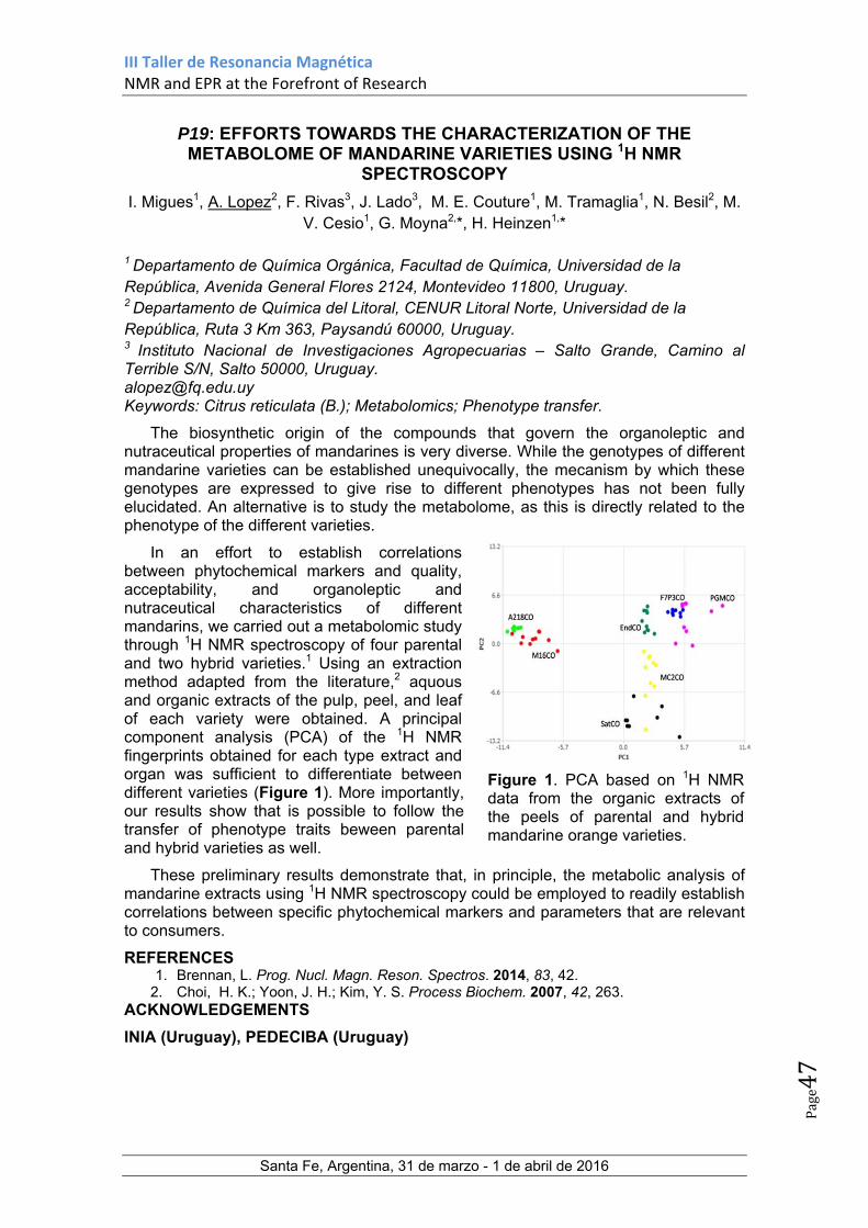
III Taller de Resonancia Magnética NMR and EPR at the Forefront of Research
Santa Fe, Argentina, 31 de marzo - 1 de abril de 2016
Page47
P19: EFFORTS TOWARDS THE CHARACTERIZATION OF THE METABOLOME OF MANDARINE VARIETIES USING 1H NMR
SPECTROSCOPY
I. Migues1, A. Lopez2, F. Rivas3, J. Lado3, M. E. Couture1, M. Tramaglia1, N. Besil2, M. V. Cesio1, G. Moyna2,*, H. Heinzen1,*
1 Departamento de Química Orgánica, Facultad de Química, Universidad de la República, Avenida General Flores 2124, Montevideo 11800, Uruguay. 2 Departamento de Química del Litoral, CENUR Litoral Norte, Universidad de la República, Ruta 3 Km 363, Paysandú 60000, Uruguay. 3 Instituto Nacional de Investigaciones Agropecuarias – Salto Grande, Camino al Terrible S/N, Salto 50000, Uruguay. [email protected] Keywords: Citrus reticulata (B.); Metabolomics; Phenotype transfer.
The biosynthetic origin of the compounds that govern the organoleptic and nutraceutical properties of mandarines is very diverse. While the genotypes of different mandarine varieties can be established unequivocally, the mecanism by which these genotypes are expressed to give rise to different phenotypes has not been fully elucidated. An alternative is to study the metabolome, as this is directly related to the phenotype of the different varieties.
In an effort to establish correlations between phytochemical markers and quality, acceptability, and organoleptic and nutraceutical characteristics of different mandarins, we carried out a metabolomic study through 1H NMR spectroscopy of four parental and two hybrid varieties.1 Using an extraction method adapted from the literature,2 aquous and organic extracts of the pulp, peel, and leaf of each variety were obtained. A principal component analysis (PCA) of the 1H NMR fingerprints obtained for each type extract and organ was sufficient to differentiate between different varieties (Figure 1). More importantly, our results show that is possible to follow the transfer of phenotype traits beween parental and hybrid varieties as well.
These preliminary results demonstrate that, in principle, the metabolic analysis of mandarine extracts using 1H NMR spectroscopy could be employed to readily establish correlations between specific phytochemical markers and parameters that are relevant to consumers.
REFERENCES 1. Brennan, L. Prog. Nucl. Magn. Reson. Spectros. 2014, 83, 42.
2. Choi, H. K.; Yoon, J. H.; Kim, Y. S. Process Biochem. 2007, 42, 263. ACKNOWLEDGEMENTS
INIA (Uruguay), PEDECIBA (Uruguay)
Figure 1. PCA based on 1H NMR data from the organic extracts of the peels of parental and hybrid mandarine orange varieties.

III Taller de Resonancia Magnética NMR and EPR at the Forefront of Research
Santa Fe, Argentina, 31 de marzo - 1 de abril de 2016
Page48
P20: THE USE OF METALS BOUND TO PEPTIDES AS GENETICALLY ENCODABLE SPIN-LABELS FOR NANOMETRIC DISTANCE
DETERMINATION IN PROTEINS
Florencia C. Mascali1, Roberta Crespo1, Tabares C. Leandro2 and Rodolfo M. Rasia1
1Instituto de Biología Molecular y Celular de Rosario; Área Biofísica, Facultad de Ciencias, Bioquímicas y Farmacéuticas, Universidad Nacional de Rosario, 2000 Rosario, Argentina
2Institute for Integrative Biology of the Cell (I2BC), Department of Biochemistry, Biophysics and Structural Biology, Université Paris-Saclay, CEA, CNRS UMR 9198, Gif-sur-Yvette, F-91198, France [email protected]
Keywords: PELDOR, spin-labels, lanthanides
Distance measurements are of importance in structural biology, allowing the understanding of quaternary structures of proteins, the arrangement of protein complexes and how proteins interact with each other. Pulse electron-electron double resonance (PELDOR or DEER) is a technique that can measure dipolar interaction between paramagnetic centers and it has been used to measure distances up to 10 nm. Such measurements can be done on systems in a wide variety of environments, even within the complex interior of cells. However, the conventional nitroxide-based DEER approaches are limited by the instability of radicals in the reducing environment of the cytosol. Because of this limitation metal-based spin-labels are more appealing. To date such labels have involved a redox stable paramagnetic metal ion encapsulated inside a well-defined ligand sphere that has a high binding efficiency for the metal. Also, rather than labelling proteins by chemical reactions, a genetically encoded spin-label would greatly simplify sample preparation, and facilitate in situ and ultimately in vivo DEER measurements. To this end we have explored the use of short peptide sequences as anchoring points for metals ions, so the spin label could be directly formed in situ by adding the metal ion to the peptide-tagged protein. Two approaches were tested: binding of Mn(II)1 to hexa-histidines (His-Tag) and binding of Gd(III) to lanthanide binding loops (LBT). Both systems were studied on overexpressed triple-helix bundles carrying a peptide tag at each end. In these peptides the two metal ion anchoring positions were separated by ~4nm. His-tags affinity for Mn(II) was low but this did not significantly impair the use of Mn(II) bound to His-tags as spin-labels for DEER measurements which were in agreement with the estimated distances. LBT affinity for Gd(III) is much higher and resulted the absence of free Gd(III). The advantages and disadvantages of these to systems as well as the perspective of they use for in situ and in vivo DEER measurements are discussed.
REFERENCES 1. H. Y. Vincent Ching, F. C. Mascali, H. C. Bertrand, E. Bruch, P. Demay-Drouhard, R. Rasia, C. Policar, L. C. Tabares, and S. Un, J. Phys. Chem. Lett., in press ACKNOWLEDGEMENTS
ECOS, CONICET, ANPCYT


![Taller de..[1]](https://static.fdocuments.us/doc/165x107/556f1dedd8b42ac8048b543b/taller-de1.jpg)


