
IHC PANEL MARKERS - biogenex.com
12
IHC PANEL MARKERS L u n g C a n c e r Antibodies for Lung Cancer Desmoglein-3, CD35, HIF-2, TTF1, p63, CK5, CK6, CK7, CK20, CEA, CK19, Napsin-A, Calretenin, PD1, PD-L1, CK Pan, Neurofilament, ALK/p80, CD34, MPO, EGFR, E-Cadherin, Epcam, Melan-A, p53, SOX2, TAG-72, Vimentin, chromogranin A, synaptophysin, CD56 BioGenex offers wide-ranging antibodies for several IHC panel for initial differentiation, tumor origin, treatment methods, and prognosis. All BioGenex antibodies are validated on human tissues to ensure sensitivity and specificity. BioGenex comprehensive IHC panels include a range of mouse monoclonal, rabbit monoclonal, and polyclonal antibodies to choose from. BioGenex offers a vast spectrum of high-quality antibodies for both diagnostic and reference laboratories. BioGenex strives to support efforts in clinical diagnostics and drug discovery development as we continue to expand our antibody product line offering in both ready-to-use and concentrated formats for both manual and automation systems.
Transcript of IHC PANEL MARKERS - biogenex.com

IHC PANEL MARKERSL u n g C a n c e r
Antibodies for Lung CancerDesmoglein-3, CD35, HIF-2, TTF1, p63, CK5, CK6, CK7, CK20, CEA, CK19, Napsin-A, Calretenin, PD1, PD-L1, CK Pan, Neurofilament, ALK/p80, CD34, MPO, EGFR, E-Cadherin, Epcam, Melan-A, p53, SOX2, TAG-72, Vimentin, chromogranin A, synaptophysin, CD56
BioGenex offers wide-ranging antibodies for several IHC panel for initial differentiation, tumor origin, treatment methods, and prognosis. All BioGenex antibodies are validated on human tissues to ensure sensitivity and specificity. BioGenex comprehensive IHC panels include a range of mouse monoclonal, rabbit monoclonal, and polyclonal antibodies to choose from.
BioGenex offers a vast spectrum of high-quality antibodies for both diagnostic and reference laboratories. BioGenex strives to support efforts in clinical diagnostics and drug discovery development as we continue to expand our antibody product line offering in both ready-to-use and concentrated formats for both manual and automation systems.

IHC PANEL MARKERS - Lung Cancer
Desmoglein-3
“Desmoglein-3 (Dsg3), also known as Cadherin family member 6 (CDHF6), is a member of the desmosomal cadherin family and plays a critical role in cell-cell adhesion. It is a calcium-binding transmembrane glycoprotein component of desmosomes in vertebrate epithelial cells. DSGs/ desmocollin (DSCs) are anchored to the intracellular plaque proteins plakoglobin, plakophilins, and desmoplakin, the latter of which mediates connection to the intermediate fil-ament cytoskeleton. Desmoglein 3 is predominately expressed in stratified squamous epithelia including epidermis, tongue, tonsil, esophagus and car-cinomas. The Desmoglein 3 antibody has been cited as a superior marker for Lung Squamous Cell Carcinomas, and helps distinguish lung squamous cell carcinoma cases from lung adenocarcinomas. Studies have also shown that a panel consisting of Desmoglein-3 utilized with Napsin A can be a useful immu-nohistochemical marker for differentiation of lung squamous cell carcinoma and adenocarcinomas from other subtypes.
Antibody Clone Localization Catalog Family
Desmoglein-3 DSG3/2839 Membrane AMA77, AXA77, MUA77
CD35
CD35, also known as complement receptor 1 (CR1), is a 220-300 kDa N-gly-cosylated member of the RCA (regulators of complement activation) family of proteins. It is a cell membrane-bound, monomeric glycoprotein and its prima-ry function is act as the receptor for complement components C3b and C4b, and it mediates the phagocytosis by neutrophils and monocytes of particles coated with C3b or C4b. CD35 binds and internalizes particles and immune complexes that are opsonized with MBL or complement components C3b, C3i, C4b, or C1q. CD35 additionally protects the cell from complement-mediated lysis by serving as a cofactor for Factor I and inhibiting the C3 and C5 conver-tases. CD35 is expressed on granulocytes, monocytes, B cells, some NK cells and erythrocytes. CD35 labels follicular dendritic cells of normal and neoplas-tic origin and is, thus, a useful marker for follicular dendritic cell sarcoma.
Antibody Clone Localization Catalog Family
CD35 To5 Membrane AMA78, AXA78, MUA78
HIF-2
HIF-2-alpha (also known as EPAS1) is a transcription factor involved in the induction of genes regulated by oxygen. It shares 48% sequence identity with HIF1-alpha (HIF1A). Like HIF1A, HIF-2-alpha regulates gene expression in response to hypoxia. It also regulates the vascular endothelial growth factor (VEGF) expression and seems to be implicated in the development of blood vessels and the tubular system of the lungs. HIF2A is expressed at relatively higher levels in villus sections of placenta and in lung samples.
Antibody Clone Localization Catalog Family
HIF-2 Polyclonal Membrane/Cytoplasm ARB28, AWB28, PUB28

IHC PANEL MARKERS - Lung Cancer
TTF1
Thyroid Transcription Factor-1 (TTF-1), also known as thyroid-specific enhancer-binding protein (T/EBP), is a 40 kD protein that is a member of NKx2 family of homeodomain transcription factors that regulates the expression of thyroid- and lung-specific genes. It is a very selective marker for adenocarcinomas of lung and thyroid origin. Nuclear localization of this protein is seen in the epithelial cells of thyroid gland and lung. The anti-TTF-1 antibody is a useful tool for differentiating pulmonary adenocarcinoma from metastatic breast carcinoma and mesothelioma.
Antibody Clone Localization Catalog Family
TTF1 SP141 Nucleus AN887, AY887, NU887
p63
This antibody will detect all isoforms of p63 since the epitope is within the DNA binding domain. The p63 protein is a member of the p53 family, which also includes p73. p63 protein is detected in proliferating cells of epithelium, cervix, urothelium and prostate. This antibody is useful to differentiate the Squamous cell carcinoma of lung.
Antibody Clone Localization Catalog Family
p63 4A4 Nucleus AM418, AX418, MU418
Cytokeratin 5
The mitotically active basal layers of most stratified squamous epithelia express 10% to 30% of their total protein as keratin. The two keratins specifically expressed in these cells are the type II keratin CK5 and its corresponding partner, type I keratin CK14, both of which are essential for the formation of 8-nm filaments. CK5 and calretinin have been useful in different studies as immunohistochemical markers suggestive of mesothelio-ma, and their expression is analyzed for the histological differential diagnosis with adenocarcinomas, especially when confronting with metastatic tumors of unknown origin. CK5 labels myoepithelial cells of breast and prostate basal cells. A cocktail of CK5, CK14 and p63, has been used as sensitive and specific basal cell marker of basal-like phenotype of breast carcinoma and to differ-entiate normal and prostate cancer. Loss-of-function mutations in the keratin 5 gene (KRT5) affected family members and in six unrelated patients with Dowling-Degos disease (DDD), an autosomal dominant genodermatosis. CK5 can be use a indicator of Squamous cell carcinoma.
Antibody Clone Localization Catalog Family
Cytokeratin 5 EP42 Cytoplasm AN853, AY853, NU853
Cytokeratin 5 EP24 Cytoplasm AN847, AY847, NU847

IHC PANEL MARKERS - Lung Cancer
Cytokeratin 6
The human type II Cytokeratin 6 (CK6; 56 kDa) is well known for its strong induction in stratified epithelia that feature an enhanced cell proliferation rate or abnormal differentiation during wound healing, in several diseases (e.g. psoriasis, actinic keratosis) and in cancer. CK6 is expressed on stratified epithelia including oral mucosa, esophagus, basal layer of epidermis, the outer root sheath of hair follicles, and in glandular epithelia. CK6 is a marker of hyperproliferative and activated keratinocytes found in psoriasis. CK6 paired with CK5 is useful to differentiate mesothelioma (positive) from lung carcinoma (negative) or metastatic carcinoma (negative) in the pleura. CK5/6 has also been used to distinguish usual ductal hyperplasia of the breast (strong staining) from solid papillary DCIS (negative) and as indicator of Squamous cell carcinoma.
Antibody Clone Localization Catalog Family
Cytokeratin 6 EP67 Cytoplasm AN845, AY845, NU845
Cytokeratin 7
Cytokeratin 7 is a 54 kD marker of simple epithelium. Antibody to Cytokeratin 7 strongly stains all cell layers of the urinary bladder transitional epithelium. However, Cytokeratin 7 is absent from gastrointestinal epithelium, hepato-cytes, proximal and distal tubules of the kidney, and myoepithelium, and also cannot be detected in the stratified epithelia of the skin, tongue, esophagus, or cervix. Cytokeratin 7 recognizes specific subtypes of adenocarcinomas and can be used to differentiate between Cytokeratin 7-positive tissues such as ovarian carcinomas and transitional cell carcinomas and Cytokeratin 7-neg-ative tissues such as carcinomas of the gastrointestinal tract and prostate cancers.
Antibody Clone Localization Catalog Family
Cytokeratin 7 OV-TL12/30 Cytoplasm AM255, AX255, MU255
Cytokeratin 20
Cytokeratin 20 (46kD) is relatively less acidic than other type I keratins. This antibody reacts with certain types of carcinomas such as adeno carcinomas of the colon, transitional cell carcinomas of the bladder and Merkel cell tumors of the skin. It does not stain breast, lung and endometrial adenocarcinomas. The differential staining pattern of this antibody makes it very useful for tumor evaluation when used in conjunction with cytokeratin 7 staining.
Antibody Clone Localization Catalog Family
Cytokeratin 20 IT-Ks20.8 Cytoplasm AM315, AX315, MU315
Cytokeratin 20 EP23 Cytoplasm AN849, AY849, NU849

IHC PANEL MARKERS - Lung Cancer
CEA
CEA consists of a heterogeneous family of related oncofetal 200 kD glycoproteins that is secreted into the glycocalyx surface of gastrointestinal cells. Usually CEA is demonstrated as a linear labeling of the apical poles of cells lining the glandular lumen and, occasionally, as weak staining near the apex of colonic epithelial cells. Pancreatic carcinomas, testicular tumor, gallbladder neoplasms and granular cell myoblastomas stain positive, where-as malignant tumors of brain, prostate, skin, lymphoreticular tissues, hepato-cellular carcinomas, esophageal squamous cell carcinomas, and mesothelio-ma fail to stain for CEA. This antibody stains carcinoembryonic antigen in the cytoplasm of positive cells.
Antibody Clone Localization Catalog Family
CEA B01-94-11M-P Cytoplasm AM009, AX009, MU009
CEA CEA88 Cytoplasm AM365, AX365, MU365
CEA Polyclonal Cytoplasm AR009, AW009
Cytokeratin 19
Cytokeratin 19 (molecular mass 40 kD) is a marker of simple epithelia. Cytokeratin 19 has been found in mesothelial and mesothelioma cells, ovarian cysts, cystadenomas, and ovarian carcinomas, in adenocarcinomas of the lung and in tumor cells of pulmonary metastases, in the ductal cells of normal pancreas and in pancreatic cancers. It has been shown to be present in the basal layer of non-keratinizing stratified squamous epithelia such as the oral cavity and the ectocervix.
Antibody Clone Localization Catalog Family
Cytokeratin 19 RCK108 Cytoplasm AM246, AX246, MU246
Napsin-A
Napsin A has specific function in normal alveolar epithelium and is proposed to play a role in the protelytic processing of surfactant precursors. Napsin A is reported to be predominantly expressed in lamellar bodies of type II pneu-mocutes, secondary lysosymes of alveolar macrophages, respiratory epithe-lium of terminal and respiratory bronchioles, plasma cells within a subset of lymphocytes in normal lung, as well as in epithellial cells of renal tubiles in normal kidney and is weakly expressed in normal spleen. Napsin-A is sensitive for primary lung adenocarcinoma.
Antibody Clone Localization Catalog Family
Napsin-A IP64 Cytoplasm AM701, AX701, MU701
IHC PANEL MARKERS - Lung Cancer
Calretinin
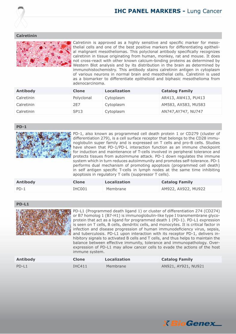
Calretinin is approved as a highly sensitive and specific marker for meso-thelial cells and one of the best positive markers for differentiating epitheli-al malignant mesotheliomas. This polyclonal antibody specifically recognizes calretinin in tissue originating from human, monkey, rat and mouse. It does not cross-react with other known calcium-binding proteins as determined by Western Blot analysis and by its distribution in the brain as determined by immunohistochemistry. This antibody stains calretinin antigen in cytoplasm of various neurons in normal brain and mesothelial cells. Calretinin is used as a biomarker to differentiate epithelioid and biphasic mesothelioma from adenocarcinoma.
Antibody Clone Localization Catalog Family
Calretinin Polyclonal Cytoplasm AR413, AW413, PU413
Calretinin 2E7 Cytoplasm AM583, AX583, MU583
Calretinin SP13 Cytoplasm AN747,AY747, NU747
PD-1
PD-1, also known as programmed cell death protein 1 or CD279 (cluster of differentiation 279), is a cell surface receptor that belongs to the CD28 immu-noglobulin super family and is expressed on T cells and pro-B cells. Studies have shown that PD-1/PD-L interaction function as an immune checkpoint for induction and maintenance of T-cells involved in peripheral tolerance and protects tissues from autoimmune attack. PD-1 down regulates the immune system which in turn reduces autoimmunity and promotes self-tolerance. PD-1 performs dual mechanism of promoting apoptosis (programmed cell death) in self antigen specific T-cells in lymph nodes at the same time inhibiting apoptosis in regulatory T cells (suppressor T cells).
Antibody Clone Localization Catalog Family
PD-1 IHC001 Membrane AM922, AX922, MU922
PD-L1
PD-L1 (Programmed death ligand 1) or cluster of differentiation 274 (CD274) or B7 homolog 1 (B7-H1) is immunoglobulin-like type I transmembrane glyco-protein that act as a ligand for programmed death 1 (PD-1). PD-L1 expression is seen on T cells, B cells, dendritic cells, and monocytes. It is critical factor in infection and disease progression of human immunodeficiency virus, sepsis, and tuberculosis. PD-L1 upon interaction with its receptor PD-1, delivers in-hibitory signals to activated B cells and T cells, and thus helps to maintain the balance between effective immunity, tolerance and immunopathology. Over-expression of PD-L1 may allow cancer cells to evade the actions of the host immune system.
Antibody Clone Localization Catalog Family
PD-L1 IHC411 Membrane AN921, AY921, NU921

IHC PANEL MARKERS - Lung Cancer
PAN Cytokeratin
Human keratins are family of water-insoluble proteins with molecular weights ranging from 40-68kD. This monoclonal cytokeratin antibody can be applied to detect cytokeratins 4, 5, 8, 10, 13, 18 in simple or stratified epithelium in most vertebrates including humans. It can be used as a marker for carcino-mas as wll as some special types of tumors which have an epithelial compo-nent or differentiation. The antibody stains cytokeratin in cytoplasm of normal or malignant epithelial cells.
Antibody Clone Localization Catalog Family
PAN Cytokeratin Lu-5 Cytoplasm AM181, AX181, MU181
PAN Cytokeratin C11 Cytoplasm AM357, AX357, MU357
Neurofilament
Neurofilaments (10 nm diameter) and microtubules (25 nm diameter) com-prise the main structural elements of neuronal axons, dendrites, and perik-erya. Neurofilaments are composed of three major polypeptides referred to as the neurofilament triplet with approximate molecular weights of 200 kD, 160 kD and 68 kD. This antibody can be used for positive identification of neurons in the central and peripheral nervous systems. In general, co-expression of keratin and neurofilament should be interpreted as indicating neuroendocrine differentiation of a given tissue or neoplasm. The antibody stains Neurofila-ment in sections of brain and other tissues.
Antibody Clone Localization Catalog Family
Neurofilament NE-14 Cytoplasm AM073, AX073, MU073
ALK/p80
This antibody recognizes a human p80 protein, identified as a hybrid of the anaplastic lymphoma kinase (ALK) gene and the nucleophosmin (NPM) gene resulting from the t(2;5)(p23;q35) translocation found in a third of large cell lymphomas. This antibody can be used to detect p80 in these lymphomas and may also be used to detect a recently described subtype of large B cell lymphoma, which expresses the full-length ALK protein.
Antibody Clone Localization Catalog Family
ALK/p80 SP8 Cytoplasmic and nuclear AN770, AY770, NU770
CD34
This is an antibody to the CD34 antigen in human endothelial and hemato-poietic cells. It stains positive in a variety of vascular and lymphatic tumors. QBEnd/10 may now prove to be a more specific method of evaluating vascu-larization than Factor VIII antibody and is an important tool for tumor eval-uation. This antibody stains endothelial cell cytoplasm and cross-reacts with basement membrane collagen.
Antibody Clone Localization Catalog Family
CD34 QBend/10 Membrane AM236, AX236, MU236
CD34 EP88 Membrane AN779, AY779, NU779

IHC PANEL MARKERS - Lung Cancer
MPO
Myeloperoxidase is an important enzyme used by granulocytes during phago-cytic lysis of foreign particles engulfed. In normal tissues and in a variety of myeloproliferative disorders, myeloid cells of both neutrophilic and eosinophil-ic types at all stages of maturation, exhibit strong cytoplasmic reactivity for MPO. Erythroid precursors, megakaryocytes, lymphoid cells, mast cells, and plasma cells are nonreactive. MPO is not observed in the neoplastic cells of a wide variety of epithelial tumors and sarcomas. MPO is useful in differentiating between myeloid and lymphoid leukemias.
Antibody Clone Localization Catalog Family
MPO Polyclonal Cytoplasm AR496, AW496, PU496
EGFR
EGFR (LRVAP) reacts with the 170 kD EGFR transmembrane glycoprotein. It binds specifically to the intracellular portion, regardless of phosphoryla-tion state. The extracellular domain binds epidermal growth factor (EGF) as a proliferation signal. The EGFR antibody is made against a sequence which is unique from related tyrosine kinase receptors and hence shows no cross-re-activity.
Antibody Clone Localization Catalog Family
EGFR Polyclonal Membrane and Cytoplasm AR335, AW335, PU335
EGFR EP22 Membrane and Cytoplasm AN781, Y781, NU781
E-Cadherin
E-Cadherin is a transmembrane glycoprotein that plays an important role in epithelial cell adhesion. In prostate cancers, the expression of E-cadherin is reported to be reduced or absent in comparison with its expression in normal prostate which is uniformly strong. A decreased expression of E-Cadherin is associated with metastatic potential and poor prognosis in breast cancer and esophagus cancer. In combination with p120 Catenin or Cytokeratin, it is use-ful for the differentiation between ductal (E-Cadherin positive) and lobular (E-Cadherin negative) breast carcinomas. It may also help in diagnosis of mesothelioma.
Antibody Clone Localization Catalog Family
E-Cadherin 36 Membrane AM390, AX390, MU390
E-Cadherin EP6 Membrane AN725, AY725, NU725

IHC PANEL MARKERS - Lung Cancer
Ep-CAM
Ep-CAM is a highly conserved type I transmembrane glycoprotein and is expressed on most normal and malignant epithelial cells. Ep-CAM is also known as epithelial cell adhesion molecule or MOC31, Ber-EP4. It is detected at the membrane/cytoplasm of the majority of epithelial tissues (all simple, pseudo-stratified and transitional epithelial), with the exception of the adult squamous epithelium and some epithelium-derived cell, such as hepatocytes, epidermal keratinocytes, gastric parietal cells, myoepithelial cells, and thymic cortical epithelium. In tumors, Ep-CAM is over expressed by the majority of human epithelial carcinomas, except hepatocellular carcinomas (HCC).
Antibody Clone Localization Catalog Family
Ep-CAM EP155 Membrane AN820, AY820, NU820
Melan-A
Melan-A, a product of the MART-1 gene, is a differentiation antigen which is expressed in 100% of melanocytes, most melanomas, and 50-60% of mela-noma cell lines. It is one of the melanoma antigens recognized by autologous cytotoxic T cells, and as an antigenic target for tumor infiltrating lymphocytes. This antibody also stains Melan-A in normal melanocytes and in the retina. It does not stain normal or tumor tissues from non-melanocyte lineages. This antibody stains positive in cytoplasm of melanocytes and other positive cells.
Antibody Clone Localization Catalog Family
Melan-A A103 Cytoplasm AM361, AX361, MU361
p53
Tumor protein p53, a nuclear protein, plays an essential role in the regulation of cell cycles, specifically in the transition from G0 to G1. It is found in very low levels in normal cells, and it functions as a tumor suppressor within a variety of tumors by either stimulating apoptosis or growth arrest in deference to cell type and physiological factors. p53 is overexpressed in over 50% of human cancers. Positive staining of p53 detected by immunohistochemistry has been observed in colon cancer, breast cancer, lung cancer, prostate cancer and ovary cancer.
Antibody Clone Localization Catalog Family
p53 EP9 Nucleus AN728, AY728,NU728
p53 BP53-12-1 Nucleus AM195, AX195,MU195
p53 DO7 Nucleus AM239, AX239,MU239
p53 1801 Nucleus AM240, AX240,MU240

IHC PANEL MARKERS - Lung Cancer
SOX2
SOX2 is a member of the SRY-related HMG-box (SOX) family of transcrip-tion factors involved in the regulation of embryonic development and in the determination of cell fate. It is required for stem cell maintenance in the central nervous system, and it also regulates gene expression in the stomach. SOX2 is necessary for regulating multiple transcription factors that affect Oct3/4 expression. An essential function of SOX2 is to stabilize embryonic stem cells in a pluripotent state by maintaining the requisite level of Oct3/4 expression. SOX2 is associated with aggressive tumor behavior, and is expressed in lung adenocarcinomas.
Antibody Clone Localization Catalog Family
SOX2 EP103 Nucleus AN833, AY833, NU833
SOX2 Polyclonal Nucleus AR788,AW788, PU788
TAG-72
Tumor-Associated Glycoprotein 72 (TAG-72) is an oncofetal mucin antigen expressed by normal secretory endometrium and most human adenocarci-nomas, including colorectal, gastric, pancreatic, mammary, and ovarian. This antigen is expressed by invasive ductal breast carcinomas, colon, pancre-atic, gastric, esophageal, lung, ovarian and endometrial adenocarcinomas. It is not expressed by leukemias, lymphomas, sarcomas, mesotheliomas, melanomas, or benign tumors. This antigen is also expressed on normal secretory endometrium, but not on other normal tissues. This antibody stains positive in the cytoplasm of specific carcinoma cells.
Antibody Clone Localization Catalog Family
TAG-72 B72.3 Cytoplasm AM054, AX054, MU054
Vimentin
Vimentin is the major intermediate filament in a variety of mesenchymal or mesenchymally derived non-muscle cell types. Vimentin is found in all types of sarcomas and lymphomas. Positive staining for vimentin is seen in most cells of fibrosarcomas, liposarcomas, malignant fibrous histocytomas, angio-sarcomas, chondrosarcomas and lymphomas. When the vimentin antibody is used in combination with other antibodies as a panel, it can aid in the histo-logical classification of normal and malignant tissues. This antibody immuno-histochemically labels a variety of mesenchymal cells.
Antibody Clone Localization Catalog Family
Vimentin V9 Cytoplasm AM074, AX074, MU074
Vimentin LN6 Cytoplasm AM163, AX163, MU163
Chromogranin A
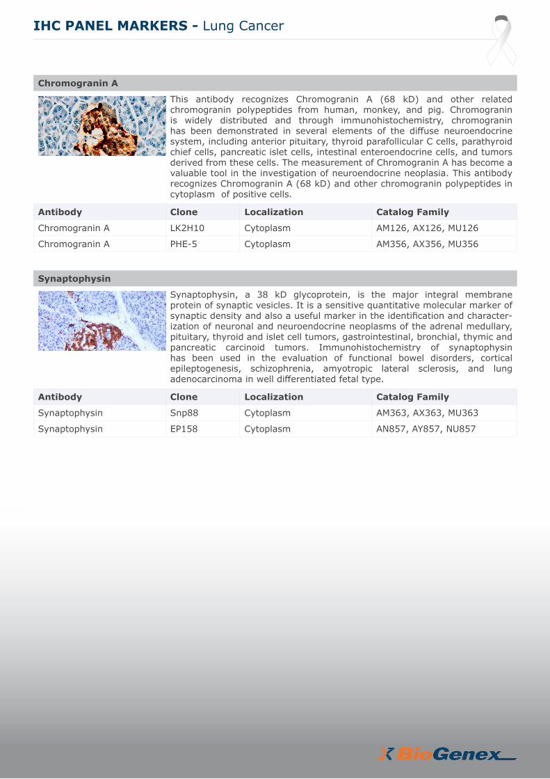
This antibody recognizes Chromogranin A (68 kD) and other related chromogranin polypeptides from human, monkey, and pig. Chromogranin is widely distributed and through immunohistochemistry, chromogranin has been demonstrated in several elements of the diffuse neuroendocrine system, including anterior pituitary, thyroid parafollicular C cells, parathyroid chief cells, pancreatic islet cells, intestinal enteroendocrine cells, and tumors derived from these cells. The measurement of Chromogranin A has become a valuable tool in the investigation of neuroendocrine neoplasia. This antibody recognizes Chromogranin A (68 kD) and other chromogranin polypeptides in cytoplasm of positive cells.
Antibody Clone Localization Catalog Family
Chromogranin A LK2H10 Cytoplasm AM126, AX126, MU126
Chromogranin A PHE-5 Cytoplasm AM356, AX356, MU356
Synaptophysin
Synaptophysin, a 38 kD glycoprotein, is the major integral membrane protein of synaptic vesicles. It is a sensitive quantitative molecular marker of synaptic density and also a useful marker in the identification and character-ization of neuronal and neuroendocrine neoplasms of the adrenal medullary, pituitary, thyroid and islet cell tumors, gastrointestinal, bronchial, thymic and pancreatic carcinoid tumors. Immunohistochemistry of synaptophysin has been used in the evaluation of functional bowel disorders, cortical epileptogenesis, schizophrenia, amyotropic lateral sclerosis, and lung adenocarcinoma in well differentiated fetal type.
Antibody Clone Localization Catalog Family
Synaptophysin Snp88 Cytoplasm AM363, AX363, MU363
Synaptophysin EP158 Cytoplasm AN857, AY857, NU857
IHC PANEL MARKERS - Lung Cancer

BioGenex Primary Antibody Format and Pack Size
BioGenex antibodies are optimized to provide a maximum signal with the minimum background for immunohistochemical staining. All our antibodies are optimized and recommended for use with all Super Sensitive™ Detection Systems to provide optimum staining.
BioGenex Ready-to-Use (RTU) antibodies are fully optimized for use with BioGenex Detection Systems without the need for further dilution or titration. BioGenex concentrated antibodies are provided with recommended dilutions for optimal use with BioGenex Detection Systems, allowing rapid titration and testing.
IHC PANEL MARKERS - Lung Cancer
For specific information on the individual antibody, please refer to the datasheets available on www.biogenex.com or call BioGenex Technical Support at 1(800)421-4149 or write to [email protected].
Prefix Type Species Suffix Volume and Format
AM/AN Monoclonal AM-Mouse/AN-Rabbit -5M/5ME 6 mL - Ready-to-use (manual)
AM/AN Monoclonal AM-Mouse/AN-Rabbit -10M/10ME 10 mL - Ready-to-use (i6000™)
AX/AY Monoclonal AX-Mouse/AY-Rabbit -YCD/YCDE and -50D/50DE 16 mL and 5 mL Ready-to-use (Xmatrx®)
AR Polyclonal Rabbit -5R/5RE 6 mL - Ready-to-use (manual)
AR Polyclonal Rabbit -10R/10RE 10 mL - Ready-to-use (i6000™)
AW Polyclonal Rabbit -YCD/YCDE and -50D/50DE 16 mL and 5 mL Ready-to-use (Xmatrx®)
MU/NU Monoclonal AM- Mouse/AN-Rabbit -UC/UCE and -5UC/5UCE 1 mL and 0.5 mL Concentrate
PU Polyclonal Rabbit -UC/UCE and -5UC/5UCE 1 mL and 0.5 mL Concentrate
Other Panel Markers from BioGenex
Breast cancer panel Pancreas tumor
B&T cell Associated Lymphoma Liver cancer
Cervical cancer Kidney cancer
Colorectal and stomach cancer Head & neck cancer
Melanoma Bladder cancer
Muscle cancer Germ cell tumor
Ovarian cancer Vascular tumor
Prostate/Testicular cancer Pituitary gland
Neuroendocrine tumor Esophagus cancer
In the U.S., call +1 (800) 421-4149Outside the U.S., call +91-40-27185500 www.biogenex.com
US: [email protected] India: [email protected] Global: [email protected]
© 2019 BioGenex Laboratories, Inc. All Rights Reserved, Doc No: 907-4057.0 Rev B
Customer Service


![IHC PPT Ancillary Productsmy1hr-public.s3.amazonaws.com/documents/enroll/IHC PPT Ancillary Products[3].pdfAncillary Products From The IHC Group. The IHC Group Corporate Overview Ø](https://static.fdocuments.us/doc/165x107/5e38c9b5e1bb9a3e4e5b3bd8/ihc-ppt-ancillary-productsmy1hr-publics3-ppt-ancillary-products3pdf-ancillary.jpg)