IgM antigen receptor complex contains phosphoprotein products of ...
5
Proc. Natl. Acad. Sci. USA Vol. 88, pp. 3982-3986, May 1991 Immunology IgM antigen receptor complex contains phosphoprotein products of B29 and mb-i genes (membrane immunoglobulin/B lymphocyte/signal transduction) KERRY S. CAMPBELL*, ELIZABETH J. HAGER*, R. JOACHIM FRIEDRICH*, AND JOHN C. CAMBIER*tt *National Jewish Center for Immunology and Respiratory Medicine, Division of Basic Research, Department of Pediatrics, Denver, CO 80206; and tUniversity of Colorado Health Sciences Center, Department of Microbiology and Immunology, Denver, CO 80206 Communicated by Philippa Marrack, February 20, 1991 ABSTRACT Membrane immunoglobulin M (mIgM) and mIgD are major B-lymphocyte antigen receptors, which func- tion by internalizing antigens for processing and presentation to T cells and by transducing essential signals for proliferation and differentiation. Although ligation of mIgM or mIgD results in rapid activation of a phospholipase C and a tyrosine ki- nase(s), these receptors have cytoplasmic tails of only three amino acid residues (Lys-Val-Lys), which seem ill suited for direct physical coupling with cytoplasmic signal transduction structures. In this report, we identify the a, 13, and Y compo- nents of the mIgM-associated phosphoprotein complex, which may play a role in signal transduction. Proteolytic peptide mapping demonstrated that the IgM-a chain differs from Ig-13 and Ig-y. The chains were purified, and amino-terminal se- quencing revealed identity with two previously cloned B-cell- specific genes. One component, IgM- a, is a product of the mb-i gene, and the two additional components, Ig-J3 and Ig-y, are products of the B29 gene. Immunoblotting analysis using rabbit antibodies prepared against predicted peptide sequences of each gene product confirmed the identification of these mIgM- associated proteins. The deduced sequence indicates that these receptor subunits lack inherent protein kinase domains but include common tyrosine-containing sequence motifs, which are likely sites of induced tyrosine phosphorylation. Membrane-bound immunoglobulin M (mIgM) and mIgD are major B-lymphocyte surface structures that specifically bind antigen and subsequently transduce growth-modulating sig- nals. Ligation of either mIgM or mIgD with polyvalent antigen or anti-immunoglobulin antibodies results in the rapid activation of a polyphosphoinositide-specific phospholipase C, which generates calcium-mobilizing inositol polyphos- phates and protein kinase C-activating diacylglycerol (1, 2). Recent evidence from several laboratories has established that a tyrosine kinase is also activated upon B-cell antigen receptor ligation (3, 4) and that ligand-induced phospholipase C activation is kinase dependent (5). Candidates for the tyrosine kinase include Lyn, which has been shown to associate with mIgM (6), and Blk, which is B-cell-specific (7). Interestingly, mIgM and mIgD have minimal cytoplasmic structure [a Lys-Val-Lys (KVK) sequence] on each of two membrane-spanning heavy chains (8-10), which is probably incapable of direct physical coupling to such cytoplasmic signaling structures. Recently B-cell antigen receptor-associated structures have been identified that may play an important role in physical coupling to signal transduction structures, analo- gous to CD3 components in association with the T-cell antigen receptor. Multiple components of the mIgM- associated glycoprotein complex have been defined biochem- ically by several laboratories (11-16) and designated IgM-a (32 kDa), Ig-,B (37 kDa), and Ig-y(34 kDa) (13, 14). All of these components are inducibly phosphorylated on tyrosine resi- dues upon receptor ligation or treatment of cells with alumi- num fluoride (ref. 13 and K.S.C. and J.C.C., unpublished observations). IgM-a is found disulfide-linked as a het- erodimer with either Ig-P or Ig-y (13, 14). Studies by Reth and colleagues (11, 12) have provided evidence that expression of the mb-i gene product, MB-1, is crucial for mIgM surface expression. Concurrently, they have correlated expression of an mIgM-associated protein, designated IgM-a, with surface expression of the receptor (11, 12). These results indicate that MB-1 is required for mIgM transport to the cell surface and suggest that it may be a component of the B-cell antigen receptor complex. In this report we have identified the primary structure of three components of the mIgM antigen receptor complex in normal B cells by using proteolytic peptide mapping, amino- terminal sequencing, and immunoblotting with anti-peptide antibodies. IgM-a (pp32) was identified as a product of the mb-i gene. Ig-A (pp37) and Ig- y (pp34) were characterized as products of the B29 gene. MATERIALS AND METHODS Cell Preparation. B lymphocytes were isolated from spleens of BDF1 mice (6-12 weeks old, The Jackson Laboratory) as previously described (17). High and intermediate-density B cells (1.066-1.092 g/cm3) were isolated by centrifugal sedi- mentation through discontinuous Percoll (Pharmacia) gradi- ents as described by Ratcliffe and Julius (18). Radiolabeling and Immunoprecipitation of the Complex. B cells were permeabilized with a-lysophosphatidylcholine, equilibrated with [y-32P]ATP, stimulated with A1F4 for 30 min at 37°C, and lysed in 1% digitonin buffer (containing phosphatase and protease inhibitors) before mIgM was im- munoprecipitated from the detergent-soluble fraction with Sepharose-derivatized monoclonal anti-, antibody b-7-6 (19), as previously described (13). The anti-major histocom- patibility complex (MHC) class I (H2K) monoclonal antibody M1/42.398 (20) was also used in control immunoprecipita- tions. V8 Protease Peptide Mapping. Immunoprecipitates (40 mil- lion cell equivalents per lane) were analyzed by SDS/PAGE under reducing conditions (21), and the gel was dried and autoradiographed. Individual mIgM-associated protein bands were excised and proteolytically digested and analyzed in a second gel according to the method of Cleveland et al. (22), using 10 pg of V8 protease (Staphylococcus aureus; ICN) per lane. The second separating gel contained 20% polyacrylamide and was prepared according to the method of Giulian and Graham (23). Abbreviations: mIgM, membrane IgM; mIgD, membrane IgD; MHC, major histocompatibility complex. fTo whom reprint requests should be addressed. 3982 The publication costs of this article were defrayed in part by page charge payment. This article must therefore be hereby marked "advertisement" in accordance with 18 U.S.C. §1734 solely to indicate this fact.
Transcript of IgM antigen receptor complex contains phosphoprotein products of ...

Proc. Natl. Acad. Sci. USAVol. 88, pp. 3982-3986, May 1991Immunology
IgM antigen receptor complex contains phosphoprotein products ofB29 and mb-i genes
(membrane immunoglobulin/B lymphocyte/signal transduction)
KERRY S. CAMPBELL*, ELIZABETH J. HAGER*, R. JOACHIM FRIEDRICH*, AND JOHN C. CAMBIER*tt*National Jewish Center for Immunology and Respiratory Medicine, Division of Basic Research, Department of Pediatrics, Denver, CO 80206; and tUniversityof Colorado Health Sciences Center, Department of Microbiology and Immunology, Denver, CO 80206
Communicated by Philippa Marrack, February 20, 1991
ABSTRACT Membrane immunoglobulin M (mIgM) andmIgD are major B-lymphocyte antigen receptors, which func-tion by internalizing antigens for processing and presentationto T cells and by transducing essential signals for proliferationand differentiation. Although ligation ofmIgM or mIgD resultsin rapid activation of a phospholipase C and a tyrosine ki-nase(s), these receptors have cytoplasmic tails of only threeamino acid residues (Lys-Val-Lys), which seem ill suited fordirect physical coupling with cytoplasmic signal transductionstructures. In this report, we identify the a, 13, and Y compo-nents of the mIgM-associated phosphoprotein complex, whichmay play a role in signal transduction. Proteolytic peptidemapping demonstrated that the IgM-a chain differs from Ig-13and Ig-y. The chains were purified, and amino-terminal se-quencing revealed identity with two previously cloned B-cell-specific genes. One component, IgM- a, is a product of the mb-igene, and the two additional components, Ig-J3 and Ig-y, areproducts of the B29 gene. Immunoblotting analysis using rabbitantibodies prepared against predicted peptide sequences ofeach gene product confirmed the identification of these mIgM-associated proteins. The deduced sequence indicates that thesereceptor subunits lack inherent protein kinase domains butinclude common tyrosine-containing sequence motifs, whichare likely sites of induced tyrosine phosphorylation.
Membrane-bound immunoglobulin M (mIgM) and mIgD aremajor B-lymphocyte surface structures that specifically bindantigen and subsequently transduce growth-modulating sig-nals. Ligation of either mIgM or mIgD with polyvalentantigen or anti-immunoglobulin antibodies results in the rapidactivation of a polyphosphoinositide-specific phospholipaseC, which generates calcium-mobilizing inositol polyphos-phates and protein kinase C-activating diacylglycerol (1, 2).Recent evidence from several laboratories has establishedthat a tyrosine kinase is also activated upon B-cell antigenreceptor ligation (3, 4) and that ligand-induced phospholipaseC activation is kinase dependent (5). Candidates for thetyrosine kinase include Lyn, which has been shown toassociate with mIgM (6), and Blk, which is B-cell-specific (7).Interestingly, mIgM and mIgD have minimal cytoplasmicstructure [a Lys-Val-Lys (KVK) sequence] on each of twomembrane-spanning heavy chains (8-10), which is probablyincapable of direct physical coupling to such cytoplasmicsignaling structures.Recently B-cell antigen receptor-associated structures
have been identified that may play an important role inphysical coupling to signal transduction structures, analo-gous to CD3 components in association with the T-cellantigen receptor. Multiple components of the mIgM-associated glycoprotein complex have been defined biochem-ically by several laboratories (11-16) and designated IgM-a
(32 kDa), Ig-,B (37 kDa), and Ig-y(34 kDa) (13, 14). All ofthesecomponents are inducibly phosphorylated on tyrosine resi-dues upon receptor ligation or treatment of cells with alumi-num fluoride (ref. 13 and K.S.C. and J.C.C., unpublishedobservations). IgM-a is found disulfide-linked as a het-erodimer with either Ig-P or Ig-y (13, 14).
Studies by Reth and colleagues (11, 12) have providedevidence that expression of the mb-i gene product, MB-1, iscrucial for mIgM surface expression. Concurrently, theyhave correlated expression of an mIgM-associated protein,designated IgM-a, with surface expression of the receptor(11, 12). These results indicate that MB-1 is required formIgM transport to the cell surface and suggest that it may bea component of the B-cell antigen receptor complex.
In this report we have identified the primary structure ofthree components of the mIgM antigen receptor complex innormal B cells by using proteolytic peptide mapping, amino-terminal sequencing, and immunoblotting with anti-peptideantibodies. IgM-a (pp32) was identified as a product of themb-i gene. Ig-A (pp37) and Ig-y (pp34) were characterized asproducts of the B29 gene.
MATERIALS AND METHODSCell Preparation. B lymphocytes were isolated from spleens
of BDF1 mice (6-12 weeks old, The Jackson Laboratory) aspreviously described (17). High and intermediate-density Bcells (1.066-1.092 g/cm3) were isolated by centrifugal sedi-mentation through discontinuous Percoll (Pharmacia) gradi-ents as described by Ratcliffe and Julius (18).
Radiolabeling and Immunoprecipitation of the Complex. Bcells were permeabilized with a-lysophosphatidylcholine,equilibrated with [y-32P]ATP, stimulated with A1F4 for 30min at 37°C, and lysed in 1% digitonin buffer (containingphosphatase and protease inhibitors) before mIgM was im-munoprecipitated from the detergent-soluble fraction withSepharose-derivatized monoclonal anti-, antibody b-7-6(19), as previously described (13). The anti-major histocom-patibility complex (MHC) class I (H2K) monoclonal antibodyM1/42.398 (20) was also used in control immunoprecipita-tions.V8 Protease Peptide Mapping. Immunoprecipitates (40 mil-
lion cell equivalents per lane) were analyzed by SDS/PAGEunder reducing conditions (21), and the gel was dried andautoradiographed. Individual mIgM-associated proteinbands were excised and proteolytically digested and analyzedin a second gel according to the method of Cleveland et al.(22), using 10 pg of V8 protease (Staphylococcus aureus;ICN) per lane. The second separating gel contained 20%polyacrylamide and was prepared according to the method ofGiulian and Graham (23).
Abbreviations: mIgM, membrane IgM; mIgD, membrane IgD;MHC, major histocompatibility complex.fTo whom reprint requests should be addressed.
3982
The publication costs of this article were defrayed in part by page chargepayment. This article must therefore be hereby marked "advertisement"in accordance with 18 U.S.C. §1734 solely to indicate this fact.

Proc. Natl. Acad. Sci. USA 88 (1991) 3983
Amino-Terminal Sequencing. Mononuclear cells were pre-pared from spleens of200 mice and lysed in 1% digitonin lysisbuffer as above. Particulate material was removed by cen-trifugation (12,000 x g for 20 min) and mIgM was affinitypurified on a Sepharose column derivatized with monoclonalanti-pt antibody (b-7-6). The Sepharose was washed exten-sively with 0.2% digitonin buffer, and the mIgM-associatedprotein complex was selectively eluted with 1% octyl gluco-side [which is dialyzable, yet dissociates the complex frommembrane immunoglobulin as effectively as 0.5% TritonX-100 (13); data not shown]. Eluate was concentrated byvacuum dialysis, dialyzed against 0.2% SDS, and lyophilized.The samples were trace labeled with a 32P-labeled anti-IgMimmunoprecipitate (prepared as above) and separated onSDS/PAGE with an agarose stacker under nonreducingconditions. Radiolabeled protein (about 70 kDa) was local-ized in the wet gel by using an automated /8 emission scanner(AMBIS Systems, San Diego) and again electrophoresed onSDS/PAGE under reducing conditions. Separating gels wereaged 2 days and the reducing gel was run with 0.1 mMthioglycolate in the cathode running buffer to avoid artificialblockage of amino termini. Proteins in the second-dimensiongel were transferred electrophoretically to Immobilon-Pmembrane (Millipore) and autoradiographed. 32P-labeledprotein bands were excised, washed extensively, and sub-jected to amino-terminal sequence analysis by the Edmandegradation method (24-26) on an Applied Biosystems 470Agas-phase sequencer using on-line phenylthiohydantoin(PTH) determination with an Applied Biosystems 120A an-alyzer. The sequencing data lacked a significant backgroundof contaminating amino acids, indicating a high degree ofpurity in the preparations.
Anti-Peptide Antibody Production. Macvector (IBI Pustell)computer analysis of MB-1 and B29 sequences, predictedthat peptide sequences PPVPLGPGQGTTQ and DMPD-DYEDENL from MB-1 (peptide 254, extracellular residues72-85 and peptide 298, cytoplasmic residues 171-181, re-spectively) and sequences DKDDGKAGMEED andFRKRGSQQPQ from B29 (peptide 296, cytoplasmic residues181-192 and peptide 300, extracellular residues 76-85, re-spectively) would have high antigenicity. These peptideswere synthesized by using t-butoxycarbonyl (t-Boc) chem-istry (27-28) on a PAM resin with an Applied Biosystems430A peptide synthesizer and purified by using reversed-phase HPLC (Beckman) on a C18 column (Vydac, Hesperia,CA). Identity was confirmed by sequence analysis, andindividual peptides were coupled to keyhole limpet hemocy-anin with 1-ethyl-3-(3-dimethylaminopropyl) carbodiimide(EDAC; Bio-Rad). Individual rabbits (New Zealand White;Hazleton Research Products, Philadelphia) were immunizedsubcutaneously with 1000 ,g of peptide-hemocyanin conju-gate in complete Freund's adjuvant (GIBCO) and boosted at2- to 3-week intervals with 100 Mg of antigen in incompleteFreund's adjuvant. Antigen-specific antibodies were affinitypurified from rabbit serum by using peptide-bovine serumalbumin-derivatized Sepharose (Pharmacia) and elution with3.5 M MgCI2.Immunoblotting. Digitonin (1%) lysates were prepared
from splenic B cells (108 cells per sample), immunoprecipi-tated with Sepharose derivatized with anti-,u monoclonalantibody (b-7-6) or anti-H2K monoclonal antibody (Mi/42.398) and subjected to reducing SDS/PAGE, and proteinswere transferred electrophoretically to nitrocellulose. Afterblocking with 2% bovine serum albumin, nitrocellulose wasincubated with affinity-purified anti-MB-1 peptide antibodyor affinity-purified anti-B29 peptide antibody (10 pg/ml) for2-4 hr at room temperature. Some blots were incubated withantibodies (10 pg/ml) that had been preincubated with theircorresponding peptide immunogen (20 ug/ml) for 15 min atroom temperature. Blots were washed with 0.01 M Tris-
buffered saline, pH 7.3 (with or without 0.05% Triton X-100),prior to incubation with alkaline phosphatase-conjugatedgoat anti-rabbit antibody (Bio-Rad) for 1 hr at room temper-ature. After washing, blots were developed with a substratekit purchased from Vector Laboratories.
RESULTSPeptide Mapping. To compare primary structure of com-
ponents of the IgM antigen receptor complex, IgM-a, Ig-,6,and Ig-y were subjected to peptide mapping analysis. As canbe seen in Fig. 1, the patterns of 32P-labeled fragments of Ig-,8(pp37) and Ig-y (pp34) are very similar (5.5- and 18.2-kDafragments), although 21.1- to 22.7-kDa fragments appear tobe unique to pp34. Alternatively, most fragments from IgM-a(pp32) (4.8, 5.2, 5.6, and 20.3 kDa) are distinct from those ofIg-f3 and Ig-y. These results indicate that the primary se-quence of IgM-a differs from the sequences of Ig-,B and Ig-y,and that Ig-,8 and Ig-y are very similar to one another. Thesepeptide patterns are highly reproducible and are essentiallyidentical to patterns of mIgD-associated components (datanot shown).Amino-Terminal Sequencing. To determine partial amino
acid sequences of IgM-a, Ig-f3, and Ig-y, these mIgM-associated proteins were purified from normal B cells andsubjected to amino-terminal sequencing using Edman degra-dation. As shown in Fig. 2, the sequence obtained for IgM-ais identical to the predicted amino-terminal sequence ofMB-1, based on nucleotide sequence of the mb-i gene (29).Sequences of Ig-,3 and Ig-y match exactly the predictedamino-terminal sequence of B29, deduced from B29 genenucleotide sequence (30). These 7- to 9-residue amino-terminal sequences lack significant homology with any otherrelevant mammalian protein sequence reported in GenBank(version 63, March 1990). MB-1 and B29, the predictedprotein products of previously cloned B-cell-specific genes(29-32), have predicted molecular masses of 20 and 25 kDa,respectively, and N-linked carbohydrate attachment sites.The protein products of B29 and mb-i genes have not beenisolated or characterized to date. As shown in Fig. 2, signal
-V8 Protease +V8 Proteasem 1 m ---I
37 34 32 37 34 32
IgoIgy
IgMc i *
-43
:-26
-18-14
-6
- 3.2
FIG. 1. Comparative V8 protease peptide mapping of the mIgM-associated 32P-labeled phosphoproteins IgM-a (pp32), Ig-f3 (pp37),and Ig-y (pp34). Shown is an autoradiograph demonstrating theelectrophoretic mobility of purified 32P-labeled IgM-associated pro-teins (-V8 protease) and proteolytic fragments generated by treat-ment of these proteins with V8 protease. Positions of markers (kDa)are given on the right.
Immunology: Campbell et al.
3984 Immunology: Campbell et al.
IgMa (pp32)MB-1
Igy (pp34)
Igp (pp37)B29
?-R-V-E-G-G-P-P-S-LP-G-C-Q-A-L-R-V-E-G-G-P-P-S-L-T-V
t
?-?-?-L-P-L-N-F-Q-G?-?-D-L-P-L-N-F-Q-G
P-V-P-A-M-T-S-S-D-L-P-L-N-F-Q-G-S-P
FIG. 2. Amino-terminal sequence analysis of purified mIgM-associated IgM-a (pp32), Ig-P (pp37), and Ig-y (pp34). The experi-mentally determined sequences are compared with the deducedsequences of the amino termini of MB-1 and B29 (29, 30). Resultingsequences are presented in one-letter code of the amino acids thatwere present in each Edman degradation cycle. Abbreviations for theamino acid residues are A, Ala; C, Cys; D, Asp; E, Glu; F, Phe; G,Gly; H, His; I, Ile; K, Lys; L, Leu; M, Met; N, Asn; P, Pro; Q,Gln;R, Arg; S, Ser; T, Thr; V, Val; W, Trp; and Y, Tyr. Question markssignify amino acids that were unidentifiable. Solid arrows mark theactual signal peptidase cleavage sites, while outlined arrows markoriginally predicted cleavage sites (29, 30).
peptidase cleavage sites which generate the actual aminotermini (solid arrows) differ from those predicted in originalreports (outlined arrows) that described these genes (29, 30).Immunoblotting. To confirm identity of the mIgM-
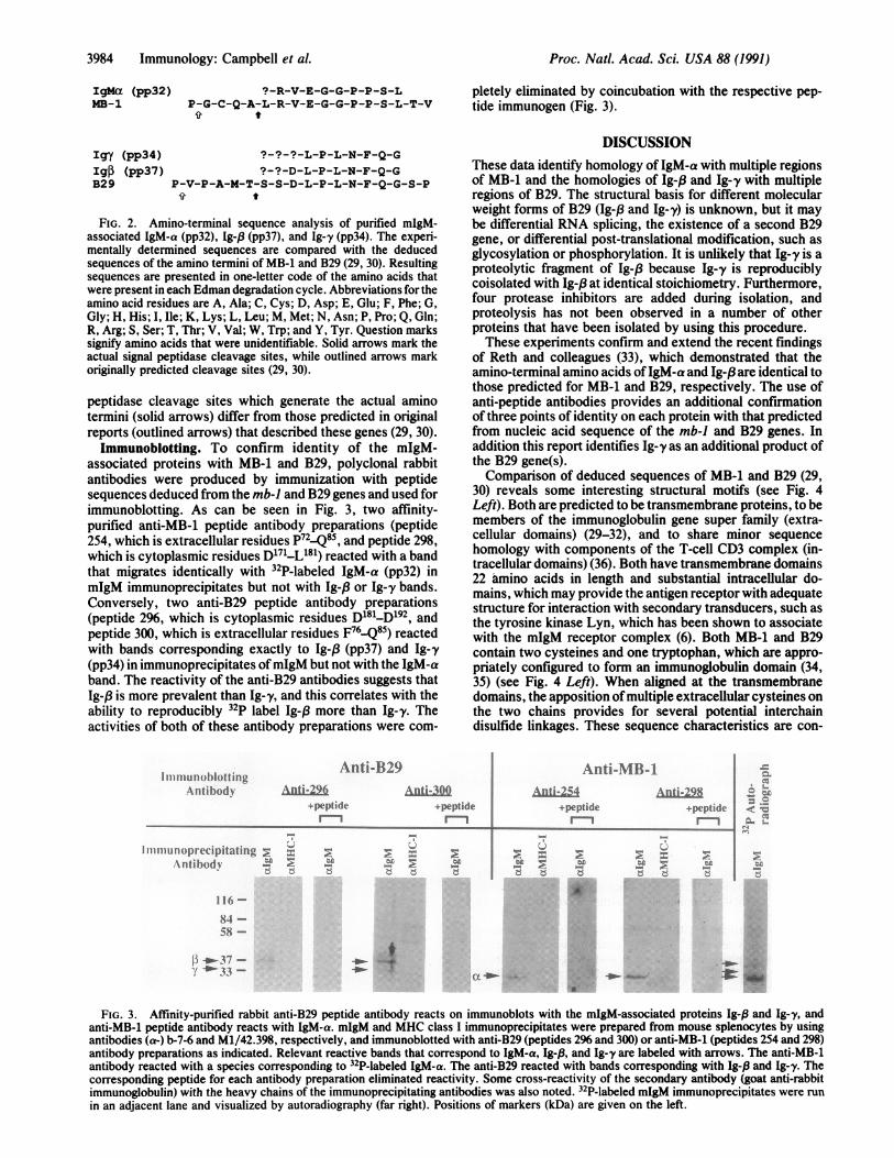
associated proteins with MB-1 and B29, polyclonal rabbitantibodies were produced by immunization with peptidesequences deduced from the mb-i and B29 genes and used forimmunoblotting. As can be seen in Fig. 3, two affinity-purified anti-MB-1 peptide antibody preparations (peptide254, which is extracellular residues P72-Q85, and peptide 298,which is cytoplasmic residues D'71-L081) reacted with a bandthat migrates identically with 32P-labeled IgM-a (pp32) inmIgM immunoprecipitates but not with Ig-,B or Ig-y bands.Conversely, two anti-B29 peptide antibody preparations(peptide 296, which is cytoplasmic residues D181-D192, andpeptide 300, which is extracellular residues F76-Q85) reactedwith bands corresponding exactly to Ig-, (pp37) and Ig-y(pp34) in immunoprecipitates ofmIgM but not with the IgM-aband. The reactivity of the anti-B29 antibodies suggests thatIg-p is more prevalent than Ig-y, and this correlates with theability to reproducibly 32p label Ig-P more than Ig-y. Theactivities of both of these antibody preparations were com-
I rllIllun (I)lottingAntihodN
Anti-B29Anti-296
+peptidem--
Anti-30Q+peptide
l
I Iu nlopretcipitating.\ntihodv
pletely eliminated by coincubation with the respective pep-tide immunogen (Fig. 3).
DISCUSSIONThese data identify homology of IgM-a with multiple regionsof MB-1 and the homologies of Ig-,f and Ig-y with multipleregions of B29. The structural basis for different molecularweight forms of B29 (Ig-f3 and Ig-y) is unknown, but it maybe differential RNA splicing, the existence of a second B29gene, or differential post-translational modification, such asglycosylation or phosphorylation. It is unlikely that Ig-y is aproteolytic fragment of Ig-/3 because Ig-y is reproduciblycoisolated with Ig-j3 at identical stoichiometry. Furthermore,four protease inhibitors are added during isolation, andproteolysis has not been observed in a number of otherproteins that have been isolated by using this procedure.These experiments confirm and extend the recent findings
of Reth and colleagues (33), which demonstrated that theamino-terminal amino acids ofIgM-a and Ig-,8 are identical tothose predicted for MB-1 and B29, respectively. The use ofanti-peptide antibodies provides an additional confirmationof three points of identity on each protein with that predictedfrom nucleic acid sequence of the mb-i and B29 genes. Inaddition this report identifies Ig-y as an additional product ofthe B29 gene(s).Comparison of deduced sequences of MB-1 and B29 (29,
30) reveals some interesting structural motifs (see Fig. 4Left). Both are predicted to be transmembrane proteins, to bemembers of the immunoglobulin gene super family (extra-cellular domains) (29-32), and to share minor sequencehomology with components of the T-cell CD3 complex (in-tracellular domains) (36). Both have transmembrane domains22 amino acids in length and substantial intracellular do-mains, which may provide the antigen receptor with adequatestructure for interaction with secondary transducers, such asthe tyrosine kinase Lyn, which has been shown to associatewith the mIgM receptor complex (6). Both MB-1 and B29contain two cysteines and one tryptophan, which are appro-priately configured to form an immunoglobulin domain (34,35) (see Fig. 4 Left). When aligned at the transmembranedomains, the apposition of multiple extracellular cysteines onthe two chains provides for several potential interchaindisulfide linkages. These sequence characteristics are con-
Anti-MB- IAnUi-254
+peptideI
AntiL298+pepliderm1
f-- L
-W As -=4-Ic_L.
r4
116-
84 -58 -
[$ -_-37 --*- 33 -
:,.~...~....
:,.. -op.sA.
FIG. 3. Affinity-purified rabbit anti-B29 peptide antibody reacts on immunoblots with the mIgM-associated proteins Ig-19 and Ig--y, andanti-MB-1 peptide antibody reacts with IgM-a. mIgM and MHC class I immunoprecipitates were prepared from mouse splenocytes by usingantibodies (a-) b-7-6 and M1/42.398, respectively, and immunoblotted with anti-B29 (peptides 296 and 300) or anti-MB-i (peptides 254 and 298)antibody preparations as indicated. Relevant reactive bands that correspond to IgM-a, Ig-,B, and Ig-y are labeled with arrows. The anti-MB-1antibody reacted with a species corresponding to 32P-labeled IgM-a. The anti-B29 reacted with bands corresponding with Ig-,3 and Ig-y. Thecorresponding peptide for each antibody preparation eliminated reactivity. Some cross-reactivity of the secondary antibody (goat anti-rabbitimmunoglobulin) with the heavy chains of the immunoprecipitating antibodies was also noted. 32P-labeled mIgM immunoprecipitates were runin an adjacent lane and visualized by autoradiography (far right). Positions of markers (kDa) are given on the left.
Proc. Natl. Acad. Sci. USA 88 (1991)
2 W.
.E.I-t t
ll.z z 2d d d

Proc. Natl. Acad. Sci. USA 88 (1991) 3985
S S D L P L N
B29 F Q G S P C S Q I W Q H P R F A A K K R S S M V KMB-1 L R V E G G P P SL T V N
* tB29 F H C Y T N H S G A L T W F R K R G S Q Q P Q E LMB-1 L G E E A R L T C E N N G R N P N I T W W F S L Q* 4
B29 V S E E G R I V Q T Q N G S V Y T L T I Q N I Q YMB-1 S N I T W P P V P L G P G Q G T T G Q L F F P E V
* * *B29 E D N G I Y F C K Q K C D S A N H N V T D S C G TMB-1 N K N T G A C T G C Q V I E N N I L K R S C G T Y
* * *
B29 E L L V L G F S T L D Q LMB-1 L R V R N P V P R P F L D
TMK R R N T L K DIG I I LM G E G T K N RI I T A
B29 I Q T L L I I L F I I V P I F L L L|D K D D G K AMB-1 E G I I L L F C A V V P G T L L L FIR K R W Q N E C,
B29 G M E E D H T|Y E G L N I D Q T AT|YED|IVTL Ig-f3MB-1 K F G V D M P D D E N LIY E G L N I D D C S (B29)
B29 R T G E V K W S V G E H P G Q EMB-1 MY EDI S R G L Q G T Y Q D V G N L H I G D A Q
MB-1 L E K P
(D
G~ CLLN
\ e~~~~~~~~~E/
IgM- a IgM- a(MB-1) (MB-1)
FIG. 4. Comparison of the predicted amino acid sequences of the mb-i and B29 gene products, and a model of MB-1/B29 interactions withmIgM. (Left) Predicted amino acid sequences of MB-1 and B29 (29, 30) aligned at their 22-amino acid transmembrane segments. Theamino-terminal sequences begin with residues as determined by sequence analysis. The common cytoplasmic tyrosine motifs are indicated inboxes as potential phosphorylation sites and the predicted transmembrane domains (TM) are bracketed. Extracellular cysteines are marked withstars. Arrows mark the extracellular tryptophan residues followed by aromatic residues, which are characteristic of immunoglobulin variabledomains and contribute to interchain dimer formation (VH to VL) (34, 35). As is characteristic of immunoglobulin-superfamily members,individual cysteines (site 1) are located 10-11 residues on the amino-terminal side of the tryptophan residues and a cluster of three cysteines(site 2) is located toward the carboxyl terminus; one of the latter presumably provides for the intrachain disulfide linkage (site 1 to one of thesite 2 cysteines) that defines the immunoglobulin-like domain (34, 35). Boldface characters mark a cytoplasmic sequence motif found in bothMB-1 and B29 that is also found in CD3 components and C chain (36). (Right) Model of IgM-a/Ig-,8 and IgM-a/Ig-y heterodimer interactionswith the two membrane-spanning heavy chains ofmIgM (,u). L, light chain. The stoichiometry of actual numbers of heterodimer interacting witheach mIgM has not been confirmed experimentally. Extracellular loops designate immunoglobulin-like domains, and common intracellulartyrosine-containing motifs are designated as potential phosphorylation sites.
sistent with the observed occurrence of MB-1 and B29 as adisulfide-linked heterodimer (13, 14), the structure of whichis modeled in Fig. 4 Right. The transmembrane segment ofMB-1 contains an acidic glutamic residue that might hydro-gen bond with hydroxyl groups of several threonine andserine residues (TTAST) found in the same region on ,u heavychains. Interestingly, Williams et al. (37) have found thatmutation of these threonine and serine residues to valine andalanine, respectively, appears to overcome the MB-1 require-ment for transport of the receptor to the cell surface.Though the role of these molecules in signal transduction
is not defined, their tyrosine phosphorylation is induced byreceptor ligation (ref. 13 and K.S.C. and J.C.C., unpublishedobservations). Importantly, B29 and MB-1 both containcytoplasmic YED and YEGLN sequences, which may besubstrate motifs for specific tyrosine kinases and tyrosinephosphatases. The YEGLN sequence is unique to MB-1 andB29 as determined by a search of GenBank (version 63,March 1990). It is intriguing to note that two of the tyrosineswithin YED and YEGLN sequences on each protein are partof a common sequence motif (Fig. 4 Left) that is also foundon CD3 components and ; chain, which associate with theT-cell antigen receptor (36). Further, phosphorylation ofMB-1 and B29 appears to be dynamically regulated byinterplay of an undefined tyrosine kinase and the phospho-tyrosine phosphatase CD45 (38). In fact, cellular expressionof CD45 and tyrosine kinase activation are both required foractivation of phospholipase C and calcium mobilization bymIgM ligation (5, 38).These findings provide evidence that MB-1 and B29 are
components of the IgM antigen receptor complex, existing asdisulfide-linked heterodimers in noncovalent association
with mIgM. In addition to our data, findings that these genesare exclusively expressed in B cells (29, 30), that expressioncorrelates with surface expression ofmIgM [in fact, MB-1 isrequired for receptor expression (11, 12) and B29 overex-pression increases receptor expression (33)], and that theproteins are inducibly phosphorylated strongly imply thatthese gene products are important for antigen receptor func-tion.
We thank Joel Boymel and Michele Justement for excellenttechnical assistance and Drs. Ralph Kubo and Louis Justement forhelpful discussions and review of the manuscript. This work wassupported by National Institutes of Health Grants A120579 andA121768 and National Research Service Award A108202 (to K.S.C.).
1. DeFranco, A. L. (1987) Annu. Rev. Cell Biol. 3, 143-178.2. Campbell, K. S., Justement, L. B. & Cambier, J. C. (1990) in
Ligands, Receptors and Signal Transduction in Regulation ofLymphocyte Function, ed. Cambier, J. C. (ASM Publications,Washington), pp. 1-50.
3. Campbell, M.-A. & Sefton, B. M. (1990) EMBO J. 9, 2125-2131.
4. Gold, M. R., Law, D. A. & DeFranco, A. L. (1990) Nature(London) 345, 810-813.
5. Cambier, J. C., Morrison, D. C., Chien, M. M. & Lehmann,K. R. (1991) J. Immunol., in press.
6. Yamanashi, Y., Kakiuchi, T., Mizuguchi, J., Yamamoto, T. &Toyoshima, K. (1991) Science 251, 192-194.
7. Dymecki, S. M., Niederhuber, J. E. & Desiderio, S. V. (1990)Nature (London) 247, 332-336.
8. Rogers, J., Early, P., Carter, C., Calame, K., Bond, M., Hood,L. & Wall, R. (1980) Cell 20, 303-312.
9. Kehry, M., Ewald, S., Douglas, R., Sibley, C., Raschke, W.,Fambrough, D. & Hood, L. (1980) Cell 21, 393-406.
B29 mIgM
Immunology: Campbell et al.

3986 Immunology: Campbell et al.
10. Cheng, H. L., Blattner, F. R., Fitzmaurice, L., Mushinski,J. F. & Tucker, P. W. (1982) Nature (London) 296, 410-415.
11. Hombach, J., Leclercq, L., Radbruch, A., Rajewsky, K. &Reth, M. (1988) EMBO J. 7, 3451-3456.
12. Hombach, J., Tsubata, T., Leclercq, L., Stappert, H. & Reth,M. (1990) Nature (London) 343, 760-762.
13. Campbell, K. S. & Cambier, J. C. (1990) EMBO J. 9,441-448.14. Wienands, J., Hombach, J., Radbruch, A., Riesterer, C. &
Reth, M. (1990) EMBO J. 9, 449-455.15. Parkhouse, R. M. E. (1990) Immunology 69, 298-302.16. Chen, J., Stall, A. M., Herzenberg, L. A. & Herzenberg, L. A.
(1990) EMBO J. 9, 2117-2124.17. Cambier, J., Chen, Z. Z., Pasternak, J., Ransom, J., Sandoval,
V. & Pickles, H. (1988) Proc. Natl. Acad. Sci. USA 85,6493-6497.
18. Ratcliffe, M. J. H. & Julius, M. H. (1982) Eur. J. Immunol. 12,634-641.
19. Julius, M. H., Heusser, C. H. & Hartmann, K.-U. (1984) Eur.J. Immunol. 14, 753-757.
20. Springer, T. A. (1980) in Monoclonal Antibodies, eds. Kennett,R. H., McKearn, T. J. & Bechtol, K. B. (Plenum, New York),pp. 185-217.
21. Laemmli, U. K. (1970) Nature (London) 227, 680-685.22. Cleveland, D. W., Fischer, S. G., Krischner, M. W. & Laemm-
li, U. K. (1977) J. Biol. Chem. 252, 1102-1106.23. Giulian, G. & Graham, J. (1990) Hoefer Scientific Instruments
Catalog (Hoefer Scientific Instruments, San Francisco), pp.126-128.
24. Edman, P. & Begg, G. (1967) Eur. J. Biochem. 1, 80-91.25. Hewick, R. M., Hunkapiller, M. W., Hood, L. E. & Dreyer,
W. J. (1981) J. Biol. Chem. 256, 7990-7997.26. Tempst, P. & Riviere, L. (1989) Anal. Biochem. 183, 290-300.27. Barany, G. & Merrifield, R. B. (1980) in The Peptides: Anal-
ysis, Synthesis, Biology, eds. Gross, E. & Meienhofer, J.(Academic, New York), Vol. 2, pp. 1-284.
28. Mitchell, A. R., Kent, S. B. H., Engelhard, M. & Merrifield,R. B. (1978) J. Org. Chem. 43, 2845-2852.
29. Sakaguchi, N., Kashiwamura, S., Kimoto, M., Thalmann, P. &Melchers, F. (1988) EMBO J. 7, 3457-3464.
30. Hermanson, G. G., Eisenberg, D., Kincade, P. W. & Wall, R.(1988) Proc. Natl. Acad. Sci. USA 85, 6890-6894.
31. Kashiwamura, S. I., Koyama, T., Matsuo, T., Steinmetz, M.,Kimoto, M. & Sakaguchi, N. (1990) J. Immunol. 145, 337-343.
32. Hermanson, G. G., Briskin, M., Sigman, D. & Wall, R. (1989)Proc. Nati. Acad. Sci. USA 86, 7341-7345.
33. Hombach, J., Lottspeich, F. & Reth, M. (1990) Eur. J. Immu-nol. 20, 2795-2799.
34. Amzel, L. M. & Poljak, R. J. (1979) Annu. Rev. Biochem. 48,961-997.
35. Williams, A. F. & Barclay, A. N. (1988) Annu. Rev. Immunol.6, 381-405.
36. Reth, M. (1989) Nature (London) 338, 383-384.37. Williams, G. T., Venkitaraman, A. R., Gilmore, D. J. & Neu-
berger, M. S. (1990) J. Exp. Med. 171, 947-952.38. Justement, L. B., Campbell, K. S., Chien, N. C. & Cambier,
J. C. (1991) Science, in press.
Proc. Natl. Acad. Sci. USA 88 (1991)