IdentificationandCharacterizationofMitochondrial ... · tus and software (Quantity One, Bio-Rad)....
12
Identification and Characterization of Mitochondrial Targeting Sequence of Human Apurinic/Apyrimidinic Endonuclease 1 * □ S Received for publication, September 24, 2009, and in revised form, March 15, 2010 Published, JBC Papers in Press, March 15, 2010, DOI 10.1074/jbc.M109.069591 Mengxia Li ‡1 , Zhaoyang Zhong ‡1 , Jianwu Zhu ‡1 , Debing Xiang § , Nan Dai ‡ , Xiaojing Cao ‡ , Yi Qing ‡ , Zhenzhou Yang ‡ , Jiayin Xie ‡ , Zengpeng Li § , Laura Baugh ¶ , Ge Wang ‡ , and Dong Wang ‡2 From the ‡ Cancer Center and § Department of Pathology of Research, Institute of Surgery, Daping Hospital, Third Military Medical University, Chongqing 400042, China and the ¶ Department of Biology, University of Dallas, Irving, Texas 75062 Dually targeted mitochondrial proteins usually possess an unconventional mitochondrial targeting sequence (MTS), which makes them difficult to predict by current bioinfor- matics approaches. Human apurinic/apyrimidinic endonu- clease (APE1) plays a central role in the cellular response to oxidative stress. It is a dually targeted protein preferentially residing in the nucleus with conditional distribution in the mitochondria. However, the mitochondrial translocation mech- anism of APE1 is not well characterized because it harbors an unconventional MTS that is difficult to predict by bioinformat- ics analysis. Two experimental approaches were combined in this study to identify the MTS of APE1. First, the interactions between the peptides from APE1 and the three purified translo- case receptors of the outer mitochondrial membrane (Tom) were evaluated using a peptide array screen. Consequently, the intracellular distribution of green fluorescent protein-tagged, truncated, or mutated APE1 proteins was traced by tag detec- tion. The results demonstrated that the only MTS of APE1 is harbored within residues 289 –318 in the C terminus, which is normally masked by the intact N-terminal structure. As a dually targeted mitochondrial protein, APE1 possesses a special distri- bution pattern of different subcellular targeting signals, the identification of which sheds light on future prediction of MTSs. Human APE1 (apurinic/apyrimidinic endonuclease) is an important multifunctional protein that plays a central role in the cellular response to oxidative stress. The two major activi- ties of APE1 are DNA repair and redox regulation of transcrip- tional factors. On one hand, APE1 functions as a critical rate- limiting enzyme in DNA base excision repair and accounts for nearly all of the AP site incision activities in cell extracts (1). On the other hand, APE1 also exerts unique redox activity to reg- ulate the DNA binding affinity of certain transcriptional factors by controlling the redox status of their DNA-binding domain (2). Inhibition of the redox function of APE1 blocks murine endothelial cell growth and angiogenesis and also blocks the growth of human tumor cell lines (3). The biological impor- tance of APE1 is highlighted by the finding that APE1 knock- out mice exhibit an embryonic lethal phenotype (4). Although APE1 has long been labeled as a nuclear protein, a growing body of evidence has shown that the subcellular distribution of APE1 can be cytoplasmic in some cell types with high metabolic or proliferative rates, with predominant localization in the mito- chondria and the endoplasmic reticulum (5–7). Recent mito- chondrial proteomic studies have further confirmed the existence of APE1 in the mitochondria (8). Considering the importance of the mitochondria in cellular response to oxida- tive stress, the roles of APE1 in the mitochondria have been extensively investigated. A number of studies have shown that the initial mitochondrial APE1 protein level is scarce (i.e. it is not easily detected by regular immunotechniques) (8, 9). When subjected to different stimuli of oxidative stress, such as hydrogen peroxide, the mitochondrial APE1 level is signifi- cantly increased in a dose- and time-dependent fashion (9, 10). Therefore, the mitochondrial targeting of APE1 seems to be conditional. The mitochondrial DNA is a small circular genome, coding a mere 37 gene products, including 24 tRNAs and 13 proteins, in contrast to the more than 1000 proteins found in the mitochon- drion (11). Hence, most mitochondrial proteins are encoded by the nuclear genome and translocated into the mitochondria. Like the proteins destined for other subcellular organelles, the mitochondrially targeted proteins possess targeting signals within their primary or secondary structure that direct them to the organelle with the assistance of elaborate protein translo- cating and folding machines (12, 13). The nucleus-encoded mitochondrially targeted preproteins are recognized by the receptors on the mitochondrial surface and are subsequently translocated across the outer mitochondrial membranes through a general import pore that assembles into a high molecular weight complex, termed the preprotein translocase of the outer mitochondrial membrane (Tom) (13, 14). The three Tom subunits, Tom20, Tom22, and Tom70, expose major portions on the cytosolic side of the outer membrane and function as import receptors for distinct classes of the prepro- teins in vivo and in organello (15). The purified cytosolic domains of these import receptors were validated to specifically bind the mitochondrial preproteins in vitro (16). Protein * This work was supported by National Natural Science Foundation of China Grant 30670628 (to D. W.) and by the Natural Science Research Foundation of the Third Military Medical University (to Z. Z.). □ S The on-line version of this article (available at http://www.jbc.org) contains supplemental Figs. S1 and S2. 1 These authors contributed equally to this work. 2 To whom correspondence should be addressed: Cancer Center, Research Institute of Surgery, Daping Hospital, Third Military Medical University, 10 Changjiang Zhi Rd., Daping Yuzhong District, Chongqing 400042, China. Tel./Fax: 86-23-68894062; E-mail: [email protected]. THE JOURNAL OF BIOLOGICAL CHEMISTRY VOL. 285, NO. 20, pp. 14871–14881, May 14, 2010 © 2010 by The American Society for Biochemistry and Molecular Biology, Inc. Printed in the U.S.A. MAY 14, 2010 • VOLUME 285 • NUMBER 20 JOURNAL OF BIOLOGICAL CHEMISTRY 14871 by guest on December 29, 2019 http://www.jbc.org/ Downloaded from
Transcript of IdentificationandCharacterizationofMitochondrial ... · tus and software (Quantity One, Bio-Rad)....

Identification and Characterization of MitochondrialTargeting Sequence of Human Apurinic/ApyrimidinicEndonuclease 1*□S
Received for publication, September 24, 2009, and in revised form, March 15, 2010 Published, JBC Papers in Press, March 15, 2010, DOI 10.1074/jbc.M109.069591
Mengxia Li‡1, Zhaoyang Zhong‡1, Jianwu Zhu‡1, Debing Xiang§, Nan Dai‡, Xiaojing Cao‡, Yi Qing‡, Zhenzhou Yang‡,Jiayin Xie‡, Zengpeng Li§, Laura Baugh¶, Ge Wang‡, and Dong Wang‡2
From the ‡Cancer Center and §Department of Pathology of Research, Institute of Surgery, Daping Hospital, Third Military MedicalUniversity, Chongqing 400042, China and the ¶Department of Biology, University of Dallas, Irving, Texas 75062
Dually targeted mitochondrial proteins usually possess anunconventional mitochondrial targeting sequence (MTS),which makes them difficult to predict by current bioinfor-matics approaches. Human apurinic/apyrimidinic endonu-clease (APE1) plays a central role in the cellular response tooxidative stress. It is a dually targeted protein preferentiallyresiding in the nucleus with conditional distribution in themitochondria.However, themitochondrial translocationmech-anism of APE1 is not well characterized because it harbors anunconventional MTS that is difficult to predict by bioinformat-ics analysis. Two experimental approaches were combined inthis study to identify the MTS of APE1. First, the interactionsbetween the peptides fromAPE1 and the three purified translo-case receptors of the outer mitochondrial membrane (Tom)were evaluated using a peptide array screen. Consequently, theintracellular distribution of green fluorescent protein-tagged,truncated, or mutated APE1 proteins was traced by tag detec-tion. The results demonstrated that the only MTS of APE1 isharbored within residues 289–318 in the C terminus, which isnormallymasked by the intact N-terminal structure. As a duallytargetedmitochondrial protein, APE1 possesses a special distri-bution pattern of different subcellular targeting signals, theidentification ofwhich sheds light on future prediction ofMTSs.
Human APE1 (apurinic/apyrimidinic endonuclease) is animportant multifunctional protein that plays a central role inthe cellular response to oxidative stress. The two major activi-ties of APE1 are DNA repair and redox regulation of transcrip-tional factors. On one hand, APE1 functions as a critical rate-limiting enzyme in DNA base excision repair and accounts fornearly all of the AP site incision activities in cell extracts (1). Onthe other hand, APE1 also exerts unique redox activity to reg-ulate theDNAbinding affinity of certain transcriptional factorsby controlling the redox status of their DNA-binding domain
(2). Inhibition of the redox function of APE1 blocks murineendothelial cell growth and angiogenesis and also blocks thegrowth of human tumor cell lines (3). The biological impor-tance of APE1 is highlighted by the finding that APE1 knock-out mice exhibit an embryonic lethal phenotype (4). AlthoughAPE1has long been labeled as a nuclear protein, a growing bodyof evidence has shown that the subcellular distribution of APE1can be cytoplasmic in some cell types with high metabolic orproliferative rates, with predominant localization in the mito-chondria and the endoplasmic reticulum (5–7). Recent mito-chondrial proteomic studies have further confirmed theexistence of APE1 in the mitochondria (8). Considering theimportance of the mitochondria in cellular response to oxida-tive stress, the roles of APE1 in the mitochondria have beenextensively investigated. A number of studies have shown thatthe initial mitochondrial APE1 protein level is scarce (i.e. it isnot easily detected by regular immunotechniques) (8, 9).Whensubjected to different stimuli of oxidative stress, such ashydrogen peroxide, the mitochondrial APE1 level is signifi-cantly increased in a dose- and time-dependent fashion (9,10). Therefore, the mitochondrial targeting of APE1 seemsto be conditional.Themitochondrial DNA is a small circular genome, coding a
mere 37 gene products, including 24 tRNAs and 13 proteins, incontrast to themore than 1000 proteins found in themitochon-drion (11). Hence, most mitochondrial proteins are encoded bythe nuclear genome and translocated into the mitochondria.Like the proteins destined for other subcellular organelles, themitochondrially targeted proteins possess targeting signalswithin their primary or secondary structure that direct them tothe organelle with the assistance of elaborate protein translo-cating and folding machines (12, 13). The nucleus-encodedmitochondrially targeted preproteins are recognized by thereceptors on the mitochondrial surface and are subsequentlytranslocated across the outer mitochondrial membranesthrough a general import pore that assembles into a highmolecular weight complex, termed the preprotein translocaseof the outer mitochondrial membrane (Tom) (13, 14). Thethree Tom subunits, Tom20, Tom22, and Tom70, exposemajor portions on the cytosolic side of the outermembrane andfunction as import receptors for distinct classes of the prepro-teins in vivo and in organello (15). The purified cytosolicdomains of these import receptorswere validated to specificallybind the mitochondrial preproteins in vitro (16). Protein
* This work was supported by National Natural Science Foundation of ChinaGrant 30670628 (to D. W.) and by the Natural Science Research Foundationof the Third Military Medical University (to Z. Z.).
□S The on-line version of this article (available at http://www.jbc.org) containssupplemental Figs. S1 and S2.
1 These authors contributed equally to this work.2 To whom correspondence should be addressed: Cancer Center, Research
Institute of Surgery, Daping Hospital, Third Military Medical University, 10Changjiang Zhi Rd., Daping Yuzhong District, Chongqing 400042, China.Tel./Fax: 86-23-68894062; E-mail: [email protected].
THE JOURNAL OF BIOLOGICAL CHEMISTRY VOL. 285, NO. 20, pp. 14871–14881, May 14, 2010© 2010 by The American Society for Biochemistry and Molecular Biology, Inc. Printed in the U.S.A.
MAY 14, 2010 • VOLUME 285 • NUMBER 20 JOURNAL OF BIOLOGICAL CHEMISTRY 14871
by guest on Decem
ber 29, 2019http://w
ww
.jbc.org/D
ownloaded from

import studies with isolatedmitochondria indicate that Tom20and Tom22 function as a heterodimer receptors for the typicalmitochondrial preproteins that carry the cleavable N-terminalmitochondrial targeting sequence (MTS).3 This signal isclassically characterized as an N-terminal motif predicted toform an amphipathic helix that is �15–70 residues in lengthand enriched in positively charged basic residues (17).Tom70 is reported to be required for the import of non-cleavable preproteins that have been shown to have specific-ity for carrier proteins destined for the inner mitochondrialmembrane (18).It is obvious that mitochondrial localization is a prerequisite
for the functions of APE1. APE1 is a 36-kDamolecule that can-not be translocated to the mitochondria through a passivetransport process and so the mitochondrial targeting of APE1involves an active mechanism. Hence, it is very important toidentify the MTS of APE1. However, the mitochondrial trans-location mechanism of APE1 is not well characterized becausethe bioinformatics analysis has failed to position the MTS (19).Previous efforts using computer-aided signal sequence-search-ing tools, including MitoProt and TargetP, have indicated thatAPE1 has a low score in MTS prediction. Instead, a classicnuclear localization signal (NLS) is identified and characterizedat the N terminus of APE1 (20). In contrast with exclusivelymitochondrially targeted proteins, APE1 is considered to be adually targeted mitochondrial protein that is mainly destinedfor the nucleus. Dinur-Mills et al. (21) found that the duallytargeted mitochondrial proteins have a weaker MTS and alower whole protein net charge. The proteins containing theseunconventional targeting signals will most probably escapeprediction using a bioinformatics-based approach. Therefore,experimental approaches are usually used to identify and char-acterize the MTS of preproteins that are localized to themitochondria.This study focuses on the identification and characterization
of the MTS of APE1 and further explores the possible importmachinery that mediate APE1 translocation. Aiming to dimin-ish the possible bias of bothmethods, we have employed a strat-egy combining two sophisticated approaches to identify andcharacterize theMTSofAPE1, including biochemical andmor-phological approaches. The results demonstrate that the MTSof APE1 resides within residues 289–318 of the C terminus,which are normally masked by the intact N-terminal structure.This may be the explanation to its predominant nuclear local-ization and conditional mitochondrial targeting after oxidativestress. Moreover, as a dually targeted mitochondrial protein,APE1 possesses a special distribution pattern of different sub-cellular targeting signals, the discovery of which sheds light onthe future identification of MTSs.
EXPERIMENTAL PROCEDURES
Materials—Dulbecco’s modified Eagle’s medium, fetalbovine serum, and transfection reagent Lipofectamine 2000were purchased from Invitrogen. The eukaryotic expression
vectors pEGFP-N2 andpEGFP-C1were fromClontech (Moun-tain View, CA). H2O2, menadione, ampicillin, penicillin, strep-tomycin, proteinase K, RNase A, isopropyl-�-D-thiogalactopy-ranoside, imidazole, and polyvinylidene difluoride transfermembraneswere fromSigma.High performance nickel-Sepha-rose was purchased from GE Healthcare. MitoTracker RedCMXRos, TO-PRO3nuclear probe, andCOX IV antibodywerefromMolecular Probes, Inc. (Eugene,OR). T4 ligase, restrictionendonucleases, and high fidelity Pfu DNA polymerase werefrom Promega (Madison,WI). Protein interaction His tag pull-down kit, SuperSignal WestPico chemiluminescent reagents,horseradish peroxidase-conjugated His probe, and horseradishperoxidase-conjugated secondary antibodies were from Pierce.The monoclonal antibody against hAPE1 was fromNovus Bio-logical (Littleton,CO). The anti-Pax5, anti-GFP and fluoresceinisothiocyanate-conjugated secondary antibodywere purchasedfrom Santa Cruz Biotechnology, Inc. (Santa Cruz, CA).Cell Culture and Transfection—The human cervical cancer
cell line HeLa was obtained from the American Type CultureCollection (Manassas, VA). The cells were maintained in highglucose Dulbecco’s modified Eagle’s medium supplementedwith 10% (v/v) fetal bovine serum, 50mg/ml penicillin/strepto-mycin, and 2 mM L-glutamine in 5% CO2 at 37 °C and passaged2–3 times/week. For transfection, 2000 cells were plated intoone well of a 96-well plate and incubated under normal condi-tions overnight until they reached �85% confluence. The dif-ferent plasmids were then transfected into human umbilicalvein endothelial cells using Lipofectamine 2000 transfectionreagent according to the manufacturer’s protocol.Immunofluorescence and Confocal Laser-scanning Micro-
scopy—HeLa cells were grown on a slide at a density of 2 � 104cells/well in a 6-well plate for 24 h. Then the HeLa cells weretreated with 100 �MH2O2 or menadione bisulfate or incubatedin a hypoxia condition with 1% oxygen content for 6 h. Theculture media were removed, and the cells were incubated witha culture medium containing 200 nM MitoTracker RedCMXRos probe for 30min at 37 °C. After washing twice in coldPBS, the cells were fixed with 2% (w/v) paraformaldehyde inPBS at 4 °C for 20 min. Then the cells were permeabilized with0.5% Triton X-100 in PBS for 15min at room temperature. Theslides were washed twice with PBS, and the cells were blockedwith normal goat serum at 37 °C for 30min, followed by immu-nobinding by APE1 antibody at a dilution of 1:200 for 2 h. Sub-sequently, the cells were incubated with fluorescein isothiocya-nate-conjugated secondary antibody for 30 min at 37 °C. Afterwashing, the cells were stainedwith TO-PRO3 nuclear probe ata dilution of 1:5000 for 5min. Then the cells weremounted andvisualized under a Leica confocal laser-scanning microscope(TCS SP5) available at the central laboratory of the Third Mil-itary Medical University. For multicolor fluorescence, the red,green, and blue signals were collected simultaneously and thensuperimposed.Organelle-enriched Fractionation—The nuclear extracts from
the treated and control groups were isolated as described pre-viously (22). Ten million HeLa cells were collected and resus-pended in 500 �l of hypotonic lysis buffer. After incubation for10 min, the nuclei were collected by centrifugation at 500 � g,and the nuclear pellet was resuspended and incubated with 100
3 The abbreviations used are: MTS, mitochondrial targeting sequence; GFP,green fluorescent protein; NLS, nuclear localization signal; PBS, phos-phate-buffered saline; EGFP, enhanced GFP.
Identification of MTS of Human APE1
14872 JOURNAL OF BIOLOGICAL CHEMISTRY VOLUME 285 • NUMBER 20 • MAY 14, 2010
by guest on Decem
ber 29, 2019http://w
ww
.jbc.org/D
ownloaded from

�l of buffer B (10 mMHEPES, 100mMNaCl, 1.5 mMMgCl2, 0.1mM EDTA, 0.1 mM dithiothreitol, 5 mM phenylmethylsulfonylfluoride, pH 7.9) for 20 min at 4 °C. After incubation, the sam-ples were centrifuged at 10,000 � g at 4 °C for 20 min.
The cytosolic and mitochondrial crude fractions were pre-pared by differential centrifugation as described previously(22). Briefly, 5 � 108 cells were collected and resuspended in 1ml of grinding medium and pulse-sonicated on ice at 30 wattsfor 15 s, ensuring 50–70% cell lysis. Then the lysate was centri-fuged for 12 min at 800 � g, and the supernatant was immedi-ately centrifuged for 20 min at 8500 � g at 4 °C. The obtainedsupernatant contained the cytosolic fraction. The mitochon-drial pellet was resuspended with 100 �l of buffer S (150 mM
sucrose, 40 mM KCl, 25 mM Tris-HCl, 1 mg/ml bovine serumalbumin, pH 7.4) and sonicated on ice at 45 watts, three timesfor 15 s each, and then precipitated by centrifugation for 20minat 10,000 � g at 4 °C.Western Blots—Twentymicrograms of protein from nuclear,
cytosolic, or mitochondrial fractions was applied to 12% SDS-polyacrylamide gels and electrophoresed to resolve proteins.The proteins were then transferred to polyvinylidene difluoridemembranes and blocked in TBST containing 5% (w/v) nonfatdry milk. The membrane was incubated with mouse anti-hAPE1primary antibody (1:2000),mouse anti-COX IVprimaryantibody (1:1000), and mouse anti-Pax5 primary antibody(1:1000) overnight at 4 °C. The membrane was then washedthree times in TBST and incubated with horseradish peroxi-dase-labeled secondary antibody for 1 h at 37 °C. The mem-brane was reacted with chemiluminescent reagents andrevealed with BioMax-Light films (Eastman Kodak Co.). Theband intensities were analyzed using the Gel Doc 2000 appara-tus and software (Quantity One, Bio-Rad).Expression and Purification of Tom Proteins in Escherichia
coli—The prokaryotic expression vectors carrying cytosolicdomains of the three Tom proteins, named pET19b-yTom20cd-His10, pET19b-yTom22cd-His10, and pET19b-yTom70cd-His10 were gifts from Prof. Nikolaus Pfanner. Theexpression and purification were performed as described withmodifications (15). Briefly, the E. coli strain BL21 (DE3) wastransformed with three plasmids, and protein expression wasinduced by the addition of 0.1 mM isopropyl-�-D-thiogalacto-pyranoside for 4 h. The bacterial pellet was resuspended in 5mM imidazole, 500 mMNaCl, and 20 mM Tris-HCl, pH 7.9, andthen sonicated. After centrifugation, the supernatant wasapplied to columns containing nickel-Sepharose resin (GEHealthcare). After five washes with different concentrations ofimidazole (20–200 mM), 500 mM NaCl, and 20 mM Tris-HCl,pH 7.9, the Tom proteins were eluted with 1 M imidazole, 500mMNaCl, and 20mMTris-HCl, pH 7.9, and dialyzed against 1�PBS, pH 7.4. The purities of Tom proteins were analyzed usingSDS-polyacrylamide gel electrophoresis, and the protein con-centrations were measured using the Bradford assay. To evalu-ate the affinity of the purified Tom proteins to His probe, theTom proteins were separated using SDS-PAGE and then sub-jected to Western blot against horseradish peroxidase-conju-gated His probe (1:10,000).Peptide Array Screen—The cellulose-bound peptide array
was designed and prepared by automated spot synthesis
(INTAVIS Bioanalytical Instruments AG, Koln, Germany). Allof the incubations were processed in a chamber (NUNCTM
4-well slide processing dish; ThermoFisher Scientific, Roskilde,Denmark). To analyze the Tom protein-binding activity, thearray was first blocked in 1% nonfat milk, TBST (w/v) solutionat 37 °C for 4 h. After washing three times with TBST, the arraywas incubated overnight with 150 nM purified Tom proteindiluted inTBST at 4 °Cwith gentle shaking. The nonspecificallybound protein was removed by washing the array with TBSTfor 5 min at room temperature and then incubated with horse-radish peroxidase-conjugated His probe (1:10,000) for 1 h at37 °C. The Tom protein bound to the peptide array wasdetected using a chemiluminescent blotting substrate (ECL,Pierce). A quantitative measurement of the binding strengthwas performed with Spotfinder software (Dana-Farber Can-cer Institute, Boston, MA), which included subtraction ofthe local background for each peptide spot. The means of atleast three independent experiments for each peptide spotwere used (average units) (23). The values for the differentmembranes were adjusted by the use of identical referencepeptides on eachmembrane. The 24-residue-longMTS frommanganese-superoxide dismutase was adopted as a positivecontrol (sequence: MLSRAVCGTSRQLAPALGYLGSRQ)and diluted in multiproportions.His Tag Pull-down Assay—The His tag pull-down assay was
performed using the ProFoundTM pull-down poly-His protein-protein interaction kit (Pierce) according to the protocol pro-vided by the manufacturer. An immobilized cobalt chelate wasadded to the HandeeTM spin columns and incubated with thepurifiedTomproteins for 4 h at room temperature to bind theirHis tag as bait proteins. Then the unbound Tom proteins wereremoved with the wash buffer. The HeLa cells were lysed byadding 1ml of lysis buffer to each culture dish and incubated onice for 30 min. The plates were scraped, and the cell lysate wasplaced in a centrifuge tube. The protein concentration of thecell lysate was analyzed using the Bradford assay. Then the celllysate was added to a column containing the bound Tom pro-teins and incubated overnight at 4 °C with gentle shaking. Afterincubation, the suspensions were centrifuged at 7000 � g at4 °C and washed three times with cold TBS. The supernatantwas removed after the third wash. Then the protein-proteincomplex was eluted using the elution buffer and analyzed using12% SDS-PAGE, followed by Western blotting using mono-clonal antibodies for APE1 (Novus Biological).Truncated and Mutated APE1 Expression Plasmid Con-
struction—Different oligonucleotides were designed as primersto amplify the target truncated APE1 cDNA fragments by PCRfrom pcDNA3.1-APE1 vector containing the full-length APE1cDNA sequence that was constructed in the previous work. Forthe vectors based onpEGFP-N2, all of the forward primerswereadded as initiation codons of theATGandEcoRI sites, and all ofthe reverse primers were added to the BamHI site at their5�-ends. For the vectors based on pEGFP-C1, all of the forwardprimers were added to the EcoRI site, and all of the reverseprimers were added as termination codons of TGA and BamHIsites at their 5�-ends. PCR reactions were processed using highfidelity Pfu DNA polymerase according to the manufacturer’sinstruction. The resulting PCR products were subcloned into
Identification of MTS of Human APE1
MAY 14, 2010 • VOLUME 285 • NUMBER 20 JOURNAL OF BIOLOGICAL CHEMISTRY 14873
by guest on Decem
ber 29, 2019http://w
ww
.jbc.org/D
ownloaded from

pEGFP-N2 or pEGFP-C1 vectors. The fidelity of the con-structed vectors was confirmed by sequencing (Sequencing andSynthesis Facility of Invitrogen (Shanghai, China)).The mutated vectors were based on the constructed trun-
cated APE1 plasmids, and the mutants were generated byQuikChange site-directed mutagenesis (Stratagene, La Jolla,CA) according to the manufacturer’s protocol. All mutationswere validated by sequencing.
RESULTS
APE1 Exhibits Dual Subcellular Targeting with the Nucleusas the Major Destination—To induce oxidative stress or DNAdamage, the HeLa cells were treated with hydrogen peroxideand menadione sodium bisulfate or cultured in hypoxic condi-tions, as indicated under “Experimental Procedures.” The sub-cellular localization of the APE1 protein was detected usingimmunofluorescence microscopy and Western blotting. The re-sults in Fig. 1A show that APE1 was localized mainly in thenucleus of the untreated HeLa cells but was translocated to thecytoplasm and especially the mitochondria after hydrogen per-oxide-, menadione-, or hypoxia-induced oxidative stress. The
mitochondrial staining of APE1 was even stronger in menadi-one (the mitochondrial DNA-specific DNA damage-inducingagent)-treated HeLa cells. Additionally, a significant decreasein the mitochondrial membrane potential was observed aftertreatment with hydrogen peroxide, menadione, or hypoxicconditions. Although, the MitoTracker Red is not the typicalmitochondrial membrane potential marker, it is hard to labelthe malfunctioning mitochondria when unfixed cells arelabeled with MitoTracker Red. Actually, the loss of staining bythe MitoTracker Red represented the abnormal mitochondrialfunction (24). To confirm the morphological findings, subcel-lular fractions of the nucleus, cytoplasm, and mitochondriafrom each group were subjected to Western blot. The resultsshown in Fig. 1B indicate that themajor localization of APE1 inHeLa cells is the nucleus and that oxidative stress ormenadioneinduced the translocation of APE1 to the mitochondria.Recombinant Cytosolic Domains of Tom20, Tom22, and
Tom70 Are Purified by Ni2�-Nitrilotriacetic Acid AffinityChromatography—To investigate the mitochondrion-target-ing sequence of APE1, we first analyzed the binding property ofthe peptides derived from APE1 to the Tom receptors in a cell-free system. The results (supplemental Fig. S1) indicate that theexpressions of all the Tom proteins were induced by 0.1 mM
isopropyl-�-D-thiogalactopyranoside (supplemental Fig. S1A),and after 4 h of induction, the foreign proteins expressed�10–20% of total cellular protein. To validate the purity of the Tomproteins that were isolated by Ni2�-nitrilotriacetic acid chro-matography, the 200 mM imidazole contained eluants sepa-rated by SDS-PAGE and then stained with Coomassie BrilliantBlue R250. The target proteins comprised �95% of the totaleluants (supplemental Fig. S1B). Then 1 �g of the eluants wassubjected to Western blot against the horseradish peroxidase-conjugated His probe. Supplemental Fig. S1C showed that onlythe target proteins that were shown as major bands insupplemental Fig. S1Bwere detected by the His probe, indicat-ing that these contaminating bacterial proteins do not signifi-cantly impact the peptide array-based screen.Tom20cd Preferentially Binds to Peptides Derived fromAPE1—
Because the presequence of the mitochondrially targeted pro-tein was recognized by the Tom proteins as a linear amino acidsequence, we designed a 15-mer peptide array for the Tompro-tein-binding site screen. Fifteen-amino acid peptides fromAPE1 that overlapped by 12 residues were covalently attachedto the cellulose membrane. The MTS from the manganese-superoxide dismutase that has been extensively investigated asa classical N-terminal mitochondrial signal sequence was usedas the positive control. The peptide consisting of the first 13residues ofAPE1,whichwas validated to be anNLS,was chosenas the negative control. The films of the peptide arrays and theirquantitative results are shown in Fig. 2. Tom20cd demon-strated the best binding efficacy among the three Tom proteinsto the peptides derived from APE1 (Fig. 2A). Tom20cd wasdistributed in the regions of the first one-third of both the Nterminus and C terminus. The binding affinity of Tom20cd tothe C terminus of APE1 was generally higher than to the Nterminus. The peak regions included peptides of residues13–39, 49–63, 64–93, 211–240, 238–258, 265–279, and 289–312. However, when compared with Tom20cd, significant dif-
FIGURE 1. Conditional mitochondrial targeting of APE1 after differentstimuli. HeLa cells were treated with 100 �M hydrogen peroxide or 100 �M
menadione sodium bisulfate or cultured in hypoxic conditions (1% oxygen)for 6 h. The subcellular distribution of the APE1 protein was detected byimmunofluorescence microscopy (A) and Western blot (B). Live cells werestained with Mito-Tracker Red mitochondrion-selective probe to visualize themitochondria in the cells. The APE1 proteins were visualized by a standardimmunofluorescence with fluorescein isothiocyanate-conjugated secondaryantibody and are shown in green. The localization of APE1 in the mitochon-dria is displayed as yellow-orange by two superimposed staining patterns.Scale bar, 30 �m. Loading controls for the Western blot were Pax5 for thenucleus, COX IV for the mitochondria, and �-actin for the cytosol. Numbers1– 4 represent control, hydrogen peroxide, menadione, and hypoxic pre-treated groups, respectively.
Identification of MTS of Human APE1
14874 JOURNAL OF BIOLOGICAL CHEMISTRY VOLUME 285 • NUMBER 20 • MAY 14, 2010
by guest on Decem
ber 29, 2019http://w
ww
.jbc.org/D
ownloaded from

ferences were observed in the peptide scans of Tom22cd andTom70cd binding. TheAPE1 peptide scan revealed that severalbinding sequences for Tom22cd were scattered over variousregions of the whole protein (Fig. 2B), whereas the Tom70cdbinding sequences were mainly located in both termini (Fig.2C). Although the binding patterns of Tom20cd, Tom22cd, and
Tom70cd were strikingly similar, the overall intensities of theinteractions with Tom22cd and Tom70cd were significantlylower.To determine which Tom protein is the most important to
APE1 mitochondrial targeting, we performed the His tag pull-down assay to validate the interactions between the purified
FIGURE 2. Binding of the cytosolic domain of Tom20, Tom22, and Tom70 to a peptide array derived from APE1. Binding of 150 nM Tom20cd (A), Tom22cd(B), and Tom70cd (C) to a peptide array consisting of 15 residues derived from APE1. The first peptide comprises amino acids 1–15 of APE1, the second peptidecomprises residues 4 –18, the third peptide comprises residues 7–21, and so on. The labeling originated from the peptide array. A quantitative measure of thebinding strength was performed with Spotfinder software, which included subtraction of the local background for each peptide spot. The values for differentmembranes were adjusted by the use of identical reference peptides on each membrane. Results were obtained from three independent experiments. Tomproteins were immobilized by cobalt chelate and then incubated with HeLa cell extracts (D). The eluants from each Tom protein His tag pull-down experimentwere separated using SDS-PAGE and then subjected to anti-APE1 antibody using Western blot (IB). The complete cellular extraction of the HeLa cell was alsoadded as an input. As shown in Fig. 4, only Tom20cd interacted with APE1 from the HeLa cell extraction.
Identification of MTS of Human APE1
MAY 14, 2010 • VOLUME 285 • NUMBER 20 JOURNAL OF BIOLOGICAL CHEMISTRY 14875
by guest on Decem
ber 29, 2019http://w
ww
.jbc.org/D
ownloaded from
Identification of MTS of Human APE1
14876 JOURNAL OF BIOLOGICAL CHEMISTRY VOLUME 285 • NUMBER 20 • MAY 14, 2010
by guest on Decem
ber 29, 2019http://w
ww
.jbc.org/D
ownloaded from
Tom proteins and the cellular APE1 protein in vitro. Becausethe His tag was added at the terminus of the purified Tomproteins, these proteins together with their interactive proteinscan be captured by the immobilized cobalt chelate. The eluants
from each Tom protein pull-downexperiment with an input of thewhole cell lysate were separatedusing SDS-PAGE and then sub-jected to an anti-APE1 antibodyusing Western blot. The resultsshown in Fig. 2D indicate that onlyTom20cd interacted with the full-length APE1 protein, which furtherconfirms the findings of the peptidearray. The results provided by thepeptide screen assay together withthe pull-down assay demonstratethat APE1 possesses higher affinityto Tom20 than to Tom22 or Tom70,which led to the hypothesis thatmitochondrial translocation of APE1may be through a Tom20-depen-dent pathway.Alanine Replacement of Three
Positively Charged Amino Acids atthe C terminus of APE1 DiminishedAffinity to Tom20cd—We furtheranalyzed the amino acid composi-tion of the “hot spot” peptides withhigher affinities to Tom20cd. Inter-estingly, most of these regions arerich in positively charged aminoacids, including lysine, arginine, andhistidine. Thus, the regions contain-ing tandems of positively chargedresidues, including residues 22–36,214–228, and 292–306, werepicked. To investigate the impact ofthese charged residues on the bind-ing efficacy to Tom20cd, we subse-quently designed another peptidearray using the “alanine walk”method, which replaced each targetamino acid position with alanineindividually. The intensity of theinteraction between the mutatedpeptides and Tom20cd was ana-lyzed by the previously mentionedmethod. As shown in Fig. 3C, thewild type peptides of residues 292–306 (Fig. 3A) possessed robust affin-ity to Tom20cd, but the alaninereplacement of Lys299, Arg301, and
Lys303 diminished the binding efficacy of residues 292–306 toTom20cd, which demonstrates that these residues may playsignificant roles in Tom20cd-APE1 interaction. Meanwhile,
FIGURE 3. Screen critical residues for APE1 binding efficacy to Tom20cd by peptide array. The peptides, containing tandems of positively charged aminoacids including residues 292–306 (A), 214 –228 (B), and 22–36 (C) were mutated using the “alanine walk” method, which replaced each target amino acidposition individually with alanine (A). All of the wild type and mutated peptides were incubated with Tom20cd, and the binding intensity was measured andquantified using the aforesaid method. The binding efficacy of each wild type and its mutated form is shown in a histogram. The results are the average valueof three independent experiments.
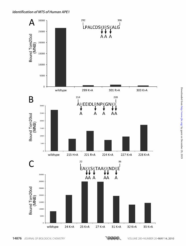
FIGURE 4. Mapping of the APE1 domains responsible for mitochondrial targeting. Immunofluorescentimages depict the subcellular localization of different, truncated APE1 proteins in the HeLa cells at 48 hafter transfection. Shown is a schematic representation of the N-terminal truncated mutants of APE1.GFP-fused positions are also indicated. Scale bar, 15 �m. The Western blots against GFP antibody innuclear (N), cytosolic (C), and mitochondrial (M) enriched fractions of some groups are also shown on theright. The bands represent distribution of the exogenously expressed truncated APE1 proteins in eachsubcellular organelle.
Identification of MTS of Human APE1
MAY 14, 2010 • VOLUME 285 • NUMBER 20 JOURNAL OF BIOLOGICAL CHEMISTRY 14877
by guest on Decem
ber 29, 2019http://w
ww
.jbc.org/D
ownloaded from

mutations in peptide 214–228 (Fig. 3B) just slightly reduced theaffinities toTom20cd. Surprisingly, the alaninemutation at res-idues Lys24, Lys25, Lys27, and Lys31 even enhanced the peptidebinding efficacy to Tom20cd (Fig. 3C).The Mitochondrial Targeting Sequence Localized at the
C-terminal of APE1—Based on previous findings, truncatedAPE1 cDNAsequenceswere generated by the stepwise deletionof the “hot regions” that were found in the peptide screen byusing PCR splicing and then subcloning in GFP-tagged eukary-otic expression vectors. TheGFPwas fused to theC terminus ofthe truncated APE1 proteins, and their subcellular distributionwas visualized by laser confocal microscopy. The subcellulardistribution of p53 and transcription factor A mitochondriawas tested as nuclear and mitochondrial targeting controls toconfirm the validity of our observation system. As shown insupplemental Fig. S2, at 48 h post-transfection, p53 was mainlytargeted to the nucleus, whereas transcription factor A mito-chondria mainly targeted to the mitochondria, indicating thatthe transfection and detection systems used in this experimentworked appropriately. We first fused the GFP tag to the C ter-minus of the truncated APE1, considering the functionalimportance of the N terminus of APE1 (Fig. 4). The full-lengthAPE1 was mainly targeted to the nucleus in accordance withprevious findings. However, when the first 13 residues of the Nterminus, which was reported to harbor the major NLS ofAPE1, were deleted, the subcellular distribution shifted to acytoplasmic dominant pattern. The truncated APE1, with thedeletion of the N-terminal hot regions, including residues
14–318 and 49–318, was mainly located in the cytoplasmrather than in the mitochondria. There was no significantnuclear localization observed, which further confirmed that theNLS of APE1 is located in the first 13 residues of the N termi-nus. However, the truncated APE1 proteins containing onlysome C-terminal hot regions, including residues 211–318,238–318, 265–318, and 289–318, were mainly targeted to themitochondria complete overlapping of the MitoTracker stain-ing. To exclude the possibility that therewere internal targetingsignals for other subcellular organs in the middle of the APE1protein sequence, we deleted both residues 1–13 and 289–318,which are the nuclear and mitochondrial targeting signals, andtransfected it into the HeLa cells. The truncated APE1 proteinwas diffusely distributed through the whole cell with no subcel-lular organelle-specific targeting observed. This distributionpattern, which is similar to the pattern of the pEGFP-N2 vectoralone, was previously reported as a loss-of-signal targeting pat-tern representing the subcellular location of proteins that lacktargeting signal sequences. Morphological observations werefurther confirmed by a subcellular organelle-enrichedWesternblot. The results of some of the critical truncatedAPE1mutantsare shown (Fig. 4). These results indicated that the MTS ofAPE1 resides in the C terminus appears masked by the N-ter-minal sequences that block the interaction between the C-ter-minal MTS of APE1 and the cytosolic subunits of the Tomprotein.To exclude the possibility that there are other MTSs that
interfered with the C-terminal sequence of APE1, we furthertested all of the possible regions according to the peptide arrayresults. C-terminal EGFP-tagged truncated protein expressionvectors carrying the residues 13–39, 49–93, 211–258 and 265–279 of APE1 were constructed and transfected into HeLa cells.The subcellular distribution of these proteins was observed bylaser confocal microscopy and confirmed byWestern blot. Theresults shown in Fig. 5 indicated these regions of APE1 have nospecific mitochondrial targeting whereas only residues 13–39may have a weak nuclear targeting signal and a preferentialnuclear distribution.Two Positively Charged Residues, Lys299 and Arg301, in the C
Terminus of APE1 Play Critical Roles in Its MitochondrialTargeting—To determine which residues are critical to themitochondrial targeting of the APE1 protein, we replaced threepositively charged residues in the C terminus of APE1 withalanine using site-directed mutagenesis generating K299A,R301A, and K303A. These point-mutated vectors are based onthe 289–318 truncated APE1 expression vectors with C-termi-nus-tagged EGFP. As shown in Fig. 6, K299A and R301A weredistributed throughout the cell without obvious subcellularorganelle-enriched locations, whereas the wild type sequenceof 289–318 appears as a mitochondrial-specific distribution.Interestingly, we noticed that K303A appeared as two distribu-tion patterns that were displayed in the same visual field in Fig.6 (third panel). Most of the cells expressing K303A showedmitochondrially targeted distribution; however, some of thesetruncated mutants were observed in the cytoplasm or nuclei,indicating that K303A had a weak effect on its mitochondrialtargeting. The K299A/R301A double mutated truncated APE1protein showed a diffuse pattern, which further confirmed the
FIGURE 5. Subcellular targeting of truncated APE1 protein of residues113–139, 49 –93, 211–258, and 265–279. The other potential Tom20cdbinding regions of APE1 were also tested for their mitochondrial targetingefficiency. Immunofluorescent images depict the subcellular localization ofdifferent, truncated APE1 proteins in the HeLa cells at 48 h after transfection.Mitochondria were visualized by MitoTacker Red probe. Scale bar, 30 �m. aa,amino acids.
Identification of MTS of Human APE1
14878 JOURNAL OF BIOLOGICAL CHEMISTRY VOLUME 285 • NUMBER 20 • MAY 14, 2010
by guest on Decem
ber 29, 2019http://w
ww
.jbc.org/D
ownloaded from

importance of Lys299 and Arg301 to the mitochondrial localiza-tion of APE1. These results further demonstrated that residues289–318 were sufficient for mitochondrial targeting, andLys299 andArg301were critical to themitochondrial targeting ofAPE1.Mutation of K299A and R301A Diminished Mitochondrial
Translocation of APE1—We then tested the menadione-in-duced APE1 mitochondrial translocation affected by mutationof the two key residues Lys299 and Arg301. The protein mito-chondrial translocation of both wild type APE1 and full-lengthAPE1with theK299A/R301Amutationswere compared.UsingEGFP, we can easily track the translocation after menadionepretreatment. Due to the existence of the NLS of APE1, bothwild type and the K299A/R301A full-length APE1 mutantmainly accumulate in the nucleus in untreated cells. However,when challenged withmenadione, appreciable fractions of wildtype APE1 relocalized to themitochondria, whereas themajor-ity of the mutant protein was in the nucleus. The nuclear andmitochondrial levels of exogenously expressed APE1 were con-firmed by Western blot using anti-GFP antibody (Fig. 7).
DISCUSSION
APE1 was discovered as a dually targeted protein that ismainly distributed in the nucleus and conditionally targeted
to the mitochondria. However, themitochondrial targeting mecha-nism of APE1 has not been well elu-cidated. In our present study, wedetect a unique MTS of APE1 byanalyzing peptide affinities to Tomproteins and the subcellular target-ing of truncated APE1 constructs.Our results indicate that the condi-tional mitochondrial targeting ofAPE1 depends on the C-terminalsequence of residues 289–318. Wefurther demonstrated that Lys299and Arg301 are the critical sites, andmutation of both sites abolishesAPE1 mitochondrial translocationafter menadione-induced oxidativestress.We first employed peptide array
to scan for a possible MTS by com-paring the affinities to the Tom pro-teins. The results showed that thepeptides with robust binding toTom are scattered throughout theentire protein, making it difficultto determine the actual MTS ofAPE1. However, the results justhighlighted some possible signalsequences for the future truncatedassay. Surprisingly, the general pat-terns of binding for the differentTom proteins are strikingly similar.Regarding the different specificitiesof the three receptors, they have dif-
ferent affinities with the typical mitochondrial targeted pro-teins. However, there are some overlaps of affinities amongthese Tom proteins when interacting with purified peptides(16). One possibility is that APE1 is not a typical exclusivemito-chondrial protein, and the MTS is not typical of a singly tar-getedmitochondrial protein.We believe theMTS of these pro-teins with multiple subcellular distributions can only bepartially positioned by a peptide affinity assay. Themutant pep-tide assay also provided some potential critical residues forbinding. The most noteworthy sites are Lys299, Arg301, andLys303, insofar as their replacementwith alanine diminished theaffinities of peptide to Tom20. Interestingly, the replacement ofresidues Lys24, Lys25, Lys27, and Lys31 enhanced the affinities toTom20. Because they are located in the N terminus of APE1, itis possible that these particular lysine residues function as sup-pressors to prevent the distribution of APE1 to themitochondrion.We then tested these possible targeting sequences by observing
the subcellular distribution of the truncated forms of APE1. Wenoticed thatwhenwedeleted a small regionof theN terminus, thetruncatedAPE1proteinswere located in the cytoplasm.The trun-cated APE1 proteins containing residues 289–318 were specifi-cally targeted to the mitochondria. Although all of the truncatedproteins contained the MTS at their C terminus, the MTS could
FIGURE 6. Subcellular targeting of K299A, R301A, K303A, or K299A/R301A mutated APE1 observed bylaser confocal microscopy. K299A, R301A, K303A, or K299A/R301A mutated APE1 vectors are based on the289 –318 truncated APE1 expression vectors with C-terminally tagged EGFP. Subcellular localization of differ-ent truncated APE1 proteins in HeLa cells was observed by laser confocal microscopy at 48 h after transfection.Scale bar, 10 �m. The Western blot against GFP antibody in nuclear (N), cytosolic (C), and mitochondrial (M)enriched fractions are also shown on the right. The bands represent distribution of the exogenously expressedmutant in each subcellular organelle.
Identification of MTS of Human APE1
MAY 14, 2010 • VOLUME 285 • NUMBER 20 JOURNAL OF BIOLOGICAL CHEMISTRY 14879
by guest on Decem
ber 29, 2019http://w
ww
.jbc.org/D
ownloaded from

be located in the core of the APE1 protein, making it difficult fortheMTS to be exposed to the Tom protein on the mitochondrialsurface. We also tested possible mitochondrial targeting signalsbesides the MTS in the C terminus, based on the results of thepeptide array. These results indicated that APE1 possesses aunique but weak MTS that is located in the C terminus and nor-mally masked by the N terminus.A previous study showed that isolated mitochondrial APE1
protein isolated from bovine liver is an N-terminally truncated
form with the deletion of its NLS (8). It is noteworthy that theMTSs of most cleavable mitochondrial preproteins are cleavedby mitochondrial processing peptidase in the matrix. Themature form of the preproteins following processing is themajor form in the mitochondrial matrix (25). According to thistheory, the deleted residues of the truncated APE1 should har-bor the MTS that is in its N terminus. However, based on theevidence provided by our present study, it is believed that thefirst 33 residues of the N terminus only harbor a classical NLSinstead of both targeting signals. A plausible explanation is thattheMTS of APE1 is overwhelmed by its NLS in the intact struc-ture of the APE1 protein. APE1 conditionally targets the mito-chondria only when its NLS is nonfunctional or deleted. Onemay ask why APE1 is targeted to the mitochondria after oxida-tive stress. Current studies are insufficient to address this ques-tion. A possible reason is that the intracellular redox imbalancealters the intactN-terminal conformation of APE1, which is themain domain exerting the redox function (26) and exposes theMTS to the Tom proteins in the mitochondrial outer mem-brane. Interestingly, the previous study has demonstrated thatthe NLS of Apn1, the APE1 homologue in yeast, resides in its Cterminus, whereas its N terminus harbors a putativeMTS. Pir1,an O-glycosylated protein required for yeast cell wall stability,was found tomask theNLS ofApn1 and also plays an active rolein the mitochondrial targeting of the protein. Thus, the pre-dicted N-terminal MTS of Apn1 is not sufficient for mitochon-drial targeting in the absence of Pir1 (27). Because we could notget an accurate crystal structure of the C terminus of APE1, wethen hypothesized that another possible mechanism of APE1conditional mitochondrial targeting could be that the putativeMTS of APE1 needs assistance to conduct the protein into themitochondria by some protein chaperones that are induced byoxidative stress.Our study further explored the possible mechanism of APE1
translocation to themitochondria. The receptor Tom proteins,including Tom20, Tom22, and Tom70, mediate the preproteintransport in two major pathways. We found that APE1 wasmainly recognized by Tom20cd, indicating that APE1 translo-cates to the mitochondria, possibly through a Tom20-depen-dent pathway. Tom20 recognizes a type of cleavable prepro-teins that possess a classical N-terminalMTS by binding via thehydrophobic portion of the signal sequence amphipathic helix.However, the identified MTS of APE1 in the present study islocated in the C terminus, which is more similar to tail-anchored preproteins. Themitochondrial tail-anchored pre-proteins, including Bcl-2 and Tom20, were inserted into themitochondrial outer membranes via a C-terminal trans-membrane domain (28, 29). Although the exact localizationof APE1 in the mitochondria has not been well character-ized, according to its function in mitochondrial DNA repair,its distribution should accompany the mitochondrial DNAin the mitochondrial matrix. Thus, we are inclined to con-sider that the MTS of APE1 is not a tail-anchored signal buta special C-terminal mitochondrial signal that conducts it tothe matrix of the mitochondria rather than to the outermembrane. Future studies to investigate APE1 locationusing submitochondrial fractions will help elucidate itsmitochondrion-targeting mechanism.
FIGURE 7. Mitochondrial translocation of K299A/301R APE1 full-lengthmutant after menadione induction. The K299A/R301A mutated APE1 full-length expression vector was transfected into HeLa cells at 48 h before 100�M menadione treatment. EGFP visualized cellular images (A) and organelle-enriched Western blots (B) were obtained at 6 h after menadione treatment.The wild type APE1 protein expression vector was simultaneously treated as acontrol. Scale bar, 50 �m. The Western blot against GFP antibody in nuclearand mitochondrially enriched fractions of some groups are shown to depictthe distribution of the exogenously expressed mutant in each organelle. Theanti-Pax5 and COX IV Western blot were included to measure the cross-con-tamination between nuclei and mitochondria.
Identification of MTS of Human APE1
14880 JOURNAL OF BIOLOGICAL CHEMISTRY VOLUME 285 • NUMBER 20 • MAY 14, 2010
by guest on Decem
ber 29, 2019http://w
ww
.jbc.org/D
ownloaded from

In summary, to our knowledge, this is the first study thatidentifies and characterizes the MTS of APE1 and explores themitochondrion-targeting mechanism of APE1. Our data sup-port the finding that theMTS of APE1 is harbored in the last 30residues of the C terminus of the protein, and Lys299 andArg301are critical to the oxidative stress-induced mitochondrial tar-geting of APE1. Our present study has also shown that APE1translocates into themitochondriamainly through the Tom20-dependent pathway, but detailedmechanisms remain to be elu-cidated.Moreover, this research sheds light on the possibility ofa targeted therapeutic strategy using peptides that block thebinding of the APE1 MTS to Tom receptors or dominant neg-ative mutation of critical sites in the APE1MTSwith the aim ofpromoting apoptosis after oxidative stress.
Acknowledgments—We thank Dr. Nikolaus Pfanner and Dr. Jan Brixfor providing the Tom protein expression vectors; Dr. Shangcheng Xufor help with the transcription factor Amitochondria and p53 vectors;Dr. Benjamin P. C. Chen, Dr. David J. Chen, Dr. Bruce Demple, andDr.Hung Fung for critical comments on themanuscript; andWei Sun,Liting Wang, Dr. Kai Fan, Dr. Zhiyong Liu, Dr. Zengtao Zhang, andLinli Zeng for excellent technical assistance.
REFERENCES1. Chen, D. S., Herman, T., and Demple, B. (1991) Nucleic Acids Res. 19,
5907–59142. Evans, A. R., Limp-Foster, M., and Kelley, M. R. (2000) Mutat. Res. 461,
83–1083. Luo, M., Delaplane, S., Jiang, A., Reed, A., He, Y., Fishel, M., Nyland, R. L.,
2nd, Borch, R. F., Qiao, X., Georgiadis, M. M., and Kelley, M. R. (2008)Antioxid. Redox Signal. 10, 1853–1867
4. Xanthoudakis, S., Smeyne, R. J., Wallace, J. D., and Curran, T. (1996) Proc.Natl. Acad. Sci. U.S.A. 93, 8919–8923
5. Tell, G., Damante, G., Caldwell, D., and Kelley, M. R. (2005) Antioxid.Redox Signal. 7, 367–384
6. Tomkinson, A. E., Bonk, R. T., and Linn, S. (1988) J. Biol. Chem. 263,12532–12537
7. Tell, G., Crivellato, E., Pines, A., Paron, I., Pucillo, C., Manzini, G., Ban-diera, A., Kelley, M. R., Di Loreto, C., and Damante, G. (2001)Mutat. Res.485, 143–152
8. Chattopadhyay, R., Wiederhold, L., Szczesny, B., Boldogh, I., Hazra, T. K.,Izumi, T., and Mitra, S. (2006) Nucleic Acids Res. 34, 2067–2076
9. Mitra, S., Izumi, T., Boldogh, I., Bhakat, K. K., Chattopadhyay, R., andSzczesny, B. (2007) DNA Repair 6, 461–469
10. Frossi, B., Tell, G., Spessotto, P., Colombatti, A., Vitale, G., and Pucillo, C.(2002) J. Cell. Physiol. 193, 180–186
11. Asin-Cayuela, J., and Gustafsson, C. M. (2007) Trends Biochem. Sci. 32,111–117
12. Karniely, S., and Pines, O. (2005) EMBO Rep. 6, 420–42513. Becker, L., Bannwarth, M., Meisinger, C., Hill, K., Model, K., Krimmer, T.,
Casadio, R., Truscott, K. N., Schulz, G. E., Pfanner, N., and Wagner, R.(2005) J. Mol. Biol. 353, 1011–1020
14. Dekker, P. J., Ryan, M. T., Brix, J., Muller, H., Honlinger, A., and Pfanner,N. (1998)Mol. Cell. Biol. 18, 6515–6524
15. Brix, J., Dietmeier, K., and Pfanner, N. (1997) J. Biol. Chem. 272,20730–20735
16. Brix, J., Rudiger, S., Bukau, B., Schneider-Mergener, J., and Pfanner, N.(1999) J. Biol. Chem. 274, 16522–16530
17. Bolender, N., Sickmann, A., Wagner, R., Meisinger, C., and Pfanner, N.(2008) EMBO Rep. 9, 42–49
18. Sollner, T., Pfaller, R., Griffiths, G., Pfanner, N., and Neupert, W. (1990)Cell 62, 107–115
19. Tsuchimoto, D., Sakai, Y., Sakumi, K., Nishioka, K., Sasaki, M., Fujiwara,T., and Nakabeppu, Y. (2001) Nucleic Acids Res. 29, 2349–2360
20. Jackson, E. B., Theriot, C. A., Chattopadhyay, R., Mitra, S., and Izumi, T.(2005) Nucleic Acids Res. 33, 3303–3312
21. Dinur-Mills, M., Tal, M., and Pines, O. (2008) PLoS ONE 3, e216122. Li, M. X., Wang, D., Zhong, Z. Y., Xiang, D. B., Li, Z. P., Xie, J. Y., Yang,
Z. Z., Jin, F., and Qing, Y. (2008) Free Radic. Biol. Med. 45, 592–60123. Saeed, A. I., Sharov, V., White, J., Li, J., Liang, W., Bhagabati, N., Braisted,
J., Klapa, M., Currier, T., Thiagarajan, M., Sturn, A., Snuffin, M., Rezant-sev, A., Popov, D., Ryltsov, A., Kostukovich, E., Borisovsky, I., Liu, Z.,Vinsavich, A., Trush, V., and Quackenbush, J. (2003) BioTechniques 34,374–378
24. Poot, M., Gibson, L. L., and Singer, V. L. (1997) Cytometry 27, 358–36425. Chacinska, A., Pfanner, N., and Meisinger, C. (2002) Trends Cell Biol. 12,
299–30326. Bhakat, K. K., Mantha, A. K., and Mitra, S. (2009) Antioxid. Redox Signal.
11, 621–63827. Vongsamphanh, R., Fortier, P. K., and Ramotar, D. (2001)Mol. Cell. Biol.
21, 1647–165528. Otera, H., Taira, Y., Horie, C., Suzuki, Y., Suzuki, H., Setoguchi, K., Kato,
H., Oka, T., and Mihara, K. (2007) J. Cell Biol. 179, 1355–136329. Setoguchi, K., Otera, H., and Mihara, K. (2006) EMBO J. 25, 5635–5647
Identification of MTS of Human APE1
MAY 14, 2010 • VOLUME 285 • NUMBER 20 JOURNAL OF BIOLOGICAL CHEMISTRY 14881
by guest on Decem
ber 29, 2019http://w
ww
.jbc.org/D
ownloaded from

WangQing, Zhenzhou Yang, Jiayin Xie, Zengpeng Li, Laura Baugh, Ge Wang and Dong
Mengxia Li, Zhaoyang Zhong, Jianwu Zhu, Debing Xiang, Nan Dai, Xiaojing Cao, YiHuman Apurinic/Apyrimidinic Endonuclease 1
Identification and Characterization of Mitochondrial Targeting Sequence of
doi: 10.1074/jbc.M109.069591 originally published online March 15, 20102010, 285:14871-14881.J. Biol. Chem.
10.1074/jbc.M109.069591Access the most updated version of this article at doi:
Alerts:
When a correction for this article is posted•
When this article is cited•
to choose from all of JBC's e-mail alertsClick here
Supplemental material:
http://www.jbc.org/content/suppl/2010/03/15/M109.069591.DC1
http://www.jbc.org/content/285/20/14871.full.html#ref-list-1
This article cites 29 references, 8 of which can be accessed free at
by guest on Decem
ber 29, 2019http://w
ww
.jbc.org/D
ownloaded from