Pathogenic antibodies induced by spike proteins of COVID ...

Identification of stress-induced mitochondrial proteins incultured tobacco cellsTomas Hajeka, David Honysa,b,*, Erwin Wittersc and Vera Capkovaa,b
aLaboratory of Pollen Biology, Institute of Experimental Botany AS CR, Rozvojova 135, CZ-165 02 Praha 6, Czech RepublicbDepartment of Plant Physiology, Faculty of Sciences, Charles University, Vinicna 5, CZ-128 44 Praha 2, Czech RepubliccLaboratorium voor Plantenbiochemie en -fysiologie, Department of Biology, University of Antwerp, Universiteitsplein 1, B-2610, Antwerp, Belgium
Correspondence
*Corresponding author,
e-mail: [email protected]
Received 11 November 2004; revised 10
January 2005
doi: 10.1111/j.1399-3054.2005.00492.x
Under stress conditions, small and large heat-shock proteins cooperativelyfulfil molecular chaperone activities within many cellular compartments, includ-ing mitochondria. Here, we report the identification and characterizationof 10 newly synthesized low-molecular-weight heat stress-induced proteins(SIPs) found solely in the mitochondrial fraction of suspension-cultured cellsof tobacco (Nicotiana tabacum L., strain Virginia Bright Italia-0). These SIPswere shown to be encoded by the nuclear genome and none was detected innon-stressed cells. Mass spectroscopy analysis revealed that eight SIPsbelonged to the group of small heat-shock proteins, whilst one protein sharedsignificant homology with transcription factors. De novo synthesis of all SIPswas detected within 10 min of the commencement of heat stress and con-tinued for the whole 12 h stress interval. Moreover, nine of the SIPs werestable for 24 h following stress termination. SIP1, which showed the mostintense synthesis profile, was found to be phosphorylated. The most intensestress response was observed mainly during the exponential growth phase ofthe cell culture. Native electrophoresis of mitochondrial protein complexesrevealed an association of all the SIPs in high-molecular-weight complexeswith no free SIPs left in mitochondria. These complexes were localized to themitochondrial membrane fraction.
Introduction
Eukaryotic cells respond to stress treatments by theexpression of heat-shock proteins (HSPs). The functionof these molecular chaperones is to prevent irreversibleprotein aggregation and to promote correct folding ofproteins after the onset of stress (Vierling 1991, Parselland Lindquist 1993, Morimoto et al. 1994). Accordingto their molecular weight, HSPs are classified into sev-eral families: HSP120, HSP100, HSP90, HSP70, HSP60and the family of small HSPs (sHSPs). While the aminoacid sequence of large HSPs (HSP60 to HSP120) is
highly conserved from yeast to animals and plants, thehomology within the large group of sHSPs is less appar-ent and is limited only to the short amino acid sequenceat the C-terminal region (De Jong 1993, Caspers et al.1995, Rassow et al. 1997).
High-molecular-weight HSPs are also present in cellsunder normal physiological conditions and theirchaperone activities have been demonstrated in a vari-ety of cell processes, in protein translocation acrossmembranes, in the formation of multiprotein complexesand in the targeting of proteins for degradation (Zhang
Abbreviations – EST, expressed sequence tag; HSP, heat-shock protein; PMSF, phenylmethylsulphonyl fluoride; PVDF, poly-
vinylidene difluoride; sHSP, small heat-shock protein; SIP, stress-induced protein; VBI-0, Virginia Bright Italia-0.
12 Physiol. Plant. 124, 2005
Physiologia Plantarum 124: 12–24. 2005 Copyright � Physiologia Plantarum 2005, ISSN 0031-9317

and Glaser 2002). Increased demand for chaperoneactivities under stress conditions turns the constitutiveHSP expression level into overexpression and inducesthe transient activation of sHSPs. The expression ofsHSPs positively correlates with strong resistance tostress treatment in both plant and animal cells (Landryet al. 1989, Downs and Heckathorn 1998). However,the main difference between the two eukaryotickingdoms lies in the number of sHSPs induced by oneagent. In human cells, the expression of sHSP27 has aprotective activity against various kinds of treatment,including heat stress, oxidative stress, chemotherapeuticdrugs and staurosporine-induced apoptosis, andphosphorylation has been shown to play an importantrole in regulating sHSP27 activity (Landry et al. 1989,Mehlen et al. 1996, Garrido et al. 1997). Plants, on theother hand, have evolved a different strategy. One stressimpulse triggers the expression of a diverse family ofsHSPs with molecular weights ranging from 12 to38 kDa (Neumann et al. 1989, Vierling 1991, Banzetet al. 1998). These molecules form homopolymericcomplexes from 200 to 800 kDa (Helm et al. 1993,Lenne and Douce 1994) that selectively bind proteinsin their non-native state to prevent their aggregation,and to maintain them in a conformation capable ofATP-dependent refolding by other chaperones (Sunet al. 2002). Under stress conditions, the synthesis andaccumulation of sHSP molecules are very high and theirabundance can reach 1% of total cellular proteins(Hsieh et al. 1992).
The presence of small and large HSPs in thecytoplasm, endoplasmic reticulum, chloroplasts andmitochondria under stress conditions demonstrates theessential cooperation of both types of HSP molecule intheir molecular chaperone activities within manycellular compartments in plants (Waters et al. 1996).The positive correlation between the stress-inducedexpression of a broad set of sHSPs and the maintenanceof cell homeostasis raises questions about the specificfunction of these particular sHSP genes. The first indica-tions of a possible functional hierarchy of sHSP species,sHSP15, sHSP17 and sHSP22, were suggested by threedifferent types of stress in tomato stem calli cells (Banzetet al. 1998). Heat stress granules formed in tobaccoprotoplasts comprise both class I and class II sHSPsarranged into class-specific dodecamers. However,class I oligomers are formed only in the presence ofclass II sHSPs (Kirschner et al. 2000). Phosphorylation,an important tool of functional variability of sHSPs inanimal cells, has been identified for the first time inmaize mitochondrial sHSP22 (Lund et al. 1998). Inanimals, mitochondria are commonly regarded as thesensor of stress (Lai et al. 1996, Arrigo 1998). However,
such a role in plants is still under discussion (Banzetet al. 1998, Jones 2000).
In this work, we report the contribution of mitochon-dria to the heat stress response of suspension-culturedcells of tobacco [Nicotiana tabacum L., strain VirginiaBright Italia-0 (VBI-0)]. A proteomic study led to theidentification of eight low-molecular-weight proteinsas stress-induced sHSPs. The kinetics of their expressionduring stress treatment, their stability and the demon-strated serine phosphorylation of one sHSP suggest thepossibility of the above-mentioned specific function orfunctional hierarchy of particular sHSP species in plantmitochondria. The presented results will be exploited asinput data for the further characterization of sHSPcomplexes.
Materials and methods
Plant material and cultivation conditions
Tobacco cell strain VBI-0, derived from the stem pith ofNicotiana tabacum L. cv. ‘Virginia Bright Italia’(Opatrny and Opatrna 1976), was cultivated in standardHeller liquid medium (Heller 1953) supplemented withthe auxins, alpha-naphthaleneacetic acid and 2,4-dichlorophenoxyacetic acid (each 5 · 10�6 M). Cellswere subcultured every 2 weeks (inoculation densityof approximately 5 · 104 cells ml�1) and cultivated indarkness on an orbital shaker (IKA KS501) at 180 rpm(diameter, 30 mm). For the heat stress treatment, thetemperature was set to 40�C. To identify stress-inducedproteins (SIPs) and to follow the kinetics of theirexpression and degradation, cell cultures were pulse orpulse-chase labelled with an L-[14C]-amino acid mixture(1.85 GBq mg�1 atom carbon, Amersham Biosciences,Little Chalfont, UK; 10 kBq ml�1 medium). The dura-tion of the stress treatments and labelling periods aregiven in the figure legends of the relevant experiments.
Isolation and subfractionation of mitochondria
Suspension cultures were separated from the cultivationmedium by filtration and immediately used for mito-chondrial isolation. Intact mitochondria were isolatedas described previously (Hajek et al. 2004). Plant mater-ial was homogenized between two nylon meshes andcentrifuged stepwise in three steps (2000 g/5 min,6000 g/10 min and 40 000 g/80 min) with PERCOLLsolution in the last step. Mitochondria forming a yellow-ish ring in PERCOLL solution were collected by filtrationand immobilized on the surface of a 0.22 mm nylon filter.
Physiol. Plant. 124, 2005 13
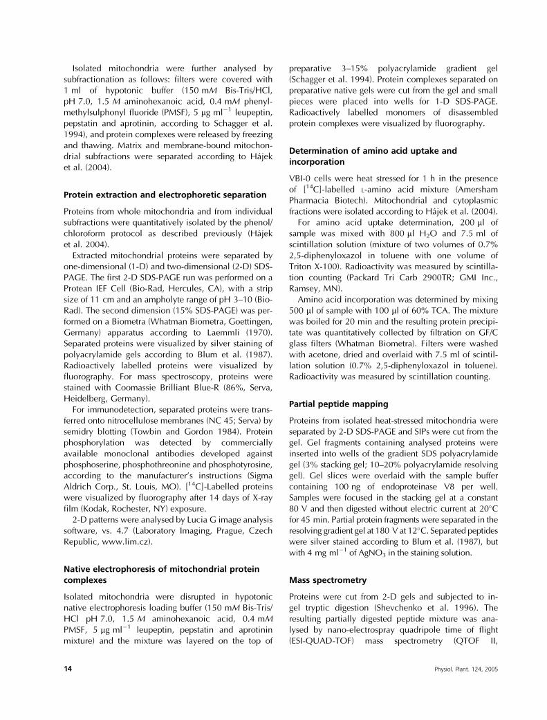
Isolated mitochondria were further analysed bysubfractionation as follows: filters were covered with1 ml of hypotonic buffer (150 mM Bis-Tris/HCl,pH 7.0, 1.5 M aminohexanoic acid, 0.4 mM phenyl-methylsulphonyl fluoride (PMSF), 5 mg ml�1 leupeptin,pepstatin and aprotinin, according to Schagger et al.1994), and protein complexes were released by freezingand thawing. Matrix and membrane-bound mitochon-drial subfractions were separated according to Hajeket al. (2004).
Protein extraction and electrophoretic separation
Proteins from whole mitochondria and from individualsubfractions were quantitatively isolated by the phenol/chloroform protocol as described previously (Hajeket al. 2004).
Extracted mitochondrial proteins were separated byone-dimensional (1-D) and two-dimensional (2-D) SDS-PAGE. The first 2-D SDS-PAGE run was performed on aProtean IEF Cell (Bio-Rad, Hercules, CA), with a stripsize of 11 cm and an ampholyte range of pH 3–10 (Bio-Rad). The second dimension (15% SDS-PAGE) was per-formed on a Biometra (Whatman Biometra, Goettingen,Germany) apparatus according to Laemmli (1970).Separated proteins were visualized by silver staining ofpolyacrylamide gels according to Blum et al. (1987).Radioactively labelled proteins were visualized byfluorography. For mass spectroscopy, proteins werestained with Coomassie Brilliant Blue-R (86%, Serva,Heidelberg, Germany).
For immunodetection, separated proteins were trans-ferred onto nitrocellulose membranes (NC 45; Serva) bysemidry blotting (Towbin and Gordon 1984). Proteinphosphorylation was detected by commerciallyavailable monoclonal antibodies developed againstphosphoserine, phosphothreonine and phosphotyrosine,according to the manufacturer’s instructions (SigmaAldrich Corp., St. Louis, MO). [14C]-Labelled proteinswere visualized by fluorography after 14 days of X-rayfilm (Kodak, Rochester, NY) exposure.
2-D patterns were analysed by Lucia G image analysissoftware, vs. 4.7 (Laboratory Imaging, Prague, CzechRepublic, www.lim.cz).
Native electrophoresis of mitochondrial proteincomplexes
Isolated mitochondria were disrupted in hypotonicnative electrophoresis loading buffer (150 mM Bis-Tris/HCl pH 7.0, 1.5 M aminohexanoic acid, 0.4 mMPMSF, 5 mg ml�1 leupeptin, pepstatin and aprotininmixture) and the mixture was layered on the top of
preparative 3–15% polyacrylamide gradient gel(Schagger et al. 1994). Protein complexes separated onpreparative native gels were cut from the gel and smallpieces were placed into wells for 1-D SDS-PAGE.Radioactively labelled monomers of disassembledprotein complexes were visualized by fluorography.
Determination of amino acid uptake andincorporation
VBI-0 cells were heat stressed for 1 h in the presenceof [14C]-labelled L-amino acid mixture (AmershamPharmacia Biotech). Mitochondrial and cytoplasmicfractions were isolated according to Hajek et al. (2004).
For amino acid uptake determination, 200 ml ofsample was mixed with 800 ml H2O and 7.5 ml ofscintillation solution (mixture of two volumes of 0.7%2,5-diphenyloxazol in toluene with one volume ofTriton X-100). Radioactivity was measured by scintilla-tion counting (Packard Tri Carb 2900TR; GMI Inc.,Ramsey, MN).
Amino acid incorporation was determined by mixing500 ml of sample with 100 ml of 60% TCA. The mixturewas boiled for 20 min and the resulting protein precipi-tate was quantitatively collected by filtration on GF/Cglass filters (Whatman Biometra). Filters were washedwith acetone, dried and overlaid with 7.5 ml of scintil-lation solution (0.7% 2,5-diphenyloxazol in toluene).Radioactivity was measured by scintillation counting.
Partial peptide mapping
Proteins from isolated heat-stressed mitochondria wereseparated by 2-D SDS-PAGE and SIPs were cut from thegel. Gel fragments containing analysed proteins wereinserted into wells of the gradient SDS polyacrylamidegel (3% stacking gel; 10–20% polyacrylamide resolvinggel). Gel slices were overlaid with the sample buffercontaining 100 ng of endoproteinase V8 per well.Samples were focused in the stacking gel at a constant80 V and then digested without electric current at 20�Cfor 45 min. Partial protein fragments were separated in theresolving gradient gel at 180 V at 12�C. Separated peptideswere silver stained according to Blum et al. (1987), butwith 4 mg ml�1 of AgNO3 in the staining solution.
Mass spectrometry
Proteins were cut from 2-D gels and subjected to in-gel tryptic digestion (Shevchenko et al. 1996). Theresulting partially digested peptide mixture was ana-lysed by nano-electrospray quadripole time of flight(ESI-QUAD-TOF) mass spectrometry (QTOF II,
14 Physiol. Plant. 124, 2005

Micromass, Manchester, UK) according to Wilm et al.(1996). The data obtained were interpreted usingMasslynx software. The resulting data were analysedby Matrix software (www.matrixscience.com/cgi/) andfragments were sequence identified by database searchagainst several publicly available databases (SwissProt,www.ebi.ac.uk/swissprot/; NCBI nr, www.ncbi.nlm.nih.gov; and dbEST, www.ncbi.nlm.nih.gov/dbEST/)using the MASCOT search engine (Perkins et al. 1999).Unannotated expressed sequence tag (EST) hits werefurther analysed by BLAST searches (www.ncbi.nlm.nih.gov/BLAST/; http://www.arabidopsis.org/Blast/) usingthe peptide fragments obtained (Altschul et al. 1990).
Results
Heat stress-induced proteins of low molecularweight
To follow the changes in the pattern of SIPs, a VBI-0 cellsuspension was treated at 40�C for 2 h in the presenceof [14C]-labelled amino acids. The application of heatstress treatment had no substantial effect on the total orcytoplasmic uptake of the radioactive label, which fellwithin the range of 300 000–350 000 dpm. In contrast,the translation efficiency under stress conditions,expressed as the level of total and cytoplasmic incor-poration of [14C], decreased by more than 50% com-pared with the non-stressed control. In mitochondria,the situation was quite different. During heat-shocktreatment, a similar decrease in the level of de novosynthesized proteins accompanied a substantialdecrease in amino acid uptake. This demonstratesimpaired free amino acid import from the cytosol tothe mitochondria by stress.
The application of stress conditions led to the emer-gence of a set of newly synthesized small proteins in themitochondrial fraction with a molecular weight of theorder of 21 kDa, as visualized by 1-D SDS-PAGE fluoro-graphy (Fig. 1). A more detailed examination by 2-DSDS-PAGE of mitochondrial protein spectra resolvedthe few protein bands visible by 1-D electrophoresisinto a set of more than 30 low-molecular-mass heatSIPs (Fig. 2A). Under identical labelling and fluorogra-phy conditions, almost no de novo synthesized proteinswere detected in a non-stressed control (Fig. 2B). Thebroad spectrum of total mitochondrial proteins in thecontrol variant on a 2-D gel silver stained prior to fluoro-graphy (Fig. 2C) verified the good quality of proteinseparation, and demonstrated the expression of SIPsonly after the onset of stress, accompanied by theweak expression of other mitochondrial proteins during30 min of labelling.
According to the intensity of SIP expression, 10 pro-teins were selected for further analysis. Using a combin-ation of 2-D and 1-D markers (Bio-Rad), the molecularweight and pI values for selected proteins weredetermined (see legend to Fig. 2A). The molecularweights of all selected SIPs ranged from 18 to 23 kDa.Eight of the 10 SIPs exhibited acidic characteristics withpI values ranging from 5.1 to 6.0. Only two proteins(SIP5 and SIP10) possessed neutral pI values (7.4 and7.5), documenting that these two proteins were notconsistent with the acidic character of the rest of theSIP group.
In order to determine whether SIPs were encoded bythe nuclear or mitochondrial genome, tobacco VBI-0cells were cultivated under stress conditions in the pre-sence of cycloheximide and chloramphenicol. Bothdrugs are potent inhibitors of translation elongation.Cycloheximide inhibits the translation of nuclear-encoded mRNAs in the cytosol of eukaryotic cells, andchloramphenicol, acting in bacteria, inhibits the trans-lation of only those eukaryotic mRNAs which areencoded by mitochondrial and chloroplast genomes(see Alberts et al. 2002). In the presence of 100 mg ml�1
cycloheximide, the synthesis of all 10 SIPs was com-pletely suppressed and the fluorogram was clear, evenwithout low expression, as shown in Fig. 2B. On theother hand, 200 mg ml�1 chloramphenicol had no effecton SIP synthesis and the fluorogram corresponded toFig. 2A. This demonstrates that all SIPs are encoded bythe nuclear genome.
Fig. 1. One-dimensional SDS-PAGE of [14C]-labelled proteins in two
subcellular fractions of control (C) and 40�C heat-stressed (HS) tobacco
(Virginia Bright Italia-0, VBI-0) cell suspension culture. Cells were heat
stressed for 2 h and were labelled for the last 30 min. cyt, cytosol; mit,
mitochondrial fraction.
Physiol. Plant. 124, 2005 15

Identification of heat stress-induced proteins
Mitochondrial SIPs separated by 2-D SDS-PAGE andimmobilized on polyvinylidene difluoride (PVDF) mem-branes were N-terminally sequenced using Edman’s degra-dation. The N-terminal microsequence of nine of the 10proteins was unsuccessful because of the modification oftheir amino termini. SIP6 was the only successfullyN-terminally sequenced protein. The resulting sequence,LMPYTRP, exhibited limited homology with three proteins,all of which are putative transcription factors. These areCaenorhabditis elegans homeobox protein YRM6_CAEELQ09602, Euglena gracilis mitochondrial inner membranecomplex III zinc finger protein CR9_EUGGR P43266and Candida albicans putative transcriptional activator1_CANAL P33181.
More precise analysis of mitochondrial heat SIPs wasperformed using mass spectrometry. Individual SIPswere separated by 2-D SDS-PAGE, excised from thegels and analysed by ESI-QUAD-TOF mass spectrome-try. The peptide fragments resulting from mass spectro-metry analyses are shown in Table 1. The analysis ofpeptide fragments revealed that SIP1, 2, 3, 4, 5 and 7fell into the family of sHSPs. Their scores in MASCOT
software analysis (www.matrixscience.com/cgi; Perkinset al. 1999) were 119, 29, 104, 35, 38 and 72, respec-tively. The lower scores obtained for proteins SIP8 andSIP9 (27 and 31) led to their classification into the sHSPfamily with a lesser degree of probability. According totheir shared peptides, SIPs were divided into two sub-groups. The first subgroup was formed by proteins SIP3,
Fig. 2. Two-dimensional SDS-PAGE separation of Virginia Bright Italia-0 (VBI-0) mitochondrial proteins. (A) Fluorogram of de novo synthesized
proteins after 2 h of heat stress and [14C]-labelling for the last 30 min. The ten most intense stress-induced proteins (SIPs) were characterized: SIP1
(20 kDa, pI 5 5.1), SIP2 (18 kDa, pI 5 5.3), SIP3 (18.5 kDa, pI 5 5.6), SIP4 (19 kDa, pI 5 6.0), SIP5 (18.5 kDa, pI 5 7.5), SIP6 (22.5 kDa, pI 5 5.8),
SIP7 (20 kDa, pI 5 5.7), SIP8 (20 kDa, pI 5 5.4), SIP9 (23 kDa, pI 5 5.2), SIP10 (21 kDa, pI 5 7.4). (B) Fluorogram of de novo synthesized proteins
after 30 min of [14C]-labelling of the control variant. (C) Silver-stained total proteins visualized on the gel used for fluorography of the control variant
(B).
Table 1. Peptide fragments of stress-induced proteins (SIPs) identified by mass spectrometry. HSP, heat-shock protein; sHSP, small HSP.
SIP Fragment GenBank Species Annotation
SIP1 AAMENGVLTVTVPKEEVK T06449 Pisum sativum Putative HSP
SIP2 VQVEDDNVLLISGER AAP33012 Citrus x paradisi HSP19 class II
EYPNSYVFIDDMPGLK AAP33012 Citrus x paradisi HSP19 class II
SIP3 AMAATPADVK CAA12390 Lycopersicon peruvianum HSP20.2
VQVEDDNVLLISGEK CAA12390 Lycopersicon peruvianum HSP20.2
DGVLTVIVQK CAA12390 Lycopersicon peruvianum HSP20.2
SIP4 VQVEDDNVLLISGER AAP33012 Citrus x paradisi HSP19 class II
EYPDSYVFVVDMPGLK AAC36312 Lycopersicon esculentum sHSP class II HCT2
SIP5 VQVEDDNVLLISGER AAP33012 Citrus x paradisi HSP19 class II
EYPDSYVFVVDMPGLK AAP33012 Citrus x paradisi HSP19 class II
SIP6 VQASIAANTWVVSGSPQTK NP 173230 Arabidopsis thaliana Transcript. factor At1g17880
SIP7 AAMENGVLTVTVPKEDVK AAM28293 Ananas comosus sHSP class I
SIP8 VLQISGER CYPZ77 Daucus carota sHSP17.7
SIP9 ASMENGVLTVTVPK CAA53286 Oryza sativa HSP17.8
SIP10 No homology to known protein
16 Physiol. Plant. 124, 2005

SIP4 and SIP5. The second subgroup comprised SIP1,SIP2 and SIP7. Two proteins, SIP6 and SIP10, did notbelong to the sHSP family. SIP6 protein shared homol-ogy with transcription factors, as also suggested by N-terminal sequencing results (see above), and SIP10exhibited no homology to any protein present in theavailable databases.
The database homology search revealed Arabidopsisthaliana homologues of SIPs. For SIPs 1, 2, 3, 4, 5, 7 and9, homology was found only to sHSPs. Their alignmentwith SIP fragments is shown in Fig. 3. Peptide fragmentsof SIP8 shared no homology to any Arabidopsis protein.Fragments of SIP6 were homologous to transcriptionfactor (At1g17880; pI 5 7.5; 17.9 kDa) and to putativetranscription factor BTF3 (At1g73230; pI 5 5.9;18 kDa).
Partial peptide mapping represented the last methodused for SIP characterization. Mass spectrometry databased on short conservative peptide fragments (Table 1)were extended to non-conserved protein regions. SIPsseparated by 2-D SDS-PAGE were excised from the geland subjected to partial peptide mapping using the V8endoproteinase. Maps of 1-D SDS-PAGE-separated pep-tides were visualized by silver staining. Eight peptidemaps were analysed; there was insufficient materialrecovered for SIP9 and SIP10 analyses (Fig. 4).According to their partial peptide maps, several groups
of SIPs were distinguished. The first group comprisedSIP3, SIP4 and SIP5, which exhibited a high degree ofsimilarity, as already suggested by mass spectrometry.The partial peptide maps of the other proteins differedfrom one another and proteins SIP1, SIP2, SIP6, SIP7and SIP8 formed individual groups.
Kinetics of synthesis and stability of heat stress-induced proteins
The kinetics of SIP expression were studied during heatstress treatment in the presence of [14C]-labelled aminoacids, and were quantified using Lucia software (Fig. 5).The first faint signal of newly synthesized SIPs in mito-chondria was detectable on the fluorogram after 10 minof stress treatment. When cell cultures were labelled forthe last 30 min of stress treatment, a distinct pattern ofproteins appeared after 30 min of stress (data notshown) and remained identical for 90 and 120 min ofstress. After 2 h of stress treatment, an increase of over90% in the quantity of all SIPs was observed (Fig. 5,2 h). Nine of the 10 selected SIPs (i.e. all except SIP5)were synthesized throughout the 12 h heat stress period.These proteins exhibited very similar expression pat-terns (Fig. 5, 12 h). The peak synthetic activity of allSIPs was observed after 6 h of stress treatment, whentheir expression levels were 1.5–15 times higher than
Fig. 3. Sequence alignment of stress-induced protein (SIP) peptide fragments identified by mass spectrometry with Arabidopsis thaliana small heat-
shock proteins.
Physiol. Plant. 124, 2005 17

those after 2 h of stress. The maximum increase in syn-thetic activity was detected in proteins SIP1 and SIP6 (6 hsignals 7.8 times and 15.5 times higher, respectively, thanthose at 2 h). Significant changes in individual expressionprofiles were observed after 12 h of stress treatment.Expression levels of SIP3, 4 and 10 decreased below thelevel at 2 h, the expression level of SIP2 decreased to the2 h level and the synthesis of SIP1, 6, 7, 8 and 9 remainedhigher than that at the 2 h level throughout.
The stability of de novo synthesized SIPs was exam-ined by pulse-chase experiments. VBI-0 cell suspensioncultures were labelled with a [14C]-amino acid mixture
for 1 h at 40�C. The remaining free radioactive aminoacids were washed out immediately after the 1 h label-ling period, cells were cultivated under standard condi-tions in medium without label and two chase intervalswere followed. The pulse-chase experiment revealedthat, 6 h after the onset of stress, the quantity of allSIPs, except SIP6, decreased to 30–15% of the quantityobserved immediately after stress (Fig. 6). In contrast,SIP6 was extremely stable and its amount remainedthe same throughout the 12 h of the pulse-chase experi-ment. After 24 h, all induced proteins were still detect-able but, with the exception of SIP6, only at negligible
Fig. 4. Partial peptide maps of
eight stress-induced mitochon-
drial proteins from Virginia
Bright Italia-0 (VBI-0) cell sus-
pension culture. Three to four
spots were cut from corres-
ponding two-dimensional SDS-
PAGE. Spots were placed into
one-dimensional SDS-PAGE
wells and cleaved by V8 endo-
proteinase for 90 min at 20�C.
20 kDa
20 kDa
20 kDa
20 kDa
20 kDa
Fig. 5. Kinetics of expression of
heat stress-induced proteins
(SIPs) in the mitochondrial frac-
tion of tobacco (Virginia Bright
Italia-0, VBI-0) cells. Cells were
heat stressed for 2, 4, 6 and
12 h, and [14C]-labelled mito-
chondrial proteins synthesized
during the last 30 min of each
stress treatment period were
identified by two-dimensional
SDS-PAGE fluorography.
Images were enhanced digitally
to increase contrast.
18 Physiol. Plant. 124, 2005

levels. SIP6 was very stable and its signal did notdecrease significantly after a 24 h chase.
The sensitivity of VBI-0 cells to heat stress during astandard cultivation cycle, as indicated by SIP synthesis,was followed. Both qualitatively and quantitatively, thepattern of expression of SIPs remained stable during thewhole culture cycle. However, the most intense synthesisof the 10 selected SIPs was observed on the seventh day ofsubculture, during the cell elongation phase (Fig. 7).
Post-translational modifications of heat stress-induced proteins
The phosphorylation of stress-induced mitochondrialproteins was tested immunologically using monoclonalantibodies developed against phosphoserine, phospho-threonine and phosphotyrosine. The antiphosphoserineantibody treatment highlighted SIP1 especially andSIP10 faintly (Fig. 8). In contrast, treatment with anti-phosphothreonine and antiphosphotyrosine antibodiesgave no positive signal for any protein studied.
Fig. 6. Stability of heat stress-induced proteins in the mitochondrial
fraction of tobacco (Virginia Bright Italia-0, VBI-0) cells was analysed
by pulse-chase experiments. A heat stress period of 12 h was followed
by [14C]-pulse labelling and subsequent 6 or 24 h chase. Images were
enhanced digitally to increase contrast.
Fig. 7. Sensitivity to heat stress treatment in individual phases of the Virginia Bright Italia-0 (VBI-0) cell cultivation interval. A stress period of 1 h in the
presence of [14C]-label was applied to cells cultivated for 4, 7, 9 and 14 days, and stress-induced proteins in the mitochondrial fraction were visualized
by two-dimensional SDS-PAGE fluorography.
Physiol. Plant. 124, 2005 19
Localization of heat stress-induced proteincomplexes in mitochondria
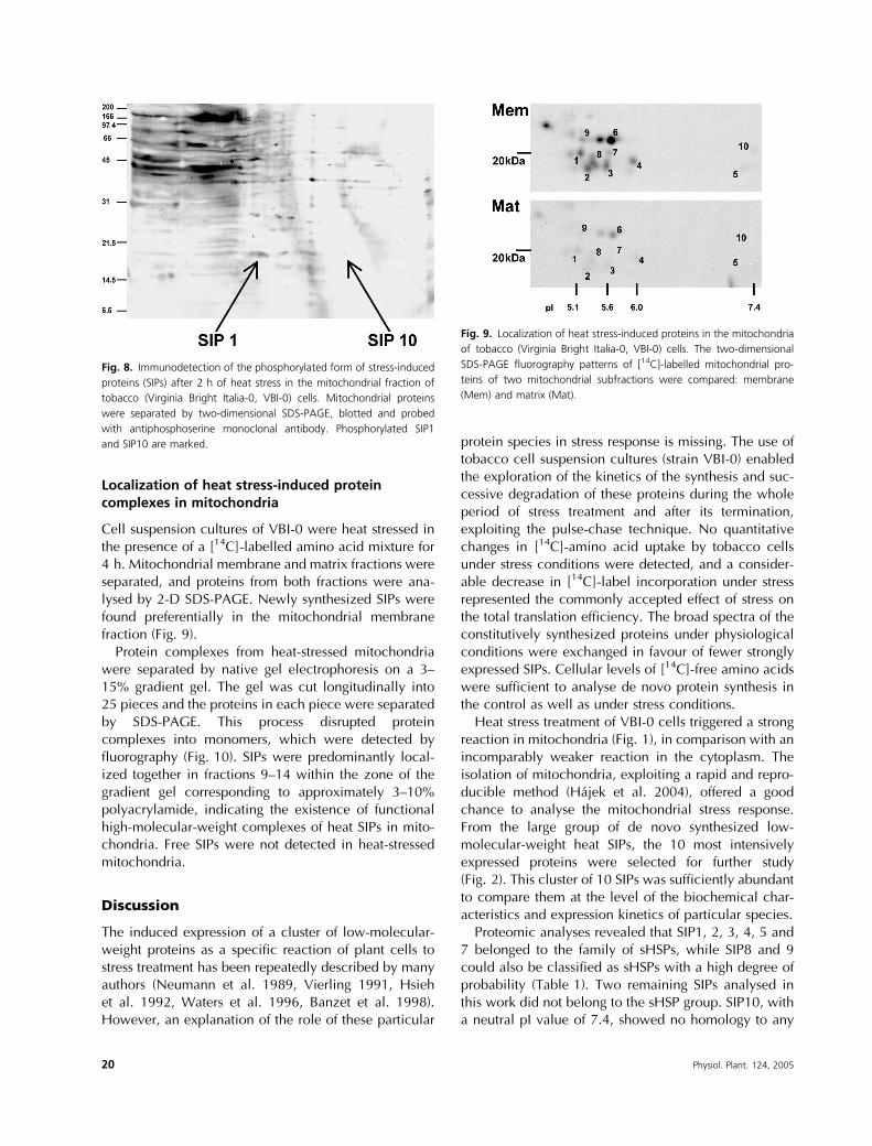
Cell suspension cultures of VBI-0 were heat stressed inthe presence of a [14C]-labelled amino acid mixture for4 h. Mitochondrial membrane and matrix fractions wereseparated, and proteins from both fractions were ana-lysed by 2-D SDS-PAGE. Newly synthesized SIPs werefound preferentially in the mitochondrial membranefraction (Fig. 9).
Protein complexes from heat-stressed mitochondriawere separated by native gel electrophoresis on a 3–15% gradient gel. The gel was cut longitudinally into25 pieces and the proteins in each piece were separatedby SDS-PAGE. This process disrupted proteincomplexes into monomers, which were detected byfluorography (Fig. 10). SIPs were predominantly local-ized together in fractions 9–14 within the zone of thegradient gel corresponding to approximately 3–10%polyacrylamide, indicating the existence of functionalhigh-molecular-weight complexes of heat SIPs in mito-chondria. Free SIPs were not detected in heat-stressedmitochondria.
Discussion
The induced expression of a cluster of low-molecular-weight proteins as a specific reaction of plant cells tostress treatment has been repeatedly described by manyauthors (Neumann et al. 1989, Vierling 1991, Hsiehet al. 1992, Waters et al. 1996, Banzet et al. 1998).However, an explanation of the role of these particular
protein species in stress response is missing. The use oftobacco cell suspension cultures (strain VBI-0) enabledthe exploration of the kinetics of the synthesis and suc-cessive degradation of these proteins during the wholeperiod of stress treatment and after its termination,exploiting the pulse-chase technique. No quantitativechanges in [14C]-amino acid uptake by tobacco cellsunder stress conditions were detected, and a consider-able decrease in [14C]-label incorporation under stressrepresented the commonly accepted effect of stress onthe total translation efficiency. The broad spectra of theconstitutively synthesized proteins under physiologicalconditions were exchanged in favour of fewer stronglyexpressed SIPs. Cellular levels of [14C]-free amino acidswere sufficient to analyse de novo protein synthesis inthe control as well as under stress conditions.
Heat stress treatment of VBI-0 cells triggered a strongreaction in mitochondria (Fig. 1), in comparison with anincomparably weaker reaction in the cytoplasm. Theisolation of mitochondria, exploiting a rapid and repro-ducible method (Hajek et al. 2004), offered a goodchance to analyse the mitochondrial stress response.From the large group of de novo synthesized low-molecular-weight heat SIPs, the 10 most intensivelyexpressed proteins were selected for further study(Fig. 2). This cluster of 10 SIPs was sufficiently abundantto compare them at the level of the biochemical char-acteristics and expression kinetics of particular species.
Proteomic analyses revealed that SIP1, 2, 3, 4, 5 and7 belonged to the family of sHSPs, while SIP8 and 9could also be classified as sHSPs with a high degree ofprobability (Table 1). Two remaining SIPs analysed inthis work did not belong to the sHSP group. SIP10, witha neutral pI value of 7.4, showed no homology to any
Fig. 8. Immunodetection of the phosphorylated form of stress-induced
proteins (SIPs) after 2 h of heat stress in the mitochondrial fraction of
tobacco (Virginia Bright Italia-0, VBI-0) cells. Mitochondrial proteins
were separated by two-dimensional SDS-PAGE, blotted and probed
with antiphosphoserine monoclonal antibody. Phosphorylated SIP1
and SIP10 are marked.
Fig. 9. Localization of heat stress-induced proteins in the mitochondria
of tobacco (Virginia Bright Italia-0, VBI-0) cells. The two-dimensional
SDS-PAGE fluorography patterns of [14C]-labelled mitochondrial pro-
teins of two mitochondrial subfractions were compared: membrane
(Mem) and matrix (Mat).
20 Physiol. Plant. 124, 2005

known protein sequence. SIP6 shared a significantdegree of homology with transcription factors fromseveral different organisms. However, both of theseproteins were always detected together with the sHSPgroup. Moreover, SIP6 was localized strictly tomitochondria. Consequently, one possible function ofthis protein may be the repression of mitochondrialtranscription under stress conditions, perhaps throughbinding of the protein to mitochondrial DNA. Thesesuggestions were based on findings that plant mitochon-dria lose their DNA during cell differentiation (Fujieet al. 1994).
Basic biochemical data from the analyses of thesHSPs with molecular masses ranging from 18 to23.5 kDa (Fig. 3) corresponded with the data previouslypublished for sHSP22 from Pisum sativum (Lenne et al.1995), sHSP22 from Zea mays (Lund et al. 1998),sHSP23 from Chenopodium rubrum (Debel et al.1997) and sHSP23.5 from Arabidopsis thaliana (Visioliet al. 1997). Seven of the eight characterizedstress-induced sHSPs exhibited acidic characteristics inagreement with previously published data on othersHSPs (Morrow et al. 2000). SIP5, with a neutral pIvalue of 7.5, was the only exception. However, SIP5shared the same peptide fragment identity with SIP3 andSIP4. Together, these proteins formed a distinct sub-group within the set of eight characterized SIPs. Theclose sequence relationship of these proteins was con-firmed by partial peptide mapping (Fig. 4). Sequencefragments obtained for these proteins were then com-pared with 28 sequences of Arabidopsis thaliana sHSPsdownloaded from the TAIR server (www.arabidopsis.org). When compared with the eight most similarsHSPs, it became clear that the amino acid sequencesof all peptide mass digest fragments suitable for data-base searches lay strictly within two of the most con-served amino acid sequence domains of sHSPs (Fig. 4).
The 2-D pattern of sHSPs detectable 30 min afterstress (Fig. 2A) remained qualitatively stable through-out the 12 h stress treatment and was maintained forup to 12 h after its termination (data not shown). Anidentical pattern was observed during the course ofthe whole subculture interval. However, higher levelsof all SIPs during the cell elongation phase suggestedhigher sensitivity to stress at this stage (Fig. 7).Quantitative analyses utilizing pulse-chase experi-ments confirmed the frequently observed intenseexpression of sHSPs induced by stress and the highstability of these molecules after the termination ofstress conditions (Lenne et al. 1995, Waters et al.1996). A 30 min pulse-label during the course ofstress demonstrated two phases of synthetic activityfor all SIPs. Synthesis increased during the first 6 h ofthe stress treatment, when it reached a maximum,and then decreased continuously for a further 6 h.However, levels of synthetic activity for individualsHSP species were found to be different. The synth-esis of sHSP22 (SIP1) was significantly high through-out the whole stress period. In contrast, the synthesisof sHSP18.5 (SIP5) was so low that, by the end of thestress period, the protein was undetectable (Fig. 5).The stability of sHSP molecules was verified by 1 hpulses followed by a 6 h and 24 h chase. After 6 h,the qualitative pattern of protein synthesis wasunchanged, with only a slight quantitative decreaseof 15–30%. A substantial decrease in synthesisoccurred 18 h later. Only SIP6, a transcription factorhomologue, which showed the highest syntheticactivity during the 12 h stress interval, exhibited anextremely stable and high quantitative level even after24 h of the pulse-chase experiment (Fig. 6)
Inspired by the previously published data on maizemitochondrial HSP22 phosphorylation (Lund et al.1998), we performed phospho-immunoassays of the
Fig. 10. Fluorogram of pro-
teins comprising heat stress-
induced macromolecular com-
plexes synthesized in tobacco
(Virginia Bright Italia-0, VBI-0)
cells. Mitochondrial protein
complexes were separated by
native 3–15% gradient gel
electrophoresis. Native gel was
cut into 25 fragments which
were run on one-dimensional
SDS-PAGE to release individual
proteins. Position of the stress-
induced protein (SIP) complex is
marked.
Physiol. Plant. 124, 2005 21

analysed SIPs. Despite the preliminary character of thephospho-immunotests performed, the observed serinephosphorylation of SIP1 represents one more key resultof this study. Moreover, the mass spectrometrysequence fragment of the 20 kDa SIP1 shared significanthomology with the C-terminal part of maize HSP22.Within their 14-amino acid aligned region, thesesequences were 71% identical with only three mis-matches (data not shown). The combination of theabove results strongly suggests that SIP1 represents atobacco homologue of the previously described maizeHSP22 (Lund et al. 1998). In animal cells, the relation-ship between the serine phosphorylation status ofsHSP27 and the oligomerization/dissociation of sHSPcomplexes has been documented (Ehrnsperger et al.1997, Haslbeck et al. 1999). The sHSP27 phosphoryla-tion is also known to influence actin cytoskeletondynamics and to modulate actin filament stability(Guay et al. 1997). Taken together, the observed strongphosphorylation of the mitochondrial HSP within SIPsintroduces heterogeneity into this coordinately inducedprotein cluster, and is likely to reveal a new perspectiveon organized sHSP function in plant mitochondria inconnection with other structures.
The exclusive presence of newly synthesized smallheat SIPs in the mitochondria of stressed VBI-0cells suggests an important role for this organelle inthe exponentially growing plant cell. The majority ofSIPs colocalized with the mitochondrial membrane frac-tions, whereas, in contrast, matrix fractions containedonly traces of their presence (Fig. 9). Moreover, no freesHSPs were detected in mitochondria and the cluster ofSIPs was shown to be stable and to form high-molecular-weight complexes. Newly synthesized SIPswere localized only in high-molecular weight com-plexes (Fig. 10). We conclude that SIPs, in agreementwith the literature, form homopolymeric complexesassembled from a broad spectrum of subunits (Noveret al. 1989, Helm et al. 1993), and these complexes arelikely to participate in the formation of high-molecular-weight HSP chaperones described previously (Lee et al.1997, Smykal et al. 2000).
Conclusions
The data presented here clearly demonstrate the com-plexity of the heat stress response in tobacco VBI-0 cellsuspension culture. In non-differentiated, proliferatingcells, heat stress preferentially induced the synthesis ofa significantly stable group of SIPs in mitochondria.Within the SIP group, eight sHSPs were accompaniedby one transcription factor and one protein withunknown function. sHSPs represented a heterogeneous
cluster of proteins synthesized in variable quantities.Moreover, the preferentially synthesized protein SIP1(sHSP22) was found to be serine phosphorylated. SIPsformed multimolecular protein complexes localized inthe mitochondrial membranes.
Acknowledgements – We thank Dr D. A. Morris (University
of Southampton, UK) for critical reading of the manuscript.
We gratefully acknowledge the financial support from the
Ministry of Education of the Czech Republic (grant no. MSM
113100003) and from the Grant Agency of the ASCR (project
no. K5052113).
References
Alberts B, Johnson A, Lewis J, Raff M, Roberts K, Walter P
(2002) Molecular Biology of the Cell. Garland Science
Publishing Inc., NY, ISBN 0-8153-4072-9
Altschul SF, Gish W, Miller W, Myers EW, Lipman DJ (1990)
Basic local alignment search tool. J Mol Biol 215: 403–410
Arrigo AP (1998) Small stress proteins: chaperones that act as
regulators of intracellular redox state and programmed
cell death. Biol Chem 379: 19–26
Banzet N, Richard C, Deveaux Y, Kazmaier M, Gagnou J,
Trintaphylides C (1998) Accumulation of small heat shock
proteins, including mitochondrial HSP22, induced by
oxidative stress and adaptive response in tomato cells.
Plant J 13: 519–527
Blum H, Beier H, Gross HJ (1987) Improved silver staining of
plant proteins, RNA and DNA in polyacrylamide gels.
Electrophoresis 8: 93–99
Caspers GJ, Leunissen JA, de Jong WW (1995) The expand-
ing small heat-shock protein family and structure predic-
tions of the conserved ‘alpha-crystallin domain’. J Mol
Evol 40: 238–248
De Jong WW (1993) Evolution of the alpha-crystallin/small
heat shock protein family. Mol Biol Evol 10: 103–126
Debel K, Sierralta WD, Braun HP, Schmitz UK, Kloppstech K
(1997) The 23-kDa light-stress-regulated heat-shock pro-
tein of Chenopodium rubrum L. is located in the mito-
chondria. Planta 201: 326–333
Downs CA, Heckathorn SA (1998) The mitochondrial small
heat shock protein protects NADH:ubiquinone oxidore-
ductase of the electron transport chain during heat stress
in plant. FEBS Lett 430: 246–250
Ehrnsperger M, Graber S, Gaestel M, Buchner J (1997)
Binding of non-native protein to Hsp25 during heat shock
creates a reservoir of folding intermediates for reactiva-
tion. EMBO J 16: 221–229
Fujie M, Kuroiwa H, Kawano S, Mutoh S, Kuroiwa T (1994)
Behavior of organelles and their nucleoids in the shoot
apical meristem during leaf development in Arabidopsis
thaliana L. Planta 194: 395–405
Garrido C, Ottavi P, Fromentin A, Hammann A, Arrigo AP,
Chauffert B, Mehlen P (1997) HSP27 as a mediator of
22 Physiol. Plant. 124, 2005

confluence-dependent resistance to cell death induced by
anticancer drugs. Cancer Res 57: 2661–2667
Guay J, Lambert H, Gingras-Breton G, Lavoie JN, Huot J,
Landry J (1997) Regulation of actin filament dynamics by
p38 map kinase-mediated phosphorylation of heat shock
protein 27. J Cell Sci 110: 357–368
Hajek T, Honys D, Capkova V (2004) New method of mito-
chondria isolation and sub-fractionation for proteomic
analyses. Plant Sci 167: 389–395
Haslbeck M, Walke S, Stromer T, Ehrnsperger M, White HE,
Chen SX, Saibil HR, Buchner J (1999) Hsp26: a tempera-
ture-regulated chaperone. EMBO J 18: 6744–6751
Heller R (1953) Studies on the mineral nutrition of in vitro plant
tissue cultures. An Sci Nat Bot Biol Veg 14: 1–223
Helm KW, La Fayette PR, Nagao RT, Key JL, Vierling E
(1993) Localization of small heat shock proteins to the
higher plant endomembrane system. Mol Cell Biol 13:
238–247
Hsieh MH, Chen JT, Jinn TL, Chen YM, Lin CY (1992) A class
of soybean low molecular weight heat shock proteins,
immunological study and quantitation. Plant Physiol 99:
1279–1284
Jones A (2000) Does the plant mitochondrion integrate cel-
lular stress and regulate programmed cell death? Trends
Plant Sci 5: 225–230
Kirschner M, Winkelhaus S, Thierfelder JM, Nover L (2000)
Transient expression and heat-stress-induced co-aggrega-
tion of endogenous and heterogenous small heat-stress
proteins in tobacco protoplasts. Plant J 24: 397–411
Laemmli UK (1970) Cleavage of structural proteins during
the assembly of the head of bacteriophage T4. Nature
227: 680–685
Lai YK, Lee WC, Hu CH, Hammpnd GL (1996) The mito-
chondria are recognition organelles of cell stress. J Surg
Res 62: 90–94
Landry J, Chretien P, Lambert H, Hickey E, Weber LAQ (1989)
Heat shock resistance conferred by expression of the
human HSP 27 gene in rodent cells. J Cell Biol 109: 7–15
Lee GJ, Roseman AM, Saibil HR, Vierling E (1997) A small
heat shock protein stably binds heat-denatured model
substrates and can maintain a substrate in a folding-
competent state. EMBO J 16: 659–671
Lenne C, Block MA, Garin J, Douce R (1995) Sequence and
expression of the messenger-RNA encoding HSP22, the
mitochondrial small heat-shock protein in pea leaves.
Biochem J 311: 805–813
Lenne C, Douce R (1994) A low molecular mass heat-shock
protein is localized to higher plant mitochondria. Plant
Physiol 105: 1255–1261
Lund AA, Blum PH, Bhattramakki D, Elthon TE (1998) Heat-
stress response of maize mitochondria. Plant Physiol 116:
1097–1110
Mehlen P, Schulze-Osthoff K, Arrigo AP (1996) Small heat
shock proteins as novel regulators of apoptosis. J Biol
Chem 271: 16 510–16 514
Morimoto R, Tissieres A, Georgopolous C (1994) The
Biology of Heat Shock Proteins and Molecular
Chaperones. Cold Spring Harbor Laboratory Press, Cold
Spring Harbor, NY
Morrow G, Inaguma Y, Kato K, Tanguay RM (2000) The
small heat shock protein Hsp22 of Drosophila melano-
gaster is a mitochondrial protein displaying oligomeric
organization. J Biol Chem 275: 31 204–31 210
Neumann D, Nover L, Partier R, Scharf KD (1989) Heat
shock and other stress response systems of plants.
Biologisches Zentralblatt 108: 1–156
Nover L, Scharf KD, Neumann D (1989) Cytoplasmic heat-
shock granules are formed from precursor particles and
are associated with a specific set of messenger-RNAs. Mol
Cell Biol 9: 1298–1308
Opatrny Z, Opatrna J (1976) The specificity of the effect of
2,4-D and NAA on the growth, micromorphology and
occurrence of starch in long-term Nicotiana tabacum L.
cell strains. Biol Plant 18: 359–365
Parsell DA, Lindquist S (1993) The function of heat-shock
proteins in stress tolerance: degradation and reactivation
of proteins. Annu Rev Genet 27: 437–496
Perkins DN, Pappin DJ, Creasy DM, Cottrell JS (1999)
Probability-based protein identification by searching
sequence databases using mass spectrometry data.
Electrophoresis 20: 3551–3567
Rassow J, von Ahsen O, Bomer U, Pfanner N (1997)
Molecular chaperones: towards a characterization of
the heat-shock protein 70 family. Trends Cell Biol 7:
129–133
Schagger H, Cramer WA, Vonjagow G (1994) Analysis of
molecular masses and oligomeric states of protein
complexes by blue native electrophoresis and isolation of
membrane-protein complexes by 2-dimensional native
electrophoresis. Anal Biochem 217: 220–230
Shevchenko A, Wilm M, Vorm O, Mann M (1996) Mass
spectrometric sequencing of proteins from silver stained
polyacrylamide gels. Anal Chem 68: 850–858
Smykal P, Masın J, Hrdy I, Konopasek I, Zarsky V (2000)
Chaperone activity of tobacco HSP18, a small heat-shock
protein, is inhibited by ATP. Plant J 23: 703–713
Sun W, Van Montagu M, Verbruggen N (2002) Small heat
shock proteins and stress tolerance in plants. Biochim
Biophys Acta 1577: 1–9
Towbin H, Gordon J (1984) Immunoblotting and dot
immunoblotting – current status and outlook. J Immunol
Methods 72: 313–340
Vierling E (1991) The roles of heat-shock proteins in plants.
Annu Rev Plant Phys 42: 579–620
Visioli G, Maestri E, Marmiroli N (1997) Differential
display-mediated isolation of a genomic sequence
for a putative mitochondrial LMW HSP specifically
expressed in condition of induced thermotolerance
in Arabidopsis thaliana (L.). Plant Mol Biol 34:
517–527
Physiol. Plant. 124, 2005 23

Waters ER, Lee GJ, Vierling E (1996) Evolution, structure and
function of the small heat shock proteins in plants. J Exp
Bot 47: 325–338
Wilm M, Shevchenko A, Houthaeve T, Breit S, Schweigerer
L, Fotsis T, Mann M (1996) Femtomole sequencing of
proteins from polyacrylamide gels by nano-electrospray
mass spectrometry. Nature 379: 466–469
Zhang XP, Glaser E (2002) Interaction of plant mitochondrial
and chloroplast signal peptides with the Hsp70 molecular
chaperone. Trends Plant Sci 7: 14–21
Edited by C. Guy
24 Physiol. Plant. 124, 2005