
Identification of potential therapeutic targets in human head & neck squamous cell carcinoma
9
BioMed Central Page 1 of 9 (page number not for citation purposes) Head & Neck Oncology Open Access Research Identification of potential therapeutic targets in human head & neck squamous cell carcinoma Jing Han 1 , Mitomu Kioi 1 , Wei-Sing Chu 2 , Jan L Kasperbauer 3 , Scott E Strome 4 and Raj K Puri* 1 Address: 1 Tumor Vaccines and Biotechnology Branch, Division of Cellular and Gene Therapies, Center for Biologics Evaluation and Research, Food and Drug Administration, Bethesda, MD 20892, USA, 2 Department of Scientific Laboratories, Armed Forces Institute of Pathology, Washington, DC 20306-6000, USA, 3 Mayo Clinic Cancer Center, 200 First Street Southwest, Rochester, Minnesota, 55905, USA and 4 Department of Otorhinolaryngology, Head & Neck Surgery, School of Medicine, University of Maryland, Baltimore, MD 21201, USA Email: Jing Han - [email protected]; Mitomu Kioi - [email protected]; Wei-Sing Chu - [email protected]; Jan L Kasperbauer - [email protected]; Scott E Strome - [email protected]; Raj K Puri* - [email protected] * Corresponding author Abstract Background: Human head and neck squamous cell carcinoma (HNSCC) is an aggressive and recurrent malignancy. Identification of unique or overexpressed cell-associated or cell surface antigens is critical for diagnosis and development of cancer vaccines and targeted therapies for HNSCC. We have used high throughput microarray technology to search for candidate targets in HNSCC. Methods: Gene expression profiling in 17 HNSCC tumors and 3 normal tonsil tissues was performed by microarray. QRT-PCR analysis was performed to validate the microarray results. The five candidate genes were further characterized by immunohistochemical technique in surgical samples and tissue arrays. Results: A total of 192 up-regulated genes at statistical significance of p < 0.01 and log2 ratio ≥ 1 were identified in HNSCC tumors compared to normal tissues. These genes belong to immune response, cell growth, cell cycle regulation, oncogenes, metabolism and others. Five potential novel target genes (FABP5, CD24, CD44, CD74, and HSP27) were identified, which were highly expressed in HNSCC tumor samples and tissue arrays. CD24, CD44, and CD74 proteins were expressed on the cell surface, and FABP5 and HSP27 proteins were predominantly expressed in the cytoplasm of HNSCC. Conclusion: Five genes and their products may serve as a diagnostic biomarker or therapeutic target for HNSCC. While additional work is needed to elucidate the biological significance of these proteins, CD24 and CD74 expressed only in small proportion of cells indicating tumor heterogeneity and subtypes of tumor initiating cells (CD24+/CD44+) present in HNSCC. Background In the United States, an estimated 40,000 new cases of head and neck cancers are diagnosed each year [1]. More than 90% of head and neck cancers are of squamous cell carcinoma, and arise from diverse anatomical locations, including lip/oral cavity, nasopharynx, oropharynx, lar- Published: 14 July 2009 Head & Neck Oncology 2009, 1:27 doi:10.1186/1758-3284-1-27 Received: 10 April 2009 Accepted: 14 July 2009 This article is available from: http://www.headandneckoncology.org/content/1/1/27 © 2009 Han et al; licensee BioMed Central Ltd. This is an Open Access article distributed under the terms of the Creative Commons Attribution License (http://creativecommons.org/licenses/by/2.0 ), which permits unrestricted use, distribution, and reproduction in any medium, provided the original work is properly cited.
Transcript of Identification of potential therapeutic targets in human head & neck squamous cell carcinoma

BioMed CentralHead & Neck Oncology
ss
Open AcceResearchIdentification of potential therapeutic targets in human head & neck squamous cell carcinomaJing Han1, Mitomu Kioi1, Wei-Sing Chu2, Jan L Kasperbauer3, Scott E Strome4 and Raj K Puri*1Address: 1Tumor Vaccines and Biotechnology Branch, Division of Cellular and Gene Therapies, Center for Biologics Evaluation and Research, Food and Drug Administration, Bethesda, MD 20892, USA, 2Department of Scientific Laboratories, Armed Forces Institute of Pathology, Washington, DC 20306-6000, USA, 3Mayo Clinic Cancer Center, 200 First Street Southwest, Rochester, Minnesota, 55905, USA and 4Department of Otorhinolaryngology, Head & Neck Surgery, School of Medicine, University of Maryland, Baltimore, MD 21201, USA
Email: Jing Han - [email protected]; Mitomu Kioi - [email protected]; Wei-Sing Chu - [email protected]; Jan L Kasperbauer - [email protected]; Scott E Strome - [email protected]; Raj K Puri* - [email protected]
* Corresponding author
AbstractBackground: Human head and neck squamous cell carcinoma (HNSCC) is an aggressive andrecurrent malignancy. Identification of unique or overexpressed cell-associated or cell surfaceantigens is critical for diagnosis and development of cancer vaccines and targeted therapies forHNSCC. We have used high throughput microarray technology to search for candidate targets inHNSCC.
Methods: Gene expression profiling in 17 HNSCC tumors and 3 normal tonsil tissues wasperformed by microarray. QRT-PCR analysis was performed to validate the microarray results.The five candidate genes were further characterized by immunohistochemical technique in surgicalsamples and tissue arrays.
Results: A total of 192 up-regulated genes at statistical significance of p < 0.01 and log2 ratio ≥ 1were identified in HNSCC tumors compared to normal tissues. These genes belong to immuneresponse, cell growth, cell cycle regulation, oncogenes, metabolism and others. Five potential noveltarget genes (FABP5, CD24, CD44, CD74, and HSP27) were identified, which were highlyexpressed in HNSCC tumor samples and tissue arrays. CD24, CD44, and CD74 proteins wereexpressed on the cell surface, and FABP5 and HSP27 proteins were predominantly expressed inthe cytoplasm of HNSCC.
Conclusion: Five genes and their products may serve as a diagnostic biomarker or therapeutictarget for HNSCC. While additional work is needed to elucidate the biological significance of theseproteins, CD24 and CD74 expressed only in small proportion of cells indicating tumorheterogeneity and subtypes of tumor initiating cells (CD24+/CD44+) present in HNSCC.
BackgroundIn the United States, an estimated 40,000 new cases ofhead and neck cancers are diagnosed each year [1]. More
than 90% of head and neck cancers are of squamous cellcarcinoma, and arise from diverse anatomical locations,including lip/oral cavity, nasopharynx, oropharynx, lar-
Published: 14 July 2009
Head & Neck Oncology 2009, 1:27 doi:10.1186/1758-3284-1-27
Received: 10 April 2009Accepted: 14 July 2009
This article is available from: http://www.headandneckoncology.org/content/1/1/27
© 2009 Han et al; licensee BioMed Central Ltd. This is an Open Access article distributed under the terms of the Creative Commons Attribution License (http://creativecommons.org/licenses/by/2.0), which permits unrestricted use, distribution, and reproduction in any medium, provided the original work is properly cited.
Page 1 of 9(page number not for citation purposes)
Head & Neck Oncology 2009, 1:27 http://www.headandneckoncology.org/content/1/1/27
ynx, and hypopharynx [1,2]. Although numerous studieshave reported on the mechanism of oncogenesis, the pre-cise cellular mechanism of tumor development, tumorprogression, and tumor aggressiveness are not known.The survival rate of patients with head & neck squamouscell carcinoma (HNSCC) has not improved significantlydespite multimodality therapy including surgery, radia-tion therapy, and chemotherapy. Recent investigationshave focused on novel therapeutic approaches, and on theidentification of molecular targets for therapy [3,4].
Microarray approaches have been widely used to identifygenes associated with tumorigenesis, metastatic potential,clinically distinct subgroups of tumors, and prognosticbiomarkers and potential targets for novel therapeuticagents. Identification of unique or overexpressed antigensor cell surface proteins is critical for the development ofcancer vaccines and targeted immunotoxins or cytotoxins,which may offer alternative approach for caner therapy.
Previously, our group has identified Interleukin-13 recep-tor α2 (IL13Rα2) as a unique marker in human head andneck cancer, which can be used to distinguish a subset ofHNSCC and may serve as a target for immunotoxin orcytotoxin therapies [5]. In attempt to identify novel thera-peutic targets for treatments of HNSCC, in the presentstudy, we analyzed differentially expressed genes inHNSCC tumors derived from clinical samples, and com-pared with normal tissues using microarray technology.
The genes significantly up-regulated in HNSCC wereselected as candidates for further evaluation. Based on theliterature and availability of antibody, highly expressedgenes (CD24, CD44, and CD74) encoding cell surfacereceptors and (FABP5 and heat shock protein Hsp27)encoding cytoplasmic proteins were evaluated at proteinlevels by IHC. The in-depth analysis of gene expressionand protein expression in tissue arrays of these candidategenes revealed that they may serve as potential prognosticbiomarkers or therapeutic targets for targeted therapy orantigen directed immunotherapy.
MethodsTissue Sample CollectionA total of 17 HNSCC and 3 normal tissues (from tonsil-lectomies) from 20 patients were collected at Mayo ClinicCancer Center. The Institutional Review Board (IRB) ofthe Mayo Clinic approved sample collection and RIHSC(Research Involving Human Subjects Committee) of theFDA had approved sample receipt at the FDA laboratory.Samples were immediately stabilized by freezing in liquidnitrogen in the operating room. All samples were patho-logically confirmed. The clinical characteristics of the tis-sue samples are summarized in Table 1. At the FDAlaboratory, the specimen was cut into two pieces, onepiece for RNA isolation and another for paraffin embed-ding. H & E staining of Paraffin-embedded tissue sectionswere performed by Histoserv, Inc (Germantown, MD).
Table 1: Patient demographics and pathology of tumors
Patient # Sex Age (yrs) Tumor Location and Grade History of ETOH or Smoking Pathological Subtypes
Normal TissuesA9 F 32 tonsillectomy No Follicular hyperplasiaB9 F 30 tonsillectomy ETOH/Smoking Follicular hyperplasiaC9 M 40 tonsillectomy No Follicular hyperplasia
TumorsA1 / / Base of tongue Grade 3 / Primary SCCAA6 / / Base of tongue Grade 3 / Primary SCCAE1 F 68 Base of tongue Grade 4 ETOH/Smoking Recurrent SCCAI1 M 38 Base of tongue ETOH/Smoking Primary SCCAF1 F 65 Larynx, Grade 2 ETOH/Smoking Primary SCCAC1 M 53 Larynx, Grade 3 ETOH/Smoking Primary SCCAD1 F 40 Larynx, Grade 3 No Metastatic, unknown primaryA3 / / Lymph node-neck / Primary SCCAA5 / / Lymph node-neck / Primary SCCAH1 F 44 Lymph node-neck, Grade 3 No Primary SCCAB1 M 72 Tonsil, Grade 3 No ETOH, Smoking Primary SCCAB2 M 60 Tonsil ETOH/Smoking Primary SCCAB5 M 62 Tonsil ETOH/Smoking Primary SCCAB4 M 53 Buccal mucosa ETOH, Minimal Smoking Recurrent SCCAG1 M 72 Buccal mucosa Minimal ETOH/Smoking Primary SCCAA2 M 60 Supraglottic / Primary SCCAB3 F 72 Supraglottic Grade 4 ETOH/Smoking Recurrent SCCA
/: patient information not availableSCCA: Squamous cell carcinoma
Page 2 of 9(page number not for citation purposes)

Head & Neck Oncology 2009, 1:27 http://www.headandneckoncology.org/content/1/1/27
Microarray ExperimentsTotal RNA from tissue samples used for microarray exper-iments were extracted by Trizol reagent according to theManufacture's instructions (Invitrogen). Briefly, tissueswere suspended in Trizol reagent (Invitrogen), homoge-nized, and RNA was isolated following the Manufacture'sinstructions. Human universal RNA (huRNA) (Strate-gene) was used as a common reference for all experi-ments. The human microarrays containing approximately17,000 oligonucleotides were used which were producedin our laboratory. The detailed information regardingarray printing, post-print processing, and testing arrayquality is described elsewhere [6]. Target preparation,microarray hybridization, image quantification, and dataanalyses have been described previously, with modifica-tions [7]. In brief, 5 μg of total RNA was reverse-tran-scribed using 5'-amino-modified primers with amino-allyl-dUTP. cDNAs synthesized from HNSCC tissues sam-ples were labeled with Cy5 dye, and cDNA from universalRNA labeled with Cy3 dye. Labeled and combined cDNAprobes were denatured, mixed in SlideHyb #1 hybridiza-tion buffer (Ambion, Austin, TX), and placed onto micro-array slides. Arrays were hybridized at 42°C in MAUIhybridization system (BioMicro Systems) for 16–18hours, and then washed with 1 × SSC with 0.05% SDS for4 min., 0.1 × SSC for 4 min. Slides were quickly spin-dried.
Data Analysis and StatisticsMicroarray slides were scanned on a GenePix 4000B scan-ner (Axon Instruments, Inc., Foster City, CA) with a 10 μmresolution. Scanned raw images were analyzed and datafiles were generated with GenePix Pro 5.1 (Axon) soft-ware. For analysis, data files were uploaded into mAdb(microarray database), and analyzed by the software toolsprovided by the Center for Information Technology (CIT),NIH. The advanced filters, spots size at least 30 μm, min-imum fluorescent intensity of 100 in both Cy3 and Cy5channels, were applied before data analysis. A standardglobal normalization approach was used for each experi-ment. All of the extracted data was normalized using a50th percentile (median) normalization method. Statisti-cal analyses were performed and group comparison t-testwas used to compare the difference of gene expressionbetween normal and tumor samples. Genes were selectedwith the following criteria: p < 0.01, and log2 fold differ-ence ≥ 1 in mean expression between HNSCC tumors andnormal tissues.
Gene-Specific Confirmation by QRT-PCRQRT-PCR was performed on selected genes identified bygene expression profiling. The first-strand cDNA was syn-thesized from 1 μg of total RNA using Superscript IIReverse Transcriptase (Invitrogen, Carlsbad, CA) accord-ing to manufactures specifications. The resulting cDNAwas amplified by using gene-specific primers. Primer
sequences are as follows: FABP5, forward, 5'-agcagct-ggaaggaagat-3', and reverse, 5'-gatacaatctggcttggc-3';CD24, forward, 5'-aggatgggattgtggaat-3', and reverse, 5'-attagtgccgtcgaaaca-3'; EIF4G2, forward, 5'-gcagaagatgcac-caaac-3', and reverse, 5'-atggctctctgttcctcc-3'; KRT18, for-ward, 5'-gtagatgcccccaaatct-3', and reverse, 5'-cactgtggtgctctcctc-3'; LGALS1, forward, 5'-tggactcaatcat-ggctt-3', and reverse, 5'-ggttgttgctgtctttgc-3'; RPLP0, for-ward, 5'-gacggattacaccttccc-3', and reverse, 5'-tggcttcaaccttagctg-3'; GAPDH, forward 5'-aaggtgaaggtcg-gagtcaa-3', and reverse 5'-gatctcgctcctggaagatg-3'. The spe-cificity of primers was first verified by RT-PCR and gelelectrophoresis. QRT-PCR reactions were performed withSYBR Q-PCR master mixture (Stratagene, La Jolla, CA) fol-lowing the manufactures protocol, and reactions were car-ried out in a Stratagene Mx3000P machine. Buffer and notemplate controls were included in each assay run. Allsamples and controls were run in triplicate.
Immunohistochemistry of HNSCCImmunohistochemical studies of selected genes were per-formed on both formalin-fixed, paraffin-embedded tissuesections from patients and HNSCC tissues arrays, whichwere purchased from Biomax Inc. Tissue diagnosis of via-ble tissue was confirmed by H and E staining for squa-mous cell carcinoma of head and neck. HNSCC tissuearrays contained 16 cases of primary HNSCC and 8 casesof cancer adjacent normal tissues in duplicate andmounted on the slides. Tissue sections or tissue arrayswere deparaffinized by xylene, and then, re-hydrated withsequential washes of 100%, 75%, and 50% ethanol, andPBS. For antigen retrieval, slides were placed in 50 mM cit-rate buffer pH6.0 (Vector Lab, CA), heated in a microwaveoven for 5 min, and then stayed in the buffer for 15 min.Endogenous peroxidase activity was inhibited with 3%hydrogen peroxidase in PBS. Non-specific binding wasblocked with 2.5% normal serum and 1% bovine serumalbumin (BSA) for 1 hr. Tissue sections and tissue arrayswere then incubated with various antibodies, CD24,CD44, and HSP27 (Chemicon; Temecular, CA), CD74(Santa Cruz Biotech; Santa Cruz, CA), FABP5 (Protein-Tech; Chicago, IL), or isotype control (IgG) (Sigma) over-night at 4°C. Immunodetection was performed usingABC staining systems according to manufacturer's instruc-tions (Santa Cruz Biotech; Santa Cruz, CA). All sectionswere counterstained with haematoxylin. After dehydra-tion with washes of 95% and 100% ethanol and xylene,tissue sections and tissue arrays with permanent mount-ing medium were covered with glass coverslips, andviewed by light microscope.
ResultsIdentification of Differentially Expressed Genes in HNSCCThe study population was representative of the generalpopulation with HNSCC, having a median age of 60 yearsat presentation. All 17 tumor tissues were squamous cell
Page 3 of 9(page number not for citation purposes)

Head & Neck Oncology 2009, 1:27 http://www.headandneckoncology.org/content/1/1/27
carcinoma from different locations, and all three tonsilsamples showed follicular hyperplasia (Table 1). Geneexpression profiling was performed in all tumor and nor-mal tissues. In order to identify the potential therapeutictargets in HNSCC, we mainly focused on the up-regulatedgenes in HNSCC. A total of 192 genes, which were signif-icantly up-regulated (with p < 0.01 and log2 fold at least ≥1) in HNSCC tumors compared with normal tonsil tis-sues, were identified. A number of up-regulated geneswere classified, which belong to various biological proc-esses including cell growth and proliferation, proteintranslation and synthesis, metabolism, signaling, andimmune response (Table 2, and see Additional file 1).
A prominent gene expression signature up-regulated inHNSCC includes genes associated with inflammation andimmune response, such as MIF, CD74 and CD24, whichwere not previously identified in HNSCC (Table 2).Another prominent signature of up-regulated genesincludes FABP5, HSP27, S100A2, EIF4G2, and RPLP0,which are associated with various biological functionssuch as metabolism, ion binding, and protein translationand synthesis (Table 2).
To confirm over expressed genes identified by microarrayin HNSCC, six genes including CD24, FABP5, EIF4G2,LGALS1, KRT18, and RPLP0, were selected and QRT-PCRanalysis was performed. The log-transformed measure-ment of gene expression levels determined by microarraypositively correlated with the QRT-PCR analysis (Fig 1).Both microarray and QRT-PCR results confirmed similartrend of gene expression profile of selected genes in theHNSCC.
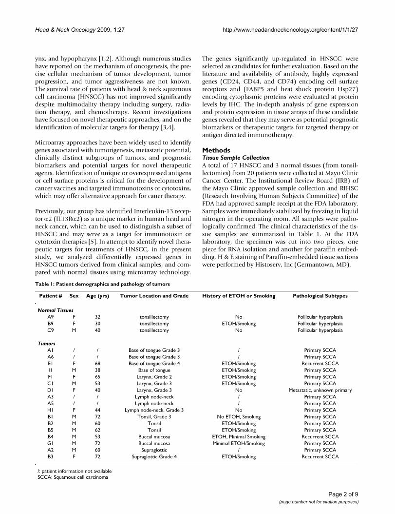
Expression of Selected Gene Products in HNSCCTo narrow down the range of potential therapeutic targetsin 192 up-regulated genes identified in our HNSCC sam-ples, we mainly focused on the genes encoding cell surfacereceptors and some cytoplasmic proteins. Among thesegenes, we selected five gene products (CD24, CD44,CD74, FABP5, and HSP27) as antibodies to these prod-ucts were available for paraffin embedded tissue sections.The expression of selected gene products was confirmedby IHC in tissue sections and tissue arrays. Since CD24,CD44, and CD74 are cell surface proteins and function asadhesion molecules, their expression was predominantlyon the cell surface. Nine out of 16 HNSCC tumors in tis-sue arrays were positive for CD24 expression. However, itsexpression was found only in small clusters of cells withintumors (Fig 2A and 2B). CD44 was strongly expressed onthe cell surface of all tumor cells in all cases of HNSCCsamples (Fig 2D and 2E). CD74 positive cells were alsoidentified in HNSCC tissue arrays (Fig 2G and 2H). Incontrast, CD24, CD44, or CD74 positive cells were notdetected in adjacent normal tissues present in tissue arrays(Fig 2C, F, and 2I).
As FABP5 and HSP27 are intra cytoplasmic proteins, theirexpression was examined in the cytoplasmic compart-ment. Both proteins showed strong positive cytoplasmicstaining in majority of cases in the HNSCC tissue arrays(Fig 2J and 2K, M and 2N). In contrast, only backgroundstaining was observed for either FABP5 or HSP27 in theadjacent normal tissues (Fig 2L and 2O), except the basallayer of dermis in normal tissue also showed FABP5 posi-tive staining (Fig 2L). Isotype control antibody showed nostaining in any section (Data not shown).
DiscussionGlobal gene expression profiling studies of humanHNSCC tumors have identified genes associated withtumorigenesis and metastatic potential [8-13]. In addi-tion, molecular classification of HNSCC has also beenproposed based on gene expression patterns [11-13]. Ithas been reported that gene expression profile can distin-guish clinically distinct subgroups of HNSCC, identifygenes potentially associated with advanced-stage of thedisease, and predict outcome of treatment [9,11,14]. Inour study, in order to identify novel therapeutic targets fordiagnosis or treatments, we compared gene expression inHNSCC with normal tissues and further characterizedprotein expression of over expressed genes in HNSCCsamples. We identified 192 up-regulated genes in HNSCCcompared with normal tissues. Based on our results, pub-lished literature, and availability of antibody, five candi-date genes (CD24, CD44, CD74, FABP5, and HSP27)were selected for further characterization in HNSCC sam-ples. These gene products were expressed in most tumorsamples but not in normal tissues. Although various stud-ies have reported on the expression of these genes in vari-ous tissues, our study presents their significance inHNSCC tumors.
Among selected genes, CD24, CD44, and CD74 are cellsurface adhesion molecules shown to be over expressed invarious cancers [15-21]. Among these, CD44 was moststrongly expressed in all HNSCC samples in tissue arrays.Interestingly, CD44+ subpopulation of cells with cancerstem cell properties has been identified in HNSCC andvarious other cancer types [21-25]. CD24 has also beenshown to be expressed in various tumors. Its expressionhas been used as a prognostic indicator of poor survival inbreast cancer, non-small cell lung carcinoma, and prostatecancers [15-18]. However, in contrast to CD44, only asmall portion of CD24+ cells were positive in HNSCCcancer cells in our study. These results suggest that a subsetof CD24+/CD44+ cells exists, which may represent cancerstem cells in HNSCC. This hypothesis is supported by areport which showed that a highly tumorigenic subpopu-lation of pancreatic cancer cells express cell surface mark-ers CD44, CD24, and epithelial-specific antigen (ESA)[23]. This subpopulation of pancreatic cancer cells withCD44+/CD24+/ESA+ phenotype (only 0.2 to 0.8% of
Page 4 of 9(page number not for citation purposes)

Head & Neck Oncology 2009, 1:27 http://www.headandneckoncology.org/content/1/1/27
Table 2: Selected up-regulated genes identified in HNSCC*
GeneBank Access ID Gene Symbol & Annotation Log2 Fold Difference**
Immune ResponseL19686 MIF, macrophage migration inhibitory factor 3.1M13560 CD74, CD74 antigen 2.6M63438 IGKC, immunoglobulin kappa constant 1.9J03909 IFI30, interferon, gamma-inducible protein 30 1.5AJ251549 IL26, interleukin 26 1.4L33930 CD24, CD24 antigen 1.4X16302 IGFBP2, insulin-like growth factor binding protein 2 1.4S75725 IGFBP7, insulin-like growth factor binding protein 7 1.4
Cell Growth, Maintenance/Cell cycle RegulationY00503 KRT19, keratin 19 3.9U43901 LAMR1, laminin receptor 1 3.2AL031670 FTL, ferritin, light polypeptide 2.5J00124 KRT14, keratin 14 2.5X07696 KRT 15, keratin 15 2.4X95404 CFL1, cofilin 1 2.3D13627 CCT8, chaperonin subunit 8 2.1AF026291 CCT4, chaperonin subunit 4 2.0Z68228 JUP, junction plakoglobin 1.9M26326 KRT18, keratin 18 1.8L42583 KRT6C, keratin 6C 1.7
Translation and Protein SynthesisAK001313 RPLP0, ribosomal protein LP0 4.1L06499 RPL37A, ribosomal protein L37A 3.9U73824 EIF4G2, translation initiation factor 4 gamma 2 3.2M64241 RPL10, ribosomal protein L10 3.2X69150 RPS18, ribosomal protein S18 3.1M84711 RPS 3A, ribosomal protein S3A 3.0NM_000996 RPL35A, ribosomal protein L35A 3.0U25789 RPL21, ribosomal protein L21 2.9L11566 RPL18, ribosomal protein L18 2.8Z21507 EEF1D, eukaryotic translation elongation factor 1D 2.3AL117412 EIF4A2, eukaryotic translation initiation factor 4A 2.2AC002544 EIF3S8, eukaryotic translation initiation factor 3, subunit 8 2.1
MetabolismZ23090 HSPB1, heat shock 27 kDa protein 4.2M94856 FABP5, fat acid binding protein 5 3.7NM_021130 PPIA, peptidylprolyl isomerase A (cyclophilin A) 2.9M26252 PKM2, pyruvate kinase, muscle 2.6NM_001679 ATP1B3, ATP synthase Na+/K+ transporting, beta 3 2.6AF061735 ATP5H, ATP synthase H+ transporting subunit 2.1Y00483 GPX1, glutathione peroxidase 1 2.0AL021546 COX6A1, cytochrome c oxidase subunit VI a1 1.9X13923 COX6B, cytochrome c oxidase subunit VI b 1.8X13794 LDHB, lactate dehydrogenase B 1.7Z85996 CDKN1A, cyclin-dependent kinase inhibitor 1A 1.7M60483 PPP2CA, protein phosphatase 2 catalytic subunit 1.7Y13936 PPM1G, protein phosphatase 1G 1.6U09813 ATP5G3, ATP synthase H+ transporting subunit 1.6D29011 PSMB5, proteasome subunit, beta 5 1.6AF047181 NDUFB5, NADH dehydrogenase beta subcomplex 5 1.5
Ion Binding ProteinsY07755 S100A2, S100 calcium binding protein A2 3.6D38583 S100A11, S100 calcium binding protein A11 2.4NM_020672 S100A14, S100 calcium binding protein A14 1.9
Page 5 of 9(page number not for citation purposes)

Head & Neck Oncology 2009, 1:27 http://www.headandneckoncology.org/content/1/1/27
pancreatic cancer cells) had a 100-fold increased tumori-genic potential compared with other cancer cells [23].Future studies will focus on the isolation and characteriza-tion of the CD24+/CD44+ and other marker positivetumor subpopulations from HNSCC.
CD74 is another cell surface membrane protein identifiedin this study, which was not previously reported inHNSCC. However, the over expression of CD74 has beenreported in many different malignancies, including gastrictumors, renal epithelial neoplasms, pancreatic cancers,certain types of sarcoma and skin cancer, and non-smallcell lung cancers [26-30]. CD74 has been identified as the
high-affinity receptor for the macrophage migrationinhibitory factor [31]. The over expression of macrophagemigration inhibitory factor has also been reported in var-ious cancers, including lung cancer, breast cancers, pros-tate cancers, ovarian cancers, colorectal cancers, andHNSCC tumors in this study (Table 2) [30,32-35]. Over-expression of tumor cell-derived macrophage migrationinhibitory factor (MIF) in solid tumors is related to tumorgrowth, progression, and angiogenesis [32-34,36]. Recentstudy on non-small cell lung cancers suggested that the co-expression of MIF and its receptor CD74 is associated withgreater tumor vascularity and greater of angiogenic CXCchemokines [30]. In vitro inhibition of MIF or its receptorresulted in reduced production of angiogenic CXC chem-okines by lung cancer cells [30]. In addition, the clinicalstudy in colorectal cancer patients found that the serumlevel of MIF was significantly increased in cancer patients,suggesting that MIF could be used as a diagnostic markerin colorectal cancers [35]. In our study, since both MIFand its receptor CD74 are highly expressed in HNSCC,these results suggest that both MIF and CD74 may poten-tially serve as valuable biomarkers in HNSCC. In addi-tion, as a cell membrane protein, CD74 may serve as anew target for anti-cancer therapy of HNSCC. MIF, on theother hand, may serve as a new diagnostic marker forHNSCC.
FABP5 is a fatty acid-binding protein and is expressed inepidermis and endothelial cells of the microvasculature ofdifferent organs [37]. FABP5 has also been identified as atumor-associated antigen, which is highly expressed invarious cancers [38,39]. FABP5 was detected in the sera ofHNSCC patients with early stage cancer [40]. Antibodiesspecific for FABP5 were significantly increased in a sub-stantial amount in patients, suggesting that FABP5 may bea potential diagnostic biomarker for HNSCC [40]. AsFABP5 is highly expressed at both mRNA and protein lev-
X99920 S100A13, S100 calcium binding protein A13 1.2
OthersM14328 ENO1, enolase 1 3.3M26880 UBC, ubiquitin C 3.2S54005 TMSB10, thymosin, beta 10 2.8D87953 NDRG1, M-myc downstream regulated gene 1 2.4M36981 NME2, non-metastatic cells 2 protein 2.3AF055008 GRN, granulin 2.3X57348 SFN, stratifin 2.3U46751 SQSTM1, sequestosome 1 2.2X67951 PRDX1, peroxiredoxin 1 2.1X65607 MT1X, metallothionein 1X 1.5X84709 FADD, Fas associated death domain 1.2
* All selected genes with p-value < 0.01.**Log2 ratio fold-differences between tumors and normal tissues were determined by subtracting the ratio of genes in normal tissue versus uRNA from the ratio of genes in HNSCC tissues versus uRNA. (Log2 ratio of 1 equals to 2-fold difference between HNSCC and normal tissue; Log2 ratio of 2 equals to 4-fold difference between HNSCC and normal tissue, and so on)
Table 2: Selected up-regulated genes identified in HNSCC* (Continued)
Real-Time PCR confirmation of selected genesFigure 1Real-Time PCR confirmation of selected genes. The open bar represents expression ratio compared to huRNA control from microarray experiments, and solid bar presents the ratio from QRT-PCR experiments. Results indicate simi-lar trend of gene expression in both microarray and QRT-PCR experiments.
00.5
11.5
22.5
33.5
44.5
Microarray
Q-PCR
Log2 Ratio
CD24EIF
4G2
FABP5
KR
T18
LGA
LS1
RPL
P0
Page 6 of 9(page number not for citation purposes)
Head & Neck Oncology 2009, 1:27 http://www.headandneckoncology.org/content/1/1/27
els in HNSCC compared to normal tissues, our resultssupport previous suggestion that FABP5 may serve as abiomarker for HNSCC.
Another interesting gene identified in HNSCC is heatshock protein (HSP27). Over expression of HSP27 inHNSCC was not reported previously, although HSPs havebeen reported to be over-expressed in a wide range ofhuman cancers, which suggest that HSPs may serve as aneffective target for therapy [41,42]. In addition, overexpression of HSP has been correlated with a poor prog-nosis in terms of survival and response to therapy in spe-
cific cancer types [41,43,44]. HSP27 is associated withpoor prognosis in gastric cancer, liver cancer, prostate car-cinoma, and non-small cell lung carcinoma [41,44].HSP27 is also implicated in resistance to chemotherapy inbreast cancer [41]. Several small-molecule drugs that tar-get the HSP27 have been identified as potential anticanceragents [45]. Thus, up-regulation of HSP27 in HNSCCtumors identified in our current study provides opportu-nities for a new biomarker of disease monitoring anddevelopment of new targeted therapy for HNSCC.
ConclusionIn this study, global gene expression profiling approachhas been used to identify a large number of differentiallyexpressed genes in HNSCC tumors compared to normaltissues. Among differentially expressed genes, we charac-terized five potential valuable targets for therapy andmonitoring. These genes are expressed in the primarytumors. Future studies will be focused on the expressionof these target genes in primary and metastatic lesions ofHNSCC, and the impact of conventional therapies, suchas chemotherapy or radiotherapy on the expression ofthese genes. While additional work is ongoing to elucidatethe biological significance, our results suggest that CD24,CD44, CD74, and HSP27 may serve as new valuable ther-apeutic targets for the treatment of HNSCC; and FABP5and MIF may be potential diagnostic markers for HNSCC.Our results also demonstrate that there may be a poten-tially new subpopulation (CD24+/CD44+) of tumor initi-ating cells in HNSCC.
Competing interestsThe authors declare that they have no competing interests.
Authors' contributionsJH carried out the experimental work, data analysis anddrafted the manuscript. MK and WSC carried out someexperiments. JLK and SES provided the clinical samplesand participated in the study design. RKP conceived anddesigned the study, performed data analysis and reviseddrafts and presentation of the manuscript and negotiatedpublication of this article. All authors read and approvedthe final manuscript.
Additional material
Additional file 1Gene expression changes in HNSCC compared to normal tonsils. Data show up-regulated genes, with p-value < 0.01 and log2 ratio ≥ 1, in HNSCC compared with normal tonsil tissues.Click here for file[http://www.biomedcentral.com/content/supplementary/1758-3284-1-27-S1.doc]
Immunohistochemical analysis of CD24, CD44, CD74, FABP5, and Hsp27 in HNSCC samplesFigure 2Immunohistochemical analysis of CD24, CD44, CD74, FABP5, and Hsp27 in HNSCC samples. HNSCC tissue arrays contained 16 tumor samples from vari-ous locations and different stages. A and B are tumor sam-ples and stained for CD24. D and E stained for CD44, and showed strong positive reactions in most tumor cells. Both CD24+ and CD44+ show cell surface staining, and CD24+ cells present in a small cluster of cells in the large tumor mass. (Magnification: ×200). G and H stained for CD74. Some of tumor cells showed CD74+. J and K stained for FABP5, and M and N stained for Hsp27. FABP5 shows strong cytoplasmic staining as well as Hsp27. C, F, I, L, O: tumor adjacent normal tissue. CD24, CD44, CD74, FABP5, and Hsp27 were negative in normal tissues, although FABP5 shows positive staining in the basal layer of dermis (Fig 2L), but negative in other areas; (Magnification: ×200). IgG was used as a negative control (Data not shown).
CD24
CD44
CD74
A B
C
C
D E F
G H I
Hsp27
FABP5
J K L
M N O
Page 7 of 9(page number not for citation purposes)

Head & Neck Oncology 2009, 1:27 http://www.headandneckoncology.org/content/1/1/27
AcknowledgementsWe thank Dr. Amy X. Yang of CBER/FDA for printing and providing human oligonucleotide microarray slides and scientific discussion; John Powell and his colleagues at Center of Information Technology, NIH for array database support, Pamela Dover for general lab support, and Drs. Steven Bauer and Syed R. Husain for reading and critiquing this manuscript.
References1. Marur S, Forastiere AA: Head and neck cancer: changing epide-
miology, diagnosis, and treatment. Mayo Clin Proc 2008,83(4):489-501.
2. Argiris A, Karamouzis MV, Raben D, Ferris RL: Head and neck can-cer. Lancet 2008, 371:1695-1709.
3. Leibowitz MS, Nayak JV, Ferris RL: Head & neck cancer immuno-therapy: clinical evaluation. Curr Oncol Rep 2008, 10:162-169.
4. Shurau K, O'Brien PE: Molecular targets in squamous cell carci-noma of the head and neck. Curr Treat Opt Oncol 2008, 8:239-251.
5. Kawakami M, Kawakami K, Kasperbauer JL, Hinkley LL, Tsukuda M,Strome SE, Puri RK: Interlukin-13 receptor a2 chain in humanhead and neck cancer serves as a unique diagnostic marker.Clin Cancer Res 2003, 9:6381-6388.
6. Yang AX, Mejido J, Bhattacharya B, Petersen D, Han J, Kawasaki ES,Puri RK: Analysis of the quality of contact pin fabricated oligo-nucleotide microarrays. Mol BioTechnol 2006, 34:303-316.
7. Han J, Lee H, Nguyen NY, Beaucage SL, Puri RK: Novel multiple 5'-amino modified primer for DNA microarrays. Genomics 2005,86:252-258.
8. Järvinen AK, Autio R, Haapa-Paananen S, Wolf M, Saarela M, Grén-mam R, Leivo I, Kallioniemi O, Mäkitie AA, Monni O: Identificationof target genes in laryngeal squamous cell carcinoma byhigh-resolution copy number and gene expression microar-ray analyses. Oncogene 2006, 25:6997-7008.
9. Cromer A, Carles A, Millon R, Ganguli G, Chalmel F, Lemaire F,Young J, Dembele D, Thibault C, Muller A, Poch O, Abecassis J,Wasylyk B: Identification of genes associated with tumorigen-esis and metastatic potential of hypopharyngeal cancer bymicroarray analysis. Oncogene 2004, 22:2484-2498.
10. Jeon GA, Lee JS, Patel V, Gutkind JS, Thorgeirsson SS, Kim EC, ChuIS, Amornphimoltham P, Park MH: Global gene expression pro-files of human head and neck squamous carcinoma cell lines.Int J Cancer 2004, 112:249-258.
11. Ginos MA, Page GP, Michalowicz BS, Patel KJ, Volker SE, PambuccianSE, Ondrey FG, Adama GL, Gaffney PM: Identification of a geneexpression signature associated with recurrent disease insquamous cell carcinoma of the head and neck. Cancer Res2004, 64:55-63.
12. Belbin TJ, Singh B, Barber I, Socci N, Wenig B, Smith R, PrystowskyMB, Childs G: Molecular classification of head and neck squa-mous cell carcinoma using cDNA microarrays. Cancer Res2002, 62:1184-1190.
13. Chung CH, Parker JS, Karaca G, Wu J, Funkhouser WK, Moore D,Butterfoss D, Xiang D, Zanation A, Yin X, Shockley WW, WeisslerMC, Dressler LG, Shores CG, Yarbrough WG, Perou CM: Molecu-lar classification of head and neck squamous cell carcinomasusing patterns of gene expression. Cancer Cell 2004, 5:489-500.
14. Pramana J, Brekel MWM Van den, Velthysen MLF, Wessels LFA,Nuyten DS, Hofland I, Atsma D, Pimentel N, Hoebers FJP, RaschCRN, Begg AC: Gene expression profiling to predict outcomeafter chemoradiation in head and neck cancer. Int J RadiatOncol Biol Phys 2007, 69(5):1544-1552.
15. Kristiansen G, Schluns K, Yongwei Y, Denkert C, Dietel M, PetersonI: CD24 expression is a new prognostic marker in breast can-cer. Clin Cancer Res 2003, 9:4906-4913.
16. Kristiansen G, Schluns K, Yongwei Y, Denkert C, Schluns K, Dietel M,Peterson I: CD24 is an independent prognostic marker of sur-vival in nonsmall cell lung cancer patients. Br J Cancer 2003,88:231-236.
17. Kristiansen G, Pilarsky C, Pervan J, Sturzbecher B, Stephan C, Jung K,Loening S, Rosenthal A, Dietel M: CD24 expression is a significantpredictor of PSA relapse and poor prognosis in low grade ororgan confined prostate cancer. Prostate 2004, 58:183-92.
18. Baumann P, Cremers N, Kroese F, Orend G, Chiquet-Ehrismann R,Uede T, Yagita H, Sleeman J: CD24 expression causes the acqui-sition of multiple cellular properties associated with tumorgrowth and metastasis. Cancer Res 2005, 65:10783-10793.
19. Stein R, Mattes MJ, Cardillo TM, Hansen HJ, Chang CH, Burton J,Govindan S, Goldenberg DM: CD74: A new candidate target forthe immunotherapy of B-cell neoplasms. Clin Cancer Res 2007,13:5556-63.
20. Lazova R, Moynes R, May D, Scott G: A marker to distinguishatypical fibroxanthoma from malignant fibrous histocytoma.Cancer 1997, 79:2115-24.
21. Prince ME, Sivanandan R, Kaczorowski A, Wolf GT, Kaplan MJ,Dalerba P, Weissman IL, Clarke MF, Ailles LE: Identification of asubpopulation of cells with cancer stem cell properties inhead and neck squamous cell carcinoma. PNAS 2007,104:973-978.
22. Bapat SA, Mali AM, Koppikar CB, Kurrey NK: Stem and progeni-tor-like cells contribute to the aggressive behavior of humanepithelial ovarian cancer. Cancer Res 2005, 65:3025-29.
23. Li C, Heidt DG, Dalerba P, Burant CF, Zhang L, Adsay V, Wicha M,Clarke MF, Simeone DM: Identification of pancreatic cancerstem cells. Cancer Res 2007, 67:1030-1037.
24. Collins AT, Berry PA, Hyde C, Stower MJ, Maitland NJ: Prospectiveidentification of tumorigenic prostate cancer stem cells. Can-cer Res 2005, 65:10946-51.
25. Ponti D, Costa A, Zaffaroni N, Pratesi G, Petrangolini G, Coradini D,Pilotti S, Pierotti MA, Daidone MG: Isolation and in vitro propa-gation of tumorigenic breast cancer cells with stem/progen-itor cell properties. Cancer Res 2005, 65:5506-11.
26. Ishigami S, Natsugoe S, Tokuda K, Nakajo A, Iwashige H, Aridome K,Hokita S, Aikou T: Invariant chain expression in gastric cancer.Cancer Lett 2001, 168:87-91.
27. Young AN, Amin MB, Moreno CS, Lim SD, Cohen C, Petros JA, Mar-shall FF, Neish AS: Expression profiling of renal epithelial neo-plasms: a method for tumor classification and discovery ofdiagnostic molecular markers. Am J Pathol 2001, 158:1639-51.
28. Hustinx SR, Cao D, Maitra A, Sato N, Martin ST, Sudhir D, Iacobuzio-Donahue C, Cameron JL, Yeo CJ, Kern SE, Goggins M, MollenhauerJ, Pandey A, Hruban RH: Differentially expressed genes in pan-creatic ductal adenocarcinomas identified through serialanalysis of gene expression. Cancer Biol Ther. 2004,3(12):1254-1261.
29. Cooper JZ, Newman SR, Scott GA, Brown MD: Metastasizingatypical fibroxanthoma (cutaneous malignant histocytoma):report of five cases. Dermatol Surg 2005, 31(2):221-225.
30. McClelland M, Zhao L, Carskadon S, Arenberg D: Expression ofCD74, the receptor for macrophage migration inhibitoryfactor, in non-small cell lung cancer. Am J Path 2009,174:638-646.
31. Leng L, Metz CN, Fang Y, Xu J, Donnelly S, Baugh J, Delohery T, ChenY, Mitchell RA, Bucala R: MIF signal transduction initiated bybinding to CD74. J Exp Med 2003, 197:1467-76.
32. Xu X, Wang B, Ye C, Yao C, Lin Y, Huang X, Zhang Y, Wang S: Over-expression of macrophage migration inhibitory factorinduces angiogenesis in human breast cancer. Cancer Lett2008, 261:147-157.
33. Meyer-Siegler KL, Iczkowski KA, Leng L, Bucala R, Vera PL: Inhibi-tion of macrophage migration inhibitory factor or its recep-tor (CD74) attenuates growth and invasion of DU-145prostate cancer cells. J Immunol 2006, 177:8730-9.
34. Hagemann T, Robinson SC, Thompson RG, Charles K, Kulbe H, Balk-will FR: Ovarian cancer cell-derived migration inhibitory fac-tor enhances tumor growth, progression, and angiogenesis.Mol Cancer Ther 2007, 6:1993-2002.
35. Lee H, Rhee H, Kang HJ, Kim HS, Min BS, Kim NK, Kim H: Macro-phage migration inhibitory factor may be used as an earlydiagnostic marker in colorectal carcinomas. Am J Clin Path2008, 129:772-779.
36. Nishihirs J, Ishibashi T, Fukushima T, Sun B, Sato Y, Todo S: Macro-phage migration inhibitory factor (MIF): its potential role intumor growth and tumor-associated angiogenesis. Ann N YAcad Sci 2003, 995:171-82.
37. Masouye J, Hagens G, Van Kuppevelt TH, Madsen P, Saurat JH,Veerkamp JH, Pepper MS, Siegenthaler G: Endothelial cells of thehuman microvasculature express epidermal fatty acid-bind-ing protein. Circ Res 1997, 81:297-303.
38. Zimmerman AW, Veerkamp JH: New insights into the structureand function of fatty acid-binding proteins. Cell Mol Life Sci2002, 59:1096-1116.
39. Adamson J, Morgan EA, Beesley C, Mei Y, Foster CS, Fujii H, RudlandPS, Smith PH, Ke Y: High-level expression of cutaneous fatty
Page 8 of 9(page number not for citation purposes)
http://www.ncbi.nlm.nih.gov/entrez/query.fcgi?cmd=Retrieve&db=PubMed&dopt=Abstract&list_uids=9179057
http://www.ncbi.nlm.nih.gov/entrez/query.fcgi?cmd=Retrieve&db=PubMed&dopt=Abstract&list_uids=9179057
http://www.ncbi.nlm.nih.gov/entrez/query.fcgi?cmd=Retrieve&db=PubMed&dopt=Abstract&list_uids=9285630
http://www.ncbi.nlm.nih.gov/entrez/query.fcgi?cmd=Retrieve&db=PubMed&dopt=Abstract&list_uids=9285630

Head & Neck Oncology 2009, 1:27 http://www.headandneckoncology.org/content/1/1/27
Publish with BioMed Central and every scientist can read your work free of charge
"BioMed Central will be the most significant development for disseminating the results of biomedical research in our lifetime."
Sir Paul Nurse, Cancer Research UK
Your research papers will be:
available free of charge to the entire biomedical community
peer reviewed and published immediately upon acceptance
cited in PubMed and archived on PubMed Central
yours — you keep the copyright
Submit your manuscript here:http://www.biomedcentral.com/info/publishing_adv.asp
BioMedcentral
acid-binding protein in prostatic carcinomas and its effect ontumorigenicity. Oncogene 2003, 22:2739-2749.
40. Rauch J, Ahlemann M, Schaffrik M, Mack B, Ertongur S, AndratschkeM, Zeidler R, Lang S, Gires O: Allogenic antibody-mediatedidentification of head and neck cancer antigens. Biochem Bio-phy Res Comm 2004, 323:156-162.
41. Ciocca DR, Calderwood SK: Heat shock proteins in cancer:diagnostic, prognostic, predictive, and treatment implica-tions. Cell Stress Chaperones. 2005, 10(2):86-103.
42. Soti C, Nagy E, Giric Z, Vigh L, Csermely P, Ferdinandy P: Heatshock proteins as emerging therapeutic targets. Br J Pharmacol2005, 146:769-780.
43. Pick E, KLuger Y, Giltnane JM, Moeder C, Camp RL, Rimm DL, KlugerHM: High HSP90 expression is associated with decreasedsurvival in breast cancer. Cancer Res 2007, 67:2932-2937.
44. Malusecka E, Krzyzowska GS, Gawrychowski J, Fiszer KA, Kolosza Z,Krawczyk Z: Stress proteins HSP27 and HSP70 predict sur-vival in non-small cell lung carcinoma. Anticancer Res 2008,28:501-6.
45. Hadaschik BA, Jackson J, Fazli L, Zoubeidi A, Burt HM, Gleave ME, SoAI: Intravesically administered antisense oligonucleotidestargeting heat-shock protein-27 inhibit the growth of non-muscle-invasive bladder cancer. BJU Int 2008, 102:610-16.
Page 9 of 9(page number not for citation purposes)


![ARID1A prevents squamous cell carcinoma initiation and ...SCCs include the skin, head and neck, esophagus, lung, and cervix [2]. Cutaneous squamous cell carcinoma (cSCC) is a nonmelanoma](https://static.fdocuments.us/doc/165x107/6012df67f7a82c062d6f1b92/arid1a-prevents-squamous-cell-carcinoma-initiation-and-sccs-include-the-skin.jpg)
