Identification of patients at risk for Opioid-Induced...
38
Identification of patients at risk for Opioid-Induced Respiratory Depression 2015
Transcript of Identification of patients at risk for Opioid-Induced...
Identification of patients at risk for Opioid-Induced Respiratory Depression
2015
Objectives:
▪ Discuss the significance of Opioid Induced Respiratory depression (OIRD)
▪ Review the patient characteristics/risk factors that can lead to OIRD
▪ Review the current Evidence Based Standards for monitoring patients at risk for OIRD
▪ Review the Pasero Opioid-induced Sedation Scale (POSS) and its application in assessing for OIRD
▪ Discuss the difference between oxygenation vs. ventilation
▪ Review capnography concepts and common terms
▪ Review abnormal capnogram waveforms and readings
▪ Demonstrate appropriate interventions for abnormal capnography readings

Background
▪ Opioid-induced respiratory depression (OIRD) is a potentially life-threatening complication of Opioid analgesia. Because of the national trend regarding the failure to recognize and respond to OIRD, in August 2012, The Joint Commission issued a sentinel event alert regarding the “safe use of opioids in hospitals”.
▪ Both the Anesthesia Patient Safety Foundation (APSF) & the Institute for Safe Medication Practices (ISMP) recommend continuous monitoring of oxygenation and/or ventilation in patients receiving opioids post-operatively.

Opioid-Induced Respiratory Depression (OIRD)
▪ OIRD is defined as a decrease in the effectiveness of an individual’s ventilatory function after opioid administration.
▪ OIRD is usually preceded by marked sedation which is a common side effect of opioids.
▪ OIRD presentation in patients include: ▪ a respiratory rate between 8 to 10 breaths per minute or less ▪ oxygen saturation <90% ▪ end tidal carbon dioxide absent or greater than 50mmHg.
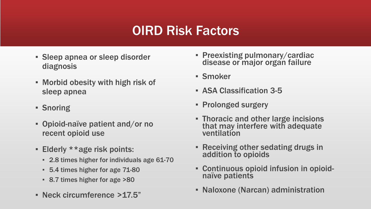
OIRD Risk Factors
▪ Sleep apnea or sleep disorder diagnosis
▪ Morbid obesity with high risk of sleep apnea
▪ Snoring
▪ Opioid-naïve patient and/or no recent opioid use
▪ Elderly **age risk points: ▪ 2.8 times higher for individuals age 61-70 ▪ 5.4 times higher for age 71-80 ▪ 8.7 times higher for age >80
▪ Neck circumference >17.5”
▪ Preexisting pulmonary/cardiac disease or major organ failure
▪ Smoker
▪ ASA Classification 3-5
▪ Prolonged surgery
▪ Thoracic and other large incisions that may interfere with adequate ventilation
▪ Receiving other sedating drugs in addition to opioids
▪ Continuous opioid infusion in opioid-naïve patients
▪ Naloxone (Narcan) administration

Evidence Based Standards for monitoring patients at
risk for OIRD
1. Identification of high-risk patients prior to opioid administration; primarily with patient controlled anesthesia (PCA) use--NEW
2. Consistent use of evidence based opioid sensitive assessment tool(s) to identify changes in patient’s status
a. Level of Pain Scale(s) b. Pasero Opioid-Induced Sedation Scale (POSS)--NEW
c. Quality of Respiratory Effort
3. The use of End Tidal Carbon Dioxide in conjunction with oxygen saturation monitoring for patients who are at risk for OIRD--NEW

Assessment Tool #1 Level of Pain Scale
▪ Evaluate patient on a scale from 0-10 or document non-verbal behavior(s).
▪ Face, Legs, Activity, Cry, Consolability scale (FLACC) for nonverbal children
▪ Wong-Baker FACES Pain Scale for children ages 4-9
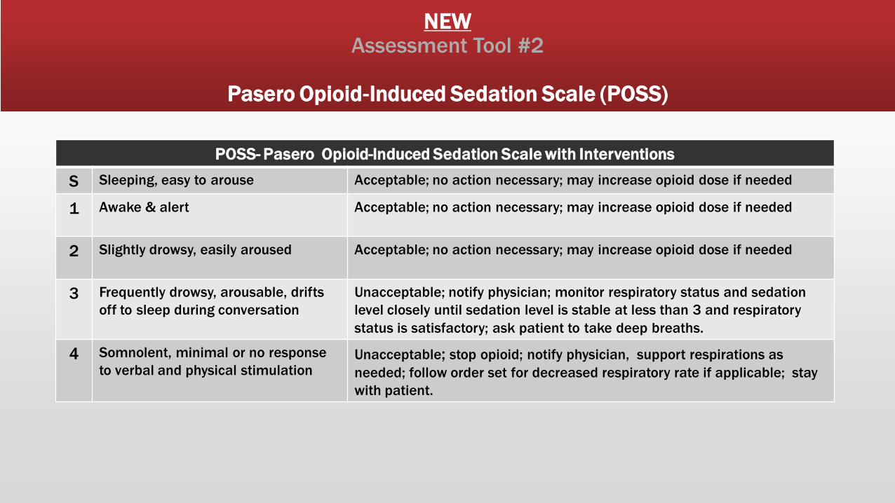
NEW Assessment Tool #2
Pasero Opioid‐Induced Sedation Scale (POSS)
POSS- Pasero Opioid-Induced Sedation Scale with Interventions
S Sleeping, easy to arouse Acceptable; no action necessary; may increase opioid dose if needed
1 Awake & alert Acceptable; no action necessary; may increase opioid dose if needed
2 Slightly drowsy, easily aroused Acceptable; no action necessary; may increase opioid dose if needed
3 Frequently drowsy, arousable, drifts off to sleep during conversation
Unacceptable; notify physician; monitor respiratory status and sedation level closely until sedation level is stable at less than 3 and respiratory status is satisfactory; ask patient to take deep breaths.
4 Somnolent, minimal or no response to verbal and physical stimulation
Unacceptable; stop opioid; notify physician, support respirations as needed; follow order set for decreased respiratory rate if applicable; stay with patient.
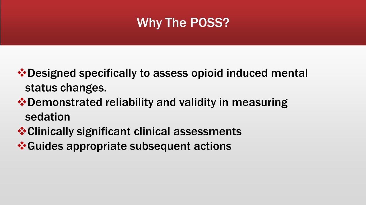
Why The POSS?
Designed specifically to assess opioid induced mental status changes. Demonstrated reliability and validity in measuring
sedation Clinically significant clinical assessments Guides appropriate subsequent actions
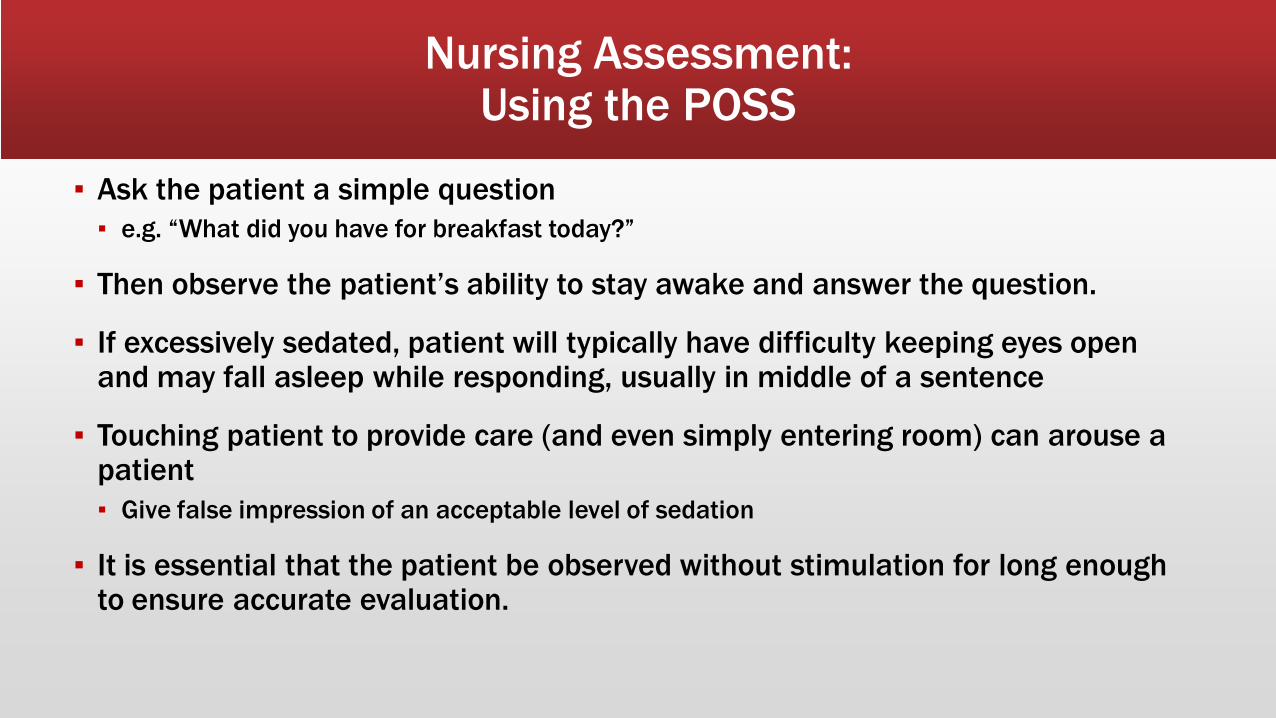
Nursing Assessment: Using the POSS
▪ Ask the patient a simple question ▪ e.g. “What did you have for breakfast today?”
▪ Then observe the patient’s ability to stay awake and answer the question.
▪ If excessively sedated, patient will typically have difficulty keeping eyes open and may fall asleep while responding, usually in middle of a sentence
▪ Touching patient to provide care (and even simply entering room) can arouse a patient ▪ Give false impression of an acceptable level of sedation
▪ It is essential that the patient be observed without stimulation for long enough to ensure accurate evaluation.
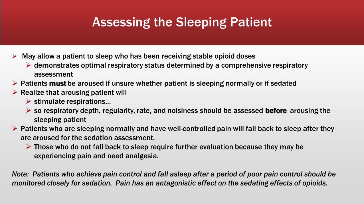
Assessing the Sleeping Patient
May allow a patient to sleep who has been receiving stable opioid doses demonstrates optimal respiratory status determined by a comprehensive respiratory
assessment Patients must be aroused if unsure whether patient is sleeping normally or if sedated Realize that arousing patient will
stimulate respirations… so respiratory depth, regularity, rate, and noisiness should be assessed before arousing the
sleeping patient Patients who are sleeping normally and have well-controlled pain will fall back to sleep after they
are aroused for the sedation assessment. Those who do not fall back to sleep require further evaluation because they may be
experiencing pain and need analgesia. Note: Patients who achieve pain control and fall asleep after a period of poor pain control should be monitored closely for sedation. Pain has an antagonistic effect on the sedating effects of opioids.
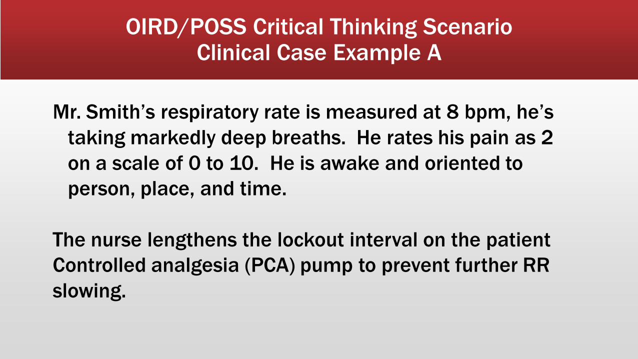
OIRD/POSS Critical Thinking Scenario Clinical Case Example A
Mr. Smith’s respiratory rate is measured at 8 bpm, he’s taking markedly deep breaths. He rates his pain as 2 on a scale of 0 to 10. He is awake and oriented to person, place, and time.
The nurse lengthens the lockout interval on the patient Controlled analgesia (PCA) pump to prevent further RR slowing.

Was the nurse’s action correct? Case A
A slow RR is a normal physiologic response to opioids. An RR of 8 bpm may not warrant intervention if the patient is awake, appropriate, and
comfortable. In this case, backing off the PCA opioid regimen is inappropriate, as it may exacerbate the
patient’s pain. And calling the Rapid Response Team (Code Rescue) based on a breach of the RR threshold of 8
bpm would constitute a false alarm. Instead, the nurse should identify and grade changes in the patient’s sedation level and
respiratory quality, including changes in respiratory pattern and depth and noisy breathing.
Mr. Smith has a sedation level of “1” on the POSS scale.
What is Mr. Smith’s POSS Score?
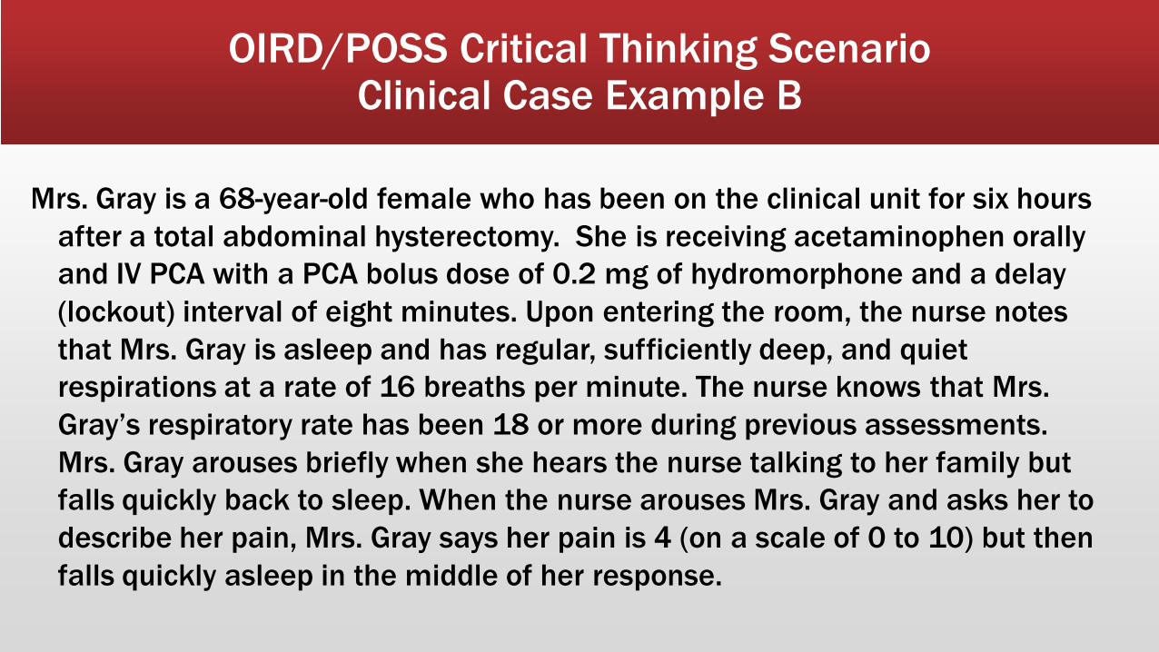
OIRD/POSS Critical Thinking Scenario Clinical Case Example B
Mrs. Gray is a 68-year-old female who has been on the clinical unit for six hours after a total abdominal hysterectomy. She is receiving acetaminophen orally and IV PCA with a PCA bolus dose of 0.2 mg of hydromorphone and a delay (lockout) interval of eight minutes. Upon entering the room, the nurse notes that Mrs. Gray is asleep and has regular, sufficiently deep, and quiet respirations at a rate of 16 breaths per minute. The nurse knows that Mrs. Gray’s respiratory rate has been 18 or more during previous assessments. Mrs. Gray arouses briefly when she hears the nurse talking to her family but falls quickly back to sleep. When the nurse arouses Mrs. Gray and asks her to describe her pain, Mrs. Gray says her pain is 4 (on a scale of 0 to 10) but then falls quickly asleep in the middle of her response.
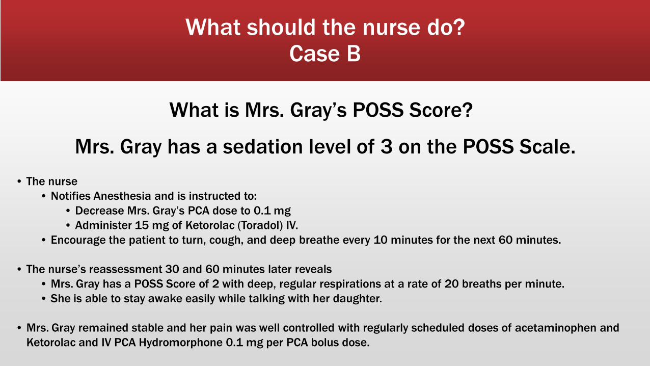
What should the nurse do? Case B
• The nurse • Notifies Anesthesia and is instructed to:
• Decrease Mrs. Gray’s PCA dose to 0.1 mg • Administer 15 mg of Ketorolac (Toradol) IV.
• Encourage the patient to turn, cough, and deep breathe every 10 minutes for the next 60 minutes. • The nurse’s reassessment 30 and 60 minutes later reveals
• Mrs. Gray has a POSS Score of 2 with deep, regular respirations at a rate of 20 breaths per minute. • She is able to stay awake easily while talking with her daughter.
• Mrs. Gray remained stable and her pain was well controlled with regularly scheduled doses of acetaminophen and Ketorolac and IV PCA Hydromorphone 0.1 mg per PCA bolus dose.
Mrs. Gray has a sedation level of 3 on the POSS Scale.
What is Mrs. Gray’s POSS Score?
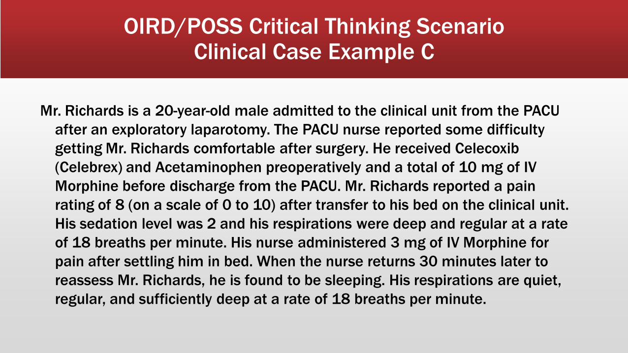
OIRD/POSS Critical Thinking Scenario Clinical Case Example C
Mr. Richards is a 20-year-old male admitted to the clinical unit from the PACU after an exploratory laparotomy. The PACU nurse reported some difficulty getting Mr. Richards comfortable after surgery. He received Celecoxib (Celebrex) and Acetaminophen preoperatively and a total of 10 mg of IV Morphine before discharge from the PACU. Mr. Richards reported a pain rating of 8 (on a scale of 0 to 10) after transfer to his bed on the clinical unit. His sedation level was 2 and his respirations were deep and regular at a rate of 18 breaths per minute. His nurse administered 3 mg of IV Morphine for pain after settling him in bed. When the nurse returns 30 minutes later to reassess Mr. Richards, he is found to be sleeping. His respirations are quiet, regular, and sufficiently deep at a rate of 18 breaths per minute.
What should the nurse do? Case C
Knowing that Mr. Richards has received a large amount of morphine over the past two to three hours for difficult- to-control pain, the nurse gently squeezes his shoulder and asks him how he feels. Mr. Richards arouses readily, which indicates an acceptable sedation level , says he feels much better, and rates his pain intensity as 3 (on a scale of 0 to 10). After talking two or three minutes longer in coherent sentences, he falls asleep and continues to demonstrate an acceptable respiratory status. Mr. Richards remained stable and his pain was well controlled with regularly scheduled doses of Celecoxib and IV Morphine boluses of 3 mg or less about every two hours.
Mr. Richards has a sedation level of “S” on the POSS Scale.
What is Mr. Richard’s POSS Score?

Assessment tool(s) #3 Quality of Respiratory Effort
Quality of Respirations Assessment:
0= Apneic
1= Labored or limited (shallow depression)
2= Can take a deep breath and cough well. Normal respiratory rate and depth
Objective Respiratory Assessment:
▪ Respiratory Rate
▪ Oxygen Saturation measurement (oxygenation)
▪ End Tidal CO2 measurement –NEW (ventilation)

Oxygenation vs. Ventilation
The respiratory cycle has two separate physiologic processes; oxygenation and ventilation:
▪ Oxygenation can be measured noninvasively through pulse
oximetry (SpO2). It detects the percentage of oxygen in the red blood cells.
▪ Ventilation can be measured noninvasively by an end-tidal CO2 sensor which detects the partial pressure (mmHg) or volume (% vol) of CO2 in the airway at the end of exhalation.

Oxygenation: Pulse Oximetry (O2 Saturation)
▪ Measures oxygen saturation
▪ Reflects oxygenation
▪ SpO2 changes lag when patient is hypoventilating or apneic
▪ Reflects change in oxygenation within 5 minutes
▪ It is affected by motion artifact, poor perfusion, etc.
▪ Should be used with Capnography in high risk patients

Ventilation: Capnography (End-Tidal CO2 Measurement)
▪ Measures Carbon Dioxide
▪ Reflects ventilation
▪ Hypoventilation/apnea detected immediately
▪ Reflects ventilation changes within 10 seconds
▪ The breath to breath measurement provides information within seconds and is not affected by motion artifact or poor perfusion.
▪ Should be used with Pulse Oximetry
What is Capnography?
History:
▪ Used by anesthesiologists since the 1970s and has been the standard of care in the operating room since 1991
▪ Used to confirm placement of ETT and quality of ventilation in intubated patients
▪ Used during anesthesia/moderate sedation to monitor patient’s ventilatory status

What is Capnography (cont.)?
Definitions:
▪ Capnography: a numerical value of the end-tidal CO2 AND a waveform of the concentration of the CO2 present in the airway and respiratory rate detected from actual airflow
▪ End-tidal CO2 (EtCO2): is the measurement of carbon dioxide (CO2) in the airway at the end of each breath.
▪ Capnometer: the numerical measurement of the carbon dioxide
▪ Capnogram: the waveform display of the carbon dioxide over time. A normal capnogram will consist of box-like waveforms directly related to the different phases of the respiratory cycle.

Common uses for Capnography:
Assessing Respiratory Status: ▪ Monitoring sedated patients or those receiving pain management
for hypoventilation. ▪ Guide the need for intubation or assisted intubation. ▪ Guide the patient with hyperventilation and anxiety symptoms.
Assessing Metabolic Emergencies: ▪ Assists in the evaluation of diabetic ketoacidosis , to differentiate
diabetic ketoacidosis and hyperglycemic hyperosmolar non-ketotic coma.
▪ Hypothermia and hyperthermia severity can be determined by capnography.
▪ Metabolic acidosis associated with gastroenteritis can have severity determined with capnography.

Understanding C02 and Respiratory Rate
▪ There is an inverse correlation between CO2 and the respiratory rate.
▪ ↓ RR = ↑ CO2 → hypoventilation
▪ ↑ RR = ↓ CO2 → hyperventilation

Hypoventilation: ↓ RR = ↑ CO2 → hypoventilation
Causes of hypoventilation in patients receiving Opioids: •Over sedation (POSS 3-4) •Respiratory depression (RR <8) •Ineffective breathing due to pain—shallow breaths (High Pain Score)
Patients who are hypoventilating will have a lower respiratory rate but higher amplitude of waveforms, resulting from the increased amount of CO2 in each breath.
0 45 90
Normal
0 45 90
Hypoventilation RR EtCO2
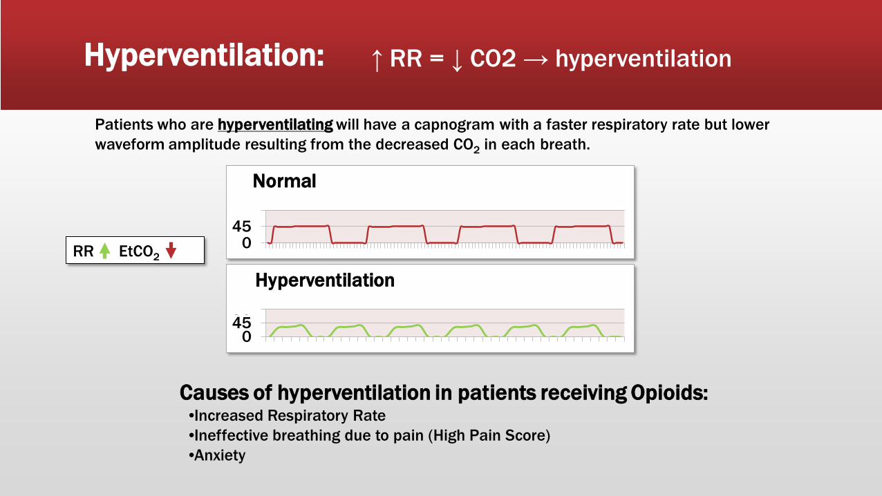
Hyperventilation: ↑ RR = ↓ CO2 → hyperventilation
Causes of hyperventilation in patients receiving Opioids: •Increased Respiratory Rate •Ineffective breathing due to pain (High Pain Score) •Anxiety
Patients who are hyperventilating will have a capnogram with a faster respiratory rate but lower waveform amplitude resulting from the decreased CO2 in each breath.
0 45 90
Normal
0 45 90
Hyperventilation RR EtCO2

Capnography Benefits for identifying OIRD:
Capnography can detect early phases of opoiod induced respiratory depression (OIRD) which can allow a more precise and safe use of medications
▪ Accurately monitors respiratory rate ▪ Monitors adequate ventilation with non-intubated patients ▪ Early indicator of airway obstruction ▪ Early warning of apnea ▪ Monitors potential risk of over-sedation resulting in hypoventilation more
effectively than pulse oximetry. ▪ Adds an additional level of patient safety

Interpretation of Capnography Results:
Normal Ranges:
▪ Capnography EtCO2 = 35-45 mmHg
When to worry:
▪ Decrease in respiratory rate
When to call the physician:
▪ EtCO2 50 mmHg or greater
▪ EtCO2 20mmHg or less
▪ Absent waveform = apnea

Interventions for elevated EtCO2 (least invasive to most invasive)
1. Reposition the patient's head to restore airway patency
2. Verbal or physical stimulation to encourage the patient to breathe
3. Decrease medication doses
4. Stopping medications
5. Administration of reversal agents
6. Bag mask ventilation

Trouble Shooting:
What is happening?
Sudden loss of waveform, EtCO2 near zero?
ALWAYS CHECK THE PATIENT FIRST!! Potential Causes: •Capnography tube disconnected, dislodged, kinked, or obstructed •Loss of circulatory function (i.e. cardiac arrest)
0
45
90

Trouble Shooting: What is happening?
Decreasing EtCO2 with loss of plateau?
ALWAYS CHECK THE PATIENT FIRST!! Potential Causes: •Partial airway obstruction
0
45
90

Trouble Shooting: What is happening?
Slow rate with gradual increase in EtCO2?
ALWAYS CHECK THE PATIENT FIRST!! Potential Causes: •Decreased tidal volume •Hypoventilation •Over sedation •Airway obstruction
0
45
90

Trouble Shooting: What is happening?
Rapid Rate with decreased EtCO2
ALWAYS CHECK THE PATIENT FIRST!! Potential Causes: •Hyperventilation •Pain •Anxiety •Increased temperature
0
45
90

Trouble Shooting: What is happening?
Decreased EtCO2; variable waveform
ALWAYS CHECK THE PATIENT FIRST!! Potential Causes: •Apnea •Shallow breaths •Sedation
0
45
90

Microstream Capnography technology is to be used for OIRD Detection:
▪ It can detect the carbon dioxide levels while the patient receives supplemental oxygen and can switch between oral and/or nasal breathing.
▪ It can be used in adult, neonatal, and pediatric populations.
▪ It can be used on non-intubated patients through a nasal cannula. The small pin holes delver oxygen around both the nose and mouth.
▪ Because of low sample flow rates, the lines are not flooded with moisture leading to occlusion or inaccuracy. The uni-junction of sampling ports prevents dilution from non-breathing source
▪ It plugs into the monitor adaptor, requires no zeroing or calibration, uses laser technology, and is not affected by other gases.
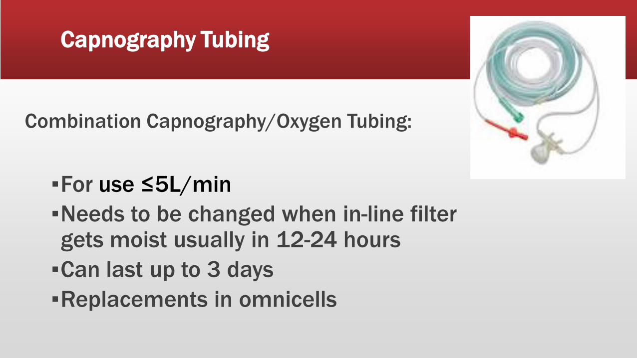
Capnography Tubing
Combination Capnography/Oxygen Tubing:
▪For use ≤5L/min ▪Needs to be changed when in-line filter gets moist usually in 12-24 hours ▪Can last up to 3 days ▪Replacements in omnicells

References
Brandt, P.A. (2009). Universal capnography: A vital asset that can improve patient care on almost any call. http://www.jems.com/resources/supplements/trend_setters/universal_capnography.html
Elsevier Inc.. (2006). Mosby's Skills. Retrieved from http://mns.elsevierperformancemanager.com/NursingSkills/Home.aspx?VirtualName=baptisthealthsouthflorida-flcoralgables
How medical equipment works explained simply. (n.d.). Retrieved from http://www.howequipmentworks.com/physics/respi_measurements/co2/capnograph/capnograph.html
Jarzyna, D., Jungquist, C.R., & Pasero, C., Willens, J.S., Nisbet, A., Oakes, L., Dempsey, S.J., Santangelo, D., & Polomano, R.C. (2011, September). American society for pain management nursing guidelines on monitoring for opioid-induced sedation and respiratory depression. Pain Management Nursing, 12(3), 118-145.
Maddox, R.R., & Williams, C.K. (2012, Winter). Clinical experience with capnography monitoring for PCA patients. Journal of the Anesthesia Patient Safety Foundation.
Oridion. (n.d.). Mainstream capnography solutions. from https://admin.adobeconnect.com/_a755547951/sedationceurn/event/login.html
Pasero, C. (2012, June). Opioid-induced sedation and respiratory depression: Evidence-based monitoring guidelines. Journal of PeriAnesthesia Nursing, 27(3), 208-211.
Safe use of opioids in hospitals. (2012, August ). The Joint Commission Sentinel Event Alert, Issue 49 . Retrieved from http://www.jointcommission.org