Identification of Allele-Specific RNAi Effectors Targeting ... · Identification of Allele-Specific...
13
Identification of Allele-Specific RNAi Effectors Targeting Genetic Forms of Parkinson’s Disease Christopher R. Sibley 1,2 , Matthew J. A. Wood 1 * 1 Department of Physiology, Anatomy and Genetics, University of Oxford, Oxford, United Kingdom, 2 MRC Laboratory of Molecular Biology, Cambridge, United Kingdom Abstract Parkinson’s disease (PD) is a progressive neurological disorder affecting an estimated 5–10 million people worldwide. Recent evidence has implicated several genes that directly cause or increase susceptibility to PD. As well as advancing understanding of the genetic aetiology of PD these findings suggest new ways to modify the disease course, in some cases through genetic manipulation. Here we generated a ‘walk-through’ series of RNA Pol III-expressed shRNAs targeting both the a-synuclein A30P and LRRK2 G2019S PD-associated mutations. Allele-specific discrimination of the a-synuclein A30P mutation was achieved with alignments at position 10, 13 and 14 in two model systems, including a heterozygous model mimicking the disease setting, whilst 59RACE was used to confirm stated alignments. Discrimination of the most common PD-linked LRRK2 G2019S mutation was assessed in hemizygous dual-luciferase assays and showed that alignment of the mutation opposite position 4 of the antisense species produced robust discrimination of alleles at all time points studied. Discrimination at this position was subsequently confirmed using siRNAs, where up to 10-fold discrimination was seen. The results suggest that RNAi-mediated silencing of PD-associated autosomal dominant genes could be a novel therapeutic approach for the treatment of the relevant clinical cases of PD in future. Citation: Sibley CR, Wood MJA (2011) Identification of Allele-Specific RNAi Effectors Targeting Genetic Forms of Parkinson’s Disease. PLoS ONE 6(10): e26194. doi:10.1371/journal.pone.0026194 Editor: Mark R. Cookson, National Institutes of Health, United States of America Received June 25, 2011; Accepted September 22, 2011; Published October 21, 2011 Copyright: ß 2011 Sibley, Wood. This is an open-access article distributed under the terms of the Creative Commons Attribution License, which permits unrestricted use, distribution, and reproduction in any medium, provided the original author and source are credited. Funding: This work was supported a Medical Research Council Studentship (CRS). This work was supported by a research grants from Parkinson’s UK to MJAW. A free gift of an siRNA was provided by Novartis Pharmaceuticals with nothing in exchange. The funders had no role in study design, data collection and analysis, decision to publish, or preparation of the manuscript. Competing Interests: A gift of an siRNA was received from a commercial source; Novartis pharmaceuticals (Basel, Switzerland). However, this does not alter our adherence to all the PLoS ONE policies on sharing data and materials. The authors have additionally filed a patent prior to submission for allele-specific silencing of the LRRK2 G2019S mutation. Again, this does not alter our adherence to all the PLoS ONE policies on sharing data and materials. * E-mail: [email protected] Introduction Genetic mapping of hereditary Parkinson’s disease (PD) over the last 12 years has revealed sixteen chromosomal ‘‘PARK’’ loci with linkage to PD. Subsequently a group of nine genes have been identified which are implicated in molecular pathways leading to PD pathogenesis [1]. The precise function and role of each of these genes in non-familial PD remains unclear since only two of these candidates were identified in recent large-scale genome-wide association studies (GWAS) [2,3]. However, collectively these hereditary cases account for 5–10% of all cases of PD and offer defined therapeutic targets for those patients bearing these genetic mutations. RNA interference (RNAi) has emerged as a highly promising strategy for conferring sequence-specific silencing of genes-of- interest. The endogenous RNAi pathway involves processing of non-coding RNA sequences, termed primary-microRNAs (pri- miRNAs), into short 21–23 nt single-stranded mature miRNAs that are antisense to targeted transcripts. Post-transcriptional regulation roles for miRNAs have been identified in development and disease [4]. Further, artificial precursors of this RNAi pathway can be generated in order to silence genes-of-interest for research and therapeutic purposes in a sequence-specific manner. Crucial- ly, changes of a single nucleotide can abrogate silencing ability of an RNAi trigger. By designing and screening RNAi triggers perfectly complementary to autosomal dominant mutant alleles at the site of a mutation, a single mismatch will exist between the antisense species and the wild-type allele which can have potential to disrupt silencing ability. In-so-doing the mutant allele can be selectively removed whilst as much as possible of the wild-type allele is retained to carry out endogenous functions. It can be argued that this is the most suitable therapy over a complete silencing of both wild-type and mutant alleles with non-allele specific silencing. Many genes have essential or presently unknown roles which could be eliminated by a complete silencing, potentially leading to damaging effects [5]. In such settings, allele-specific silencing has the obvious advantage that some of the wild-type gene product remains, whilst the pathogenic mutant is eliminated. This allele-specific silencing approach has been exploited to target disease-linked mutations linked to fronto- temporal dementia [6], Alzheimer’s disease [7], Huntington’s disease [8], amyotrophic lateral sclerosis [9], spino-cerebellar ataxia type 7 [10] and pachyonychia congenital [11]. Autosomal dominant, gain-of-function mutations linked to be PD have also been identified in the a-synuclein and leucine-rich repeat kinase 2 (LRRK2) genes, yet just one study has investigated allele-specific silencing of the A53T a-synuclein mutation [12]. Three pathogenic mutations in a-synuclein lead to increased rates of formation of a-synuclein fibrils and/or intermediate toxic proto- fibrils to suggest a toxic gain-of-function to these mutations [13– 15]. Over 30 PD-linked mutations have been identified in LRRK2 which has both kinase and GTPase activity [1]. The G2019S PLoS ONE | www.plosone.org 1 October 2011 | Volume 6 | Issue 10 | e26194
Transcript of Identification of Allele-Specific RNAi Effectors Targeting ... · Identification of Allele-Specific...

Identification of Allele-Specific RNAi Effectors TargetingGenetic Forms of Parkinson’s DiseaseChristopher R. Sibley1,2, Matthew J. A. Wood1*
1 Department of Physiology, Anatomy and Genetics, University of Oxford, Oxford, United Kingdom, 2 MRC Laboratory of Molecular Biology, Cambridge, United Kingdom
Abstract
Parkinson’s disease (PD) is a progressive neurological disorder affecting an estimated 5–10 million people worldwide. Recentevidence has implicated several genes that directly cause or increase susceptibility to PD. As well as advancingunderstanding of the genetic aetiology of PD these findings suggest new ways to modify the disease course, in some casesthrough genetic manipulation. Here we generated a ‘walk-through’ series of RNA Pol III-expressed shRNAs targeting boththe a-synuclein A30P and LRRK2 G2019S PD-associated mutations. Allele-specific discrimination of the a-synuclein A30Pmutation was achieved with alignments at position 10, 13 and 14 in two model systems, including a heterozygous modelmimicking the disease setting, whilst 59RACE was used to confirm stated alignments. Discrimination of the most commonPD-linked LRRK2 G2019S mutation was assessed in hemizygous dual-luciferase assays and showed that alignment of themutation opposite position 4 of the antisense species produced robust discrimination of alleles at all time points studied.Discrimination at this position was subsequently confirmed using siRNAs, where up to 10-fold discrimination was seen. Theresults suggest that RNAi-mediated silencing of PD-associated autosomal dominant genes could be a novel therapeuticapproach for the treatment of the relevant clinical cases of PD in future.
Citation: Sibley CR, Wood MJA (2011) Identification of Allele-Specific RNAi Effectors Targeting Genetic Forms of Parkinson’s Disease. PLoS ONE 6(10): e26194.doi:10.1371/journal.pone.0026194
Editor: Mark R. Cookson, National Institutes of Health, United States of America
Received June 25, 2011; Accepted September 22, 2011; Published October 21, 2011
Copyright: � 2011 Sibley, Wood. This is an open-access article distributed under the terms of the Creative Commons Attribution License, which permitsunrestricted use, distribution, and reproduction in any medium, provided the original author and source are credited.
Funding: This work was supported a Medical Research Council Studentship (CRS). This work was supported by a research grants from Parkinson’s UK to MJAW. Afree gift of an siRNA was provided by Novartis Pharmaceuticals with nothing in exchange. The funders had no role in study design, data collection and analysis,decision to publish, or preparation of the manuscript.
Competing Interests: A gift of an siRNA was received from a commercial source; Novartis pharmaceuticals (Basel, Switzerland). However, this does not alter ouradherence to all the PLoS ONE policies on sharing data and materials. The authors have additionally filed a patent prior to submission for allele-specific silencingof the LRRK2 G2019S mutation. Again, this does not alter our adherence to all the PLoS ONE policies on sharing data and materials.
* E-mail: [email protected]
Introduction
Genetic mapping of hereditary Parkinson’s disease (PD) over the
last 12 years has revealed sixteen chromosomal ‘‘PARK’’ loci with
linkage to PD. Subsequently a group of nine genes have been
identified which are implicated in molecular pathways leading to
PD pathogenesis [1]. The precise function and role of each of
these genes in non-familial PD remains unclear since only two of
these candidates were identified in recent large-scale genome-wide
association studies (GWAS) [2,3]. However, collectively these
hereditary cases account for 5–10% of all cases of PD and offer
defined therapeutic targets for those patients bearing these genetic
mutations.
RNA interference (RNAi) has emerged as a highly promising
strategy for conferring sequence-specific silencing of genes-of-
interest. The endogenous RNAi pathway involves processing of
non-coding RNA sequences, termed primary-microRNAs (pri-
miRNAs), into short 21–23 nt single-stranded mature miRNAs
that are antisense to targeted transcripts. Post-transcriptional
regulation roles for miRNAs have been identified in development
and disease [4]. Further, artificial precursors of this RNAi pathway
can be generated in order to silence genes-of-interest for research
and therapeutic purposes in a sequence-specific manner. Crucial-
ly, changes of a single nucleotide can abrogate silencing ability of
an RNAi trigger. By designing and screening RNAi triggers
perfectly complementary to autosomal dominant mutant alleles at
the site of a mutation, a single mismatch will exist between the
antisense species and the wild-type allele which can have potential
to disrupt silencing ability. In-so-doing the mutant allele can be
selectively removed whilst as much as possible of the wild-type
allele is retained to carry out endogenous functions. It can be
argued that this is the most suitable therapy over a complete
silencing of both wild-type and mutant alleles with non-allele
specific silencing. Many genes have essential or presently unknown
roles which could be eliminated by a complete silencing,
potentially leading to damaging effects [5]. In such settings,
allele-specific silencing has the obvious advantage that some of the
wild-type gene product remains, whilst the pathogenic mutant is
eliminated. This allele-specific silencing approach has been
exploited to target disease-linked mutations linked to fronto-
temporal dementia [6], Alzheimer’s disease [7], Huntington’s
disease [8], amyotrophic lateral sclerosis [9], spino-cerebellar
ataxia type 7 [10] and pachyonychia congenital [11].
Autosomal dominant, gain-of-function mutations linked to be
PD have also been identified in the a-synuclein and leucine-rich
repeat kinase 2 (LRRK2) genes, yet just one study has investigated
allele-specific silencing of the A53T a-synuclein mutation [12].
Three pathogenic mutations in a-synuclein lead to increased rates
of formation of a-synuclein fibrils and/or intermediate toxic proto-
fibrils to suggest a toxic gain-of-function to these mutations [13–
15]. Over 30 PD-linked mutations have been identified in LRRK2
which has both kinase and GTPase activity [1]. The G2019S
PLoS ONE | www.plosone.org 1 October 2011 | Volume 6 | Issue 10 | e26194
mutation, located in the active site of the kinase domain, is the
most common PD-associated mutation accounting for 2–8% of all
hereditary cases of PD and 0.6–1.6% of PD cases with no obvious
signs of familial inheritance implying that it could be a mutational
hotspot for sporadic PD [16]. Collectively it suggests that this
mutation accounts for ,1% of all PD cases making it a high-
profile therapeutic target.
In this study we experimentally validate and optimise RNAi
triggers which selectively target the a-synuclein A30P and LRRK2
G2019S mutations linked to PD. We report successful discrimi-
nation of both these mutations using short-hairpin RNAs
(shRNAs) with mutations aligned opposite certain, but not all,
positions of the antisense species. In both cases, the discriminating
abilities of some shRNAs could be further improved by
incorporation of secondary mismatches to the wild-type allele.
Finally we show that a-synuclein A30P discriminating effectors are
functional in full-length hemizygous and heterozygous models
more closely resembling the disease setting indicating the
suitability of the identified allele-specific sequences for future in
vivo and potentially clinical work.
Materials and Methods
ConstructsAll shRNA and target sequences can be found in supplementary
data (Table S1). Oligonucleotides used to generate constructs were
ordered from Sigma Genosys (Sigma Genosys, UK). All constructs
were verified by sequencing prior to use.
shRNA expression plasmidsshRNA expression plasmids were designed with antisense
species located in the 39 arm of stem-loop hairpins as described
previously. To place shRNA sequences downstream of the U6
promoter, PCR was performed with Pol-III U6 promoter as
template, a U6 forward primer (U6F’: 59-GATCGGGCCCGTC-
GACAAGGTCGGGCAGGAAGAGGGCCT-39) and a U6 re-
verse primer containing shRNA sequences (U6R’: 59-AAAAAA..
Anti-sense ..TGGGTCAGG.. Sense ..GGTGTTTCGTCCTTT-
CCACAA-39). PCR products were subsequently ligated into the
pGEM-T Easy vector (Promega, USA) according to the manu-
facturer’s instructions to generate RNA Pol-III expressed shRNA
plasmids.
siRNAsAll siRNAs were designed as 19-mers with 2-nt dT overhangs.
The a-synuclein targeting siRNA p1314 was a kind gift from
Novartis pharmaceuticals (Basel, Switzerland) with anti-sense
sequence: 59-UUGUCUUUCCUGGCGCUUCdTdT-39. LRRK2
siRNAs p3-5 were from thermoscientific with the following antisense
sequences: p3 59-GCUGUAGUCAGCAAUCUUUdTdT-39, p4
59-UGCUGUAGUCAGCAAUCUUdTdT-39, p5 59-AUGCU-
GUAGUCAGCAAUCUdTdT-39.
Dual-luciferase targetsComplementary oligonucleotides containing partial length
target sequences for dual-luciferase screening were annealed
together and ligated into the multiple cloning site 39 of the
Renilla Luciferase gene in the psiCheck2.2 dual-luciferase cassette
(Promega, USA) using Xho1 and Not1 restriction sites.
Full-length a-synuclein constructsFull-length wild-type and A30P mutant a-synuclein sequences
cloned in the pcDNA-3.1 (Clontech, USA) plasmid were a kind
gift from Dr J.Galvin (Washington, USA). Wild-type a-synuclein
cloned in the peGFP-N1 plasmid was a kind gift from Dr A.
Exposito (Cambridge, UK). The A30P mutant transcript was
accordingly PCR amplified with forward and reverse primers
containing SalI and SacII restriction sites respectively before being
sub-cloned into the peGFP-N1 expression plasmid (Clontech,
USA) using SalI and SacII restriction sites. To generate mCherry
tagged constructs, mCherry was PCR amplified with forward and
reverse primers containing SacII and NotI restriction sites
respectively. Concomitantly, eGFP was removed by SacII and
NotI digestion from a-synuclein eGFP-N1 plasmids. The mCherry
PCR product was subsequently sub-cloned into a-synuclein
plasmids with SacII and NotI restriction sites to generate a-
synuclein mCherry-N1 expression plasmids. Finally, to generate
heterozygous expression plasmids expressing eGFP-tagged wild-
type a-synuclein and A30P mutant mCherry-tagged a-synuclein,
mCherry-tagged A30P mutant a-synuclein together with a 59
CMV promoter and 39 poly-A tail were PCR amplified with
forward and reverse primers containing PciI restriction sites. PCR
products were subsequently sub-cloned into the wild-type a-
synuclein peGFP-N1 plasmid using a single PciI restriction, and
correct insert orientation verified by sequencing.
Cell culture and transfectionsHEK-293 cells (ATCC, CRL-1573) were cultured in DMEM
supplemented with 10% FCS. For transfection, cells were grown in
24-well plates to 80% confluence and transfected with Lipofecta-
mine 2000 (Invitrogen, USA) according to manufacturer’s
instruction. shRNAs and dual-luciferase targets were transfected
at 1 mg/ml unless otherwise stated. For hemizygous assays,
shRNAs and eGFP-tagged a-synuclein constructs were transfected
at 1 mg/ml. For heterozygous assays shRNAs were transfected at
1 mg/ml, pri-miR-30 mimics at 0.5 mg/ml and the heterozygous
expression plasmid at 2 mg/ml. For siRNA-1314 transfection, the
heterozygous expression plasmid and siRNA-1314 were co-
transfected at 2 mg/ml and 100 nM respectively. For LRRK2
siRNA transfections, 250 ng of psiCheck target plasmid was
transfected with 50 nM of indicated siRNA unless stated
otherwise.
Luciferase assaysCells were lysed for 20 minutes at time-points stated in the text
using 100 ml passive lysis buffer (Promega, USA). A total of 20 ml
of protein lysate was subsequently assayed for dual-luciferase
readings using a dual-luciferase kit (Promega, USA) and Wallac-
Victor 2 plate reader as per manufacturer’s instructions. Ratios of
renilla luciferase:firefly luciferase were obtained and normalised to
respective non-specific control samples.
Cell counting and viability assaysCells were trypsinised 48 hrs post-transfection before cell
number and typan blue determined cell viability were assessed
with the Vi-Cell XR cell viability analyzer (Beckman Coulter) as
per manufacturer’s instructions.
Rapid amplification of cDNA ends (RACE)RNA was harvested at stated time-points using Trizol (Invitro-
gen, USA) according to manufacturers instructions. To define 59
nucleotides of a-synuclein RNAi degradation products, 1 mM of
RNA adapter with 59 and 39 hydroxyl groups (59 OH-
ACACUCUUUCCCUACACGACGCUCUUCCGAUCU-OH)
was used in a ligation reaction with 250 ng of total RNA from a-
synuclein mCherry-N1 and respective shRNA transfected HEK-
293 cells. Following 1 hr reaction at 37uC with T4 RNA ligase
Allele-Specific RNAi for Familial Parkinson’s
PLoS ONE | www.plosone.org 2 October 2011 | Volume 6 | Issue 10 | e26194
(NEB, USA), 200 ng of RNA was reverse transcribed with
thermoscript (Invitrogen, USA) and a-synuclein specific reverse
primer (59 CTGCTCCCTCCACTGTCTTC) according to the
manufacturer’s instruction. Resulting cDNA was diluted 1:400
and 15 ml used as template in a 50 ml PCR reaction as previously
described using adapter specific forward primer and nested a-
synuclein specific reverse primer (F: 59 AATGATACGGCGAC-
CACCGAGATCTACACTCTTTCCCTACACGACGCTCTT-
CCGATCT, R: 59 CCACTGCTCCTCCAACATTT). Products
were visualised on a 2% agarose gel and cleavage products
amplified prior to PCR clean-up and sequencing using band-stab
PCR. RNAi cleavage sites were subsequently determined from
sequencing reads by identifying junctions between adapter and
a-synuclein transcripts.
Fluorescent microscopyCells transfected with fluorescent constructs were visualised at
stated time-points using a Leica DM-IRB inverted microscope
(Leica, Germany). Images were taken with Carl Zeiss AxioCam
MRm monochrome digital camera (Carl Zeiss, Germany) using
AxioVision digital imaging software (Carl Zeiss, Germany). All
images are 3600 mm in width.
Fluorescent quantificationCells transfected with fluorescent constructs were lysed at time
points stated in the text and protein content determined using the
micro BCA protein assay as per manufacturer’s instructions
(Pierce Biotechnology, USA). A volume of 100 ml was prepared in
black-walled, clear-bottom 96-well plates (BD falcom) which
contained 50 mg of protein lysate. Samples were subsequently
assayed for eGFP and/or mCherry fluorescence using a
fluorescent plate reader (Wallac Victor). Background fluorescence
determined from cells transfected with target alone was subtracted.
Fluorescence levels were subsequently normalised to non-specific
control samples.
Statistical analysisStatistical significance between control and experimental values
was determined using Student’s t test (un-paired, 2-tailed). All data
are expressed as mean 6 standard deviation (S.D.).
Results
Identification of RNAi effectors for allele-specifictargeting of a-synuclein A30P mutation
Mutations to the a-synuclein encoding SNCA gene have been
reported to cause early-onset PD in an autosomal dominant
manner. To investigate the potential of RNAi as a therapeutic
strategy for mutation carriers at the route of their disease, a panel
of U6-transcribed shRNAs was designed which were fully
complementary to the A30P mutant allele of a-synuclein and
had a single G:G mismatch to the wild-type allele. Based on
previous reports [8,10], shRNAs were designed with the A30P
mutation aligned at sequential positions along the 39-region of the
antisense strand from p10-16, with P1 representing the most 5 nt
of the antisense species (Fig. 1A). Some studies have demonstrated
that discrimination can be improved when secondary mismatches
to the wild-type allele are incorporated despite this leading to a
single mismatch with the targeted mutant allele [6,17]. To test this
hypothesis, a second pool of shRNAs was designed in which a
single URC swap to create a single A:C mismatch to the mutant
A30P allele, and a secondary mismatch to the wild-type allele, was
placed immediately 39 of the mutation alignment in the antisense
strand (Fig. 1A).
Initially, full-length eGFP-tagged wild-type and A30P mutant a-
synuclein constructs were designed and expressed in HEK-293
cells in hemizygous experiments. Western blot analysis confirmed
expected sizes of the fusion proteins, and showed that the
constructs represented a comprehensive overexpression of a-
synuclein since no endogenous protein could be detected at the
exposure times used (data not shown). Further, fluorescence
microscopy confirmed robust expression whilst no intracellular
aggregates of a-synuclein could be seen with either wild-type or
mutant constructs at 72 hrs, agreeing with previously reported
equivalent models [18]. Finally, cell counting and cell viability
assays demonstrated no toxicity associated with these constructs
relative to mock-transfected cells or relative to one another (Fig
S1).
To screen for allele-specific silencing, shRNAs were co-
transfected with either wild-type or A30P mutant a-synuclein
targets and eGFP expression was assessed and quantified at 48 hrs
(Fig. S2) or 72 hrs (Fig. 1B, C). A positive control eGFP-targeting
shRNA detectably reduced eGFP expression, and this was
reproducibly quantified as a ,60% reduction of both wild-type
and mutant targets (Fig. 1C and S2). Importantly this demon-
strates that both target constructs have the potential to be knocked
down by non-mutation targeting shRNAs to almost identical
levels, and implies that any difference seen in the silencing of the
two different targets by A30P-targeted shRNAs is likely the result
of the single-mismatch generated between the antisense species
and the wild-type target. Analysis at both 48 and 72 hrs
demonstrated that multiple constructs had the ability to silence
one or both of the a-synuclein full-length targets (Fig. 1B and S2).
In particular, constructs p10, p13 and p14 demonstrated striking
discrimination when co-transfected with either the wild-type or
A30P mutant constructs at both time points. The trends observed
from eGFP-microscopy were verified by direct eGFP-quantifica-
tion (Fig. 1C and S2). All constructs, with the exception of p11,
displayed discrimination between mutant and wild-type constructs
with the maximum difference being 4.65-fold (p,0.001) discrim-
ination by construct p13 at 72 hrs. Further, each construct, with
the exception of p11, displayed increased discrimination with time.
p13 had the most substantial amount of mutant knockdown to
18%. Yet whilst p13 also showed silencing of the wild-type
construct by 12%, both constructs p10 and p14 demonstrated high
specificity with absent wild-type silencing accompanied by
silencing of the mutant transcripts to 42% and 51% respectively.
Finally, screening of double-mismatch shRNAs was also carried
out. The secondary mismatches both qualitatively (Fig. 1D and S2)
and quantitatively improved the levels of wild-type retention
relative to single mismatch shRNAs, but in nearly all cases this was
accompanied by a greater retention of the mutant allele implying
that the single mismatch to this sequence was not tolerated well by
the RNAi machinery (Fig. 1E and S2). However, secondary-
mismatch alignments to the wild-type allele at p1314 directed a
marked reduction in the mutant protein to 20% and 12% of
original levels at 48 and 72 hrs respectively, whilst the wild-type
protein was completely retained at 48 hrs, but reduced to 60% at
72 hrs (Fig. 1E and S2). The resulting 5.8-fold (p,0.001)
discrimination seen with this construct at 48 rs was the most
impressive difference seen in this full-length assay and confirms
that incorporation of an additional secondary mismatch can
improve allele-specific discrimination of certain RNAi effectors.
Allele-specific discrimination of the A30P mutant targetin a heterozygous cell model
The autosomal dominant A30P mutation will result in both
wild-type and mutant alleles being transcribed in patient cells. The
Allele-Specific RNAi for Familial Parkinson’s
PLoS ONE | www.plosone.org 3 October 2011 | Volume 6 | Issue 10 | e26194
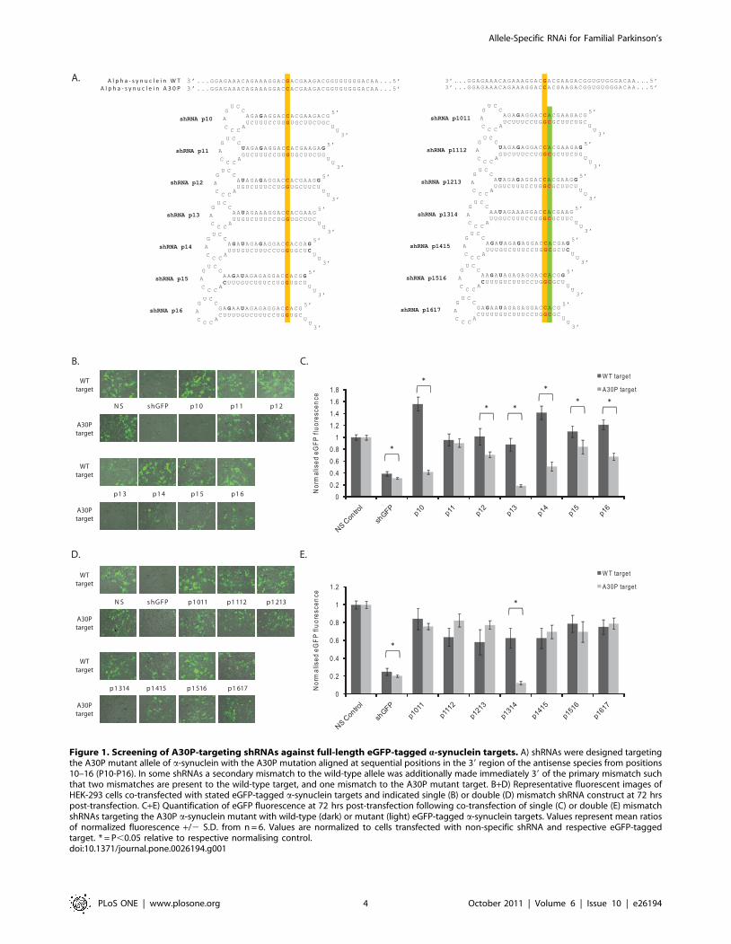
Figure 1. Screening of A30P-targeting shRNAs against full-length eGFP-tagged a-synuclein targets. A) shRNAs were designed targetingthe A30P mutant allele of a-synuclein with the A30P mutation aligned at sequential positions in the 39 region of the antisense species from positions10–16 (P10-P16). In some shRNAs a secondary mismatch to the wild-type allele was additionally made immediately 39 of the primary mismatch suchthat two mismatches are present to the wild-type target, and one mismatch to the A30P mutant target. B+D) Representative fluorescent images ofHEK-293 cells co-transfected with stated eGFP-tagged a-synuclein targets and indicated single (B) or double (D) mismatch shRNA construct at 72 hrspost-transfection. C+E) Quantification of eGFP fluorescence at 72 hrs post-transfection following co-transfection of single (C) or double (E) mismatchshRNAs targeting the A30P a-synuclein mutant with wild-type (dark) or mutant (light) eGFP-tagged a-synuclein targets. Values represent mean ratiosof normalized fluorescence +/2 S.D. from n = 6. Values are normalized to cells transfected with non-specific shRNA and respective eGFP-taggedtarget. * = P,0.05 relative to respective normalising control.doi:10.1371/journal.pone.0026194.g001
Allele-Specific RNAi for Familial Parkinson’s
PLoS ONE | www.plosone.org 4 October 2011 | Volume 6 | Issue 10 | e26194
silencing of either mRNA transcript will therefore be competitive
under these conditions, and it is unknown how this will affect the
allele-specific outcome. To investigate this a more complex model
of a-synuclein overexpression was generated in which a eGFP-
tagged wild-type and a mCherry-tagged A30P mutant transcript
were expressed from the same plasmid using identical promoters
located side-by-side, but directing anti-parallel transcription
(Fig. 2A). This heterozygous A30P (HetA30P) arrangement was
considered the most favourable for transcribing two constructs
from one plasmid at equal levels with minimal interference of
promoter activity on neighboring transcription units. Fluorescence
microscopy at 48 hrs post-transfection revealed strong eGFP and
mCherry fluorescence in HEK-293 cells to create a merged yellow
pattern of expression (Fig. 2B), viability assays demonstrated that
this plasmid presented no detectable toxicity relative to mock-
transfections (Fig. S3) whilst qPCR confirmed that levels of both
wild-type and mutant transcripts were comparable (data not
shown).
Screening of all single and double mismatch shRNAs was
performed against the HetA30P plasmid and both fluorescence
microscopy and quantification for eGFP and mCherry were
determined at 48 hrs post-transfection. An eGFP-targeting shRNA
was used as a positive control for this system. This construct
initiated a change from yellow to red colour in fluorescent images,
reproducibly demonstrating ,80% silencing of the eGFP-tagged
wild-type construct whilst having no effect on the mCherry-tagged
A30P mutant construct following quantification of fluorescence
(Figs. 2C–F). The results highlight the sequence-specificity of
RNAi and also the ability of this system to identify allele-specific
silencing of the co-expressed target transcripts. Single mismatch
shRNAs demonstrated that p10, p13 and p14 were once again the
best discriminators between the wild-type and mutant alleles, with
each construct leading to a predominant retention of green
fluorescence in microscopy images (Fig. 2C). However, the
discrimination was not as comprehensive as in hemizygous assays.
Quantification of fluorescence revealed that mutant target
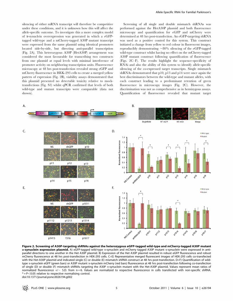
Figure 2. Screening of A30P-targeting shRNAs against the heterozygous eGFP-tagged wild-type and mCherry-tagged A30P mutanta-synuclein expression plasmid. A) eGFP-tagged wild-type a-synuclein and mCherry tagged A30P mutant a-synuclein were expressed in anti-parallel directions to one another in the Het A30P plasmid. B) Expression of the Het A30P plasmid resulted in robust eGFP fluorescence and robustmCherry fluorescence at 48 hrs post-transfection in HEK-293 cells. C+E) Representative merged fluorescent images of HEK-293 cells co-transfectedwith the Het-A30P plasmid and indicated single (C) or double (E) mismatch shRNA construct at 48 hrs post-transfection. D+F) Quantification of wild-type a-synuclein eGFP (green bars) or A30P mutant a-synuclein mCherry (red bars) fluorescence at 48 hrs post-transfection following co-transfectionof single (D) or double (F) mismatch shRNAs targeting the A30P a-synuclein mutant with the Het-A30P plasmid. Values represent mean ratios ofnormalized fluorescence +/2 S.D. from n = 6. Values are normalized to respective fluorescence in cells transfected with non-specific shRNA.* = P,0.05 relative to respective normalising control.doi:10.1371/journal.pone.0026194.g002
Allele-Specific RNAi for Familial Parkinson’s
PLoS ONE | www.plosone.org 5 October 2011 | Volume 6 | Issue 10 | e26194
expression was now reduced to 23%, 17% and 36% of control
levels for p10, p13 and p14 respectively, but reductions in the wild-
type transcript were now to 73%, 59% and 72% of control levels
respectively (Fig. 2D). P13 still had the greatest discrimination of
3.4-fold (p,0.001) in this model, but this was not as substantial as
the 4.3-fold discrimination at the same time point in hemizygous
assays (Fig. S2). In recent years reports have suggested shRNAs
may elicit toxic effects in vivo [19,20]. To rule out the possibility
that toxicity of individual shRNAs leading to cell death or cell
cycle arrest was responsible for the observed changes in
fluorescence, cell counting and viability assays were performed
at the time of analysis on a variety of shRNAs including the most
successful constructs, p10, p13 and p14. No significant differences
were seen in cell number relative to mock-transfected cells, and
only shRNA p10 showed a modest 12% decline in cell viability
which is not expected to lead to significant changes in mutant
fluorescence (Fig S3).
Likewise, construct p1314 was the only double mismatch
construct capable of allele-specific silencing using this model
(Figs. 2E, F), but the 3.6-fold (p,0.001) discrimination between
alleles was not as great as the 5.8-fold change seen at the same time
point in the corresponding hemizygous assay due to the 30%
reduction of the wild-type construct seen in this heterozygous
model. As with single mismatch constructs, shRNA p1314 had no
detectable effect on cell viability or cell number relative to mock-
transfected cells (Fig. S3). Collectively the results demonstrate that
RNAi effectors have been identified that discriminate impressively
between wild-type a-synuclein and the A30P a-synuclein mutant
in a heterozygous model which closely mimics the disease setting.
Furthermore, the analysis of the silencing and discriminating
ability of the most successful constructs across the two cellular
models highlights the influence that competing wild-type and
mutant transcripts may have on results, whilst additionally
emphasising that constructs p13 and p1314 are the most consistent
discriminators and potentially suitable for future in vivo and clinical
application.
siRNA analysis and 59RACE of target degradationproducts confirms the alignments of the mutation inantisense species
In order to put the findings with respect to allele-specific
silencing of mutant A30P a-synuclein into perspective it is
important that the precise alignment of mutations in the antisense
strand is verified. Unlike siRNAs, shRNAs are subject to
processing by Dicer to produce the dsRNA duplex from which
the active antisense species is selected. Whilst the shRNAs were
designed to release a sequence with the mutation alignment at the
stated position based on known criteria for Dicer recognition, this
remains to be verified. Here, two different approaches were
utilised to verify the sequence alignments of mutations in the
generated antisense species.
First, a siRNA bearing a primary mutation at p13 of the
antisense species, and secondary mismatch at p14 was tested for
allele-specific silencing ability to compare activity with the
previously identified shRNA construct p1314 (Fig. 3A). The
double mismatch siRNA-p1314 was chosen over a single
mismatch variant since it was clear from results in the previous
models that only one double mismatch construct was capable of
allele-specific silencing whilst all other double mismatch variants
failed to silence the wild-type and mutant transcripts. Thus, if
siRNA-p1314 was capable of allele-specific discrimination then it
is highly likely that shRNA p1314 has the primary mutation
aligned correctly at p13 whilst other constructs have the mutation
aligned accordingly. Transfection of siRNA-p1314 with the
HetA30P plasmid demonstrated that the mutant A30P target
was silenced by 79% and the wild-type target by 31% to produce a
3.4-fold (p,0.001) allele-specific discrimination (Figs. 3B, C).
These results correlate closely with the 81% silencing of the
mutant target and 30% reduction in wild-type target observed
with shRNA p1314 at 48 hrs which strongly suggests that the
alignments of mutations in the shRNAs are as predicted.
Rapid amplification of cDNA ends (RACE) has previously been
used to determine the precise cleavage site of antisense species by
determining the 59 terminal nt of 39 target cleavage products.
Assuming cleavage of fully complementary targets is always
directed opposite from p10 and p11 as reported [21] (Fig. 3E),
comparison of alignments to shRNA sequences can subsequently
be used to confirm the cleavage site of Dicer within the shRNA
and hence the alignments of specific nucleotides in the antisense
species. A 59RACE protocol was used in which an RNA adapter
was ligated to 39 degradation products of the mCherry-tagged
A30P mutant construct following co-transfection of constructs
siRNA-p1314, p13, p1314 and p14, and then RT-PCR carried
out with adaptor and a-synuclein specific primers. This adapter
ligation exploits unique RNA ends and is only expected to ligate to
RNA populations with 59 phosphate groups. This includes RNAi
degradation products but not full-length mRNAs which have 7-
methylguanosine caps at the 59 end.
Following 59 RACE, gel-electrophoresis revealed that a band of
the expected size for degradation products, but not the full-length
transcript, was seen with all constructs (Fig. 3D). Importantly this
was absent if the RNA-adaptor was excluded from ligation
reactions. Sequencing of PCR products revealed first that the
product was indeed that of a-synuclein degradation products, and
second that the 5 nt of the degradation products varied between
effectors with the primary mutation aligned at P13 and P14 to
confirm that the cleavage site within a-synuclein changes between
constructs (Figs. 3F, G). More specifically the 5nt of the cleavage
product aligned to the 11th nt of each specific antisense species,
including with siRNA-p1314 with which the alignment is verified.
This demonstrates that cleavage is occurring opposite nucleotides
p10 and p11 for each construct, and that the sequence alignments
are as predicted.
Dual-luciferase screening reveals shRNAs that candiscriminate the LRRK2 G2019S mutant allele from thewild-type allele
Following the successful allele-specific discrimination achieved
with shRNAs targeting the A30P a-synuclein mutation, a second
PD candidate mutation was chosen for shRNA screening. The
LRRK2 G2019S mutation is the most common PD-linked
mutation currently known and therefore represents the most
attractive mutation for allele-specific silencing. This mutation leads
to a G:A conversion in the LRRK2 mRNA which results in a G:U
mismatch between the antisense species of targeting RNAi
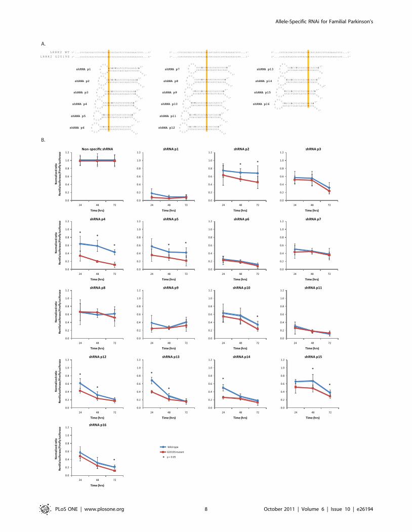
effectors and the wild-type allele. A pool of shRNAs was initially
designed which was fully complementary to the G2019S mutant
allele of LRRK2 resulting in a single G:U mismatch to the wild-
type allele mutation aligned at sequential positions from p10-16 of
the antisense arm (Fig. 4A). However, kinetic studies on RNAi
suggest that alignments in the 59 region of the antisense species
could lead to improved discrimination of the G2019S mutation.
Specifically, G:U wobbles between the targeting shRNA and wild-
type allele, have been reported to strongly interfere with pairings
of antisense species to mRNA when placed either 59 or centrally in
the RNAi effector sequence [22,23]. Accordingly, shRNAs were
subsequently designed with mutations at sequential positions from
P1-9 of the antisense arm.
Allele-Specific RNAi for Familial Parkinson’s
PLoS ONE | www.plosone.org 6 October 2011 | Volume 6 | Issue 10 | e26194
There remains an acknowledged paucity of experimental
models of LRRK2 that have made its study troublesome to date
[24]. In order to rapidly screen shRNAs for allele-specific silencing
ability, short wild-type and mutant allele target sequences
corresponding to the 52nts of LRRK2 mRNA immediately
surrounding the location of the G2019S mutation were therefore
inserted into the 3 UTR of the Renilla luciferase gene in a dual-
luciferase vector. Screening of shRNAs against partial-length
LRRK2 dual-luciferase targets revealed that at 24 hrs post-
transfection, four shRNAs displayed significant allele-specific
discrimination (Fig. 4B). Alignments of the G2019S mutation at
p4, p12, p13 and p14 displayed 1.88- (p,0.005), 1.44- (p,0.005),
1.70- (p,0.001) and 1.93-fold (p,0.001) discrimination respec-
tively, with p14 producing the maximum levels of mutant silencing
of these three constructs to 26% whilst retaining 50% of the wild-
type allele expression. Analysis at different points post-transfection
revealed that all shRNAs led to increased levels of silencing of both
the mutant and wild-type alleles over time (Fig. 4B). At 72 hrs, six
shRNAs displayed significant discrimination between the mutant
and wild-type alleles; p2, p4, p5, p10, p15 and p16. Construct p4
displayed the greatest discrimination (3.7-fold; p,0.001) between
mutant and wild-type alleles, and was the only construct which
maintained discrimination at all time points. Further, this shRNA
p4 had no effect on cell number or cell viability relative to mock-
transfected cells to rule out changes in luminescence being
dependent on non-specific effects of cell death or cell cycle arrest
(Fig S3).
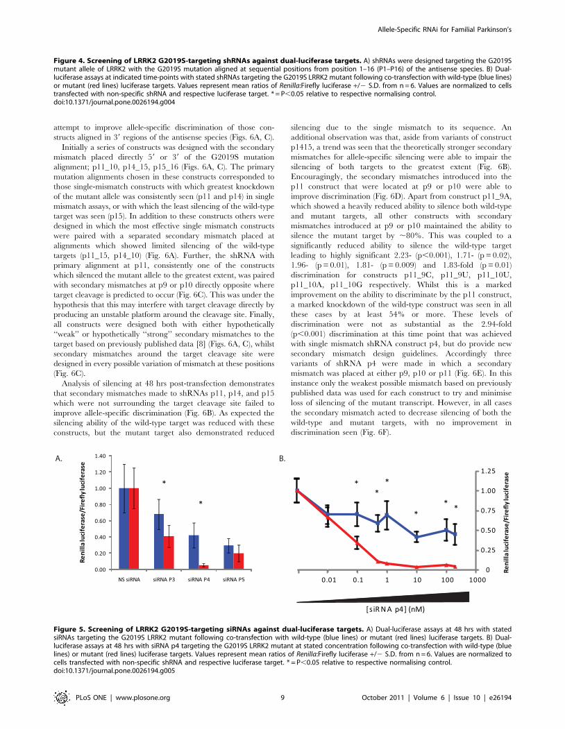
In order to verify the sequence alignment of the G2019S
mutation in the generated antisense species of the p4 construct,
siRNAs with alignment of the mutations at p3, p4 and p5 were
screened against the luciferase targets. At 48 hrs post-transfection
siRNA p4 displayed a 7.7-fold (p,0.001) discrimination that was
improved upon that seen with shRNA p4 at this time point
(Fig 5A). In contrast siRNAs p3 and p5 displayed limited, if any,
discrimination between the two alleles, again agreeing with the
trends from previous shRNA data which showed alignment at p4
to be superior to these two constructs. Further, discrimination by
siRNA p4 was evident using siRNA concentrations as low as
0.1 nM, and was increased to .8-fold by concentrations of siRNA
greater than 1 nM in separate experiments (Fig 5B). The greatest
discrimination was 10.8-fold (p,0.001) when using 10 nM siRNA,
and at this concentration a 96% silencing of the mutant target was
seen which was accompanied by a modest 58% silencing of the
wild-type target. However, the greatest difference in target
silencing was the 61% difference between the 92% silencing of
the mutant target and 31% silencing of the wild-type target when
using 1 nM siRNA. Collectively this data strongly suggests that the
alignment of the G2019S mutation in shRNA p4 was as stated,
whilst additionally demonstrating that siRNA p4 has impressive
and potent discriminating ability that could be useful in future pre-
clinical models of G2019S associated pathology.
Secondary mismatches at nucleotide positions oppositethe target cleavage site can improve the discriminationof G2019S targeting shRNAs
Despite allele-discrimination being evident with single-mis-
match shRNAs targeting the G2019S mutation, particularly at
p4, substantial silencing of the wild-type allele is also observed.
Following the previous demonstration that secondary mismatches
could improve the discrimination between mutant and wild-type
targets in this and other reports [6,17], a series of shRNAs
containing one mismatch to the G2019S mutant allele and a
secondary mismatch to the wild-type allele was initially designed to
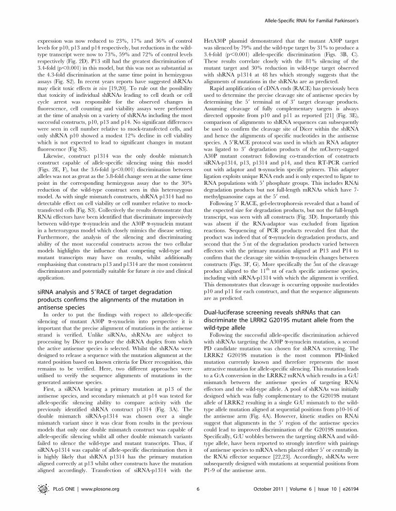
Figure 3. Determination of A30P mutation alignment with siRNA and 59RACE. A) siRNA design with A30P mutation aligned opposite P13 ofthe antisense species, and secondary mismatch to the wild-type a-synuclein allele at P14. B) Representative merged fluorescent images of HEK-293cells co-transfected with the Het-A30P plasmid siRNA-1314 at 48 hrs post-transfection. C) Quantification of wild-type a-synuclein eGFP (green bars) orA30P mutant a-synuclein mCherry (red bars) fluorescence at 48 hrs post-transfection following co-transfection of siRNA-1314 with the Het-A30Pplasmid. Values represent mean ratios of normalized fluorescence +/2 S.D. from n = 6. Values are normalized to respective fluorescence in cellstransfected with non-specific siRNA. * = P,0.05 relative to respective normalising control. D) Visualisation of PCR products following 59RACE usingRNA from cells transfected with mCherry-tagged A30P mutant a-synuclein and stated constructs. Products were run on a 2% agarose gel. E) Expectedtarget cleavage site for siRNA-1314. F) Sequencing of siRNA-1314 PCR product following 59RACE. G) Mapping of 59 adaptor ligations sites,determination of target cleavage sites and determination of A30P mutation alignments in stated constructs.doi:10.1371/journal.pone.0026194.g003
Allele-Specific RNAi for Familial Parkinson’s
PLoS ONE | www.plosone.org 7 October 2011 | Volume 6 | Issue 10 | e26194
Allele-Specific RNAi for Familial Parkinson’s
PLoS ONE | www.plosone.org 8 October 2011 | Volume 6 | Issue 10 | e26194
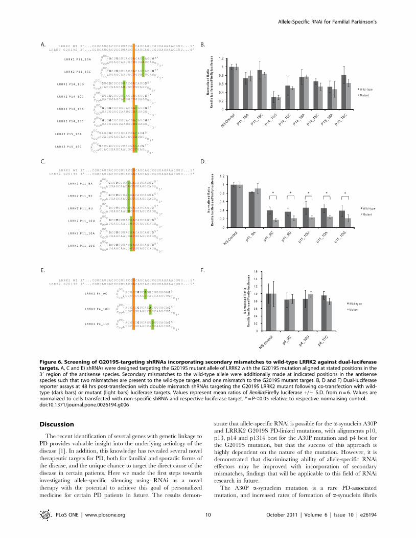
attempt to improve allele-specific discrimination of those con-
structs aligned in 39 regions of the antisense species (Figs. 6A, C).
Initially a series of constructs was designed with the secondary
mismatch placed directly 59 or 39 of the G2019S mutation
alignment; p11_10, p14_15, p15_16 (Figs. 6A, C). The primary
mutation alignments chosen in these constructs corresponded to
those single-mismatch constructs with which greatest knockdown
of the mutant allele was consistently seen (p11 and p14) in single
mismatch assays, or with which the least silencing of the wild-type
target was seen (p15). In addition to these constructs others were
designed in which the most effective single mismatch constructs
were paired with a separated secondary mismatch placed at
alignments which showed limited silencing of the wild-type
targets (p11_15, p14_10) (Fig. 6A). Further, the shRNA with
primary alignment at p11, consistently one of the constructs
which silenced the mutant allele to the greatest extent, was paired
with secondary mismatches at p9 or p10 directly opposite where
target cleavage is predicted to occur (Fig. 6C). This was under the
hypothesis that this may interfere with target cleavage directly by
producing an unstable platform around the cleavage site. Finally,
all constructs were designed both with either hypothetically
‘‘weak’’ or hypothetically ‘‘strong’’ secondary mismatches to the
target based on previously published data [8] (Figs. 6A, C), whilst
secondary mismatches around the target cleavage site were
designed in every possible variation of mismatch at these positions
(Fig. 6C).
Analysis of silencing at 48 hrs post-transfection demonstrates
that secondary mismatches made to shRNAs p11, p14, and p15
which were not surrounding the target cleavage site failed to
improve allele-specific discrimination (Fig. 6B). As expected the
silencing ability of the wild-type target was reduced with these
constructs, but the mutant target also demonstrated reduced
silencing due to the single mismatch to its sequence. An
additional observation was that, aside from variants of construct
p1415, a trend was seen that the theoretically stronger secondary
mismatches for allele-specific silencing were able to impair the
silencing of both targets to the greatest extent (Fig. 6B).
Encouragingly, the secondary mismatches introduced into the
p11 construct that were located at p9 or p10 were able to
improve discrimination (Fig. 6D). Apart from construct p11_9A,
which showed a heavily reduced ability to silence both wild-type
and mutant targets, all other constructs with secondary
mismatches introduced at p9 or p10 maintained the ability to
silence the mutant target by ,80%. This was coupled to a
significantly reduced ability to silence the wild-type target
leading to highly significant 2.23- (p,0.001), 1.71- (p = 0.02),
1.96- (p = 0.01), 1.81- (p = 0.009) and 1.83-fold (p = 0.01)
discrimination for constructs p11_9C, p11_9U, p11_10U,
p11_10A, p11_10G respectively. Whilst this is a marked
improvement on the ability to discriminate by the p11 construct,
a marked knockdown of the wild-type construct was seen in all
these cases by at least 54% or more. These levels of
discrimination were not as substantial as the 2.94-fold
(p,0.001) discrimination at this time point that was achieved
with single mismatch shRNA construct p4, but do provide new
secondary mismatch design guidelines. Accordingly three
variants of shRNA p4 were made in which a secondary
mismatch was placed at either p9, p10 or p11 (Fig. 6E). In this
instance only the weakest possible mismatch based on previously
published data was used for each construct to try and minimise
loss of silencing of the mutant transcript. However, in all cases
the secondary mismatch acted to decrease silencing of both the
wild-type and mutant targets, with no improvement in
discrimination seen (Fig. 6F).
Figure 4. Screening of LRRK2 G2019S-targeting shRNAs against dual-luciferase targets. A) shRNAs were designed targeting the G2019Smutant allele of LRRK2 with the G2019S mutation aligned at sequential positions from position 1–16 (P1–P16) of the antisense species. B) Dual-luciferase assays at indicated time-points with stated shRNAs targeting the G2019S LRRK2 mutant following co-transfection with wild-type (blue lines)or mutant (red lines) luciferase targets. Values represent mean ratios of Renilla:Firefly luciferase +/2 S.D. from n = 6. Values are normalized to cellstransfected with non-specific shRNA and respective luciferase target. * = P,0.05 relative to respective normalising control.doi:10.1371/journal.pone.0026194.g004
Figure 5. Screening of LRRK2 G2019S-targeting siRNAs against dual-luciferase targets. A) Dual-luciferase assays at 48 hrs with statedsiRNAs targeting the G2019S LRRK2 mutant following co-transfection with wild-type (blue lines) or mutant (red lines) luciferase targets. B) Dual-luciferase assays at 48 hrs with siRNA p4 targeting the G2019S LRRK2 mutant at stated concentration following co-transfection with wild-type (bluelines) or mutant (red lines) luciferase targets. Values represent mean ratios of Renilla:Firefly luciferase +/2 S.D. from n = 6. Values are normalized tocells transfected with non-specific shRNA and respective luciferase target. * = P,0.05 relative to respective normalising control.doi:10.1371/journal.pone.0026194.g005
Allele-Specific RNAi for Familial Parkinson’s
PLoS ONE | www.plosone.org 9 October 2011 | Volume 6 | Issue 10 | e26194
Discussion
The recent identification of several genes with genetic linkage to
PD provides valuable insight into the underlying aetiology of the
disease [1]. In addition, this knowledge has revealed several novel
therapeutic targets for PD, both for familial and sporadic forms of
the disease, and the unique chance to target the direct cause of the
disease in certain patients. Here we made the first steps towards
investigating allele-specific silencing using RNAi as a novel
therapy with the potential to achieve this goal of personalized
medicine for certain PD patients in future. The results demon-
strate that allele-specific RNAi is possible for the a-synuclein A30P
and LRRK2 G2019S PD-linked mutations, with alignments p10,
p13, p14 and p1314 best for the A30P mutation and p4 best for
the G2019S mutation, but that the success of this approach is
highly dependent on the nature of the mutation. However, it is
demonstrated that discriminating ability of allele-specific RNAi
effectors may be improved with incorporation of secondary
mismatches, findings that will be applicable to this field of RNAi
research in future.
The A30P a-synuclein mutation is a rare PD-associated
mutation, and increased rates of formation of a-synuclein fibrils
Figure 6. Screening of G2019S-targeting shRNAs incorporating secondary mismatches to wild-type LRRK2 against dual-luciferasetargets. A, C and E) shRNAs were designed targeting the G2019S mutant allele of LRRK2 with the G2019S mutation aligned at stated positions in the39 region of the antisense species. Secondary mismatches to the wild-type allele were additionally made at indicated positions in the antisensespecies such that two mismatches are present to the wild-type target, and one mismatch to the G2019S mutant target. B, D and F) Dual-luciferasereporter assays at 48 hrs post-transfection with double mismatch shRNAs targeting the G2019S LRRK2 mutant following co-transfection with wild-type (dark bars) or mutant (light bars) luciferase targets. Values represent mean ratios of Renilla:Firefly luciferase +/2 S.D. from n = 6. Values arenormalized to cells transfected with non-specific shRNA and respective luciferase target. * = P,0.05 relative to respective normalising control.doi:10.1371/journal.pone.0026194.g006
Allele-Specific RNAi for Familial Parkinson’s
PLoS ONE | www.plosone.org 10 October 2011 | Volume 6 | Issue 10 | e26194
and/or intermediate toxic proto-fibrils in addition to other
pathogenic mechanisms resulting from this mutation suggest that
silencing would be beneficial patients with this variant [25–27].
Non-allele specific a-synuclein RNAi has been investigated as a
potential approach for PD through in vivo delivery of shRNAs to
the rat substantia nigra pars compacta [28]. However this
indiscriminate silencing was associated with nigrostriatal degener-
ation which agrees with findings from transgenic a-synuclein null
mice showing disturbances to the nigrostriatal system [29]. Thus
whilst silencing of a-synuclein is expected to ameliorate a-
synuclein pathology it may enhance the dopaminergic deficit
responsible for the characteristic motor phenotypes of PD patients.
Targeted reduction of the mutant a-synuclein transcripts should
therefore be considered the ideal strategy for treating these
hereditary forms of PD bearing mutations in a-synuclein due to
sparing of the essential wild-type function.
Several shRNA constructs with mutations aligned across 39
regions of the antisense species were capable of silencing the a-
synuclein A30P mutation whilst sparing the wild-type allele in both
models tested, including a heterozygous model designed to
replicate the disease setting in which both wild-type and mutant
alleles are present [14]. Success with 39 alignments is consistent
with previous studies showing good discrimination at such
positions [8,10], although good discrimination was seen with
several alignments and not just the strong preference for alignment
at p16 suggested by Schwarz et al. [8]. Whilst phenotypic models
of A30P pathology [30,31] could not be replicated in our hands, it
is likely that the strong discrimination seen against the A30P
mutation with some constructs would be therapeutically beneficial.
It will be interesting to test these constructs in transgenic mouse
models carrying this mutation in future to see if phenotypic
correction can be achieved [32,33].
Estimates for the incidence of the LRRK2 G2019S mutation
are presently ,1% of all PD patients making it an attractive
mutation to target [1,16]. The mutation leads to a gain-in-function
of the kinase domain to suggest that reducing kinase activity would
be therapeutically beneficial [34]. Despite transgenic null mice
displaying no gross central nervous system abnormalities [35], at
present little is known about the function of LRRK2 whilst GWAS
studies imply abnormalities at the LRRK2 locus are a cause of
non-hereditary PD to stress its potential importance in disease
pathology [2,3]. As such allele specific silencing would be
considered preferable over non-allele specific silencing due to
LRRK29s presently unknown activity, and this is the first report
where this mutation has been successfully targeted. A recent
publication of allele-specific discrimination of this mutation with
siRNAs has been made. However, the G2019S model initially
reported online contains a 6056GRA transition rather than the
6055GRA transition originally reported in the LRRK2 coding
sequence [36], and siRNAs appear designed based on this
incorrect mutation [37]. A mistake in manuscript preparation
has been confirmed, and this likely explains why discrimination
was reported at P10, P11, P14 and P16 which each failed to show
discrimination in this study.
In this study discrimination was seen between wild-type and
mutant alleles in hemizygous dual-luciferase assays, but the
differences observed were relatively modest and reduced with
time. The exception to this was when the mutation was aligned at
p4 of the antisense strand. Discrimination of 2.94- and 3.7-fold
were seen at 48 and 72 hrs respectively with shRNA p4, and
greater than 10-fold was observed when using siRNA p4 at 48 hrs.
It remains to be seen if the discrimination levels observed would
have therapeutic benefit. One concern with the dual-luciferase
system is the absence of similar secondary structure to the desired
targets which could have an effect on silencing ability [38,39]. It is
possible that in this study the secondary structure surrounding the
G2019S target sequence is particularly relaxed and accessible to
RNAi. Whether this is the same with the endogenous full-length
LRRK2 sequence surrounding the G2019S mutation is unclear.
However, influence of secondary structure on RNAi is still debated
since the formation of the A-form helix recognised by the RNA
induced silencing complex (RISC) dictates that any secondary
structure would have to be released prior to RNAi [40,41]. It will
therefore be imperative to test this p4 construct and future
secondary mismatch variants displaying good discrimination in
more advanced models such as primary patient cells or the
recently reported G2019S transgenic mouse [42].
Reports have been published of shRNAs leading to significant
toxicity in vivo which can have fatal consequences in some animal
models [19,20]. Whilst no toxicity was associated with shRNA p4,
or indeed any of the shRNAs tested in this study, the incorporation
of antisense species into pri-miRNA backbones has been
highlighted as the safest expressed RNAi effector presently
available due to an apparent regulatory role of the Drosha/
DGCR8 microprocessor in limiting accumulation of toxic pre-
miRNA species [20,43]. Whilst shRNAs targeting the A30P
mutation may not merit translation into pri-miRNA mimics, the
incidence of the LRRK2 G2019S mutation suggests that
development of the p4 antisense sequence into pri-miRNA mimics
should be strongly considered in future studies.
The success in targeting the a-synuclein A30P mutation is
unsurprising given the resulting G:G mismatch that is present
between the wild-type allele and antisense species, a mismatch that
has been demonstrated previously to be one of the most favourable
for allele-specific silencing purposes [6,8,9]. It is expected that the
purine:purine alignment strongly disrupts the A-form helix that is
usually formed following pairing of the antisense species to the
target due to the presence of two dual-ring nitrogenous bases
aligned between the sugar-phosphate backbones where usually a
dual-ringed purine and a single-ringed pyrimidine exist.
It is expected that the reason for limited discrimination between
alleles when targeting the LRRK2 G2019S mutation with most
alignments was the result of a weaker mismatch being present
between the antisense species and the wild-type allele. The
resulting G:U alignment is commonly encountered within RNA
biology [44], and has thermodynamic stability approaching that of
normal Watson–Crick base pairs which exceeds almost all other
mismatches [45]. Indeed previous rules determined for allele-
specific silencing found the G:U wobble to be the second weakest
alignment for discrimination that wasn’t Watson-Crick base-
pairing [8], although others have demonstrated allele-specific
silencing of G:U mismatches [6,7,10,12]. However, alignment at
p4 in the 59 region of the antisense species showed improved
discrimination of the G2019S mutation when using both shRNAs
and siRNAs. This fits with previous reports indicating introduction
of G:U wobbles strongly interferes with pairings of antisense
species to mRNA when placed either 59 or centrally in the RNAi
effector [22,23], although this is not a consensus view at present
[46].
Finally, it was confirmed that secondary mismatches could
improve allele-specific discrimination of RNAi effectors. Given the
improved discrimination by the a-synuclein targeting construct
P1314 over the single mismatch construct P13, it was disappoint-
ing not to see a more marked improvement with the tested
secondary mismatches in shRNAs targeting the G2019S mutation.
No definitive rules have previously been established for incorpo-
ration of secondary mismatches to enhance allele-specific
discrimination despite previous success with this approach
Allele-Specific RNAi for Familial Parkinson’s
PLoS ONE | www.plosone.org 11 October 2011 | Volume 6 | Issue 10 | e26194
[6,17], and attempts were made here to determine these by testing
multiple design strategies against the G2019S mutation. Placing a
secondary mismatch opposite the target cleavage site improved
discrimination relative to the single mismatch constructs in some
shRNAs, but not all, and this trend supports early allele-specific
studies which hypothesised that alignment of mutations at such
positions was likely to have the most disruptive effect on RNAi
activity [6,7,9,12]. Importantly it also demonstrates that extensive
screening of variants may often be needed before suitable variants
are found. Finally, in the successful examples, the strongest effect
was seen when this secondary position was changed from a G:C
alignment to a C:C mismatch, and this agrees with this alignment
being more disruptive than both a A:C mismatch and a U:C
mismatch in previous studies [8]. This suggests that if secondary
mismatches are to be used in future, then the ‘‘strength’’ of the
secondary mismatch should be considered as this can influence the
success.
In summary we have identified several RNAi constructs with
the ability to discriminate wild-type and mutant alleles of two PD-
linked genes. The results agree with previous allele-specific
silencing rules suggesting that the nature of the mutations has a
strong influence on the success of this approach, whilst we confirm
that secondary mismatches can be used to improve discrimination
further in some cases. It remains to be seen whether the constructs
identified are therapeutically beneficial, and the identified
constructs now await evaluation and development in more
advanced pre-clinical models to test for therapeutic efficacy.
Supporting Information
Figure S1 Cell counting and cell viability from cellsexpressing eGFP-tagged a-synuclein targets. A) Cell
counts from HEK-293 cells transfected with stated eGFP-tagged
a-synuclein targets or mock transfection at 48 hrs post-transfec-
tion. B) Trypan blue cell viability assay of HEK-293 cells
transfected with stated eGFP-tagged a-synuclein targets or mock
transfection at 48 hrs post-transfection.
(EPS)
Figure S2 Screening of A30P-targeting shRNAs againstfull-length eGFP-tagged a-synuclein targets. A+C) Repre-
sentative fluorescent images of HEK-293 cells co-transfected with
stated eGFP-tagged a-synuclein targets and indicated single (A) or
double (C) mismatch shRNA construct at 48 hrs post-transfection.
B+D) Quantification of eGFP fluorescence at 48 hrs post-
transfection following co-transfection of single (B) or double (D)
mismatch shRNAs targeting the A30P a-synuclein mutant with
wild-type (dark bars) or mutant (light bars) eGFP-tagged a-
synuclein targets. Values represent mean ratios of normalized
fluorescence +/2 S.D. from n = 6. Values are normalized to cells
transfected with non-specific shRNA and respective eGFP-tagged
target. * = P,0.05 relative to respective normalising control.
(EPS)
Figure S3 Cell counting and cell viability from cellsexpressing shRNAs and corresponding targets. A) Cell
counts from mock transfected HEK-293 cells or HEK-293 cells
transfected with stated het-A30P plasmid and indicated shRNAs at
48 hrs post-transfection. B) Trypan blue cell viability assay of
mock transfected HEK-293 cells or HEK-293 cells transfected
with stated het-A30P plasmid and indicated shRNAs at 48 hrs
post-transfection. C) Cell counts from mock transfected HEK-293
cells or HEK-293 cells transfected with G2019S dual-luciferase
target plasmid and indicated shRNAs at 48 hrs post-transfection.
D) Trypan blue cell viability assay of mock transfected HEK-293
cells or HEK-293 cells transfected with G2019S dual-luciferase
target plasmid and indicated shRNAs at 48 hrs post-transfection.
(EPS)
Table S1 Oligonucleotide sequences used.
(DOC)
Author Contributions
Conceived and designed the experiments: CRS MJAW. Performed the
experiments: CRS. Analyzed the data: CRS. Contributed reagents/
materials/analysis tools: CRS MJAW. Wrote the paper: CRS MJAW.
References
1. Lesage S, Brice A (2009) Parkinson’s disease: from monogenic forms to genetic
susceptibility factors. Hum Mol Genet 18: R48–59.
2. Simon-Sanchez J, Schulte C, Bras JM, Sharma M, Gibbs JR, et al. (2009)
Genome-wide association study reveals genetic risk underlying Parkinson’s
disease. Nat Genet 41: 1308–1312.
3. Satake W, Nakabayashi Y, Mizuta I, Hirota Y, Ito C, et al. (2009) Genome-wide
association study identifies common variants at four loci as genetic risk factors for
Parkinson’s disease. Nat Genet 41: 1303–1307.
4. Erson AE, Petty EM (2008) MicroRNAs in development and disease. Clin Genet
74: 296–306.
5. Kubodera T, Yokota T, Ishikawa K, Mizusawa H (2005) New RNAi strategy for
selective suppression of a mutant allele in polyglutamine disease. Oligonucle-
otides 15: 298–302.
6. Miller VM, Xia H, Marrs GL, Gouvion CM, Lee G, et al. (2003) Allele-specific
silencing of dominant disease genes. Proc Natl Acad Sci USA 100: 7195–7200.
7. Miller VM, Gouvion CM, Davidson BL, Paulson HL (2004) Targeting
Alzheimer’s disease genes with RNA interference: an efficient strategy for
silencing mutant alleles. Nucleic Acids Res 32: 661–668.
8. Schwarz DS, Ding H, Kennington L, Moore JT, Schelter J, et al. (2006)
Designing siRNA that distinguish between genes that differ by a single
nucleotide. PLoS Genet 2: e140.
9. Ding H, Schwarz DS, Keene A, Affar el B, Fenton L, et al. (2003) Selective
silencing by RNAi of a dominant allele that causes amyotrophic lateral sclerosis.
Aging Cell 2: 209–217.
10. Scholefield J, Greenberg LJ, Weinberg MS, Arbuthnot PB, Abdelgany A, et al.
(2009) Design of RNAi hairpins for mutation-specific silencing of ataxin-7 and
correction of a SCA7 phenotype. PLoS ONE 4: e7232.
11. Hickerson RP, Smith FJ, Reeves RE, Contag CH, Leake CH, et al. (2008)
Single-nucleotide-specific siRNA targeting in a dominant-negative skin model.
J Invest Dermatol 128: 594–605.
12. Sapru MK, Yates JW, Hogan S, Jiang L, Halter J, et al. (2006) Silencing of
human alpha-synuclein in vitro and in rat brain using lentiviral-mediated RNAi.
Exp Neurol 198: 382–390.
13. Polymeropoulos MH, Lavedan C, Leroy E, Ide SE, Dehjia A, et al. (1997)
Mutation in the alpha-synuclein gene identified in families with Parkinson’s
disease. Science 276: 2045–2047.
14. Kruger R, Kuhn W, Muller T, Woitalla D, Graeber M, et al. (1998) Ala30Pro
mutation in the gene encoding alpha-synuclein in Parkinson’s disease. Nat Genet
18: 106–108.
15. Zarranz JJ, Alegre J, Gomez-Esteban JC, Lezcano E, Ros R, et al. (2004) The
new mutation, E46K, of alpha-synuclein causes Parkinson and Lewy body
dementia. Ann Neurol 55: 164–173.
16. Giasson BI, Van Deerlin VM (2008) Mutations in LRRK2 as a cause of
Parkinson’s disease. Neurosignals 16: 99–105.
17. Ohnishi Y, Tamura Y, Yoshida M, Tokunaga K, Hohjoh H (2008)
Enhancement of allele discrimination by introduction of nucleotide mismatches
into siRNA in allele-specific gene silencing by RNAi. PLoS ONE 3: e2248.
18. Pandey N, Schmidt RE, Galvin JE (2006) The alpha-synuclein mutation E46K
promotes aggregation in cultured cells. Exp Neurol 197: 515–520.
19. Grimm D, Streetz KL, Jopling CL, Storm TA, Pandey K, et al. (2006) Fatality in
mice due to oversaturation of cellular microRNA/short hairpin RNA pathways.
Nature 441: 537–541.
20. McBride JL, Boudreau RL, Harper SQ, Staber PD, Monteys AM, et al. (2008)
Artificial miRNAs mitigate shRNA-mediated toxicity in the brain: implications
for the therapeutic development of RNAi. Proc Natl Acad Sci USA 105:
5868–5873.
21. Haley B, Zamore PD (2004) Kinetic analysis of the RNAi enzyme complex.
Nat Struct Mol Biol 11: 599–606.
22. Doench JG, Sharp PA (2004) Specificity of microRNA target selection in
translational repression. Genes Dev 18: 504–511.
Allele-Specific RNAi for Familial Parkinson’s
PLoS ONE | www.plosone.org 12 October 2011 | Volume 6 | Issue 10 | e26194
23. Holen T, Moe SE, Sorbo JG, Meza TJ, Ottersen OP, et al. (2005) Tolerated
wobble mutations in siRNAs decrease specificity, but can enhance activity invivo. Nucleic Acids Res 33: 4704–4710.
24. Yue Z (2009) LRRK2 in Parkinson’s disease: in vivo models and approaches for
understanding pathogenic roles. FEBS J 276: 6445–6454.25. Conway KA, Lee SJ, Rochet JC, Ding TT, Williamson RE, et al. (2000)
Acceleration of oligomerization, not fibrillization, is a shared property of bothalpha-synuclein mutations linked to early-onset Parkinson’s disease: implications
for pathogenesis and therapy. Proc Natl Acad Sci USA 97: 571–576.
26. Lashuel HA, Petre BM, Wall J, Simon M, Nowak RJ, et al. (2002) Alpha-synuclein, especially the Parkinson’s disease-associated mutants, forms pore-like
annular and tubular protofibrils. J Mol Biol 322: 1089–1102.27. Cuervo AM, Stefanis L, Fredenburg R, Lansbury PT, Sulzer D (2004) Impaired
degradation of mutant alpha-synuclein by chaperone-mediated autophagy.Science 305: 1292–1295.
28. Gorbatyuk OS, Li S, Gorbatyuk M, Lewin AS, Sullivan LF, et al. (2010) In vivo
RNAi-mediated alpha-synuclein silencing induces nigrostriatal degeneration.Mol Ther 18: 1450–1457.
29. Abeliovich A, Schmitz Y, Farinas I, Choi-Lundberg D, Ho WH, et al. (2000)Mice lacking alpha-synuclein display functional deficits in the nigrostriatal
dopamine system. Neuron 25: 239–252.
30. Kanda S, Bishop JF, Eglitis MA, Yang Y, Mouradian MM (2000) Enhancedvulnerability to oxidative stress by alpha-synuclein mutations and C-terminal
truncation. Neuroscience 97: 279–284.31. Nonaka T, Hasegawa M (2009) A cellular model to monitor proteasome
dysfunction by alpha-synuclein. Biochemistry 48: 8014–8022.32. Rathke-Hartlieb S, Kahle PJ, Neumann M, Ozmen L, Haid S, et al. (2001)
Sensitivity to MPTP is not increased in Parkinson’s disease-associated mutant
alpha-synuclein transgenic mice. J Neurochem 77: 1181–1184.33. Gomez-Isla T, Irizarry MC, Mariash A, Cheung B, Soto O, et al. (2003) Motor
dysfunction and gliosis with preserved dopaminergic markers in human alpha-synuclein A30P transgenic mice. Neurobiol Aging 24: 245–258.
34. West AB, Moore DJ, Choi C, Andrabi SA, Li X, et al. (2007) Parkinson’s
disease-associated mutations in LRRK2 link enhanced GTP-binding and kinaseactivities to neuronal toxicity. Hum Mol Genet 16: 223–232.
35. Tong Y, Yamaguchi H, Giaime E, Boyle S, Kopan R, et al. (2010) Loss of
leucine-rich repeat kinase 2 causes impairment of protein degradation pathways,
accumulation of alpha-synuclein, and apoptotic cell death in aged mice.
Proc Natl Acad Sci USA 107: 9879–9884.
36. Kachergus J, Mata IF, Hulihan M, Taylor JP, Lincoln S, et al. (2005)
Identification of a novel LRRK2 mutation linked to autosomal dominant
parkinsonism: evidence of a common founder across European populations. Am
J Hum Genet. 2005 Apr; 76(4): 672–80.
37. Wang JJ, Li QS, Li Y, Zheng YR (2011) Discrimination of Parkinson-associated
LRRK2 alleles by introduction of a single nucleotide mismatch into siRNA.
Neurosci Lett. (2011). doi:10.1016/j.neulet.2011.04.064.
38. Shao Y, Chan CY, Maliyekkel A, Lawrence CE, Roninson IB, et al. (2007)
Effect of target secondary structure on RNAi efficiency. RNA 13: 1631–1640.
39. Zhou H, Zeng X (2009) Energy profile and secondary structure impact shRNA
efficacy. BMC Genomics 10 Suppl 1: S9.
40. Song J-J, Liu J, Tolia NH, Schneiderman J, Smith SK, et al. (2003) The crystal
structure of the Argonaute2 PAZ domain reveals an RNA binding motif in
RNAi effector complexes. Nat Struct Biol 10: 1026–1032.
41. Heale BSE, Soifer HS, Bowers C, Rossi JJ (2005) siRNA target site secondary
structure predictions using local stable substructures. Nucleic Acids Res 33: e30.
42. Winner B, Melrose HL, Zhao C, Hinkle KM, Yue M, et al. (2011) Adult
neurogenesis and neurite outgrowth are impaired in LRRK2 G2019S mice.
Neurobiol Dis 41: 706–716.
43. Sibley CR, Seow Y, Wood MJA (2010) Novel RNA-based strategies for
therapeutic gene silencing. Mol Ther 18: 466–476.
44. Varani G, McClain WH (2000) The G x U wobble base pair. A fundamental
building block of RNA structure crucial to RNA function in diverse biological
systems. EMBO Rep 1: 18–23.
45. Strazewski P, Biala E, Gabriel K, McClain WH (1999) The relationship of
thermodynamic stability at a G x U recognition site to tRNA aminoacylation
specificity. RNA 5: 1490–1494.
46. Du Q, Thonberg H, Wang J, Wahlestedt C, Liang Z (2005) A systematic analysis
of the silencing effects of an active siRNA at all single-nucleotide mismatched
target sites. Nucleic Acids Res 33: 1671–1677.
Allele-Specific RNAi for Familial Parkinson’s
PLoS ONE | www.plosone.org 13 October 2011 | Volume 6 | Issue 10 | e26194